Observing the Asymmetry of Amygdaloid Complex in Patients with Complex Partial Attacks
|
|
|
- Wilfrid Shaw
- 6 years ago
- Views:
Transcription
1 & Observing the Asymmetry of Amygdaloid Complex in Patients with Complex Partial Attacks Aida Sarač Hadžihalilović¹*, Faruk Dilberović¹, Abdulah Kučukalić² 1. Department of Anatomy, Faculty of Medicine, University of Sarajevo, Čekaluša 90, Sarajevo, Bosnia and Herzegovina 2. Psychiatric Clinic, University of Sarajevo Clinics Centre, Bolnička 25, Sarajevo, Bosnia and Herzegovina * Corresponding author Abstract Lobus limbicus is an anatomical basis for understanding the temporal epilepsy because it includes not only the focus of temporal lobe infection but of the frontal lobe as well. With it we can explain many of the phenomena accompanying epilepsy (hallucinations, the change of the effects, and so on). The goal of this assignment was to explore the asymmetry of amygdaloidal complex in the patients with complex partial attacks. The results show that the smallest number of patients with epilepsy have a symmetric (same size) amygdaloidal complex on the left side and the right. According to the asymmetry direction the difference in the number of patients with epilepsy is not statistically significant. Coefficient of asymmetry shows that the asymmetry on the left side is more frequent in men, while women have the same distribution on both sides. The greatest differences were found when considering the age factor. So, in all the three groups of evaluated data the differences in average age of patients with epilepsy according to total symm. / asymm. were not statistically significant. But, the differences in average age depending on the direction of asymmetry were significant. Patients with longer amygdaloidal complex on the left side are significantly younger, both male and female (related to the axial slice, ant. post. diameter). Thus, we propose the application of MRI technique in examining the asymmetry of the amygdaloidal complex that we used in this assignment as a template for future examinations in a sense of shedding light on the anatomical functions that underlie neuro-psychiatric dysfunctions. KEY WORDS: limbic system, amygdaloidal complex, asymmetry, temporal epilepsy 21
 and motor symptoms (oral automatism).")

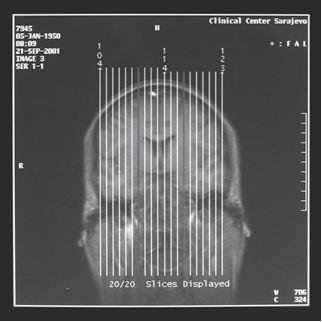
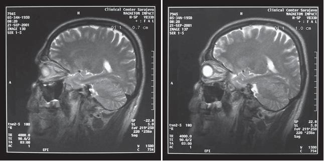
2 Introduction In the limbic system visceral functions are integrated with emotional behavior. That is best illustrated by clinical picture of complex partial attack with vegetative, psychiatric (dysfunction of a sensible behavior) and motor symptoms (oral automatism). Bilateral injury of the temporal lobe that has a great effect on amygdaloidal complex that causes a series of behavioral changes named Kluver Bucy syndrome. This is evident in patients with temporal lobe trauma or after surgical operation on the temporal lobe due to epilepsy. Lobus limbicus is anatomical basis for understanding the temporal epilepsy because it includes not only the focus of temporal lobe infection but of the frontal lobe as well. With it we can explain many of the phenomena accompanying epilepsy (hallucinations, the change of the effects, and so on). Many bodily functions maintain regular rhythms with cycles of different length. The role of «biological clock«that regulates these rhythms is a part of the limbic system function. Therefore, psychomotor epilepsy is frequently associated with epilepsy focused in temporal region. Material and Methods 35 MRI scans of patient with complex partial attacks were used as a background material for this study. The size of amygdaloidal complex was measured in two projections: horizontal (axial) and sagittal. In axial projections we measured anterior posterior and lateral medial diameters of amygdaloidal complex. We did not measure the amygdaloidal complex in coronal projections, because other cerebral structures make clear diffraction impossible. MRI scans were obtained using MAGNET IMPACT SIEMENS 1,0 TESLA in T1 relaxation ( TR / TE 15 / field of view 180 x 260, the layer thickness SL 5 mm ) and T2 relaxation ( TR 4000 / TE 90 field of view 188 x 250 for axial and 173 x 230 for coronal, 210 x 240 for sagittal scans in 5 mm layer ). Dual sequences are used - PD and T2. In PD TR is 4000, and TE 22. We used head neck spiral, as well as head spiral. For the measurement of amygdaloidal complex sizes, and their comparison from the right to the left we used a program that evaluates the distance in MRI from Sarajevo Clinics Center Institute of Radiology. This study conforms to all the standards of research ethics as proposed by the Ethics Comity of Sarajevo School of Medicine. For amygdaloidal complex in two projections (axial and sagittal) we gave: 1. The number of patients with epilepsy according to symmetry/asymmetry on the right and left side. 2. Analysis of patients with epilepsy according to the approximate size of left and right side. Significance of differences was tested with t-test. 3. Distribution of patients with epilepsy according to the difference between the right and left side. The results are given in tables and diagrams. 4. The approximate age of patients with epilepsy according to symmetry / asymmetry on the right and left side. Significance of differences was tested with t-test. Methods of statistical analysis used in this assignment are: 1. Arithmetic mean 2. Standard deviation 3. Standard error 4. Median 5. Mod 6. Chi-square test 7. t-test differences of arithmetic mean 8. t-test of frequencies 9. Coefficient of asymmetry 22
3 Results 23
4 24
5 Coefficient of asymmetry indicates asymmetry on the right side, for right side and for both sex, and left asymmetry, for left side and for both sex. Asymmetry is particularly pronounced on the right side in female. 25
6 26
7 Discussion The precise location of the incorrect anatomical function that underlies neurological and psychiatric dysfunctions just recently became the subject of intensive research in this filed. In that sense it is important to apply structural and functional techniques of MRI, for future pinpointing of the problems in working with epilepsy. Therefore, MRI offers the most sensitive volumetric measurements of hippocampal formation. Chronister R. C. et al. (1) use limbic system as a reference that pertains to emotions and interconnecting pathways. Authors differentiate centers of aversions and centers of pleasures (gratifications). If we stimulate centers of aversions person will face fear or sadness. On the other hand, stimulation of centers of pleasures will have pleasure as a result. Functional interconnections among aversion and gratification centers, according to the authors, contribute to emotional stability. As a consequence, amygdale stimulation can cause fear, while nucleus accumbens stimulation leads to a feeling of happiness and pleasure. Hadziselimovic et al. (2,3,4,5) confirmed that hippocampus and amygdaloidal complex with their positions depend on the position of temporal lobe; that their positions are followed by brain asymmetry and, when analysis of their positions are made it has to be taken into account. Vogeley K. et al. (6) in Brain image study show loss of upper temporal gyri (smaller size) and abnormal asymmetry example in SCH, but concerning this, there are no sustainable diagnoses. Belin P. et al. (7) in paper Lateralization of speech and hearing temporal processing prove that auditoria processing of fast acoustic passes is lateralized in human brain as well as that functional asymmetry in temporal processing is most likely contributed to lingual lateralization to the lower level of cortical empting. In their works Tzourio N. et al. (8) came to the conclusions that size of left temporal plane is relative anatomic indicator for lingual domination and demonstrate that anatomic asymmetry is a part of functional lingual variation. In morphological studies of temporal lobe and the related central temporal structures in children and adolescents from 27
8 14 to 18 years of age, which were conducted by Jay N. Giedd et al. (9), the total volume of temporal lobe was stable, while amygdale increased its volume only in males and hippocampus increased its volume only in females. This example coexists with the distribution of sex hormonal receptors for this structure; amygdale predominantly hosts androgen receptors while hippocampus predominantly hosts estrogens receptors. Conclusion On the basis of our analysis we can conclude: 1. In all the three slices the least number of patients with epilepsy have symmetric (same size) amygdaloidal complex on the left and the right side. That number is statistically significantly lower then the number of patients with epilepsy with asymmetric (different size) amygdaloidal complex from both sides. In this group we found no statistically significant difference according to the direction of the asymmetry. 2. In all the three groups, differences in average length of right and left amygdaloidal complex are statistically not significant concerning neither sex nor the direction of asymmetry. 3. The coefficient of asymmetry shows that the asymmetry on the left side is more frequent in men, while women have the same distribution on both sides. 4. Besides the previous parameters which are mainly harmonized, age as a factor shows the highest differences. So, in all the three groups of evaluated data differences in average age of patients with epilepsy according to total symmetry / asymmetry are not statistically significant. But, differences in average age depending on the direction of asymmetry are significant. Patients with longer amygdaloidal complex on the left side are significantly younger, both male and female (related to axial slice, ant. post. diameter). 5. Every one of the analyzed asymmetries shows the same characteristics in the group, in which we emphasize variations. 6. We also emphasize the importance of presence of consensus in individual characteristics of every one of the parameters in the shading light on asymmetry of amygdaloidal complex 7. We have to be careful in what projection we observe amygdaloidal complex because the results will depend on that. We can suggest the prospective studies in more projections because of the value of the statistically significant conclusions. 8. MRI volumetric measurement have their values. 9. We suggest the usage of MRI techniques in examining the asymmetry of amygdaloidal complex that we used, as a tool in the future studies in the sense of shading light on the anatomical functions that underlie neuropsychiatry dysfunctions. References (1) Chronister R. B., Hardy S. G. P. The Limbic System, chapter thirty, 2000; Churchill Livingstone, USA. (2) Hadžiselimović H., Čuš M. The appearance of internal structures of the brain in relation to configuration of the human skull. Acta Anat. 1966; 63: (3) Hadžiselimović H., Ruždić N. Appearance of the base of the brain in relation to the configuration of human skull. Acta Anat. 1966; 65: (4) Hadžiselimović H., Andželić M. On the appearance of some interior brain structures in relation to the exterior configuration of the brain. Acta Anat. 1966; 63: (5) Hadžiselimović H., M. Čuš, Dilberović F.. Hipokamp i amigdaloidni kompleks novoređenčeta. Folia Anatomica Jug. 1979: (6) Vogele K., Hobson T., Schneider Axmann T., Honer W.G., Bogerts B., Falkai P. Compartmental volumetry of the superior temporal gyrus reveal sex differences in schizophrenia a post mortem study. Schizophrenia Research 1998; 31: (7) Belin P., Zilbovicius M., Crozier S., Thivard L., Fontaine A., Masure M.C., Samson Y. Lateralization of speech and auditory temporal processing, J. Congn. Neurosci. 1998; 10 (4): (8) Tzourio N., Nkanga Ngila B., Mazoyer B. Left planum temporale surface correlates with functional dominance during story listening. Neuroreport. 1998; 30: (9) Giedd J.N., Vaituzis A.C., Hamburger S.D., Lange N., Rajapakse J.C., Kaysen D., Vauss YC., Rapoport J.L. Quantitative MRI of the lobe, amygdale and hippocampus in normal human development: ages 4 18 years. Journal of Comparative Neurology ; 336 (2):
Observing of the asymmetry of hippocampal formation on patients with complex partial attacks
 Observing of the asymmetry of hippocampal formation on patients with complex partial attacks Aida Sara~ - Had`ihalilovi}, Faruk Dilberovi} Department of Anatomy, Faculty of Medicine, University of Sarajevo,
P. Hitchcock, Ph.D. Department of Cell and Developmental Biology Kellogg Eye Center. Wednesday, 16 March 2009, 1:00p.m. 2:00p.m.
 Normal CNS, Special Senses, Head and Neck TOPIC: CEREBRAL HEMISPHERES FACULTY: LECTURE: READING: P. Hitchcock, Ph.D. Department of Cell and Developmental Biology Kellogg Eye Center Wednesday, 16 March
Chapter 2 Test. 1. Evolutionary structures within the are the most primitive. *a. hindbrain b. thalamus c. forebrain d. midbrain e.
 Cognitive Psychology In and Out of the Laboratory 5th Edition Galotti TEST BANK Full clear download (no formatting errors) at: https://testbankreal.com/download/cognitive-psychology-laboratory-5thedition-galotti-test-bank/
Frontal Contributions to Memory Encoding Before and After Unilateral Medial Temporal Lobectomy
 Frontal Contributions to Memory Encoding Before and After Unilateral Medial Temporal Lobectomy Jeff Ojemann, MD Department of Neurological Surgery University of Washington Children s Hospital & Regional
Brain Mechanisms of Emotion 1 of 6
 Brain Mechanisms of Emotion 1 of 6 I. WHAT IS AN EMOTION? A. Three components (Oately & Jenkins, 1996) 1. caused by conscious or unconscious evaluation of an event as relevant to a goal that is important
CISC 3250 Systems Neuroscience
 CISC 3250 Systems Neuroscience Levels of organization Central Nervous System 1m 10 11 neurons Neural systems and neuroanatomy Systems 10cm Networks 1mm Neurons 100μm 10 8 neurons Professor Daniel Leeds
Neuroimaging for Diagnosis of Psychiatric Disorders
 Psychiatric Disorder Neuroimaging for Diagnosis of Psychiatric Disorders JMAJ 45(12): 538 544, 2002 Yoshio HIRAYASU Associate Professor, Department of Neuropsychiatry Kyorin University School of Medicine
CNS Tour (Lecture 12)
") A. Introduction CNS Tour (Lecture 12) There are to a chemical pathways in the nervous system. These pathways also form different neurological structures B. Spinal Cord Receives sensory neurons from skin
A. Introduction CNS Tour (Lecture 12) There are to a chemical pathways in the nervous system. These pathways also form different neurological structures B. Spinal Cord Receives sensory neurons from skin
Differences in brain structure and function between the sexes has been a topic of
 Introduction Differences in brain structure and function between the sexes has been a topic of scientific inquiry for over 100 years. In particular, this topic has had significant interest in the past
Auditory fmri correlates of loudness perception for monaural and diotic stimulation
 PROCEEDINGS of the 22 nd International Congress on Acoustics Psychological and Physiological Acoustics (others): Paper ICA2016-435 Auditory fmri correlates of loudness perception for monaural and diotic
Telencephalon (Cerebral Hemisphere)
") Telencephalon (Cerebral Hemisphere) OUTLINE The Cortex - Lobes, Sulci & Gyri - Functional Subdivisions - Limbic Lobe & Limbic System The Subcortex - Basal Ganglia - White Matter (Internal Capsule) - Relations
Telencephalon (Cerebral Hemisphere) OUTLINE The Cortex - Lobes, Sulci & Gyri - Functional Subdivisions - Limbic Lobe & Limbic System The Subcortex - Basal Ganglia - White Matter (Internal Capsule) - Relations
XIXth Century: Localization of Functions to Different Parts of the Brain
 XIXth Century: Localization of Functions to Different Parts of the Brain Studies by Bell and Magendie initiated an extremely important scientific procedure,, where a specific part of the nervous system
DEFINING EMOTION 11/19/2009 THE BIOLOGY OF EMOTION & STRESS. A change in physiological arousal, ranging from slight to intense.
 DEFINING EMOTION Emotion A feeling that differs from a person s normal affective state; a biological function of the nervous system. A change in physiological arousal, ranging from slight to intense. An
Prof. Saeed Abuel Makarem & Dr.Sanaa Alshaarawy
 Prof. Saeed Abuel Makarem & Dr.Sanaa Alshaarawy 1 Objectives By the end of the lecture, you should be able to: Describe the anatomy and main functions of the thalamus. Name and identify different nuclei
Cerebral MRI as an important diagnostic tool in temporal lobe epilepsy
 Cerebral MRI as an important diagnostic tool in temporal lobe epilepsy Poster No.: C-2190 Congress: ECR 2014 Type: Educational Exhibit Authors: A. Puiu, D. Negru; Iasi/RO Keywords: Neuroradiology brain,
STRESS, MEMORY AND BOSNIAN WAR VETERANS
 & STRESS, MEMORY AND BOSNIAN WAR VETERANS Aida Sarač Hadžihalilović ¹*, Amela Kulenović¹, Abdulah Kučukalić² ¹ Department for Anatomy, Faculty of Medicine, University of Sarajevo, Čekaluša 90, 71000 Sarajevo,
Brain, language, and handedness: a family affair
 Brain, language, and handedness: a family affair Bernard Mazoyer 1,2,3, Gregory Simon 1,2, Fabrice Crivello 1, Laure Zago, Laurent Petit, Emmanuel Mellet, Nathalie Tzourio-Mazoyer 1* 1 CI-NAPS, CNRS, CEA,
XIXth Century: Localization of Functions to Different Parts of the Brain
 XIXth Century: Localization of Functions to Different Parts of the Brain Studies by Bell and Magendie initiated an extremely important scientific procedure,, where a specific part of the nervous system
Limbic system. Lecture 29, November 10, 2017
 Limbic system Lecture 29, November 10, 2017 Circadian rhythms (Latin, approximately a day ) Regulation of our daily rhythm Eating Sleeping Defecating Periods of activity Suprachiasmatic n. http://slideplayer.com/slide/6351731/
Introduction to Systems Neuroscience. Nov. 28, The limbic system. Daniel C. Kiper
 Introduction to Systems Neuroscience Nov. 28, 2017 The limbic system Daniel C. Kiper kiper@ini.phys.ethz.ch http: www.ini.unizh.ch/~kiper/system_neurosci.html LIMBIC SYSTEM The term limbic system mean
LIMBIC SYSTEM. Dr. Amani A. Elfaki Associate Professor Department of Anatomy
 LIMBIC SYSTEM Dr. Amani A. Elfaki Associate Professor Department of Anatomy Learning Objectives Define the limbic system Identify the parts of the limbic system Describe the circulation of the limbic system
CEREBRUM. Dr. Jamila EL Medany
 CEREBRUM Dr. Jamila EL Medany Objectives At the end of the lecture, the student should be able to: List the parts of the cerebral hemisphere (cortex, medulla, basal nuclei, lateral ventricle). Describe
NIH Public Access Author Manuscript Proc SPIE. Author manuscript; available in PMC 2014 February 07.
 NIH Public Access Author Manuscript Published in final edited form as: Proc SPIE. 2007 March 5; 6512: 651236. doi:10.1117/12.708950. Semi-Automatic Parcellation of the Corpus Striatum Ramsey Al-Hakim a,
High spatial resolution reveals excellent detail in pediatric neuro imaging
 Publication for the Philips MRI Community Issue 46 2012/2 High spatial resolution reveals excellent detail in pediatric neuro imaging Achieva 3.0T with 32-channel SENSE Head coil has become the system
The Brain and Behavior
 PNS Chapter 1 The Brain and Behavior 18-698 / 42-632 Neural Signal Processing Spring 2017 Prof. Byron Yu Roadmap Introduction to neuroscience Chapter 1 The brain and behavior Chapter 2 Nerve cells and
The Central Nervous System I. Chapter 12
 The Central Nervous System I Chapter 12 The Central Nervous System The Brain and Spinal Cord Contained within the Axial Skeleton Brain Regions and Organization Medical Scheme (4 regions) 1. Cerebral Hemispheres
Music, Epilepsy and the Brain. Stephen Brown
 Music, Epilepsy and the Brain Stephen Brown this may increase your IQ by a few points for a few minutes (or not) Music What s it for? Mood & music Musical morbidity Cognitive aspects What s it for? Music
Text to brain: predicting the spatial distribution of neuroimaging observations from text reports (submitted to MICCAI 2018)
") 1 / 22 Text to brain: predicting the spatial distribution of neuroimaging observations from text reports (submitted to MICCAI 2018) Jérôme Dockès, ussel Poldrack, Demian Wassermann, Fabian Suchanek, Bertrand
1 / 22 Text to brain: predicting the spatial distribution of neuroimaging observations from text reports (submitted to MICCAI 2018) Jérôme Dockès, ussel Poldrack, Demian Wassermann, Fabian Suchanek, Bertrand
Gross Organization I The Brain. Reading: BCP Chapter 7
 Gross Organization I The Brain Reading: BCP Chapter 7 Layout of the Nervous System Central Nervous System (CNS) Located inside of bone Includes the brain (in the skull) and the spinal cord (in the backbone)
Exam 1 PSYC Fall 1998
 Exam 1 PSYC 2022 Fall 1998 (2 points) Briefly describe the difference between a dualistic and a materialistic explanation of brain-mind relationships. (1 point) True or False. George Berkely was a monist.
Cerebral Cortex Structure, Function, Dysfunction Reading Ch 10 Waxman Dental Neuroanatomy Lecture. Suzanne Stensaas, Ph.D.
 Cerebral Cortex Structure, Function, Dysfunction Reading Ch 10 Waxman Dental Neuroanatomy Lecture Suzanne Stensaas, Ph.D. March 7, 2012 Anatomy Review Lobes and layers Brodmann s areas Vascular Supply
We are IntechOpen, the world s leading publisher of Open Access books Built by scientists, for scientists. International authors and editors
 We are IntechOpen, the world s leading publisher of Open Access books Built by scientists, for scientists 4,100 116,000 120M Open access books available International authors and editors Downloads Our
Overview of the Nervous System (some basic concepts) Steven McLoon Department of Neuroscience University of Minnesota
 Steven McLoon Department of Neuroscience University of Minnesota") Overview of the Nervous System (some basic concepts) Steven McLoon Department of Neuroscience University of Minnesota 1 Coffee Hour Tuesday (Sept 11) 10:00-11:00am Friday (Sept 14) 8:30-9:30am Surdyk s
Overview of the Nervous System (some basic concepts) Steven McLoon Department of Neuroscience University of Minnesota 1 Coffee Hour Tuesday (Sept 11) 10:00-11:00am Friday (Sept 14) 8:30-9:30am Surdyk s
Department of Human Anatomy GUIDELINES. nuclei. The lateral ventricles. White substance of cerebral hemispheres. course 1
 Department of Human Anatomy GUIDELINES Academic discipline Human Anatomy Module 2 Content module 11 Study subject The olfactory brain. Basal nuclei. The lateral ventricles. White substance of cerebral
Analysis between clinical and MRI findings of childhood and teenages with epilepsy after hypoxic-ischemic encephalopathy in neonates periods
 Analysis between clinical and MRI findings of childhood and teenages with epilepsy after hypoxic-ischemic encephalopathy in neonates periods Poster No.: C-0401 Congress: ECR 2015 Type: Scientific Exhibit
Chapter 3. Structure and Function of the Nervous System. Copyright (c) Allyn and Bacon 2004
 Allyn and Bacon 2004") Chapter 3 Structure and Function of the Nervous System 1 Basic Features of the Nervous System Neuraxis: An imaginary line drawn through the center of the length of the central nervous system, from the
Chapter 3 Structure and Function of the Nervous System 1 Basic Features of the Nervous System Neuraxis: An imaginary line drawn through the center of the length of the central nervous system, from the
Systems Neuroscience Dan Kiper. Today: Wolfger von der Behrens
 Systems Neuroscience Dan Kiper Today: Wolfger von der Behrens wolfger@ini.ethz.ch 18.9.2018 Neurons Pyramidal neuron by Santiago Ramón y Cajal (1852-1934, Nobel prize with Camillo Golgi in 1906) Neurons
fmri (functional MRI)
") Lesion fmri (functional MRI) Electroencephalogram (EEG) Brainstem CT (computed tomography) Scan Medulla PET (positron emission tomography) Scan Reticular Formation MRI (magnetic resonance imaging) Thalamus
Lesion fmri (functional MRI) Electroencephalogram (EEG) Brainstem CT (computed tomography) Scan Medulla PET (positron emission tomography) Scan Reticular Formation MRI (magnetic resonance imaging) Thalamus
Anatomy and Physiology (Bio 220) The Brain Chapter 14 and select portions of Chapter 16
 The Brain Chapter 14 and select portions of Chapter 16") Anatomy and Physiology (Bio 220) The Brain Chapter 14 and select portions of Chapter 16 I. Introduction A. Appearance 1. physical 2. weight 3. relative weight B. Major parts of the brain 1. cerebrum 2.
Anatomy and Physiology (Bio 220) The Brain Chapter 14 and select portions of Chapter 16 I. Introduction A. Appearance 1. physical 2. weight 3. relative weight B. Major parts of the brain 1. cerebrum 2.
Title:Atypical language organization in temporal lobe epilepsy revealed by a passive semantic paradigm
 Author's response to reviews Title:Atypical language organization in temporal lobe epilepsy revealed by a passive semantic paradigm Authors: Julia Miro (juliamirollado@gmail.com) Pablo Ripollès (pablo.ripolles.vidal@gmail.com)
Regional and Lobe Parcellation Rhesus Monkey Brain Atlas. Manual Tracing for Parcellation Template
 Regional and Lobe Parcellation Rhesus Monkey Brain Atlas Manual Tracing for Parcellation Template Overview of Tracing Guidelines A) Traces are performed in a systematic order they, allowing the more easily
Myers Psychology for AP*
 Myers Psychology for AP* David G. Myers PowerPoint Presentation Slides by Kent Korek Germantown High School Worth Publishers, 2010 *AP is a trademark registered and/or owned by the College Board, which
1. Processes nutrients and provides energy for the neuron to function; contains the cell's nucleus; also called the soma.
 1. Base of brainstem; controls heartbeat and breathing 2. tissue destruction; a brain lesion is a naturally or experimentally caused destruction of brain tissue 3. A thick band of axons that connects the
Department of Cognitive Science UCSD
 Department of Cognitive Science UCSD Verse 1: Neocortex, frontal lobe, Brain stem, brain stem, Hippocampus, neural node, Right hemisphere, Pons and cortex visual, Brain stem, brain stem, Sylvian fissure,
Medical Neuroscience Tutorial Notes
 Medical Neuroscience Tutorial Notes Finding the Central Sulcus MAP TO NEUROSCIENCE CORE CONCEPTS 1 NCC1. The brain is the body's most complex organ. LEARNING OBJECTIVES After study of the assigned learning
PHYSIOLOGY of LIMBIC SYSTEM
 PHYSIOLOGY of LIMBIC SYSTEM By Dr. Mudassar Ali Roomi (MBBS, M.Phil.) Assistant Professor Physiology Limbic system: (shown in dark pink) Limbic = border Definition: limbic system means the entire neuronal
Biological Bases of Behavior. 3: Structure of the Nervous System
 Biological Bases of Behavior 3: Structure of the Nervous System Neuroanatomy Terms The neuraxis is an imaginary line drawn through the spinal cord up to the front of the brain Anatomical directions are
Assessing Brain Volumes Using MorphoBox Prototype
 MAGNETOM Flash (68) 2/207 33 Assessing Brain Volumes Using MorphoBox Prototype Alexis Roche,2,3 ; Bénédicte Maréchal,2,3 ; Tobias Kober,2,3 ; Gunnar Krueger 4 ; Patric Hagmann ; Philippe Maeder ; Reto
Resistance to forgetting associated with hippocampus-mediated. reactivation during new learning
 Resistance to Forgetting 1 Resistance to forgetting associated with hippocampus-mediated reactivation during new learning Brice A. Kuhl, Arpeet T. Shah, Sarah DuBrow, & Anthony D. Wagner Resistance to
Test Bank. Multiple Choice
 Chapter 2: The Brain: An Overview of Structure and Function Test Bank Multiple Choice 1. Evolutionary structures within the are the most primitive. a. hindbrain b. thalamus c. forebrain d. midbrain Answer
Quantitative Analysis of Vascular Canals in Vertebral Endplate
 Quantitative Analysis of Vascular Canals in Vertebral Endplate Kristine Tan 1, Won C. Bae, PhD 1, Tomonori Yamaguchi, MS 1,2, Kelli Xu, BS 1, Iris Shieh, BS 1, Jade He, BS 1, Robert L. Sah, MD, ScD 1,
Lecture 35 Association Cortices and Hemispheric Asymmetries -- M. Goldberg
 Lecture 35 Association Cortices and Hemispheric Asymmetries -- M. Goldberg The concept that different parts of the brain did different things started with Spurzheim and Gall, whose phrenology became quite
Meniscus T2 Relaxation Time at Various Stages of Knee Joint Degeneration
 Meniscus T2 Relaxation Time at Various Stages of Knee Joint Degeneration Richard Kijowski, Michael Fazio, Benjamin Beduhn, and Fang Liu Department of Radiology University of Wisconsin School of Medicine
Ways we Study the Brain. Accidents Lesions CAT Scan PET Scan MRI Functional MRI
 The Brain Ways we Study the Brain Accidents Lesions CAT Scan PET Scan MRI Functional MRI Accidents Phineas Gage Story Personality changed after the accident. What this this tell us? That different part
Ex. 1 :Language of Anatomy
 Collin College BIOL 2401 : Human Anatomy & Physiology Ex. 1 :Language of Anatomy The Anatomical Position Used as a reference point when referring to specific areas of the human body Body erect Head and
Announcement. Danny to schedule a time if you are interested.
 Announcement If you need more experiments to participate in, contact Danny Sanchez (dsanchez@ucsd.edu) make sure to tell him that you are from LIGN171, so he will let me know about your credit (1 point).
Supplementary Material S3 Further Seed Regions
 Supplementary Material S3 Further Seed Regions Figure I. Changes in connectivity with the right anterior insular cortex. (A) wake > mild sedation, showing a reduction in connectivity between the anterior
SpeechEasy: Altered auditory feedback in Adults with Persistent Developmental Stuttering
 SpeechEasy: Altered auditory feedback in Adults with Persistent Developmental Stuttering Anne L. Foundas, M.D., Principal Investigator (PI) Tulane University & Tulane Neuroscience Program Phone: 504-988-2241
Auditory and Vestibular Systems
 Auditory and Vestibular Systems Objective To learn the functional organization of the auditory and vestibular systems To understand how one can use changes in auditory function following injury to localize
Learning Objectives.
 Emilie O Neill, class of 2016 Learning Objectives 1. Describe the types of deficits that occur with lesions in association areas including: prosopagnosia, neglect, aphasias, agnosia, apraxia 2. Discuss
Ch. 18. Brain Mechanisms of Emotion. Bear et al., Neuroscience: Exploring the Brain, 3 rd ed., Lippincott Williams & Wilkins, 2006, pp
 Ch. 18. Brain Mechanisms of Emotion Bear et al., Neuroscience: Exploring the Brain, 3 rd ed., Lippincott Williams & Wilkins, 2006, pp 564-583. WHAT IS EMOTION? Love, hate, disgust, joy, shame, envy, guilt,
Lesson 14. The Nervous System. Introduction to Life Processes - SCI 102 1
 Lesson 14 The Nervous System Introduction to Life Processes - SCI 102 1 Structures and Functions of Nerve Cells The nervous system has two principal cell types: Neurons (nerve cells) Glia The functions
LORETA Coherence and Phase Differences
 LORETA Coherence and Phase Differences Robert W. Thatcher, Ph.D. 4/25/12 Example from the Neuroguide Demo from a high functioning business professional prior to right hemisphere brain damage by being struck
DISSECTION OF THE SHEEP'S BRAIN
 Sheep Brain Dissection Guide Page 1 DISSECTION OF THE SHEEP'S BRAIN Introduction The purpose of the sheep brain dissection is to familiarize you with the threedimensional structure of the brain and teach
PRESERVE: How intensively should we treat blood pressure in established cerebral small vessel disease? Guide to assessing MRI scans
 PRESERVE: How intensively should we treat blood pressure in established cerebral small vessel disease? Guide to assessing MRI scans Inclusion Criteria Clinical syndrome Patients must have clinical evidence
Medical Neuroscience Tutorial Notes
 Medical Neuroscience Tutorial Notes Blood Supply to the Brain MAP TO NEUROSCIENCE CORE CONCEPTS 1 NCC1. The brain is the body's most complex organ. LEARNING OBJECTIVES After study of the assigned learning
Cognitive Neuroscience Cortical Hemispheres Attention Language
 Cognitive Neuroscience Cortical Hemispheres Attention Language Based on: Chapter 18 and 19, Breedlove, Watson, Rosenzweig, 6e/7e. Cerebral Cortex Brain s most complex area with billions of neurons and
Background & Rationale Great apes share many behavioral, cognitive, and neuroanatomical similarities with humans. Yet, there are also significant
 Background & Rationale Great apes share many behavioral, cognitive, and neuroanatomical similarities with humans. Yet, there are also significant differences that distinguish humans from apes in terms
NEURO PROTOCOLS MRI NEURO PROTOCOLS (SIEMENS SCANNERS)
") Page 1 NEURO PROTOCOLS Brain Stroke Brain Brain with contrast Brain for seizures Brain for MS Brain for Pineal gland Sella FAST Scan for hydrocephalus MRA/MRV Brain MRA carotids 8 th nerve Cranial nerves
Page 1 NEURO PROTOCOLS Brain Stroke Brain Brain with contrast Brain for seizures Brain for MS Brain for Pineal gland Sella FAST Scan for hydrocephalus MRA/MRV Brain MRA carotids 8 th nerve Cranial nerves
Essentials of Human Anatomy & Physiology. Seventh Edition. The Nervous System. Copyright 2003 Pearson Education, Inc. publishing as Benjamin Cummings
 Essentials of Human Anatomy & Physiology Seventh Edition The Nervous System Copyright 2003 Pearson Education, Inc. publishing as Benjamin Cummings Functions of the Nervous System 1. Sensory input gathering
The heterogeneous space distribution of Superior Longitudinal Fascicle in human telencephalon. Neuronal imaging and forensic implications
 Rom J Leg Med [0] 69-78 [0] DOI: 0./rjlm.0.69 0 Romanian Society of Legal Medicine The heterogeneous space distribution of Superior Longitudinal Fascicle in human telencephalon. Neuronal imaging and forensic
THE CENTRAL NERVOUS SYSTEM. The Brain & Spinal Cord
 THE CENTRAL NERVOUS SYSTEM The Brain & Spinal Cord Review: Nervous System Parallel Distributed Processing Composition of the CNS Nuclei: Clusters of neurons in the CNS ( neighborhoods ) Fiber Tracts/Pathways:
Supplementary Digital Content
 Supplementary Digital Content Contextual modulation of pain in masochists: involvement of the parietal operculum and insula Sandra Kamping a, Jamila Andoh a, Isabelle C. Bomba a, Martin Diers a,b, Eugen
Cerebrum-Cerebral Hemispheres. Cuneyt Mirzanli Istanbul Gelisim University
 Cerebrum-Cerebral Hemispheres Cuneyt Mirzanli Istanbul Gelisim University The largest part of the brain. Ovoid shape. Two incompletely separated cerebral hemispheres. The outer surface of the cerebral
Supplementary Online Content
 Supplementary Online Content Hooshmand B, Magialasche F, Kalpouzos G, et al. Association of vitamin B, folate, and sulfur amino acids with brain magnetic resonance imaging measures in older adults: a longitudinal
Neocortex. Hemispheres 9/22/2010. Psychology 472 Pharmacology of Psychoactive Drugs. Structures are divided into several section or lobes.
 Neocortex Psychology 472 Pharmacology of Psychoactive Drugs 1 Is the most developed in Humans Has many folds and fissures The folds of tissue are called gyri or a gyrus (single) The fissures or valleys
Cerebral Cortex 1. Sarah Heilbronner
 Cerebral Cortex 1 Sarah Heilbronner heilb028@umn.edu Want to meet? Coffee hour 10-11am Tuesday 11/27 Surdyk s Overview and organization of the cerebral cortex What is the cerebral cortex? Where is each
41 year old female with headache. Elena G. Violari MD and Leo Wolansky MD
 41 year old female with headache Elena G. Violari MD and Leo Wolansky MD ? Dural Venous Sinus Thrombosis with Hemorrhagic Venous Infarct Acute intraparenchymal hematoma measuring ~3 cm in diameter centered
Parts of the Brain. Hindbrain. Controls autonomic functions Breathing, Heartbeat, Blood pressure, Swallowing, Vomiting, etc. Upper part of hindbrain
 Parts of the Brain The human brain is made up of three main parts: 1) Hindbrain (or brainstem) Which is made up of: Myelencephalon Metencephalon 2) Midbrain Which is made up of: Mesencephalon 3) Forebrain
Author's response to reviews
 Author's response to reviews Title: MRI-negative PET-positive Temporal Lobe Epilepsy (TLE) and Mesial TLE differ with Quantitative MRI and PET: a case control study Authors: Ross P Carne (carnero@svhm.org.au)
Brain tissue and white matter lesion volume analysis in diabetes mellitus type 2
 Brain tissue and white matter lesion volume analysis in diabetes mellitus type 2 C. Jongen J. van der Grond L.J. Kappelle G.J. Biessels M.A. Viergever J.P.W. Pluim On behalf of the Utrecht Diabetic Encephalopathy
The Brain Studying & Structures. Unit 3
 The Brain Studying & Structures Unit 3 Modified PowerPoint from: Aneeq Ahmad -- Henderson State University. Worth Publishers 2007 Learning Objectives Describe the nervous system and its subdivisions and
Chapter 6 Section 1. The Nervous System: The Basic Structure
 Chapter 6 Section 1 The Nervous System: The Basic Structure Essential Question: How does studying the biology of the brain give us an understanding of our behavior? Draw or type 2 things you already know
Shape Modeling of the Corpus Callosum for Neuroimaging Studies of the Brain (Part I) Dongqing Chen, Ph.D.
 Dongqing Chen, Ph.D.") The University of Louisville CVIP Lab Shape Modeling of the Corpus Callosum for Neuroimaging Studies of the Brain (Part I) Dongqing Chen, Ph.D. Computer Vision & Image Processing (CVIP) Laboratory Department
The University of Louisville CVIP Lab Shape Modeling of the Corpus Callosum for Neuroimaging Studies of the Brain (Part I) Dongqing Chen, Ph.D. Computer Vision & Image Processing (CVIP) Laboratory Department
Organization of the nervous system. The withdrawal reflex. The central nervous system. Structure of a neuron. Overview
 Overview The nervous system- central and peripheral The brain: The source of mind and self Neurons Neuron Communication Chemical messengers Inside the brain Parts of the brain Split Brain Patients Organization
CEREBRUM Dr. Jamila Elmedany Dr. Essam Eldin Salama
 CEREBRUM Dr. Jamila Elmedany Dr. Essam Eldin Salama Objectives At the end of the lecture, the student should be able to: List the parts of the cerebral hemisphere (cortex, medulla, basal nuclei, lateral
Psychology, 3 Department of Anatomy, Histology and Embryology,
 PROCEEDINGS OF THE BALKAN SCIENTIFIC CONFERENCE OF BIOLOGY IN PLOVDIV (BULGARIA) FROM 19 TH TILL 21 ST OF MAY 2005 (EDS B. GRUEV, M. NIKOLOVA AND A. DONEV), 2005 (P. 115 124) QUANTITATIVE CEREBRAL ANATOMY
Prof. Greg Francis 5/23/08
 Brain parts The brain IIE 269: Cognitive Psychology Greg Francis Lecture 02 The source of cognition (consider transplant!) Weighs about 3 pounds Damage to some parts result in immediate death or disability
Memory. Lynn Yen, class of 2009
 Memory Lynn Yen, class of 2009 Objectives 1. Understand the different types of memory. 2. Describe where different types of memory are stored and the CNS structures involved in storage. 3. Describe how
What s the difference between EEG and MEG in practice?
 International Workshop on Advanced Epilepsy Treatment March 28-30, 2009, Kitakyushu, Japan (Invited Talk #2) What s the difference between EEG and MEG in practice? Nobukazu Nakasato, MD, PhD Department
Biocomputer Wired for Action MWABBYH CTBIR LOBES
 Biocomputer Wired for Action MWABBYH CTBIR LOBES 100 100 100 100 100 200 200 200 200 200 300 300 300 300 300 400 400 400 400 400 500 500 500 500 500 Biocomputer Wired for Action MWABBYH CTBIR LOBES 100
Sex difference and laterality in the volume of mouse dentate gyrus granule cell layer
 Brain Research 827 1999 41 45 Research report Sex difference and laterality in the volume of mouse dentate gyrus granule cell layer Golnaz Tabibnia, Bradley M. Cooke, S. Marc Breedlove ) Accepted 16 February
IV. The Divisions of the Brain. Slide # 1
 IV. The Divisions of the Brain Slide # 1 The Hindbrain Hindbrain, located at the rear base of the skull, controlling automatic functions Contains: Cerebellum (balance & coordination) Medulla (heartbeat,
Functional Anatomy of Dominance for Speech Comprehension in Left Handers vs Right Handers 1
 NEUROIMAGE 8, 1 16 (1998) ARTICLE NO. NI980343 Functional Anatomy of Dominance for Speech Comprehension in Left Handers vs Right Handers 1 N. Tzourio, F. Crivello, E. Mellet, B. Nkanga-Ngila, and B. Mazoyer
CNS Imaging. Dr Amir Monir, MD. Lecturer of radiodiagnosis.
 CNS Imaging Dr Amir Monir, MD Lecturer of radiodiagnosis www.dramir.net Types of radiological examinations you know Plain X ray X ray with contrast GIT : barium (swallow, meal, follow through, enema) ERCP
Limbic system. Dr Devendra Save
 Limbic system Dr Devendra Save Named by Paul Broca. Introduction Limbic = border (Greek word) It is structure forming border between hypothalamus and cerebral cortex Is functional anatomic system of interconnected
Investigations in Resting State Connectivity. Overview
 Investigations in Resting State Connectivity Scott FMRI Laboratory Overview Introduction Functional connectivity explorations Dynamic change (motor fatigue) Neurological change (Asperger s Disorder, depression)
The human brain. of cognition need to make sense gives the structure of the brain (duh). ! What is the basic physiology of this organ?
. ! What is the basic physiology of this organ?") The human brain The human brain! What is the basic physiology of this organ?! Understanding the parts of this organ provides a hypothesis space for its function perhaps different parts perform different
The human brain The human brain! What is the basic physiology of this organ?! Understanding the parts of this organ provides a hypothesis space for its function perhaps different parts perform different
Neurophysiology of systems
 Neurophysiology of systems Motor cortex (voluntary movements) Dana Cohen, Room 410, tel: 7138 danacoh@gmail.com Voluntary movements vs. reflexes Same stimulus yields a different movement depending on context
DIADEM Instructions for Use
 Software Version: 1.0.0 Document Version: 3.0 July 2017 Contents 1 Intended Use... 4 2 Scanning... 5 2.1 Automatic Operation... 5 2.2 Suitable Scans... 5 2.2.1 Protocols... 5 2.2.2 Brain Coverage... 5