Sahand University of Technology
|
|
|
- Amelia Ellis
- 6 years ago
- Views:
Transcription

1 Sahand University of Technology
2 Bioelectric Signals Bioelectrical potential is a result of electrochemical activity across the membrane of the cell. Bioelectrical signals are generated by excitable cells such as nervous, muscular, and glandular cells. The resting potential of the cell is -40 to -90 mv relative to the outside and +60 mv during action potential. Volume conductor electric field is an electric field generated by many excitable cells of the specific organ such as the heart. Typical types of bioelectric signals Electrocardiogram (ECG, EKG) Electroencephalogram (EEG) Electromyogram (EMG) Electroretinogram (ERG) 2
3 Bioelectric Signals L: latent period= transmission time from stimulus to recording site. Potential inside cells -40 to -90 mv relative to the outside. Cell membrane is lipoprotein complex that is impermeable to intracellular protein and other organic anions (A - ) 3
4 The Resting State Membrane at resting state is -slightly permeable to Na + and freely permeable to K + and Cl - -permeability of potassium P K is 50 to 100 times larger than the permeability to sodium ion P Na. 2.5 mmol/liter of K mmol/liter of K mmol/liter of K mmol/liter of K + Cl - K + Cl K + Electric Field External media Internal media External media Internal media Frog skeletal muscle membrane Diffusional force > electrical force Frog skeletal muscle membrane Diffusional force = electrical force 4
5 Sodium-Potassium Pump Keeping the cell at resting state requires active transport of ionic species against their normal electrochemical gradients. Sodium-potassium pump is an active transport that transports Na + out of the cell and K + into the cell in ration 3Na + :2K + Energy for the pump is provided by a cellular energy adenosine triphosphate (ATP) 2.5 mmol/liter of K mmol/liter of K + 2K + 3Na Electric Field + - External media Internal media Frog skeletal muscle membrane 5
6 Equilibrium Potential- Nernst Equation RT nf K o Ek ln log10 K i Where n is the valence of K +. K K i o At 37 o C E RT F P ln P K K K o PNa Na o PCl Cl i K P Na P Cl o i Na i Cl E: Equilibrium transmembrane resting potential, net current is zero P M : permeability coefficient of the membrane for ionic species M [M] i and [M] o : the intracellular and extracellular concentrations of M in moles/ liter R: Universal gas constant (8.31 j/mol.k) T: Absolute temperature in K F: Faraday constant (96500 c/equivalent) 6
7 Example 4.1 For the frog skeletal muscle, typical values for the intracellular and extracellular concentrations for the major ion species (in millimoles per liter) are as follows. Species Intracellular Extracellular Na K Cl Assuming room temperature (20 o C) and typical values of permeability coefficient for the frog skeletal muscle (P Na = 2*10-8 cm/s, P k = 2*10-6 cm/s, and P Cl = 4*10-6 cm/s), calculate the equilibrium resting potential for this membrane, using the Goldman equation. 7
8 The Active State Membrane at resting state is polarized (more negative inside the cell) Depolarization : lessening the magnitude of cell polarization by making inside the cell less negative. Hyperpolarization : increasing the magnitude of cell polarization by making inside the cell more negative. A stimulus that depolarize the cell to a potential higher than the threshold potential causes the cell to generate an action potential. Action Potential: - Rate: 1000 action potential per second for nerve - All-or-none - v = 120 mv for nerve 8
9 Action Potential If stimulus depolarize the cell such that V cell > V threshold an action potential is generated. External media Internal media 2.5 mmol/liter of K + Na mmol/liter of K+ Electric Field K + Electric Field
10 Action Potential Absolute refractory period: membrane can not respond to any stimulus. Relative refractory period: membrane can respond to intense stimulus. 10
11 Action Potential Action potential travel at one direction Resting membrane Direction of propagation External medium Active region + - Local closed (solenoidal) lines of current flow Axon + - Depolarized membrane Repolarized membrane Periaxonal space Active node Axon + - Myelin sheath Schwann Cell Node of Ranvier Myelination reduces leakage currents and improve transmission rate by a factor of approximately
12 Diagram of network equivalent circuit of a small length ( z) of an unmyelinated nerve fiber or a skeletal muscle fiber The membrane proper is characterized by specific membrane capacitance C m (mf/cm 2 ) and specific membrane conductances g Na, g K, and g Cl in ms/cm 2 (millisiemens/cm 2 ). Here an average specific leakage conductance is included that corresponds to ionic current from sources other than Na + and K + (for example, Cl -). This term is usually neglected. The cell cytoplasm is considered simply resistive, as is the external bathing medium; these media may thus be characterized by the resistance per unit length r i and r o ( /cm), respectively. Here i m is the transmembrane current per unit length (A/cm), and i and o are the internal and external potentials at point z, respectively. 12
13 Volume Conductor Fields Volume conductor fields is an electric field generated by active cell (current source) or cells immersed in a volume conductor medium of resistivity such as the body fluids. Potential Waveform at the outer surface of membrane for monophasic action potential: 1- triphasic in nature 2- greater spatial extent than the action potential 3- much smaller in peak to peak magnitude 4- relatively constant in propagation along the excited cell. - Potential in the extracellular medium of a single fiber fall off exponentially in magnitude with increasing radial distance from the fiber (potential zero within fifteen fiber radii) - Potential depends on medium Properties. External medium Local closed (solenoidal) lines of current flow Active region Axon Resting Repolarized membrane membrane Direction of Depolarized propagation membrane 13
14 Volume Conductor Fields The extracellular field of an active nerve trunk with its thousands of component nerve fibers simultaneously activated is similar to the field of a single fiber. Figure 4.5 Extracellular field potentials (average of 128 responses) were recorded at the surface of an active (1-mm-diameter) frog sciatic nerve in an extensive volume conductor. The potential was recorded with (a) both motor and sensory components excited (S m + S s ), (b) only motor nerve components excited (S m ), and (c) only sensory nerve components excited (S s ). Sensory branch Motor branch 14
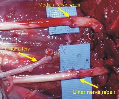
15 Peripheral Nervous System Spinal nervous system is functionally organized on the basis of what is called the reflex arc: 1. A sense organ: (ear-sound, eye-light, skin-temperature) 2. A sensory nerve: (transmit information to the CNS) 3. The CNS: serves as a central integrating station 4. Motor nerve: communication link between CNS and peripheral muscle 5. Effector organ: skeletal muscle fibers 15

16 Example of reflex arc Example of reflex arc 16
17 (Feedback) Schematic diagram of a muscle-length control system for a peripheral muscle (biceps) (a) Anatomical diagram of limb system, showing interconnections. (b) Block diagram of control system. 17
 release neurotransmitter substance Acetylcholine (Ach) Time delay due to junction is 0.")
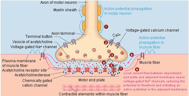
18 Junctional Transmission Synapses: intercommunicating links between neurons Neuromuscular junctions: communicating links between neurons and muscle fibers at end-plate region. Neuromuscular junction (20nm thickness) release neurotransmitter substance Acetylcholine (Ach) Time delay due to junction is 0.5 to 1 msec Excitation-contraction time delay due to muscle contraction Muscle Neuron end-plate region At high stimulation rates, the mechanical response fuse into one continuous contraction called a tetanus (mechanical response summates). 18
19 Neuromuscular junction 19
20 Electroneurogram (ENG) Recording the field potential of an excited nerve. Neural field potential is generated by - Sensory component - Motor component Parameters for diagnosing peripheral nerve disorder - Conduction velocity - Latency - Characteristic of field potentials evoked in muscle supplied by the stimulated nerve (temporal dispersion) Amplitude of field potentials of nerve fibers < extracellular potentials from muscle fibers. 20
21 1 mv Conduction Velocity of a Nerve V (t) S 1 S D R Muscle Reference V (t) V (t) S 2 S 1 L 2 t D Velocity = u = L 1 - L 2 L 1 2 ms Figure 4.7 Measurement of neural conduction velocity via measurement of latency of evoked electrical response in muscle. The nerve was stimulated at two different sites a known distance D apart. 21
22 Field Potential of Sensory Nerves Extracellular field response from the sensory nerves of the median or ulnar nerves To excite the large, rapidly conducting sensory nerve fibers but not small pain fibers or surrounding muscle, apply brief, intense stimulus ( square pulse with amplitude 100-V and duration msec). To prevent artifact signal from muscle movement position the limb in a comfortable posture. Figure 4.8 Sensory nerve action potentials evoked from median nerve of a healthy subject at elbow and wrist after stimulation of index finger with ring electrodes. The potential at the wrist is triphasic and of much larger magnitude than the delayed potential recorded at the elbow. Considering the median nerve to be of the same size and shape at the elbow as at the wrist, we find that the difference in magnitude and waveshape of the potentials is due to the size of the volume conductor at each location and the radial distance of the measurement point from the neural source. 22
23 23
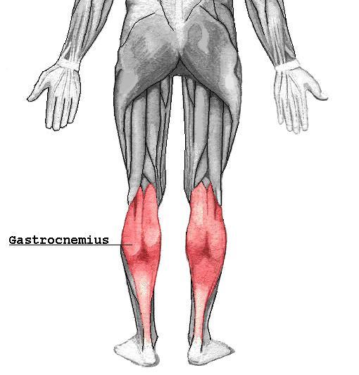
24 Reflexly Evoked Field Potentials Some times when a peripheral nerve is stimulated, a two evoked potentials are recorded in the muscle the nerve supplies. The time difference between the two potentials determined by the distance between the stimulus and the muscle. Stimulated nerve: posterior tibial nerve Muscle: gastrocnemius 24
25 25
26 Reflexly Evoked Field Potentials Medium intensity stimulus stimulate smaller motor fibers in addition to the large sensory fibers. Motor fibers produce a direct muscle response the M wave. Low intensity stimulus stimulate only the large sensory fibers that conduct toward the CNS. No M wave With strong stimuli, the excited motor fibers are in their refractory period so only the M wave is produced. Figure 4.9 The H reflex The four traces show potentials evoked by stimulation of the medial popliteal nerve with pulses of increasing magnitude (the stimulus artifact increases with stimulus magnitude). The later potential or H wave is a low-threshold response, maximally evoked by a stimulus too weak to evoke the muscular response (M wave). As the M wave increases in magnitude, the H wave diminishes. 26

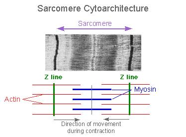
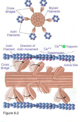
27 Electromyogram (EMG) Skeletal muscle is organized functionally on the basis of the single motor unit (SMU). SMU is the smallest unit that can be activated by a volitional effort where all muscle fibers are activated synchronously. SMU may contain 10 to 2000 muscle fibers, depending on the location of the muscle. Factors for muscle varying strength 1. Number of muscle fibers contracting within a muscle 2. Tension developed by each contracting fiber 27
")
28 Muscle Fiber (Cell) 28
29 Figure 4.10 Diagram of a single motor unit (SMU), which consists of a single motoneuron and the group of skeletal muscle fibers that it innervates. Length transducers [muscle spindles, Figure 4.6(a)] in the muscle activate sensory nerve fibers whose cell bodies are located in the dorsal root ganglion. These bipolar neurons send axonal projections to the spinal cord that divide into a descending and an ascending branch. The descending branch enters into a simple reflex arc with the motor neuron, while the ascending branch conveys information regarding current muscle length to higher centers in the CNS via ascending nerve fiber tracts in the 29 spinal cord and brain stem. These ascending pathways are discussed in Section 4.8.
30 Electromyogram (EMG) Field potential of the active fibers of an SMU 1- triphasic form 2- duration 3-15 msec 3- discharge rate varies from 6 to 30 per second 4- Amplitude range from 20 to 2000 mv Surface electrode record field potential of surface muscles and over a wide area. Monopolar and bipolar insertion-type needle electrode can be used to record SMU field potentials at different locations. The shape of SMU potential is considerably modified by disease such as partial denervation. 30
31 Figure 4.11 Motor unit action potentials from normal dorsal interosseus muscle during progressively more powerful contractions. In the interference pattern (c ), individual units can no longer be clearly distinguished. (d) Interference pattern during very strong muscular contraction. Time scale is 10 ms per dot. 31
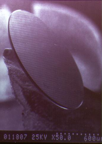
32 Electroretinogram (ERG) ERG is a recording of the temporal sequence of changes in potential in the retina when stimulated with a brief flash of light. Aqueous humor Glaucoma High pressure A transparent contact lens contains one electrode and the reference electrode can be placed on the right temple. 32
 Ag/AgCl")

33 Electroretinogram (ERG) Ag/AgCl electrode impeded in a special contact lens. 33


34 Source of Retinal Potential There are more photoreceptors than ganglion cells so there is a convergence pattern. Many photoreceptors terminate into one bipolar cell and many bipolar cells terminate into one ganglion cell. The convergence rate is greater at peripheral parts of the retina than at the fovea. Rod (10 million) is for vision in dim light and cone (3 million) is for color vision in brighter light. 34
.")
35 Electroretinogram (ERG) The a-wave, sometimes called the "late receptor potential," reflects the general physiological health of the photoreceptors in the outer retina. In contrast, the b- wave reflects the health of the inner layers of the retina, including the ON bipolar cells and the Muller cells (Miller and Dowling, 1970). Two other waveforms that are sometimes recorded in the clinic are the c-wave originating in the pigment epithelium (Marmor and Hock, 1982) and the d-wave indicating activity of the OFF bipolar cells (see Figure 3). 35
36 <> 36
37 Electro-Oculogram (EOG) EOG is the recording of the corneal-retinal potential to determine the eye movement. By placing two electrodes to the left and the right of the eye or above and below the eye one can measure the potential between the two electrode to determine the horizontal or vertical movement of the eye. The potential is zero when the gaze is straight ahead. Applications 1- Sleep and dream research, 2- Evaluating reading ability and visual fatigue. 37

38 Electro-Oculogram (EOG) 38

39 Bionic Eyes 39
 flows from the lung into the left atrium and then to the left ventricle. The left ventricle pump the blood to the rest of the body.")
40 Electrocardiogram (ECG) Blood (poor with oxygen) flows from the body to the right atrium and then to the right ventricle. The right ventricle pump the blood to the lung. Blood (rich with oxygen) flows from the lung into the left atrium and then to the left ventricle. The left ventricle pump the blood to the rest of the body. Diastole: is the resting or filling phase (atria chamber) of the heart cycle. Systole: is the contractile or pumping phase (ventricle chamber) of the heart cycle. The electrical events is intrinsic to the heart itself. See website below for the animation of the heart. rts/html/crm/heart/index.html 40
 node, located at the junction of the superior vena cava and the right atrium.")
41 Electrocardiogram (ECG) Distribution of specialized conductive tissues in the atria and ventricles, showing the impulse-forming and conduction system of the heart. The rhythmic cardiac impulse originates in pacemaking cells in the sinoatrial (SA) node, located at the junction of the superior vena cava and the right atrium. Note the three specialized pathways (anterior, middle, and posterior internodal tracts) between the SA and atrioventricular (AV) nodes. Bachmann's bundle (interatrial tract) comes off the anterior internodal tract leading to the left atrium. The impulse passes from the SA node in an organized manner through specialized conducting tracts in the atria to activate first the right and then the left atrium. Passage of the impulse is delayed at the AV node before it continues into the bundle of His, the right bundle branch, the common left bundle branch, the anterior and posterior divisions of the left bundle branch, and the Purkinje network. The right bundle branch runs along the right side of the interventricular septum to the apex of the right ventricle before it gives off significant branches. The left common bundle crosses to the left side of the septum and splits into the anterior division (which is thin and long and goes under the aortic valve in the outflow tract to the anterolateral papillary muscle) and the posterior division (which is wide and short and goes to 41 the posterior papillary muscle lying in the inflow tract).
 contracting (depol.")
42 SA node activates first the right and then the left atrium. AV node delays a signal coming from the SA node before it distribute it to the Bundle of His. Bundle of His and Purkinie fibers activate the right and left ventricles A typical QRS amplitude is 1-3 mv The P-wave shows the heart's upper chambers (atria) contracting (depol.) The QRS complex shows the heart's lower chambers (ventricles) contracting The T-wave shows the heart's lower chambers (ventricles) relaxing (repol.) The U-wave believed to be due repolarization of ventricular papillary muscles. P-R interval is caused by delay in the AV node S-T segment is related to the average duration of the plateau regions of the 42 individual ventricular cells.

43 Steps of action potential of the ventricular cell -Prior to excitation the resting potential is -90 mv -Rapid Depolarization at a rate 150 V/s -Initial rapid repolarization that leads to a fixed depolarization level for 200 t0 300 msec -Final repolarization phase that restore membrane potential to the resting level for the remainder of the cardiac cycle Myofibrils Centroid Nuclei The cellular architecture of myocardial fibers. 43

44 Isochronous lines of ventricular activation of the human heart Note the nearly closed activation surface at 30 ms into the QRS complex. 44
45 The electrocardiography problem Points A and B are arbitrary observation points on the torso, R AB is the resistance between them, and R T1, R T2 are lumped thoracic medium resistances. The bipolar ECG scalar lead voltage is A - B, where these voltages are both measured with respect to an indifferent reference potential. 45
46 Heart Block (dysfunctional His Bundle) Figure 4.17 Atrioventricular 60 to 70 bps block (a) Complete heart block. Cells in the AV node are dead 30 to 45 bps and activity cannot pass from atria to ventricles. Atria and ventricles beat independently, ventricles being driven by an ectopic (other-than-normal) pacemaker. (B) AV block wherein the node is diseased (examples include rheumatic heart disease and viral infections of the heart). Although each wave from the atria reaches the ventricles, the AV nodal delay is greatly increased. This is first-degree heart block. - When one branch of the bundle of His is interrupted, then the QRS 46 complexes are prolonged while the heart rate is normal.
47 Arrhythmias A portion of the myocardium sometimes becomes irritable and discharge independently. Figure 4.18 Normal ECG followed by an ectopic beat An irritable focus, or ectopic pacemaker, within the ventricle or specialized conduction system may discharge, producing an extra beat, or extrasystole, that interrupts the normal rhythm. This extrasystole is also referred to as a premature ventricular contraction (PVC). 47
48 Figure 4.19 (a) Paroxysmal tachycardia. An ectopic focus may repetitively discharge at a rapid regular rate for minutes, hours, or even days. (B) Atrial flutter. The atria begin a very rapid, perfectly regular "flapping" movement, beating at rates of 200 to 300 beats/min. 48
49 Figure 4.20 (a) Atrial fibrillation. The atria stop their regular beat and begin a feeble, uncoordinated twitching. Concomitantly, low-amplitude, irregular waves appear in the ECG, as shown. This type of recording can be clearly distinguished from the very regular ECG waveform containing atrial flutter. (b) Ventricular fibrillation. Mechanically the ventricles twitch in a feeble, uncoordinated fashion with no blood being pumped from the heart. The ECG is likewise very uncoordinated, as shown 49
50 Alteration of Potential Waveforms in Ischemia Figure 4.21 (a) Action potentials recorded from normal (solid lines) and ischemic (dashed lines) myocardium in a dog. Control is before coronary occlusion. (b) During the control period prior to coronary occlusion, there is no ECG S-T segment shift; after ischemia, there is such a shift. 50
51 Electroencephalogram (EEG) EEG is a superposition of the volume-conductor fields produced by a variety of active neuronal current generators. The three type of electrodes to make the measurements are scalp, cortical, and depth. Superior Topics in this section -Gross anatomy and function of the brain -Ultrastructure of the cerebral cortex Anterior -The potential fields of single neuron -Typical clinical EEG waveform -Abnormal EEG waveform Diencephalon Cerebrum Posterior Midbrain The three main parts of the brain: -Cerebrum -Conscious functions -Brainstem -primitive functions such as controlling heart beat -Integration center for motor reflexes -Thalamus is integration center for sensory system (a) Pons Ventral Medulla oblongata -Cerebellum (balance and voluntary muscle movement) 51 Inferior Caudal Cerebellum
52 The cerebrum, showing the four lobes (frontal, parietal, temporal, and occipital), the lateral and longitudinal fissures, and the central sulcus. The cortex receives sensory information from skin, eyes, ears, and other receptors. This information is compared with previous experience and produces movements in response to these stimuli. SER: somatosensory evoked response AER: auditory evoked response VER: visual evoked response 52
53 The outer layer ( mm) of the cerebrum is called cerebral cortex and consist of a dense collection of nerve cells that appear gray in color (gray matter). The deeper layer consists of axons (or white matter) and collection of cell body. 53
54 Neuron Cell in the Cortex Excitatory synaptic input EEG wave activity Two type of cells in the cortex -Pyramidal cell -Nonpyramidal cell - small cell body - Dendrites spring in all direction - Axons most of the times don t leave the cortex Cell body (soma) - Lines of current flow Apical dendritic tree Electrogenesis of cortical field potentials for a net excitatory input to the apical dendritic tree of a typical pyramidal cell. For the case of a net inhibitory input, polarity is reversed and the apical region becomes a source (+). Current flow to and from active fluctuating synaptic knobs on the dendrites produces wave-like activity. + Basilar dendrites Axon 54
55 Bioelectric Potential From the Brain Conducted action potentials in axons contribute little to surface cortical records, because they usually occur asynchronously in time and at different spatial directions. Pyramid cells of the cerebral cortex are oriented vertically, with their long apical dendrites running parallel to one another. So, the surface records obtained signal principally the net effect of local postsynaptic potentials of cortical cells. Nonpyramidal cells in the neocortex are unlikely to contribute substantially to surface records because their dendritic trees are radially arranged around their cells, so the current sum to zero when viewed by electrode at a distance. When the sum of dendritic activity is negative relative to the cell, the cell is depolarized and quite excitable. When it is positive, the cell is hyperpolarized and less excitable. 55
56 Bioelectric Potential From the Brain Wave group of the normal cortex -Alpha wave - 8 to 13 Hz, mv, - Recorded mainly at the occipital region -disappear when subject is sleep, change when subject change focus, see Fig. 4.27b -Beta wave (I and II) - 14 to 30Hz, - during mental activity f=50hz, beta I disappear during brain activity while beta II intensified. - Recorded mainly at the parietal and frontal regions -Theta wave 4 to 7 Hz, appear during emotional stress such as disappointment and frustration Recorded at the parietal and temporal regions 56
57 Bioelectric Potential From the Brain -Delta wave -Below 3.5 Hz, occur in deep sleep, occur independent of activity - Occur solely within the cortex, independent of activities in lower regions of the brain. -Synchronization is the underline process that bring a group of neurons into unified action. Synaptic interconnection and extracellular field interaction cause Synchronization. - Although various regions of the cortex capable of exhibiting rhythmic activity they require trigger inputs to excite rhythmicity. The reticular activation system (RAS) provide this pacemaker function. 57
58 EEG Waves Fig 4.27 (a) Different types of normal EEG waves. (b) Replacement of alpha rhythm by an asynchronous discharge when patient opens eyes. (c) Representative abnormal EEG waveforms in different types of epilepsy. 58

59 International Federation System Type of electrode connections 1- Between each member of a pair (bipolar) 2- Between one monopolar lead and a distant reference 3- Between one monopolar lead and the average of all. 59
60 EEG Waves During Sleep The electroencephalographic changes that occur as a human subject goes to sleep The calibration marks on the right represent 50 mv. 60
 The Abnormal EEG EEG is used to diagnose different type of epilepsy and in the location of the focus in the brain causing the epilepsy.")
61 Two type of epilepsy 1- Generalized epilepsy a- Grand mal b- petit mal (myoclonic form and absence form) 2- Partial epilepsy a- Jacksonian epilepsy b- Psychomotor seizure (amnesia, abnormal rage, sudden anxiety or fear, incoherent speech) The Abnormal EEG EEG is used to diagnose different type of epilepsy and in the location of the focus in the brain causing the epilepsy. Causes of epilepsy could be intrinsic hyperexcitability of the neurons that make up the reticular activation system (RAS) or by abnormality of the local neural pathways of this system. 61
Bioelectric Signals. MYcsvtu Notes.
 Bioelectric Signals Bioelectrical potential is a result of electrochemical activity across the membrane of the cell. Bioelectrical signals are generated by excitable cells such as nervous, muscular, and
Typical types of bioelectric signals
 Bioelectric Signals Bioelectrical potential is a result of electrochemical activity across the membrane of the cell. Bioelectrical signals are generated by excitable cells such as nervous, muscular, and
PD233: Design of Biomedical Devices and Systems
 PD233: Design of Biomedical Devices and Systems (Lecture-7 Biopotentials- 2) Dr. Manish Arora CPDM, IISc Course Website: http://cpdm.iisc.ac.in/utsaah/courses/ Electromyogram (EMG) Skeletal muscles are
Neural Basis of Motor Control
 Neural Basis of Motor Control Central Nervous System Skeletal muscles are controlled by the CNS which consists of the brain and spinal cord. Determines which muscles will contract When How fast To what
Chapter 9. Nervous System
 Chapter 9 Nervous System Central Nervous System (CNS) vs. Peripheral Nervous System(PNS) CNS Brain Spinal cord PNS Peripheral nerves connecting CNS to the body Cranial nerves Spinal nerves Neurons transmit
Chapter 7. The Nervous System: Structure and Control of Movement
 Chapter 7 The Nervous System: Structure and Control of Movement Objectives Discuss the general organization of the nervous system Describe the structure & function of a nerve Draw and label the pathways
Chapter 7. Objectives
 Chapter 7 The Nervous System: Structure and Control of Movement Objectives Discuss the general organization of the nervous system Describe the structure & function of a nerve Draw and label the pathways
EE 791 Lecture 2 Jan 19, 2015
 EE 791 Lecture 2 Jan 19, 2015 Action Potential Conduction And Neural Organization EE 791-Lecture 2 1 Core-conductor model: In the core-conductor model we approximate an axon or a segment of a dendrite
Dendrites Receive impulse from the axon of other neurons through synaptic connection. Conduct impulse towards the cell body Axon
 Dendrites Receive impulse from the axon of other neurons through synaptic connection. Conduct impulse towards the cell body Axon Page 22 of 237 Conduct impulses away from cell body Impulses arise from
Chapter 20 (2) The Heart
 The Heart") Chapter 20 (2) The Heart ----------------------------------------------------------------------------------------------------------------------------------------- Describe the component and function of
Chapter 20 (2) The Heart ----------------------------------------------------------------------------------------------------------------------------------------- Describe the component and function of
THE CARDIOVASCULAR SYSTEM. Heart 2
 THE CARDIOVASCULAR SYSTEM Heart 2 PROPERTIES OF CARDIAC MUSCLE Cardiac muscle Striated Short Wide Branched Interconnected Skeletal muscle Striated Long Narrow Cylindrical PROPERTIES OF CARDIAC MUSCLE Intercalated
CARDIOVASCULAR SYSTEM
 CARDIOVASCULAR SYSTEM Overview Heart and Vessels 2 Major Divisions Pulmonary Circuit Systemic Circuit Closed and Continuous Loop Location Aorta Superior vena cava Right lung Pulmonary trunk Base of heart
NEURONS Chapter Neurons: specialized cells of the nervous system 2. Nerves: bundles of neuron axons 3. Nervous systems
 NEURONS Chapter 12 Figure 12.1 Neuronal and hormonal signaling both convey information over long distances 1. Nervous system A. nervous tissue B. conducts electrical impulses C. rapid communication 2.
ELECTROCARDIOGRAPHY (ECG)
") ELECTROCARDIOGRAPHY (ECG) The heart is a muscular organ, which pumps blood through the blood vessels of the circulatory system. Blood provides the body with oxygen and nutrients, as well as assists in
ELECTROCARDIOGRAPHY (ECG) The heart is a muscular organ, which pumps blood through the blood vessels of the circulatory system. Blood provides the body with oxygen and nutrients, as well as assists in
Chapter 7 Nerve Cells and Electrical Signaling
 Chapter 7 Nerve Cells and Electrical Signaling 7.1. Overview of the Nervous System (Figure 7.1) 7.2. Cells of the Nervous System o Neurons are excitable cells which can generate action potentials o 90%
Axon Nerve impulse. Axoplasm Receptor. Axomembrane Stimuli. Schwann cell Effector. Myelin Cell body
 Nervous System Review 1. Explain a reflex arc. 2. Know the structure, function and location of a sensory neuron, interneuron, and motor neuron 3. What is (a) Neuron Axon Nerve impulse Axoplasm Receptor
Module H NERVOUS SYSTEM
 Module H NERVOUS SYSTEM Topic from General functions of the nervous system Organization of the nervous system from both anatomical & functional perspectives Gross & microscopic anatomy of nervous tissue
Cardiovascular System
 Cardiovascular System The Heart Cardiovascular System The Heart Overview What does the heart do? By timed muscular contractions creates pressure gradients blood moves then from high pressure to low pressure
The cardiovascular system is composed of the heart and blood vessels that carry blood to and from the body s organs. There are 2 major circuits:
 1 The cardiovascular system is composed of the heart and blood vessels that carry blood to and from the body s organs. There are 2 major circuits: pulmonary and systemic. The pulmonary goes out to the
Chapter 17 Nervous System
 Chapter 17 Nervous System 1 The Nervous System Two Anatomical Divisions Central Nervous System (CNS) Brain and Spinal Cord Peripheral Nervous System (PNS) Two Types of Cells Neurons Transmit nerve impulses
STRUCTURAL ELEMENTS OF THE NERVOUS SYSTEM
 STRUCTURAL ELEMENTS OF THE NERVOUS SYSTEM STRUCTURE AND MAINTENANCE OF NEURONS (a) (b) Dendrites Cell body Initial segment collateral terminals (a) Diagrammatic representation of a neuron. The break in
Neurons, Synapses and Signaling. Chapter 48
 Neurons, Synapses and Signaling Chapter 48 Warm Up Exercise What types of cells can receive a nerve signal? Nervous Organization Neurons- nerve cells. Brain- organized into clusters of neurons, called
Cardiac Cycle. Each heartbeat is called a cardiac cycle. First the two atria contract at the same time.
 The Heartbeat Cardiac Cycle Each heartbeat is called a cardiac cycle. First the two atria contract at the same time. Next the two ventricles contract at the same time. Then all the chambers relax. http://www.youtube.com/watch?v=frd3k6lkhws
: Biomedical Signal Processing
 : Biomedical Signal Processing 0. Introduction: Biomedical signal processing refers to the applications of signal processing methods, such as Fourier transform, spectral estimation and wavelet transform,
What is Anatomy and Physiology?
 Introduction BI 212 BI 213 BI 211 Ecosystems Organs / organ systems Cells Organelles Communities Tissues Molecules Populations Organisms Campbell et al. Figure 1.4 Introduction What is Anatomy and Physiology?
Neural Basis of Motor Control. Chapter 4
 Neural Basis of Motor Control Chapter 4 Neurological Perspective A basic understanding of the physiology underlying the control of voluntary movement establishes a more comprehensive appreciation and awareness
11/10/2014. Muscular pump Two atria Two ventricles. In mediastinum of thoracic cavity 2/3 of heart's mass lies left of midline of sternum
 It beats over 100,000 times a day to pump over 1,800 gallons of blood per day through over 60,000 miles of blood vessels. During the average lifetime, the heart pumps nearly 3 billion times, delivering
CHAPTER 48: NERVOUS SYSTEMS
 CHAPTER 48: NERVOUS SYSTEMS Name I. AN OVERVIEW OF NERVOUS SYSTEMS A. Nervous systems perform the three overlapping functions of sensory input, integration, and motor output B. Networks of neurons with
Nervous System Dr. Naim Kittana Department of Biomedical Sciences Faculty of Medicine & Health Sciences An-Najah National University
 Nervous System Department of Biomedical Sciences Faculty of Medicine & Health Sciences An-Najah National University Declaration The content and the figures of this seminar were directly adopted from the
If I Only Had a Brain
 If I Only Had a Brain A Heart. (The Nerve!) Regions of the Brain Cerebral hemisphere Diencephalon Cerebellum (b) Adult brain Brain stem Regions of the Brain: Cerebrum Precentral gyrus Frontal lobe Central
The Heart. Size, Form, and Location of the Heart. 1. Blunt, rounded point; most inferior part of the heart.
 12 The Heart FOCUS: The heart is composed of cardiac muscle cells, which are elongated, branching cells that appear striated. Cardiac muscle cells behave as a single electrical unit, and the highly coordinated
Primary Functions. Monitor changes. Integrate input. Initiate a response. External / internal. Process, interpret, make decisions, store information
 NERVOUS SYSTEM Monitor changes External / internal Integrate input Primary Functions Process, interpret, make decisions, store information Initiate a response E.g., movement, hormone release, stimulate/inhibit
4. The two inferior chambers of the heart are known as the atria. the superior and inferior vena cava, which empty into the left atrium.
 Answer each statement true or false. If the statement is false, change the underlined word to make it true. 1. The heart is located approximately between the second and fifth ribs and posterior to the
Neurons Chapter 7 2/19/2016. Learning Objectives. Cells of the Nervous System. Cells of the Nervous System. Cells of the Nervous System
 Learning Objectives Neurons Chapter 7 Identify and describe the functions of the two main divisions of the nervous system. Differentiate between a neuron and neuroglial cells in terms of structure and
STRUCTURAL ORGANIZATION OF THE NERVOUS SYSTEM
 STRUCTURAL ORGANIZATION OF THE NERVOUS SYSTEM STRUCTURAL ORGANIZATION OF THE BRAIN The central nervous system (CNS), consisting of the brain and spinal cord, receives input from sensory neurons and directs
Chapter 11: Nervous System and Nervous Tissue
 Chapter 11: Nervous System and Nervous Tissue I. Functions and divisions of the nervous system A. Sensory input: monitor changes in internal and external environment B. Integrations: make decisions about
From last week: The body is a complex electrical machine. Basic Electrophysiology, the Electroretinogram ( ERG ) and the Electrooculogram ( EOG )
 and the Electrooculogram ( EOG )") From last week: Differential Amplification This diagram shows a low frequency signal from the patient that differs between the two inputs and is therefore amplified, with an interfering high frequency
From last week: Differential Amplification This diagram shows a low frequency signal from the patient that differs between the two inputs and is therefore amplified, with an interfering high frequency
Omar Sami. Muhammad Abid. Muhammad khatatbeh
 10 Omar Sami Muhammad Abid Muhammad khatatbeh Let s shock the world In this lecture we are going to cover topics said in previous lectures and then start with the nerve cells (neurons) and the synapses
211MDS Pain theories
 211MDS Pain theories Definition In 1986, the International Association for the Study of Pain (IASP) defined pain as a sensory and emotional experience associated with real or potential injuries, or described
10. Thick deposits of lipids on the walls of blood vessels, called, can lead to serious circulatory issues. A. aneurysm B. atherosclerosis C.
 Heart Student: 1. carry blood away from the heart. A. Arteries B. Veins C. Capillaries 2. What is the leading cause of heart attack and stroke in North America? A. alcohol B. smoking C. arteriosclerosis
Thursday, January 22, Nerve impulse
 Nerve impulse Transmembrane Potential caused by ions moving through cell membrane at different rates Two main ions of concern Na + - Sodium K + - potassium Cell membrane not freely permeable therefore
Version A. AP* Biology: Nervous System. Questions 1 and 2. Name: Period
 Name: Period Version A AP* Biology: Nervous System Directions: Each of the questions or incomplete statements below is followed by four suggested answers or completions. Select the one that is best in
10.1: Introduction. Cell types in neural tissue: Neurons Neuroglial cells (also known as neuroglia, glia, and glial cells) Dendrites.
 Dendrites.") 10.1: Introduction Copyright The McGraw-Hill Companies, Inc. Permission required for reproduction or display. Cell types in neural tissue: Neurons Neuroglial cells (also known as neuroglia, glia, and glial
10.1: Introduction Copyright The McGraw-Hill Companies, Inc. Permission required for reproduction or display. Cell types in neural tissue: Neurons Neuroglial cells (also known as neuroglia, glia, and glial
CASE 10. What would the ST segment of this ECG look like? On which leads would you see this ST segment change? What does the T wave represent?
 CASE 10 A 57-year-old man presents to the emergency center with complaints of chest pain with radiation to the left arm and jaw. He reports feeling anxious, diaphoretic, and short of breath. His past history
Nervous System C H A P T E R 2
 Nervous System C H A P T E R 2 Input Output Neuron 3 Nerve cell Allows information to travel throughout the body to various destinations Receptive Segment Cell Body Dendrites: receive message Myelin sheath
Introduction to Neurobiology
 Biology 240 General Zoology Introduction to Neurobiology Nervous System functions: communication of information via nerve signals integration and processing of information control of physiological and
Neurophysiology scripts. Slide 2
 Neurophysiology scripts Slide 2 Nervous system and Endocrine system both maintain homeostasis in the body. Nervous system by nerve impulse and Endocrine system by hormones. Since the nerve impulse is an
ANATOMY AND PHYSIOLOGY OF NEURONS. AP Biology Chapter 48
 ANATOMY AND PHYSIOLOGY OF NEURONS AP Biology Chapter 48 Objectives Describe the different types of neurons Describe the structure and function of dendrites, axons, a synapse, types of ion channels, and
The Nervous System. Nervous System Functions 1. gather sensory input 2. integration- process and interpret sensory input 3. cause motor output
 The Nervous System Nervous System Functions 1. gather sensory input 2. integration- process and interpret sensory input 3. cause motor output The Nervous System 2 Parts of the Nervous System 1. central
Organization of the nervous system. [See Fig. 48.1]
![Organization of the nervous system. [See Fig. 48.1]](/thumbs/90/103926552.jpg "Organization of the nervous system. [See Fig. 48.1]") Nervous System [Note: This is the text version of this lecture file. To make the lecture notes downloadable over a slow connection (e.g. modem) the figures have been replaced with figure numbers as found
Nervous System [Note: This is the text version of this lecture file. To make the lecture notes downloadable over a slow connection (e.g. modem) the figures have been replaced with figure numbers as found
Homework Week 2. PreLab 2 HW #2 Synapses (Page 1 in the HW Section)
") Homework Week 2 Due in Lab PreLab 2 HW #2 Synapses (Page 1 in the HW Section) Reminders No class next Monday Quiz 1 is @ 5:30pm on Tuesday, 1/22/13 Study guide posted under Study Aids section of website
Homework Week 2 Due in Lab PreLab 2 HW #2 Synapses (Page 1 in the HW Section) Reminders No class next Monday Quiz 1 is @ 5:30pm on Tuesday, 1/22/13 Study guide posted under Study Aids section of website
Heart. Heart 2-Tunica media: middle layer (media ='middle') muscle fibers (smooth or cardiac).
 muscle fibers (smooth or cardiac).") t. innermost lumenal General Circulatory system heart and blood vessels walls have 3 layers (inside to outside) 1-Tunica interna: aka tunica intima layer--lumenal layer epithelium--endothelium simple squamous
t. innermost lumenal General Circulatory system heart and blood vessels walls have 3 layers (inside to outside) 1-Tunica interna: aka tunica intima layer--lumenal layer epithelium--endothelium simple squamous
Nervous System. Unit 6.6 (6 th Edition) Chapter 7.6 (7 th Edition)
 Chapter 7.6 (7 th Edition)") Nervous System Unit 6.6 (6 th Edition) Chapter 7.6 (7 th Edition) 1 Learning Objectives Identify the main parts (anatomy) of a neuron. Identify the 2 divisions of nervous system. Classify the major types
Nervous System Unit 6.6 (6 th Edition) Chapter 7.6 (7 th Edition) 1 Learning Objectives Identify the main parts (anatomy) of a neuron. Identify the 2 divisions of nervous system. Classify the major types
Chapter 11: Functional Organization of Nervous Tissue
 Chapter 11: Functional Organization of Nervous Tissue I. Functions of the Nervous System A. List and describe the five major nervous system functions: 1. 2. 3. 4. 5. II. Divisions of the Nervous System
AnS SI 214 Practice Exam 2 Nervous, Muscle, Cardiovascular
 AnS SI 214 Practice Exam 2 Nervous, Muscle, Cardiovascular Select the best answer choice in the questions below. 1) On the electrocardiogram, repolarization of the atria is represented by the: A) P wave
10/23/2017. Muscular pump Two atria Two ventricles. In mediastinum of thoracic cavity 2/3 of heart's mass lies left of midline of sternum
 It beats over 100,000 times a day to pump over 1,800 gallons of blood per day through over 60,000 miles of blood vessels. During the average lifetime, the heart pumps nearly 3 billion times, delivering
The Nervous System II Neurons
 The Nervous System II Neurons Review Nervous System What is it? The system that receives, processes, stores and transmits information that comes from various parts of the body and the external world. Composed
Nervous System. Master controlling and communicating system of the body. Secrete chemicals called neurotransmitters
 Nervous System Master controlling and communicating system of the body Interacts with the endocrine system to control and coordinate the body s responses to changes in its environment, as well as growth,
Nerve. (2) Duration of the stimulus A certain period can give response. The Strength - Duration Curve
 Duration of the stimulus A certain period can give response. The Strength - Duration Curve") Nerve Neuron (nerve cell) is the structural unit of nervous system. Nerve is formed of large numbers of nerve fibers. Types of nerve fibers Myelinated nerve fibers Covered by myelin sheath interrupted
Nerve Neuron (nerve cell) is the structural unit of nervous system. Nerve is formed of large numbers of nerve fibers. Types of nerve fibers Myelinated nerve fibers Covered by myelin sheath interrupted
Chapter 18 - Heart. I. Heart Anatomy: size of your fist; located in mediastinum (medial cavity)
") Chapter 18 - Heart I. Heart Anatomy: size of your fist; located in mediastinum (medial cavity) A. Coverings: heart enclosed in double walled sac called the pericardium 1. Fibrous pericardium: dense connective
Chapter 18 - Heart I. Heart Anatomy: size of your fist; located in mediastinum (medial cavity) A. Coverings: heart enclosed in double walled sac called the pericardium 1. Fibrous pericardium: dense connective
Brain and behaviour (Wk 6 + 7)
") Brain and behaviour (Wk 6 + 7) What is a neuron? What is the cell body? What is the axon? The basic building block of the nervous system, the individual nerve cell that receives, processes and transmits
Brain and behaviour (Wk 6 + 7) What is a neuron? What is the cell body? What is the axon? The basic building block of the nervous system, the individual nerve cell that receives, processes and transmits
Chapter 2: Cellular Mechanisms and Cognition
 Chapter 2: Cellular Mechanisms and Cognition MULTIPLE CHOICE 1. Two principles about neurons were defined by Ramón y Cajal. The principle of connectional specificity states that, whereas the principle
Ameen Alsaras. Ameen Alsaras. Mohd.Khatatbeh
 9 Ameen Alsaras Ameen Alsaras Mohd.Khatatbeh Nerve Cells (Neurons) *Remember: The neural cell consists of: 1-Cell body 2-Dendrites 3-Axon which ends as axon terminals. The conduction of impulse through
Cardiovascular System Notes: Physiology of the Heart
 Cardiovascular System Notes: Physiology of the Heart Interesting Heart Fact Capillaries are so small it takes ten of them to equal the thickness of a human hair. Review What are the 3 parts of the cardiovascular
Neurons. Pyramidal neurons in mouse cerebral cortex expressing green fluorescent protein. The red staining indicates GABAergic interneurons.
 Neurons Pyramidal neurons in mouse cerebral cortex expressing green fluorescent protein. The red staining indicates GABAergic interneurons. MBL, Woods Hole R Cheung MSc Bioelectronics: PGEE11106 1 Neuron
Chapter 9 Nervous System Test Review
 Chapter 9 Nervous System Test Review Multiple Choice Choose the best answer from the choices given 1. The central nervous system consists of: a) sensory nerves b) nerves that run throughout the body c)
PART I. Disorders of the Heart Rhythm: Basic Principles
 PART I Disorders of the Heart Rhythm: Basic Principles FET01.indd 1 1/11/06 9:53:05 AM FET01.indd 2 1/11/06 9:53:06 AM CHAPTER 1 The Cardiac Electrical System The heart spontaneously generates electrical
Chapter 17. Nervous System Nervous systems receive sensory input, interpret it, and send out appropriate commands. !
 Chapter 17 Sensory receptor Sensory input Integration Nervous System Motor output Brain and spinal cord Effector cells Peripheral nervous system (PNS) Central nervous system (CNS) 28.1 Nervous systems
Warm-up. Warm-up. Warm-up. Chapter 48. Why do animals need a nervous system? 3/9/2012. Nervous System
 Warm-up Objective: Explain how membrane potentials arise from differences in ion concentrations between cells' content and the extracellular fluid. Warm-up: Cells from this structure migrate to other parts
Neural Communication. Central Nervous System Peripheral Nervous System. Communication in the Nervous System. 4 Common Components of a Neuron
 Neural Communication Overview of CNS / PNS Electrical Signaling Chemical Signaling Central Nervous System Peripheral Nervous System Somatic = sensory & motor Autonomic = arousal state Parasympathetic =
Chapter 11 Introduction to the Nervous System and Nervous Tissue Chapter Outline
 Chapter 11 Introduction to the Nervous System and Nervous Tissue Chapter Outline Module 11.1 Overview of the Nervous System (Figures 11.1-11.3) A. The nervous system controls our perception and experience
ACTIVITY2.15 Text:Campbell,v.8,chapter48 DATE HOUR NERVOUS SYSTEMS NEURON
 AP BIOLOGY ACTIVITY2.15 Text:Campbell,v.8,chapter48 NAME DATE HOUR NERVOUS SYSTEMS NEURON SIMPLE REFLEX RESTING POTENTIAL ACTION POTENTIAL ACTION POTENTIAL GRAPH TRANSMISSION ACROSS A SYNAPSE QUESTIONS:
Storage is accomplished through the following mechanisms:
 NROSCI/BIOSC 1070 and MSNBIO 2070 September 13, 2017 Examples of Coordinated Autonomic and Motor Responses and Return to the Cardiovascular System 1) Micturition Micturition, or the process of emptying
Introduction to Physiological Psychology
 Introduction to Physiological Psychology Review Kim Sweeney ksweeney@cogsci.ucsd.edu www.cogsci.ucsd.edu/~ksweeney/psy260.html Today n Discuss Final Paper Proposal (due 3/10) n General Review 1 The article
Lesson 14. The Nervous System. Introduction to Life Processes - SCI 102 1
 Lesson 14 The Nervous System Introduction to Life Processes - SCI 102 1 Structures and Functions of Nerve Cells The nervous system has two principal cell types: Neurons (nerve cells) Glia The functions
Basic Electrophysiology, the Electroretinogram (ERG) and the Electrooculogram (EOG) - Signal origins, recording methods and clinical applications
 and the Electrooculogram (EOG) - Signal origins, recording methods and clinical applications") Basic Electrophysiology, the Electroretinogram (ERG) and the Electrooculogram (EOG) - Signal origins, recording methods and clinical applications The body is a complex machine consisting of the central
Basic Electrophysiology, the Electroretinogram (ERG) and the Electrooculogram (EOG) - Signal origins, recording methods and clinical applications The body is a complex machine consisting of the central
-Ensherah Mokheemer. -Amani Nofal. -Loai Alzghoul
 -1 -Ensherah Mokheemer -Amani Nofal -Loai Alzghoul 1 P a g e Today we will start talking about the physiology of the nervous system and we will mainly focus on the Central Nervous System. Introduction:
Collin County Community College. ! BIOL Anatomy & Physiology! WEEK 5. The Heart
 Collin County Community College! BIOL. 2402 Anatomy & Physiology! WEEK 5 The Heart 1 (1578-1657) A groundbreaking work in the history of medicine, English physician William Harvey s Anatomical Essay on
The Nervous System 12/11/2015
 The Nervous System Biology 12 Unit 3: Homeostasis December 11, 2015 The nervous system is an elaborate communication system that contains more than 100 billion nerve cells in the brain alone There are
Biology 3201 Quiz on Nervous System. Total 33 points
 Biology 3201 Quiz on Nervous System Total 33 points Name: Circle the best response to the following: (33 points) 1. What do we call the long fibre that carries impulses away from the nerve cell body? A.
Chapter 20: Cardiovascular System: The Heart
 Chapter 20: Cardiovascular System: The Heart I. Functions of the Heart A. List and describe the four functions of the heart: 1. 2. 3. 4. II. Size, Shape, and Location of the Heart A. Size and Shape 1.
The Nervous System. B. The Components: 1) Nerve Cells Neurons are the cells of the body and are specialized to carry messages through an process.
 Nerve Cells Neurons are the cells of the body and are specialized to carry messages through an process.") The Nervous System A. The Divisions: 1) The Central Nervous System includes the and. The brain contains billions of nerve cells called, and trillions of support cells called. 2) The Peripheral Nervous
The Nervous System A. The Divisions: 1) The Central Nervous System includes the and. The brain contains billions of nerve cells called, and trillions of support cells called. 2) The Peripheral Nervous
The Heart. Happy Friday! #takeoutyournotes #testnotgradedyet
 The Heart Happy Friday! #takeoutyournotes #testnotgradedyet Introduction Cardiovascular system distributes blood Pump (heart) Distribution areas (capillaries) Heart has 4 compartments 2 receive blood (atria)
3) Most of the organelles in a neuron are located in the A) dendritic region. B) axon hillock. C) axon. D) cell body. E) axon terminals.
 Most of the organelles in a neuron are located in the A) dendritic region. B) axon hillock. C) axon. D) cell body. E) axon terminals.") Chapter 48 Neurons, Synapses, and Signaling Multiple-Choice Questions 1) A simple nervous system A) must include chemical senses, mechanoreception, and vision. B) includes a minimum of 12 ganglia. C) has
Chapter 48 Neurons, Synapses, and Signaling Multiple-Choice Questions 1) A simple nervous system A) must include chemical senses, mechanoreception, and vision. B) includes a minimum of 12 ganglia. C) has
Neurons, Synapses, and Signaling
 Neurons, Synapses, and Signaling The Neuron is the functional unit of the nervous system. Neurons are composed of a cell body, which contains the nucleus and organelles; Dendrites which are extensions
All questions below pertain to mandatory material: all slides, and mandatory homework (if any).
.") ECOL 182 Spring 2008 Dr. Ferriere s lectures Lecture 6: Nervous system and brain Quiz Book reference: LIFE-The Science of Biology, 8 th Edition. http://bcs.whfreeman.com/thelifewire8e/ All questions below
ECOL 182 Spring 2008 Dr. Ferriere s lectures Lecture 6: Nervous system and brain Quiz Book reference: LIFE-The Science of Biology, 8 th Edition. http://bcs.whfreeman.com/thelifewire8e/ All questions below
1. NERVOUS SYSTEM FUNCTIONS OF THE NERVOUS SYSTEM. FUNCTION The major function of the nervous system can be summarized as follows (Figure 1-1).
.") 1. NERVOUS SYSTEM FUNCTION The major function of the nervous system can be summarized as follows (Figure 1-1). Sensory input. Multiple signals from both, internal and external environment are detected
1. NERVOUS SYSTEM FUNCTION The major function of the nervous system can be summarized as follows (Figure 1-1). Sensory input. Multiple signals from both, internal and external environment are detected
Cardiovascular system
 BIO 301 Human Physiology Cardiovascular system The Cardiovascular System: consists of the heart plus all the blood vessels transports blood to all parts of the body in two 'circulations': pulmonary (lungs)
The neurvous system senses, interprets, and responds to changes in the environment. Two types of cells makes this possible:
 NERVOUS SYSTEM The neurvous system senses, interprets, and responds to changes in the environment. Two types of cells makes this possible: the neuron and the supporting cells ("glial cells"). Neuron Neurons
II. Nervous System (NS) Organization: can be organized by location/ structure or by function A. Structural Organization 1. Central N.S.
 Organization: can be organized by location/ structure or by function A. Structural Organization 1. Central N.S.") Nervous System I. Nervous system Functions A. Detect Changes in the environment (stimuli) B. Interpret/evaluate those stimuli C. Initiate responses (trigger muscle contractions or glandular response) II.
Nervous System I. Nervous system Functions A. Detect Changes in the environment (stimuli) B. Interpret/evaluate those stimuli C. Initiate responses (trigger muscle contractions or glandular response) II.
Page 1. Neurons Transmit Signal via Action Potentials: neuron At rest, neurons maintain an electrical difference across
 Chapter 33: The Nervous System and the Senses Neurons: Specialized excitable cells that allow for communication throughout the body via electrical impulses Neuron Anatomy / Function: 1) Dendrites: Receive
Full file at
 MULTIPLE CHOICE. Choose the one alternative that best completes the statement or answers the question. 1) What electrical event must occur for atrial kick to occur? 1) A) Atrial repolarization B) Ventricular
Chapter 13 The Cardiovascular System: Cardiac Function
 Chapter 13 The Cardiovascular System: Cardiac Function Overview of the Cardiovascular System The Path of Blood Flow through the Heart and Vasculature Anatomy of the Heart Electrical Activity of the Heart
Warm-Up. Label the parts of the neuron below.
 Warm-Up Label the parts of the neuron below. A B C D E F G Warm-Up 1. One neuron transmits a nerve impulse at 40 m/s. Another conducts at the rate of 1 m/s. Which neuron has a myelinated axon? 2. List
NEURAL TISSUE (NEUROPHYSIOLOGY) PART I (A): NEURONS & NEUROGLIA
 PART I (A): NEURONS & NEUROGLIA") PART I (A): NEURONS & NEUROGLIA Neural Tissue Contains 2 kinds of cells: neurons: cells that send and receive signals neuroglia (glial cells): cells that support and protect neurons Neuron Types Sensory
PART I (A): NEURONS & NEUROGLIA Neural Tissue Contains 2 kinds of cells: neurons: cells that send and receive signals neuroglia (glial cells): cells that support and protect neurons Neuron Types Sensory
Collin County Community College
 Collin County Community College BIOL. 2402 Anatomy & Physiology WEEK 5 The Heart 1 The Heart Beat and the EKG 2 1 The Heart Beat and the EKG P-wave = Atrial depolarization QRS-wave = Ventricular depolarization
Fig Copyright 2002 Pearson Education, Inc., publishing as Benjamin Cummings
 Fig. 48.1 Fig. 48.2 Axon endings are called synaptic terminals. They contain neurotransmitters which conduct a signal across a synapse. A synapse is the junction between a presynaptic and postsynaptic
Muscle and Muscle Tissue
 Muscle and Muscle Tissue Make up about half of total body mass Exerts force by converting chemical energy, ATP, to mechanical energy Muscle tissue is classified based on Shape Number and position of nuclei
1. 01/20/15 Ch 8: Muscular System /09/15 Ch 9: Nervous System 16
 Table of Contents # Date Title Page # 1. 01/20/15 Ch 8: Muscular System 1 2. 02/09/15 Ch 9: Nervous System 16 i 1 Anatomy and Physiology Sem 2 Ch 9 Nervous System.notebook 02/09/15 Ch. 9 Nervous System
Function of the Nervous System
 Nervous System Function of the Nervous System Receive sensory information, interpret it, and send out appropriate commands to form a response Composed of neurons (functional unit of the nervous system)