The Muscular System. Jim Swan
|
|
|
- Tobias Gallagher
- 6 years ago
- Views:
Transcription

1 The Muscular System Jim Swan 1 These slides are from class presentations, reformatted for static viewing. The content contained in these pages is also in the Class Notes pages in a narrative format. Best screen resolution for viewing is 1024 x 768. To change resolution click on start, then control panel, then display, then settings. If you are viewing this in Adobe Reader version 7 and are connected to the internet you will also be able to access the enriched links to notes and comments, as well as web pages including animations and videos. You will also be able to make your own notes and comments on the pages. Download the free reader from [Adobe.com] 1
2 3 Types of Muscle Tissue Muscle Type Location Characteristics Control Skeletal Attached to the bones for movement Long, cynlindrical cells; multinucleated, striated Voluntary Cardiac Muscle of the Heart Short, branching cells, mononucleated, faintly striated. Forms functional syncytia. Involuntary myogenic Smooth Muscle Single Unit: GI, Respiratory, & Genitourinary tract mucous membranes. Multi-unit: smooth muscle in blood vessel walls. Small oblong cells, mononucleated, also may form a functional syncytium. Involuntary myogenic 2 2

3 Skeletal Muscle nuclei Striations = dark bands Connective endomysium separates cells. Myofibrils fill sarcoplasm 3 The nuclei and other organelles of skeletal muscle cells are found next to the sarcolemms and the majority of the sarcoplasm is filled with the contractile machinery of the cell, the myofibrils. Skeletal muscle cells are derived from individual myocytes which fuse to produce a mature multinucleated muscle fiber. There are few if any of the precursor myocytes found in a mature muscle, and so muscles produce no new cells after maturity. Individual cells respond to training by enlarging and building myofibrils and other components. 3

4 Skeletal Muscle photomicrographs Dark striations = A-bands, the light areas between are the I-bands. Z-line. From one Z line to the next is a sarcomere. Z-line The sarcolemma is the cell membrane 4 4

5 Cardiac Muscle Intercalated disks Faint striations Branching cells connect to form network. mononucleated The action potential travels through all cells connected together in a syncytium causing them to function as a unit. 5 Cardiac muscle cells are much shorter than cells in skeletal muscle and they branch to connect to neighboring cells through specialized membranes called intercalated disks to form a network called a syncytium. 5

6 Smooth Muscle nucleus Spindle -shaped mononucleated smooth muscle cell. 6 Smooth muscle cells connect to form single-unit syncytia similar to cardiac muscle. But impulses and contractions occur much more slowly in smooth than in cardiac muscle. 6

7 Smooth Muscle Arrangement In the intestine smooth muscle forms two distinct layers, one running along, the other running around the organ. Together these layers cause movements which propel the contents. The circular layer runs around the intestine and its contraction causes segmentation The longitudinal layer runs along the 7 intestine; it causes wave-like contractions. 7
8 Types of Smooth Muscle Fibers Single unit smooth muscle cells connected to function as a single unit (syncytium) e.g. in GI tract Multiunit smooth muscle cells grouped into many contractile units controlled by the nervous system. e.g. in blood vessel walls and sphincters in GI tract. 8 8
 endomysium Surrounds cells (fibers) 9 The hierarchy of connective tissues associated with a skeletal muscle provide a continuous connection")
9 Structure of a Skeletal Muscle Tendon attachment Belly contains cells epimysium Fibrous covering Figure 9.1 fascicle { perimysium Surrounds fascicle Cell (fiber) endomysium Surrounds cells (fibers) 9 The hierarchy of connective tissues associated with a skeletal muscle provide a continuous connection between muscle cells and their action on a bone or other attachment. At the same time cells are effectively separated from one another and each is controlled by a separate nerve fiber. 9
10 Functional Characteristics of Skeletal Muscle Excitability (responsiveness) muscles can be stimulated by electrical, chemical, and physical means. Contractility a muscle responds to stimuli by contracting. Elasticity muscles tend to recoil to their resting length. Extensibility muscles can be stretched beyond their resting length
11 Muscle Attachments Tendons attach muscle to bone. Aponeuroses broad, flat, tendinous attachment. Origin more fixed point of attachment. Insertion more movable point of attachment. Muscle action pulls insertion toward the origin. A muscle can only pull, it cannot push
12 Types of Muscle Contractions Agonist the prime mover; the muscle which performs the movement in question. Antagonist the muscle that performs the opposing movement to that of the agonist. Both muscles contract (exert tension) regardless of which is the agonist or antagonist. On-center movement that of the agonist Off-center (eccentric) movement that of the antagonist 12 The antagonist may actually be stretching while it is generating tension (contracting). 12
13 Knee Extension Agonist: the rectus femoris (quadriceps femoris) Antagonist: the biceps femoris (hamstrings) 13 13
 14")
14 Knee Flexion Agonist: the biceps femoris (hamstrings) Antagonist: the rectus femoris (quadriceps femoris) 14 14
15 Synergists muscles which work together to perform a movement; often differs from the movement either performs when working alone. Fixators muscles which work to keep a part from moving; stabilizers, neutralizers. Assignment: make a list of 5 each antagonists, synergists, and fixators. 15 For each example you will need a couple of muscles: for antagonists you will need the two muscles which are antagonistic, for synergists the two or more muscles which work together, and for fixators the part of the body which is fixed and under what conditions. 15

16 nucleus A Muscle Cell = A Fiber myofibril striations I Band sarcolemma { Titin proteins Striation = A Band A sarcomere actin myosin 16 Look at the video clip showing muscle hierarchy. From the largest to smallest the hierarchy is as follows: whole muscle is composed of bundles of cells called fasciculi, individual cells are composed of myofibrils, which are organized of myofilaments of actin and myosin and other proteins arranged in a specific way. 16

17 { The Sarcomere Z-disk A-Band H-zone I-Band Titin Actin filaments (thin bands) Myosin filaments (thick bands) 17 Titin proteins are part of the structural support for the myofilaments of the sarcomere and also a part of the series elastic elements along with other proteins and connective tissues. 17


18 Relaxed Partially contracted Fully contracted 18 In the relaxed state you can see the resting-length overlap of actin and myosin fibers. Note how the actin myofilaments move together in the partially contracted muscle, and themselves overlap when fully contracted. As the muscle contracts the z-disk of the sarcomere move toward one another and the sarcomere shortens. The actin and myosin myofilaments themselves do not shorten, nor does the Z-disk, despite its appearance in this view which is an artifact from making the graphic. 18
19 The Sliding Filament Mechanism of Muscle Contraction The shortening of sarcomeres, and the resulting muscle contraction, is due to the sliding of the actin and myosin myofilaments against one another

20 The Myosin Molecule Tail Heads Figure Each myosin molecule is actually composed of two tails and two heads. 20

21 The Thick Filament 21 Myosin molecules are arranged into the thick filaments with their tails parallel and their heads projecting toward the adjacent actin filaments. 21

22 Thick and Thin Filaments in a Sarcomere Thin filament Thick filament H zone Connections (crossbridge attachments) can form between myosin heads and actin. 22 Myosin heads project toward the acting filaments, with the heads angled toward the Z-disks on each side of the sarcomere. In the center of the thick myofilaments the H zone is bare of heads, with myosin tails only. 22

23 The Thin Filament Troponin complex Tropomyosin G Actin Tropomyosin blocks the crossbridge attachment sites on actin. Troponin shifts to move tropomyosin and expose the active sites. 23 Tropomyosin molecules wrap around the fibrous actin myofilaments blocking the active sites where myosin heads could attach. Troponin complexes hold the tropomyosin in position, and when stimulated by the presence of Ca +2 ions the troponin complex moves, causing tropomyosin to shift and expose the active sites. 23
 Ca +2 binds to TnC 2) Troponin comlex shifts, moving tropomyosin, and exposing the active sites on actin.")
24 Cross-Section at Troponin Complex TnT = attaches to tropomyosin TnC = binds calcium ions TnI = shifts when Ca +2 binds tropomyosin Ca Figure 9.6 1) Ca +2 binds to TnC 2) Troponin comlex shifts, moving tropomyosin, and exposing the active sites on actin. 3) Crossbridges attached from myosin heads to actin molecules. 24
 The myosin heads then swivel, the Working Stroke, pulling the Z- lines closer together and shortening the sarcomeres. As this occurs the products of ATP hydrolysis, ADP and Pi, are released.")
25 Ca +2 Figure ) In response to Ca +2 release into the sarcoplasm, the troponintropomyosin complex removes its block from actin, and the myosin heads immediately bind to active sites. 2) The myosin heads then swivel, the Working Stroke, pulling the Z- lines closer together and shortening the sarcomeres. As this occurs the products of ATP hydrolysis, ADP and Pi, are released. 3) ATP is taken up by the myosin heads as the crossbridges detach. If ATP is unavailable at this point the crossbridges cannot detach and release. Such a condition occurs in rigor mortis, the tensing seen in muscles after death, and in extreme forms of contracture in which muscle metabolism can no longer provide ATP. 4) ATP is hydrolyzed and the energy transferred to the myosin heads as they cock and reset for the next stimulus. 25
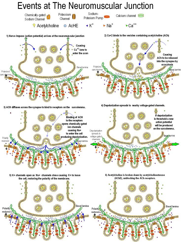
26 Neuromuscular Junction Flash Video Chemically-gated Sodium Channel Potassium Channel SodiumPotasium Pump The Neuromuscular Junction Acetylcholine AcHE K+ Na+ Ca++ Calcium channel Basal lamina Polarized sarcolemma Na+ is pumped out, K+ in at 3:2 producing unequal distribution which leads to a polarized membrane, ~ -65mv.1) An impulse arrives at the neuromuscular junction causing Ca+2 to enter the axon terminus. 2) Ca+2 causes exocytosis of Ach vesicles into synaptic cleft. 3) Ach diffuses across the synapse to contact post-synaptic receptors on the sarcolemma. Ach causes Na+ to enter sarcolemma causing depolarization. 4) If depolarization is threshold a new impulse is produced on the sarcolemma by depolarization of voltage-regulated ion gates. 5) Sarcolemma repolarizes, K+ leaves the cell, pump restores distribution.6) Achase (a.k.a. ACh-E) breaks down Ach so the NMJ can function again. See the next slide for illustrations. 15
27
28 Synaptic Blockers chlinesterase inhibitors: cause paralysis by leading to blockage of receptors by ACh, e.g. insecticides and nerve gas. The antidote to these toxins is atropine which blocks ACh. Curare is an ACh competitor derived from plants, which has been used to relax muscles

29 Neuromuscular Junction: Motor neuron Movement of impulse (action potential) Sarcolemma 28 Each skeletal muscle cell is stimulated by branches from a neuron s axon. 28

30 The Axon Terminal at the Motor End Plate Axon terminal Synaptic cleft T tubule myofibril 29 At each axon terminal is a motor end plate at which the neuromuscular junction occurs. 29

31 The Motor End Plate Axon terminal ACh Synaptic cleft ACh bound to receptor opens chemically-gated ion channel. 30 The ACh receptor binds to a chemically-gated ion channel which opens due to contact with ACh. 30

32 Skeletal Muscle Impulse Conduction T - tubules Sarcoplasmic reticulum myofibrils Figure The T-tubules and sarcoplasmic reticulum represent membranes which penetrate the sarcoplasm of the cell, taking the action potential to the inside. 31
33 Resting membrane potential Figure An action potential is produced when voltage-gated ion channels open in response to threshold depolarization, causing Na + ions to rush into the cell. 32

34 Action potential: Self-propagated All-or-none Na + gates open at at each location allowing Na + in, then K + gates open to allow K + out of the cell causing repolarization. 33 The action potential is a self-propagated, all-or-none movement of depolarization along the membrane. All-or-none means that there are not different size action potentials. You either have one or you don't. As the action potential passes along the sarcolemma it causes release of Na + into the cell by voltage-regulated ion gates, just as at the chemically-regulated gates when stimulated by ACH. Then K + gates open to repolarize that section of the membrane. The opening of Na + gates then K + gates happens at each location along the sarcolemma to propagate the action potential. 33
. Figure 9.9 34 The resting membrane potential occurs because of the unequal distribution of sodium and potassium ions (a).")
35 3 2 Resting membrane potential Threshold depolarization Voltage-gated ion channels: open in in response to threshold depolarization, allow Na + to rush into the cell producing an action potential (impulse). Figure The resting membrane potential occurs because of the unequal distribution of sodium and potassium ions (a). An action potential is produced when voltage-gated ion channels open in response to threshold depolarization, causing Na + ions to rush into the cell (b). 34
 3) K + rushes out causing repolarization 35 As the action potential passes along the sarcolemma it causes release of Na + into the cell by voltage-regulated ion gates, just as at the")
36 Propagation of the Action Potential Na + K + Na + K + Na + K + Na + K + Na + K + 1) 1) Na + rushes in in from depolarization 2) 2) Depolarization spreads to next set of gates. 3) 3) K + rushes out causing repolarization 35 As the action potential passes along the sarcolemma it causes release of Na + into the cell by voltage-regulated ion gates, just as at the chemically-regulated gates when stimulated by ACH. This depolarization spreads to the next set of gates causing them to open, and so forth all along the membrane. K + gates open as the Na + gates close to repolarize each section of the membrane. The opening of Na + gates then K + gates happens at each location along the sarcolemma to propagate the action potential. 35
37 +30 3 } }A B The Action Potential 0 Membrane Potential (mv) Threshold Resting potential Time (ms) 35 The action potential is a self-propagated, all-or-none movement of depolarization along the membrane.all-or-none means that there are not different size action potentials. You either have one or you don't. 1) Depolarization speads from previous ion channel. 2) When this depolarization reaches threshold, the Na+ channel opens, allowing Na+ ions into the cell and causing a reversal of potential. 3) As the Na+ channels close, K+ channels open allowing K+ ions to leave the cell causing repolarization. 4) Resting potential is reached, but so much K+ leaves the cell that there is a brief hyperpolarization 5) The hyperpolarization ends as K+ channels close. 35
38 K + Na+ 37 The sequential opening of first sodium, then potassium, ion channels produces depolarization which spreads along the membrane as an action potential. 37
39 Impulse travels along the sarcolemma. 38 As the action potential passes along the sarcolemma it enters the T- tubules which occur at each Z-line. (See Figure 9.4) The T-tubules are membranes which run across the cell (T for transverse) connecting to the sarcolemma. The T-tubules allow the action potential to continue into the cell interior. 38
40 Impulse travels along the sarcolemma, enters the T-tubules. 39 At points along the T tubules they attach to the sarcoplasmic reticulum, a system of membrane channels inside the sarcoplasm. 39

41 Impulse travels along the sarcolemma, enters the T-tubules, and triggers the release of Ca +2 from the S.R. Ca+2 pumped into the S.R. as crossbridges unattach. Ca +2 Ca +2 Ca When the action potential moves along the T tubules it causes the sarcoplasmic reticulum to release Ca +2 which is sequestered by the SR. The SR pumps calcium like the sarcolemma pumps sodium and releases it into the sarcoplasm when stimulated by the action potential. This causes the sliding of filaments as outlined earlier. The SR then pumps the Ca +2 back out of the sarcoplasm. 40

42 41 Here is shown Excitation-Contraction Coupling from the beginning to end, including each of the components examined previously. 41

43 Spinal cord Motor neuron 1 The Motor Unit Motor neuron 2 Motor unit = a group of of muscle cells stimulated by by the same neuron. < cells per motor unit = very precise control e.g. extraocular muscles to to 100 cells per unit = most muscles, average control >100 cells per unit = gross movements, e.g. postural muscles 42 42
44 a) b) 58 There are two ways in which a muscle can contract: isotonically and isometrically. Isotonic contraction involves a shortening of the muscle as tension is generated, exhibited by diagram a) above, in which the muscle is moving a load. The tension remains constant as long as the load doesn t change. Isotonic contractions are the majority of our muscular movements. In b) above, the muscle cannot shorten but may increase its tension continuously against the force transducer. Isometric contractions are seen when a muscle generates tension against an immovable object. Physiologically the contractions are identical. The heat produced by isotonic contractions is greater due to the action of movement of the internal myofilaments, but not significantly. Isotonic contractions provide a more complete workout of both the muscle and supportive systems such as the cardiovascular and respiratory. Isometric contractions can be effective in building muscle tone and size. 58

45 active passive 43 The Muscle Twitch Figure 9.12 The muscle twitch is a single response to a single stimulus. In a diagram of the muscle twitch can be seen the latent period, the period of a few ms encompassing the chemical and physical events preceding actual contraction. This is not the same as the absolute refractory period, the even briefer period when the sarcolemma is depolarized and cannot be stimulated. The relative refractory period occurs after this when the sarcolemma is briefly hyperpolarized and requires a greater than normal stimulus. [See Refractory Periods Diagram in Slide #34] Following the latent period is the contraction phase in which the shortening of the sarcomeres and cells occurs. Then comes the relaxation phase, a longer period because it is passive, the result of recoil due to the series elastic elements of the muscle. 43
.")
46 Fast twitch = 3 to 5 ms. Intermediate = 10 to 20 ms. Slow twitch = > 40 ms. Twitch Lengths 44 Muscle twitches vary in length according to the type of muscle cells involved. Fast twitch muscles such as those which move the eyeball have twitches which reach maximum contraction in 3 to 5 ms (milliseconds). See [superior eye] and [lateral eye] These muscles were mentioned earlier as also having small numbers of cells in their motor units for precise control. The cells in slow twitch muscles like the postural muscles (e.g. back muscles, soleus) have twitches which reach maximum tension in 40 ms or so. The muscles which exhibit most of our body movements have intermediate twitch lengths of 10 to 20 ms. These three types also represents different metabolic patterns as will be discussed later. 44
47 Quantal Summation (Recruitment) Response Maximum response Quantal Summation Degree of muscle contraction increases with increasing stim ulus strength. More cells are recruited with each increase in stimulus strength, up to maximum response at which time all cells are contracting Stimulus Strength (volts) 46 Quantal Summation or Recruitment - this refers to increasing the number of cells contracting. This is done experimentally by increasing the voltage used to stimulate a muscle, thus reaching the thresholds of more and more cells. In the human body quantal summation is accomplished by the nervous system, stimulating increasing numbers of cells or motor units to increase the force of contraction. 46

48 Frequency Summation Muscle is stimulated before it relaxes from the previous stimulus. Figure If you stimulate a muscle cell, or muscle, before it has relaxed from a previous stimulus, it will contract some more. This is called frequency summation. 47

49 Incomplete and Complete Tetanus High frequency stimuli don t permit the muscle to relax, producing a sustained contraction. Tetanus = tetany = sustained contraction 48 Wave Summation (a.k.a. frequency summation) and Tetanization- this results from stimulating a muscle cell before it has relaxed from a previous stimulus. This is possible because the contraction and relaxation phases are much longer than the refractory period. This causes the contractions to build on one another producing a wave pattern or, if the stimuli are high frequency, a sustained contraction called tetany or tetanus. (The term tetanus is also used for an illness caused by a bacterial toxin which causes contracture of the skeletal muscles.) This form of tetanus is perfectly normal and in fact is the way you maintain a sustained contraction. 48
50 Two Types of Summation Quantal summation the number of cells or motor units varies to produce contractions of different strengths. a.k.a. recruitment Frequency summation a given set of cells or motor units increases the degree of contraction to produce a sustained contraction. Wave summation or incomplete tetanus Complete tetanus
51 Example of Tetanus Contraction Stimuli per second 50 50

52 Treppe: Warmup Figure 9.14 muscle cells initially stimulated when cold will exhibit gradually Stimuli are all of the same voltage. increasing responses until they have warmed 51 up. Treppe is not a way muscles exhibit graded contractions. It is a warmup phenomenon in which when muscle cells are initially stimulated when cold, they will exhibit gradually increasing responses until they have warmed up. The phenomenon is due to the increasing efficiency of the ion gates as they are repeatedly stimulated. Treppe can be differentiated from quantal summation because the strength of stimulus remains the same in treppe, but increases in quantal summation. 51

53 Length-Tension Relationship in a Sarcomere Figure Another way in which the tension of a muscle can vary is due to the length-tension relationship. This relationship expresses the characteristic that within about 10% the resting length of the muscle, the tension the muscle exerts is maximum. At lengths above or below this optimum length the tension decreases. In practical terms a muscle will be its strongest at midpoint in its extensibility. For the heart, you will later learn, it means the muscle will adjust its output to normal increases in blood supply. 52
54 Myofilament Overlap Related to % of Resting Length 53 Note how the overlap of actin and myosin is maximized near the resting length, and reduced when the sarcomere is fully contracted or fully stretched. The overlap of myosin and actin produces the ability to generate tension. 53
55 Length-Tension Relationship in a Muscle Muscle Tension Optimum length for maximum tension Muscle Length 54 The length-tension relationship is derived from the anatomy of the sarcomere, but it can be applied to the whole muscle as well. A muscle can exhibit its greatest tension within 10% of its resting length. 54
56 Adenosine Triphosphate (ATP) High Energy bonds 55 Muscle cells, like all others, use ATP as their energy currency. During periods of rest ATP is built from ADP and Pi (inorganic phosphate), then during energy-requiring activities the ATP is hydrolyzed to release the energy for doing work, along with the ADP and Pi. But some muscle cells must exhibit activity levels in which they cannot make ATP as fast as it is consumed. So muscle cells have several mechanisms to provide the ATP they need. 55
57 Primary Energy for Muscle Contraction Energy for muscle contraction 56 The phosphagen system - this is the use of immediately available ATP. This is not from stored ATP itself, muscle cells can store only very limited amounts. It is from energy stored as the related molecule CP, creatine phosphate. Creatine phosphate can be stored and is made from ATP during periods of rest. Then during periods of high activity CP is broken down quickly and its energy converted to ATP. But this source of ATP can only supply a cell for 8 to 10 seconds during the most strenuous exercise. Creatine released during muscle activity shows up in the urine as creatinine, a combination of two creatine molecules. 56
58 The Phosphagen System Used during the first 15 sec. of speed and strength exercises. Creatinine molecules released during strenuous exercise are released as creatinine in the urine. 57 The phosphagen system - this is the use of immediately available ATP. This is not from stored ATP itself, muscle cells can store only very limited amounts. It is from energy stored as the related molecule CP, creatine phosphate. Creatine phosphate can be stored and is made from ATP during periods of rest. Then during periods of high activity CP is broken down quickly and its energy converted to ATP. But this source of ATP can only supply a cell for 8 to 10 seconds during the most strenuous exercise. Creatine released during muscle activity shows up in the urine as creatinine, a combination of two creatine molecules. 57
59 Anerobic Glycolysis 58 Glycolysis is the initial way of utilizing glucose in all cells, and is used exclusively by certain cells to provide ATP when insufficient oxygen is available for aerobic metabolism. Glycolysis doesn't produce much ATP in comparison to aerobic metabolism, but it has the advantage that it doesn't require oxygen. In addition, glycolysis occurs in the cytoplasm, not the mitochondria. So it is used by cells which are responsible for quick bursts of speed or strength. Like most chemical reactions, glycolysis slows down as its product, pyruvic acid, builds up. In order to extend glycolysis the pyruvic acid is converted to lactic acid in a process known as fermentation. Lactic acid itself eventually builds up, slowing metabolism and contributing to muscle fatigue. 58
60 Anerobic Glycolysis Allows speed and strength fibers to perform for 1 to 1.5 minutes. 59 Eventually the lactic acid must be reconverted to pyruvic acid and metabolized aerobically, either in the muscle cell itself, or in the liver. The oxygen which is "borrowed" by anaerobic glycolysis is called oxygen debt and must be paid back. 59
61 Oxygen Debt Rate of Oxygen Uptake L/min Exercise 0 4 Alactacid oxygen debt = 3.5 L Extra oxygen from tissues and blood. Lactic acid oxygen debt = 8.0 L Oxygen required to to oxidize the lactic acid 32 produced Minutes 60 This graph shows the rate of oxygen uptake by the lungs during maximal exercise for 4 minutes and then for one hour after the exercise is over. This figure demonstrates the principle of oxygen debt. Oxygen debt is partly oxygen reserves in the lungs, tissues, and myoglobin in the lungs (alactacid oxygen debt). But mostly it is the amount of oxygen which will be required to metabolize the lactic acid produced, the lactic acid oxygen debt. 60
62 61 Ultimately, the product of glycolysis, pyruvic acid, must be metabolized aerobically. Aerobic metabolism is performed exclusively in the mitochondria. Pyruvic acid is converted to a molecule called an acetyl group and put into a pathway known as the Krebs Cycle. Energy is released in the form of ATP and, especially, as high energy electrons. These high energy electrons are sent to a process within the mitochondria known as the electron transport system which produces the vast majority of the ATP. The waste products of aerobic metabolism are CO 2 and H 2 O. The reactant other than glucose is O 2. 61
![Summary of Metabolic Processes in Muscles: The Phosphagen System See [Facts About Performance Boosters] Figure 9.](/docs-images/72/67844345/images/63-0.jpg "16 a 62 Training can increase the amount of creatine phosphate stored, but this alone does not increase the strength of a muscle, just the length of time before it runs out of CP,")
63 Summary of Metabolic Processes in Muscles: The Phosphagen System See [Facts About Performance Boosters] Figure 9.16 a 62 Training can increase the amount of creatine phosphate stored, but this alone does not increase the strength of a muscle, just the length of time before it runs out of CP, and that by only a few seconds. See [Facts About Performance Boosters] 62

64 Summary of Metabolic Processes in Muscles: Anerobic Glycolysis Figure 9.16 b 63 Strength training increases the myofilaments in muscle cells and therefore the number of crossbridge attachments which can form. Training does not increase the number of muscle cells in any real way. (Sometimes a cell will tear and split resulting in two cells when healed). Lactic acid removal by the cardiovascular system improves with training which increases the anaerobic capacity. Even so, the glycolysis-lactic acid system can produce ATP for active muscle cells for only about a minute and a half. 63

65 Summary of Metabolic Processes in Muscles: Aerobic Metabolism Figure 9.16 c 64 Aerobic metabolism is used for endurance activities and has the distinct advantage that it can go on for hours. Aerobic training increases the length of endurance activities by increasing the number of mitochondria in the muscle cells, increasing the availability of enzymes, increasing the number of blood vessels, and increasing the amount of an oxygenstoring molecule called myoglobin. 64
66 Types of Muscle Fibers: White Fibers Fast twitch Large diameter, used for speed and strength. Depends on the phosphagen system and on glycolysis-lactic acid. Stores glycogen for conversion to glucose. Fewer blood vessels. Little or no myoglobin. Training increases the number of of myofibrils and, as a result, the muscle size. 65 Different types of cells perform the differing functions of endurance activities and speed- strength activities. There are three types, red, white, and intermediate. The main differences can be exemplified by looking at red and white fibers and remembering that intermediate fibers have properties of the other two. 65
67 Slow twitch Red Fibers Small diameter, used for endurance. Depends on aerobic metabolism. Utilize fats as well as glucose. Little glycogen storage. Many blood vessels and much myoglobin give this muscle its reddish appearance. Training increases the number of of mitochondria and enzymes for aerobic metabolism. Also increases cardiovascular and respiratory capacity. 66 Intermediate Fibers: sometimes called "fast twitch red", these fibers have faster action but rely more on aerobic metabolism and have more endurance. Most muscles are mixtures of the different types. Muscle fiber types and their relative abundance cannot be varied by training, although there is some evidence that prior to maturation of the muscular system the emphasis on certain activities can influence their development. 66
68 Energy Systems Used in Various S ports Phosphagen System, almost entirely: 100 meter dash jumping weight lifting diving football dashes Phosphagen and Glycogen-lactic Acid systems: 200 meter dash basketball baseball home run 4 M ATP/Min for 8 to 10 Sec. ice hockey dashes Glycogen-lactic Acid System, mainly 400 meter dash 100 meter swim tennis soccer Glycogen-lactic Acid System and Aerobic System 800 meter dash 200 meter swim 1500 meter run boxing 2000 meter rowing 1500 meter run 1 mile run 400 meter swim Aerobic System: 10,000 meter skating cross-country skiing marathon run jogging 2.5 M ATP/Min for 1.3 to 1.6 Min. Pathways Used in Various Activities 67 67
69 % Carbohydrate Usage High Fat Diet High Carb Diet Mixed Diet Sec. Min. Hours Exhaustion In exercise, high carbohydrate diets lead to longer sustained performance before exhaustion sets in. 68 Effect of duration and type of diet on relative percentages of carbohydrate or fat used for muscle energy. Percentage carbohydrate used is proportional to glycogen reserves and inversely proportional to fat usage. 68
70 Muscle glycogen mg/kg High carbohydrate 2 hours of Recovery diets lead vs. to Diet quicker recovery of muscle glycogen than do exercise other diets. High carbohydrate diet No food Hours of Recovery Fat & protein diet 5 days 69 Effect of diet on the rate of muscle glycogen replenishment after prolonged exercise. This study compares athletes who have had a high carb diet with those who have had high fat & protein diets and with those who have fasted. 69
Muscle and Muscle Tissue
 Muscle and Muscle Tissue Make up about half of total body mass Exerts force by converting chemical energy, ATP, to mechanical energy Muscle tissue is classified based on Shape Number and position of nuclei
MUSCLE TISSUE (MUSCLE PHYSIOLOGY) PART I: MUSCLE STRUCTURE
 PART I: MUSCLE STRUCTURE") PART I: MUSCLE STRUCTURE Muscle Tissue A primary tissue type, divided into: skeletal muscle cardiac muscle smooth muscle Functions of Skeletal Muscles Produce skeletal movement Maintain body position Support
PART I: MUSCLE STRUCTURE Muscle Tissue A primary tissue type, divided into: skeletal muscle cardiac muscle smooth muscle Functions of Skeletal Muscles Produce skeletal movement Maintain body position Support
Chapter 10 Muscle Tissue Lecture Outline
 Chapter 10 Muscle Tissue Lecture Outline Muscle tissue types 1. Skeletal muscle = voluntary striated 2. Cardiac muscle = involuntary striated 3. Smooth muscle = involuntary nonstriated Characteristics
CHAPTER 6 2/9/2016. Learning Objectives List the four traits that all muscle types have in common.
 Learning Objectives List the four traits that all muscle types have in common. CHAPTER 6 The Muscular System Demonstrate and explain the use of antagonistic muscle pairs. Describe the attachment of muscle
Anatomy and Physiology 1 Chapter 10 self quiz Pro, Dima Darwish,MD.
 Anatomy and Physiology 1 Chapter 10 self quiz Pro, Dima Darwish,MD. 1) Which of the following is a recognized function of skeletal muscle? A) produce movement B) maintain posture C) maintain body temperature
MUSCULAR TISSUE. Dr. Gary Mumaugh
 MUSCULAR TISSUE Dr. Gary Mumaugh MUSCLE OVERVIEW The three types of muscle tissue are skeletal, cardiac, and smooth These types differ in structure, location, function, and means of activation FUNCTIONAL
Muscles and Muscle Tissue
 1 Muscles and Muscle Tissue Chapter 9 2 Overview of Muscle Tissues Compare and Contrast the three basic types of muscle tissue List four important functions of muscle tissue 3 Muscle Terminology Muscle
About This Chapter. Skeletal muscle Mechanics of body movement Smooth muscle Cardiac muscle Pearson Education, Inc.
 About This Chapter Skeletal muscle Mechanics of body movement Smooth muscle Cardiac muscle Skeletal Muscle Usually attached to bones by tendons Origin: closest to the trunk or to more stationary bone Insertion:
Smooth Cardiac Skeletal Location Around tubes Heart tissue attached to skeleton Moves stuff thru Heart beat pumps Moves body parts
 Biology 067 - Muscular system A. Type of muscles: Smooth Cardiac Skeletal Location Around tubes Heart tissue attached to skeleton Function Moves stuff thru Heart beat pumps Moves body parts tubes blood
Chapter 10 Muscle Tissue and Physiology Chapter Outline
 Chapter 10 Muscle Tissue and Physiology Chapter Outline Module 10.1 Overview of muscle tissue (Figures 10.1 10.2) A. Types of Muscle Tissue (Figure 10.1) 1. The three types of cells in muscle tissue are,,
Chapter 9 - Muscle and Muscle Tissue
 Chapter 9 - Muscle and Muscle Tissue I. Overview of muscle tissue A. Three muscle types in the body: B. Special characteristics 1. Excitability: able to receive and respond to a stimulus 2. Contractility:
1/4/2017. Introduction. Connective Tissue Coverings. 9.1: Structure of a Skeletal Muscle. Skeletal Muscle Fibers. Connective Tissue Coverings
 Introduction Chapter 09 Lecture Outline See separate PowerPoint slides for all figures and tables preinserted into PowerPoint without notes. Copyright McGraw-Hill Education. Permission required for reproduction
Skeletal Muscle. Connective tissue: Binding, support and insulation. Blood vessels
 Chapter 12 Muscle Physiology Outline o Skeletal Muscle Structure o The mechanism of Force Generation in Muscle o The mechanics of Skeletal Muscle Contraction o Skeletal Muscle Metabolism o Control of Skeletal
Muscle Cell Anatomy & Function (mainly striated muscle tissue)
") Muscle Cell Anatomy & Function (mainly striated muscle tissue) General Structure of Muscle Cells (skeletal) several nuclei (skeletal muscle) skeletal muscles are formed when embryonic cells fuse together
Muscle Cell Anatomy & Function (mainly striated muscle tissue) General Structure of Muscle Cells (skeletal) several nuclei (skeletal muscle) skeletal muscles are formed when embryonic cells fuse together
CLASS SET Unit 4: The Muscular System STUDY GUIDE
 NPHS Anatomy & Physiology Questions to answer: 1) List three functions of the muscular system. 1) movement 2) thermogenesis (generates heat) 3) posture & body/joint support CLASS SET Unit 4: The Muscular
Bio 103 Muscular System 61
 61 Lecture Outline: MUSCULAR SYSTEM [Chapter 9] A. Functions of Skeletal Muscle 1. Movement 2. Maintain posture 3. Support 4. Guard openings 5. Maintain body temperature (thermogenesis) B. Muscle Tissue
Muscle Tissue. PowerPoint Lecture Presentations prepared by Jason LaPres. Lone Star College North Harris Pearson Education, Inc.
 10 Muscle Tissue PowerPoint Lecture Presentations prepared by Jason LaPres Lone Star College North Harris An Introduction to Muscle Tissue Muscle Tissue A primary tissue type, divided into: Skeletal muscle
Chapter 8 Notes. Muscles
 Chapter 8 Notes Muscles 8.1 Intro Three muscle types Skeletal Smooth cardiac 8.2 Structure of Skeletal Muscle Composition Skeletal muscle tissue Nervous tissue Blood Connective tissue Connective tissue
Skeletal Muscle Tissue
 Functions of Skeletal Muscle Skeletal Muscle Tissue Keri Muma Bio 6 Movement muscles attach directly or indirectly to bone, pull on bone or tissue when they contract Maintain posture / body position muscles
Muscle Tissue- 3 Types
 AN INTRODUCTION TO MUSCLE TISSUE Muscle Tissue- 3 Types Skeletal muscle (focus on these) Cardiac muscle Smooth muscle FUNCTIONS OF SKELETAL MUSCLES Produce movement of the skeleton Maintain posture and
2/19/2018. Learn and Understand:
 Muscular System with Special Emphasis on Skeletal Muscle Anatomy and Physiology Learn and Understand: The definition of cell changes again The contractile unit of muscle is the sarcomere. ATP and Ca 2+
Chapter 8: Skeletal Muscle: Structure and Function
 Chapter 8: Skeletal Muscle: Structure and Function Objectives Draw & label the microstructure of skeletal muscle Outline the steps leading to muscle shortening Define the concentric and isometric Discuss:
10 - Muscular Contraction. Taft College Human Physiology
 10 - Muscular Contraction Taft College Human Physiology Muscular Contraction Sliding filament theory (Hanson and Huxley, 1954) These 2 investigators proposed that skeletal muscle shortens during contraction
Ch.10 Muscle Tissue. Copyright 2009, John Wiley & Sons, Inc.
 Ch.10 Muscle Tissue Preview Chapter 10 In groups we will define the following terms 1. Skeletal muscle 2. Smooth muscle 3. Cardiac muscle 4. Sarcomere 5. Myofibril 6. Myofilament 7. Sarcoplasmic reticulum
MODULE 6 MUSCLE PHYSIOLOGY
 MODULE 6 MUSCLE PHYSIOLOGY III SEMESTER BOTANY Syllabi: Striated, Non striated and Cardiac muscle, Ultra structure of striated muscle fibre, Mechanism of muscle contraction, Threshold and spike potential,
Fig Copyright McGraw-Hill Education. Permission required for reproduction or display. Nucleus. Muscle fiber. Endomysium. Striations.
 Fig. 11.1 Nucleus Muscle fiber Endomysium Striations Ed Reschke 1 Fig. 11.2 Muscle fiber Nucleus I band A band Z disc Mitochondria Openings into transverse tubules Sarcoplasmic reticulum Triad: Terminal
Lecture Overview. Muscular System. Marieb s Human Anatomy and Physiology. Chapter 9 Muscles and Muscle Tissue Lecture 16
 Marieb s Human Anatomy and Physiology Marieb Hoehn Chapter 9 Muscles and Muscle Tissue Lecture 16 1 Lecture Overview Types, characteristics, functions of muscle Structure of skeletal muscle Mechanism of
Anatomy & Physiology Muscular System Worksheet
 Anatomy & Physiology Muscular System Worksheet 1. What are the three categories of muscle tissue? a) b) c) 2. The smallest functional unit of a muscle fiber is called a. 3. What are the four characteristics
Essentials of Human Anatomy & Physiology. The Muscular System
 Essentials of Human Anatomy & Physiology The Muscular System The Muscular System Muscles are responsible for all types of body movement they contract or shorten and are the machine of the body Three basic
Microanatomy of Muscles. Anatomy & Physiology Class
 Microanatomy of Muscles Anatomy & Physiology Class Three Main Muscle Types Objectives: By the end of this presentation you will have the information to: 1. 2. 3. 4. 5. 6. Describe the 3 main types of muscles.
Muscle Tissue. Muscle Tissue Outline. General Function of Muscle Tissue
 Muscle Tissue Muscle Tissue Outline General Functions of Muscle Tissue Characteristics of Muscle Tissue Classification of Muscle Tissue Skeletal Muscle Structure and Function Muscle Energetics Muscle Mechanics
Chapter 10 -Muscle Tissue
 Chapter 10 -Muscle Tissue Muscles: 1. Overview of Muscle Tissue A. Review 5 functions of muscle tissue. B. Review the 5 properties of muscle tissue. WHICH do they share with nervous tissue? (2, plus the
Page 1. Chapter 9: Muscle Tissue. Types of Muscle Tissue: Skeletal Muscle Cardiac Muscle Smooth Muscle. Characteristics of Muscle:
 1 Chapter 9: Muscle Tissue Muscle little mouse Types of Muscle Tissue: Skeletal Muscle Cardiac Muscle Smooth Muscle Characteristics: Attaches to skeleton Voluntary control Striated / multi-nucleated Characteristics:
Page 1. Chapter 9: Muscle Tissue. Types of Muscle Tissue: Skeletal Muscle Cardiac Muscle Smooth Muscle. Gross Anatomy of Muscle:
 1 Chapter 9: Muscle Tissue Types of Muscle Tissue: Skeletal Muscle Cardiac Muscle Smooth Muscle Characteristics: Attaches to skeleton Voluntary control Striated / multi-nucleated Characteristics: Composes
Muscle Physiology. Dr. Ebneshahidi Ebneshahidi
 Muscle Physiology Dr. Ebneshahidi Skeletal Muscle Figure 9.2 (a) Functions of the muscular system 1. Locomotion body movements are due to skeletal muscle contraction. 2. Vasoconstriction and vasodilatation
Skeletal Muscle. Skeletal Muscle
 Skeletal Muscle Skeletal Muscle Types of muscle Skeletal muscle-moves the skeleton by pulling on the tendons that are connected to the bones Cardiac muscle-pumps blood through the heart and blood vessels
1. Locomotion. 2. Repositioning. 3. Internal movement
 MUSCLE and MOVEMENT Chapters 20, 8, 21 1. Locomotion A. Movement B. 2. Repositioning A. 3. Internal movement A. 1 Muscle Cells 1. Contractile 2. Myocytes 3. Striated A. Skeletal B. Cardiac 4. Smooth 5.
1. Locomotion. 2. Repositioning. 3. Internal movement
 MUSCLE and MOVEMENT Chapters 20, 8, 21 1. Locomotion A. Movement B. 2. Repositioning A. 3. Internal movement A. Muscle Cells 1. Contractile 2. Myocytes 3. Striated A. Skeletal B. Cardiac 4. Smooth 5. Striated
PSK4U THE NEUROMUSCULAR SYSTEM
 PSK4U THE NEUROMUSCULAR SYSTEM REVIEW Review of muscle so we can see how the neuromuscular system works This is not on today's note Skeletal Muscle Cell: Cellular System A) Excitation System Electrical
Muscular System. This chapter will focus on muscle cells and tissues. Muscle tissue has several functions:
 Muscular System Slide 2 This chapter will focus on muscle cells and tissues. Muscle tissue has several functions: Movement: Muscles work as pulleys on bones to help create changes in body position. Muscles
Hole s Human Anatomy and Physiology Eleventh Edition. Mrs. Hummer. Chapter 9 Muscular System
 Hole s Human Anatomy and Physiology Eleventh Edition Mrs. Hummer Chapter 9 Muscular System 1 Chapter 9 Muscular System Skeletal Muscle usually attached to bones under conscious control striated Three Types
Chapter 10: Muscle Tissue
 Chapter 10: Muscle Tissue Muscle is one of the 4 primary types of tissue. It is subdivided into skeletal, cardiac and smooth muscle. I. Skeletal Muscle Tissue and the Muscular System, p. 284 Objective
Muscles & Muscle Tissue
 Muscles & Muscle Tissue Chapter 6 I. Overview of Muscle 1 A. MUSCLE TYPES SKELETAL: striated, voluntary CARDIAC: only in heart involuntary striated SMOOTH: walls of organs involuntary nonstriated All Muscle
Principles of Anatomy and Physiology
 Principles of Anatomy and Physiology 14 th Edition CHAPTER 10 Muscular Tissue Introduction The purpose of the chapter is to: 1. Learn about the structure and function of the 3 types of muscular tissue
MUSCULAR SYSTEM CHAPTER 09 BIO 211: ANATOMY & PHYSIOLOGY I
 1 BIO 211: ANATOMY & PHYSIOLOGY I 1 CHAPTER 09 MUSCULAR SYSTEM Part 2 of 2 Dr. Dr. Lawrence G. G. Altman www.lawrencegaltman.com Some illustrations are courtesy of McGraw-Hill. Some illustrations are courtesy
Session 3-Part 2: Skeletal Muscle
 Session 3-Part 2: Skeletal Muscle Course: Introduction to Exercise Science-Level 2 (Exercise Physiology) Presentation Created by Ken Baldwin, M.ED, ACSM-H/FI Copyright EFS Inc. All Rights Reserved. Skeletal
Chapter 9 Muscle. Types of muscle Skeletal muscle Cardiac muscle Smooth muscle. Striated muscle
 Chapter 9 Muscle Types of muscle Skeletal muscle Cardiac muscle Smooth muscle Striated muscle Chapter 9 Muscle (cont.) The sliding filament mechanism, in which myosin filaments bind to and move actin
Types of Muscle. Skeletal striated & voluntary Smooth involuntary Cardiac - heart
 Muscular System Types of Muscle Skeletal striated & voluntary Smooth involuntary Cardiac - heart The word striated means striped. Skeletal muscle appears striped under a microscope. Muscles and Muscle
I. Overview of Muscle Tissues
 I. Overview of Muscle Tissues A. Types of Muscle Tissue 1. Terminology 1. Muscle fibers = muscle cells are greatly elongated therefore known as fibers; true for skeletal and smooth muscles only 2. Myo
Human Anatomy. Muscle Tissue and Organization. DR.SADIQ ALI (K.E Medalist) 10-1
 10-1") Human Anatomy Muscle Tissue and Organization DR.SADIQ ALI (K.E Medalist) 10-1 Tissue and Organization Over 700 skeletal muscles have been named. Form the muscular system. Muscle tissue is distributed almost
Human Anatomy Muscle Tissue and Organization DR.SADIQ ALI (K.E Medalist) 10-1 Tissue and Organization Over 700 skeletal muscles have been named. Form the muscular system. Muscle tissue is distributed almost
BIOH111. o Cell Module o Tissue Module o Integumentary system o Skeletal system o Muscle system o Nervous system o Endocrine system
 BIOH111 o Cell Module o Tissue Module o Integumentary system o Skeletal system o Muscle system o Nervous system o Endocrine system Endeavour College of Natural Health endeavour.edu.au 1 TEXTBOOK AND REQUIRED/RECOMMENDED
The All-or-None Principle Motor units also comply to a rule known as the all-ornone principle (or law).
.") The All-or-None Principle Motor units also comply to a rule known as the all-ornone principle (or law). This principle stipulates that, when a motor unit is stimulated to contract, it will do so to its
The All-or-None Principle Motor units also comply to a rule known as the all-ornone principle (or law). This principle stipulates that, when a motor unit is stimulated to contract, it will do so to its
The Muscular System. Objective: The student will become familiar with the structure and function of the muscular system
 The Muscular System Objective: The student will become familiar with the structure and function of the muscular system 1 Question of the day: What moves you? Composition: The musclar system makes up 40-50%
Skeletal Muscle Qiang XIA (
 Skeletal Muscle Qiang XIA ( 夏强 ), PhD Department of Physiology Rm C518, Block C, Research Building, School of Medicine Tel: 88208252 Email: xiaqiang@zju.edu.cn Course website: http://10.71.121.151/physiology
Muscle Tissue. C h a p t e r. PowerPoint Lecture Slides prepared by Jason LaPres Lone Star College - North Harris
 C h a p t e r 10 Muscle Tissue PowerPoint Lecture Slides prepared by Jason LaPres Lone Star College - North Harris Copyright 2009 Pearson Education, Inc., publishing as Pearson Benjamin Cummings An Introduction
Chapter 10! Chapter 10, Part 2 Muscle. Muscle Tissue - Part 2! Pages !
 ! Chapter 10, Part 2 Muscle Chapter 10! Muscle Tissue - Part 2! Pages 308-324! SECTION 10-5! Sarcomere shortening and muscle fiber stimulation produce tension! 2! Tension Production - Muscle FIBER! All-or-none
Muscle Tissue. Alternating contraction and relaxation of cells. Chemical energy changed into mechanical energy
 Know these muscles Muscle Tissue Alternating contraction and relaxation of cells Chemical energy changed into mechanical energy 3 Types of Muscle Tissue Skeletal muscle attaches to bone, skin or fascia
Ch 10: Skeletal Muscle Tissue (Myology)
") Ch 10: Skeletal Muscle Tissue (Myology) main objectives: Describe the distinguishing characteristics of the different muscle tissues Discuss the organization of skeletal muscle Explain the micro-anatomy
Ch 10: Skeletal Muscle Tissue (Myology) main objectives: Describe the distinguishing characteristics of the different muscle tissues Discuss the organization of skeletal muscle Explain the micro-anatomy
Ch 12: Muscles sarcolemma, t-tubules, sarcoplasmic reticulum, myofibrils, myofilaments, sarcomere...
 Ch 12: Muscles Review micro-anatomy of muscle tissue Terminology examples: sarcolemma, t-tubules, sarcoplasmic reticulum, myofibrils, myofilaments, sarcomere... SLOs Differentiate levels of muscle structure:
Muscular System- Part 1. Unit 5 Miss Wheeler
 Muscular System- Part 1 Unit 5 Miss Wheeler Fun Facts! The tongue is the strongest muscle in your body The smallest muscles in the body are in the middle ear The largest muscle in the body is the gluteus
Outline. Bio 105: Muscular System. Muscular System. Types of Muscles. Smooth Muscle. Cardiac Muscle 4/6/2016
 Outline Bio 105: Muscular System Lecture 11 Chapter 6 Characteristics of muscles 3 types of muscles Functions of muscles Structure of skeletal muscles Mechanics of muscle contraction Energy sources for
Nerve Cell (aka neuron)
") Nerve Cell (aka neuron) Neuromuscular Junction Nerve cell Muscle fiber (cell) The Nerve Stimulus and Action Potential The Nerve Stimulus and Action Potential Skeletal muscles must be stimulated by a motor
Nerve Cell (aka neuron) Neuromuscular Junction Nerve cell Muscle fiber (cell) The Nerve Stimulus and Action Potential The Nerve Stimulus and Action Potential Skeletal muscles must be stimulated by a motor
Skeletal Muscle and the Molecular Basis of Contraction. Lanny Shulman, O.D., Ph.D. University of Houston College of Optometry
 Skeletal Muscle and the Molecular Basis of Contraction Lanny Shulman, O.D., Ph.D. University of Houston College of Optometry Like neurons, all muscle cells can be excited chemically, electrically, and
BIOH111. o Cell Module o Tissue Module o Integumentary system o Skeletal system o Muscle system o Nervous system o Endocrine system
 BIOH111 o Cell Module o Tissue Module o Integumentary system o Skeletal system o Muscle system o Nervous system o Endocrine system Endeavour College of Natural Health endeavour.edu.au 1 Textbook and required/recommended
Concept 50.5: The physical interaction of protein filaments is required for muscle function
 Concept 50.5: The physical interaction of protein filaments is required for muscle function Muscle activity is a response to input from the nervous system The action of a muscle is always to contract Vertebrate
Nerve Muscle Relationship and Neural Muscular Junction Quiz. Remember, you need to know the structure and the function!
 Nerve Muscle Relationship and Neural Muscular Junction Quiz Remember, you need to know the structure and the function! What is this called? What is this? Schwann cell What is this called? Basal lamina
Nerve regeneration. Somatic nervous system
 Somatic nervous system Signals from CNS are sent to skeletal muscles. Final result is a muscle contraction. Motor neuron starts in CNS and its axon ends at a muscle cell. Alpha motor neuron Alpha motor
Nerve meets muscle. Nerve regeneration. Somatic nervous system
 Somatic nervous system Signals from CNS are sent to skeletal muscles. Final result is a muscle contraction. Alpha motor neurons branch into several terminals (can be over 1000), each contacting a separate
Human Anatomy and Physiology - Problem Drill 09: The Muscular System
 Human Anatomy and Physiology - Problem Drill 09: The Muscular System Question No. 1 of 10 The muscular system of the human body fulfills many different roles. Which of the following statements about the
Chapter 10: Muscles. Vocabulary: aponeurosis, fatigue
 Chapter 10: Muscles 37. Describe the structural components of skeletal muscle tissue from the molecular to the organ level. 38. Describe the structure, function, and importance of sarcomeres. 39. Identify
A and P CH 8 Lecture Notes.notebook. February 10, Table of Contents # Date Title Page # /30/17 Ch 8: Muscular System
 Table of Contents # Date Title Page # 1. 01/30/17 Ch 8: Muscular System 2. 1 3. 4. 5. 6. 7. i 1 Turnitin.com Class #: 13551662 Password: GoViks 2 01/30/17 Ch 8: Muscular System Objective: Students will
2/28/18. Muscular System. 1 Copyright 2016 by Elsevier Inc. All rights reserved. Introduction. Physiology. Anatomy. Muscle Fiber
 Introduction Muscular System Chapter 20 Shortening or lengthening of a muscle results from changes in relative positions of one small part of a muscle cell to another To understand contraction, we will
7/10/18. Introduction. Muscular System. Anatomy. Physiology. Skeletal Muscle Anatomy. Muscle Fiber
 Introduction Muscular System Chapter 20 Shortening or lengthening of a muscle results from changes in relative positions of one small part of a muscle cell to another To understand contraction, we will
2/28/18. Muscular System. Introduction. Anatomy. Chapter 20
 Muscular System Chapter 20 1 Introduction Shortening or lengthening of a muscle results from changes in relative positions of one small part of a muscle cell to another To understand contraction, we will
(c) sarcolemma with acethylcholine (protein) receptors
 sarcolemma with acethylcholine (protein) receptors") (slide 1) Lecture Notes: Muscular System I. (slide 2) Introduction to Muscular System A) Tissues of the Muscular System: 1) Connective Tissues (a) dense fibrous (tendons and ligaments) 2) Nervous Tissue
(slide 1) Lecture Notes: Muscular System I. (slide 2) Introduction to Muscular System A) Tissues of the Muscular System: 1) Connective Tissues (a) dense fibrous (tendons and ligaments) 2) Nervous Tissue
Muscular System. 3 types of muscle tissue. How skeletal muscles arrange CARDIAC SMOOTH SKELETAL
 Muscular System Functions Support the body by allowing us to stay upright Allow for movement by attaching to the skeleton Help maintain a constant body temperature Assist in movement in the cardiovascular
Anatomy & Physiology. Unit Two. Muscular System URLs Frog Dissection
 Anatomy & Physiology 9 Muscular System URLs Frog Dissection http://curry.edschool.virginia.edu/go/frog/home.html Cat Dissection http://www.mhhe.com/biosci/ap/cat_dissect/index.htm List of Muscles http://www.meddean.luc.edu/lumen/meded/
Muscle Tissue. PowerPoint Lecture Presentations prepared by Jason LaPres. Lone Star College North Harris Pearson Education, Inc.
 10 Muscle Tissue PowerPoint Lecture Presentations prepared by Jason LaPres Lone Star College North Harris 10-1 An Introduction to Muscle Tissue Learning Outcomes 10-1 Specify the functions of skeletal
Musculoskeletal Systems. Anatomy: Arrangement of Cells Physiology: Contractions
 Musculoskeletal Systems Anatomy: Arrangement of Cells Physiology: Contractions Characteristics of all muscle Contractile: it shortens Excitable: receives & responds to electrical signals Extensible: stretches
Muscle Tissue. Dr. Heba Kalbouneh Associate Professor of Anatomy and Histology
 Muscle Tissue Dr. Heba Kalbouneh Associate Professor of Anatomy and Histology Functions of muscle tissue Movement Maintenance of posture Joint stabilization Heat generation Tendon Belly Tendon Types of
Skeletal Muscle. Smooth Muscle. Cardiac Muscle. I. 3 Types of Muscle Tissue. 1. Smooth 2. Cardiac 3. Skeletal
 I. 3 Types of Muscle Tissue 1. Smooth 2. Cardiac 3. Skeletal Smooth Muscle Found in body organs,vessels, respiratory passages Not striated, tapered, single cell nucleus involuntary, peristaltic contractions
Muscular Tissue. Functions of Muscular Tissue. Types of Muscular Tissue. Skeletal Muscular Tissue. Properties of Muscular Tissue
 Muscular Tissue Functions of Muscular Tissue Muscle makes up a large percentage of the body s weight (40-50%) Their main functions are to: Create motion muscles work with nerves, bones, and joints to produce
The Nervous and Muscular Systems and the role of ATP
 The Nervous and Muscular Systems and the role of ATP Overview of the Nervous System General parts: The brain The spinal cord The nerves and sense organs General functions: controls and coordinates body
Muscle Tissue. General concepts. Classification of muscle. I. Functional classification is based on the type of neural control.
 Muscle Tissue LEARNING OBJECTIVES 1. Identify the three types of muscle tissue at the light microscopic level. 2. List and compare the structural and functional features of each of the three muscle fiber
Chapter 10! Muscle Tissue - Part 2! Pages ! SECTION 10-5! Sarcomere shortening and muscle fiber stimulation produce tension!
 ! Chapter 10, Part 2 Muscle Chapter 10! Muscle Tissue - Part 2! Pages 308-324! SECTION 10-5! Sarcomere shortening and muscle fiber stimulation produce tension! 2! 1 Tension Production - MUSCLE FIBER! All-or-none
Muscle Tissue. Alternating contraction and relaxation of cells Chemical energy changed into mechanical energy 10:32
 Muscle Tissue Alternating contraction and relaxation of cells Chemical energy changed into mechanical energy 1 Properties of Muscle Tissue Excitability responds to chemical messengers (neurotransmitters)
Biology 201-Worksheet on Muscle System (Answers are in your power point outlines-there is no key!)
") Bio 201 Tissues and Skin 1 February 23, 2011 Biology 201-Worksheet on Muscle System (Answers are in your power point outlines-there is no key!) 1. Name and define 5 characteristics of the muscle system.
Bio 201 Tissues and Skin 1 February 23, 2011 Biology 201-Worksheet on Muscle System (Answers are in your power point outlines-there is no key!) 1. Name and define 5 characteristics of the muscle system.
Muscular System. Honors Anatomy & Physiology. Susan Chabot Lemon Bay High School
 Muscular System Honors Anatomy & Physiology Susan Chabot Lemon Bay High School Skeletal, Smooth, or Cardiac SKELETAL Striated Voluntary Multinucleated Bound to bones Moves skeleton SMOOTH Not striated
Energy for Muscle Contractions: Direct phosphorylation. Creatine phosphate loses a phosphate to ADP to create ATP
 Energy for Muscle Contractions: Direct phosphorylation Aerobic respiration Anaerobic respiration (lactic acid fermentation) Creatine phosphate loses a phosphate to ADP to create ATP Requires oxygen to
SKELETAL MUSCLE CHARACTERISTICS
 THE MUSCULAR SYSTEM SKELETAL MUSCLE CHARACTERISTICS Most are attached by tendons to bones Cells are multinucleate Striated have visible banding Voluntary subject to conscious control Cells are surrounded
The Muscular System PART A
 6 The Muscular System PART A PowerPoint Lecture Slide Presentation by Jerry L. Cook, Sam Houston University ESSENTIALS OF HUMAN ANATOMY & PHYSIOLOGY EIGHTH EDITION ELAINE N. MARIEB The Muscular System
STRUCTURE OF A SKELETAL MUSCLE
 89 STRUCTURE OF A SKELETAL MUSCLE Skeletal muscles are not made of muscle cells alone Skeletal muscle contains blood vessels that supply muscle cells with oxygen and glucose, and remove wastes, and nerves
Functions of Muscle Tissue
 The Muscular System Functions of Muscle Tissue Movement Facilitation Thermogenesis Postural Support Regulation of Organ Volume Protects Internal Organs Pumps Blood (HEART) Characteristics of Muscle Tissue
Muscles and Animal Movement
 Muscles and Animal Movement Evolution of Muscle and Movement Animals are the only multicellular organisms that actively move. Movement is due to muscle cells (motor proteins) Muscle proteins have homologues
Skeletal Muscle. Cardiac Muscle. Smooth Muscle. II. Muscular System. The Muscular System
 Chapter CHAPTER 8 8 The Muscular System College Prep NOTES Smooth Muscle Found in body organs,vessels, respiratory passages Not striated, tapered, single cell nucleus involuntary, peristaltic contractions
BIOLOGY - CLUTCH CH.49 - MUSCLE SYSTEMS.
 !! www.clutchprep.com BIOLOGY - CLUTCH Muscle system organ system that includes skeletal, cardiac, and smooth muscle Muscle tissue capable of contracting through the interaction of actin and myosin proteins
Human Anatomy and Physiology I Laboratory
 Human Anatomy and Physiology I Laboratory Microscopic Anatomy and Organization of Skeletal Muscle This lab involves study of the laboratory exercise Microscopic Anatomy and Organization of Skeletal Muscle,
Organismic Biology Bio 207. Lecture 6. Muscle and movement; sliding filaments; E-C coupling; length-tension relationships; biomechanics. Prof.
 Organismic Biology Bio 207 Lecture 6 Muscle and movement; sliding filaments; E-C coupling; length-tension relationships; biomechanics Prof. Simchon Today s Agenda Skeletal muscle Neuro Muscular Junction
Types of Muscle. Skeletal striated & voluntary Smooth involuntary Cardiac - heart
 Muscular System Types of Muscle Skeletal striated & voluntary Smooth involuntary Cardiac - heart The word striated means striped. Skeletal muscle appears striped under a microscope. Muscles and Muscle
Warm-Up. 2. What structure connects muscle to bone?
 Warm-Up 1. Based on what you know about Latin root words, what do you think these terms refer to? Sarcomere Sarcoplasm Myofibril Epimysium Perimysium Endomysium 2. What structure connects muscle to bone?