ACE s Essentials of Exercise Science for Fitness Professionals Chapter 1: Human Anatomy
|
|
|
- Pearl Pauline Short
- 6 years ago
- Views:
Transcription
1 ACE s Essentials of Exercise Science for Fitness Professionals Chapter 1: Human Anatomy 1
2 Learning Objectives This session, which is based on Chapter 1 of ACE s Essentials of Exercise Science for Fitness Professionals, covers the seven physiological systems of the human body that all fitness professionals must understand: the cardiovascular, respiratory, digestive, skeletal, neuromuscular, muscular, and endocrine systems. After completing this session, you will have a better understanding of: Basic anatomical terminology The functional anatomy of the heart and blood flow through the heart The components of the respiratory system The function of the skeletal system The structure and type of movements allowed by joints The role of the nervous system in muscular actions Fundamental movements of the human body Muscle names and locations The principal endocrine glands
3 Introduction A working knowledge of human anatomy requires an understanding of the body s structures and how these structures operate in various systems. With knowledge of the important anatomical, directional, and regional terms associated with the structures of the body, people often find that most tissues are named quite descriptively, as seen on the following slide.

4 Anatomical Position Anatomical position is the reference point for describing structures of the body in relation to each other. Anatomical position refers to a person standing erect with the head, eyes, and palms facing forward.

5 Anatomical, Directional, and Regional Terms

6 Anatomical Terminology Knowing the meaning of common root words will help in understanding the bodily structures and related terminology.
7 Structural Levels of the Body There are four structural levels of the body: cells, tissues, organs, and systems. Cells are the most basic structure and combine to form tissue. Two or more tissues make up an organ. Organs that function together make up a system. The fitness professional must understand the cardiovascular, respiratory, digestive, skeletal, nervous, muscular, and endocrine systems.
.")
8 Cardiovascular System The cardiovascular system, also called the circulatory system, is composed of the heart, blood vessels, and blood. Blood is the fluid component that transports necessary substances throughout the body. Blood is composed of plasma and formed elements (red blood cells, white blood cells, platelets). Blood is transported via blood vessels: arteries, veins, and capillaries.
9 The Heart Blood travels continuously through the heart into the arteries, then to the capillaries and into the veins, and then back to the heart. The heart, which is about the size of an adult fist, pumps blood throughout the body. It is divided into four chambers: right atrium, right ventricle, left atrium, and left ventricle. The atria are the receiving chambers and the ventricles are the propulsion chambers. Valves are necessary to prevent backflow between the atria and ventricles, and between the ventricles and the pulmonary arteries and aorta.
10 Blood Flow Through the Heart The pathway of blood through the heart Oxygen-poor blood coming from the body (via the veins) enters the right atrium. From the right atrium, it is pumped to the right ventricle, which sends it to the lungs (via the pulmonary arteries) to give off carbon dioxide and pick up fresh oxygen. Oxygenated blood returns to the heart (via the pulmonary veins) entering the left atrium. It is then pumped to the left ventricle, which pumps it through the aorta to the rest of the body (except the lungs).
11 The Cardiac Cycle The series of cardiovascular events occurring from the beginning of one heartbeat to the beginning of the next is called the cardiac cycle. The left and right sides of the heart work simultaneously. When the heart beats, both atria contract. Approximately 0.1 second after the atria contract, both ventricles contract. The repeated contraction and relaxation is known as systole and diastole. Systole: contraction phase (ventricles contract) Diastole: relaxation phase (ventricles fill)
12 Respiratory System The functions of the respiratory system include: Replacing oxygen and removing carbon dioxide from the blood Vocalization Regulation of the acid-base balance during exercise Components of the respiratory system include the nose, nasal cavity, pharynx, larynx, trachea, bronchi, and lungs. They form a passage that filters air and transports it to the lungs. Gas exchange occurs in the lungs in the alveoli.
13 Air Flow Through the Respiratory System Air flow Air enters through the mouth and nostrils. It is warmed and passed through the pharynx (throat) and the larynx. It continues through the trachea (windpipe) to the right and left primary bronchi, which divide further: Into secondary bronchi (in each lobe), into many tertiary bronchi, into tiny bronchioles, into terminal bronchioles, into smaller respiratory bronchioles, into clusters of alveoli (approximately 300 million) The breathing rate through the nose increases from 5 to 6 liters of air per minute at rest to 20 to 30 liters per minute during exercise. During exercise, additional muscles are recruited to aid in both inspiration and expiration.

14 Digestive System The digestive system is activated as soon as a substance enters the mouth, and is responsible for moving the food along the digestive tract, preparing it for digestion, chemically digesting it, absorbing the food, and eliminating the waste products. After entering the cells, the digested food molecules may be reassembled into proteins, carbohydrates, and fats, or may be used in the production of energy to support body activity. This diagram shows key organs of the digestive system.
15 Skeletal System The human skeleton performs the following functions: Supports soft tissues and provides attachment sites for muscles Movement at joints when muscles are contracted Protects organs (e.g., skull encases the brain) Stores calcium, phosphorus, fat, sodium, potassium, and other minerals Production of blood cells The skeletal system is divided into two parts: The axial skeleton The appendicular skeleton An illustration of the skeletal system is presented on the following slide.

16 Skeletal System Illustration
17 Bones Bones take on different shapes (i.e., flat, long, short, irregular). The majority of bones in the body are long bones. The figure below presents the anatomy of long bones. Bone is continuously being remodeled via osteoclasts (cells that break down bone) and osteoblasts (cells that build bone). Wolff s law states that changes in bone structure coincide with changes in bone function. Form follows function When bone is subjected to stress, more tissue is created. When bone is not stressed (e.g., during prolonged inactivity, injury, or illness), bone density decreases.
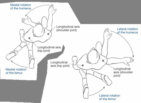
18 Movement of the Skeleton There are three main types of joints: Fibrous joints Cartilaginous joints Synovial joints Synovial joint movement occurs within the three planes of motion: sagittal, frontal, and transverse. Movement occurs along the joint s axis of rotation, where the plane of movement is generally perpendicular to the axis. Uniplanar joints (hinge joints) allow movement in only one plane. Biplanar joints allow movement in two planes that are perpendicular to each other. Multiplanar joints allow movement in all three planes.
19 Movement of Synovial Joints

20 Movement in the Sagittal Plane
21 Movement in the Frontal Plane The frontal plane runs laterally, dividing the body into anterior and posterior sections. Movements that involve rotation about an anteroposterior axis occur in the frontal plane. Examples include: Abduction Adduction Elevation Depression Inversion Eversion

22 Movement in the Transverse Plane
23 Multiplanar Movement Circumduction and opposition are two specific actions that occur in multiple planes. Circumduction: cone motion; combines flexion, extension, abduction, and adduction in sequence Opposition: thumb movement specific to humans and primates
24 Nervous System The nervous system connects the muscles to the brain and spinal cord through a network of nerve circuits that direct the ebb and flow of muscular energy. Structurally, it is divided into the central nervous system (CNS) and peripheral nervous system (PNS). The CNS consists of the brain and spinal cords, while the PNS consists of all the nerve structures outside the brain and spinal cord. Nerves are made up of multiple nerve cells called neurons. Sensory nerves carry impulses to the CNS, while motor nerves carry impulses from the CNS to the PNS.
25 Proprioception Proprioception is the sense of knowing where the body is in relation to its various segments and the external environment. Receptors in the skin, in and around the joints and muscles, and in the inner ear transmit the information. The primary receptors involved in muscular control and coordination are the Golgi tendon organs (GTO) and the muscle spindles.
26 Musculotendinous Receptors Muscle spindle Located in the muscle belly lying parallel to the fibers Causes a reflexive contraction (stretch reflex) in the muscle when the muscle senses a force. It simultaneously causes the relax (reciprocal inhibition). GTO Located between the muscle belly and its tendon Causes muscle inhibition (autogenic inhibition) when it senses tension. stretch antagonist to
27 Muscular System Three types of muscle: Skeletal Attaches to the skeleton via tendons, contracts to move bones Voluntary Striated appearance Smooth Found on the walls of hollow organs and tubes (e.g., stomach, blood vessels) Involuntary Smooth appearance Cardiac Forms the walls of the heart Involuntary Smooth appearance
28 Skeletal Muscle Fiber Types Skeletal fibers can be divided into two general categories based on how quickly they contract. Slow-twitch muscle fibers (also called slow oxidative or type I muscle fibers) contain relatively large amounts of mitochondria and are surrounded by more capillaries than fast-twitch fibers. As the name implies, slow-twitch fibers contract more slowly than fast-twitch fibers. They have lower force outputs, but are more efficient and fatigueresistant than fast-twitch fibers. Fast-twitch muscle fibers (also called type II muscle fibers) are further subdivided into fast-glycolytic (type IIx) and fast-oxidative glycolytic (type IIa) fibers. Type IIx muscle fibers contain a relatively small amount of mitochondria, have a limited capacity for aerobic metabolism, and fatigue more easily than slow-twitch fibers. They have considerable anaerobic capacity, and are the largest and fastest, and are capable of producing the most force, of all the skeletal muscle fibers. Type IIa muscle fibers possess speed, fatigue, and force-production capabilities somewhere between type I and type IIx fibers. For this reason, type IIa fibers are also called intermediate fibers.
29 Comparison of Muscle Fiber Types The following table compares the three types of muscle fiber using the relative terms low, medium, and high. Type I Type IIa Type IIx Speed of Low Medium High contraction Force capacity Low Medium High Fatigue resistance High Medium Low Mitochondrial High Medium Low content Size Low Medium High Efficiency High Medium Low Aerobic capacity High Medium Low Anaerobic capacity Low Medium High
30 Muscle-fiber Microanatomy Skeletal muscles are made up of many muscle fibers held in place by connective tissue (fascia). Muscle fibers are made up of myofibrils (protein filaments) composed of a series of repeating segments called sarcomeres. Sarcomeres, made up of thick (myosin) and thin (actin) myofilaments, are the functional contracting unit of skeletal muscle.
 and the muscle fiber")
31 Muscle Contraction Sliding filament model When acetylcholine is released from the CNS and detected, calcium is released. Calcium exposes binding sites along the actin for the myosin to attach. If sufficient ATP is present, cross-bridges are formed and the myosin pulls the actin toward the center, thereby shortening the sarcomere (all sarcomeres shorten simultaneously) and the muscle fiber itself. If multiple muscle fibers are stimulated to contract at the same time, the muscle will try to actively shorten by contracting.
32 Connective Tissue There are two types of connective tissue directly related to joint movement: Collagen Made up of proteins that provide tensile strength and relative inextensibility, therefore limiting motion and resisting stretch Found in tendons and ligaments Elastic fibers Made up of amino acids and allow for extensibility Surround the sarcomere and are found in other organs Tendons are tough, cord-like tissues that transmit force from the muscle to the bone, causing movement. Ligaments contain a greater mixture of collagen and elastic fibers, taking on various shapes that support a joint by attaching bone to bone.

33 Factors That ImpactFlexibility Soft tissues contribute to the total resistance to joint movement as follows: Joint capsule: 47% Muscle (fasciae): 41% Tendons: 10% Skin: 2% Other factors that impact flexibility include: Age Muscle strength, endurance, flexibility, and agility naturally decrease with age due to muscle atrophy that coincides with increased collagen. Gender In general, females are more flexible than males due to anatomical and physiological differences. Joint structure and past injury The rebuilding of broken bones and the build-up of scar tissue can limit joint movement.
34 The Shoulder Girdle The muscles of the shoulder girdle act on the scapula, primarily to stabilize it. There are six major muscles that anchor the scapula. Four posterior muscles: trapezius, rhomboid major, rhomboid minor, and levator scapulae Two anterior muscles: pectoralis minor and serratus anterior

35 Major Muscles That Act at the Shoulder Girdle This table lists the origins, insertions, primary functions, and examples of exercises for the six major muscles that act at the shoulder girdle.
36 The Shoulder The shoulder joint is the most mobile joint in the body. There are a total of nine muscles that cross the shoulder joint (inserting on the humerus). Seven muscles originate from the scapulae: supraspinatus, infraspinatus, subscapularis, teres minor, deltoid, teres minor, and coracobrachialis Two muscles originate from the axial skeleton (no attachment on the scapula): pectoralis major and latissimus dorsi
37 The Rotator Cuff Four of the muscles that act at the shoulder are commonly called the rotator cuff. The rotator cuff s primary stabilizing function is to hold the humeral head in the glenoidfossa to prevent subluxation (dislocation). The muscles of the rotator cuff can be remembered using the acronym SITS: Supraspinatus Infraspinatus Teres minor Subscapularis

38 Major Muscles That Act at the Shoulder This table lists the origins, insertions, primary functions, and examples of exercises for five major muscles that act at the shoulder.
39 The Elbow Flexion and extension of the elbow are controlled by muscles in the upper arm: biceps brachii, brachialis, brachioradialis, and triceps brachii. Pronation and supination of the forearm are controlled by muscles in the upper arm (biceps brachii and brachioradialis), as well as several muscles in the forearm (pronator teres, pronatorquadratus, and supinator).
40 The Wrist The majority of the muscles that act at the wrist cross the elbow (only slight actions occur at the elbow) and are responsible for flexion and extension of the wrist and pronation and supination of the forearm. The muscles that flex the wrist originate primarily from or near the medial epicondyle of the humerus. The muscles that extend the wrist originate primarily from or near the lateral epicondyle of the humerus.

41 Major Muscles That Act at the Elbow and Forearm This table lists the origins, insertions, primary functions, and examples of exercises of the seven major muscles that act at the elbow and forearm.

42 Major Muscles That Act at the Wrist This table lists the origins, insertions, primary functions, and examples of exercises of the five major muscles involved in flexion and extension of the wrist.
43 The Trunk The major muscles of the trunk support, stabilize, and move the spine. These muscles include the rectus abdominis, external obliques, internal obliques, transverse abdominis, erector spinae, and multifidi. The abdominal wall, made up of the rectus abdominis, obliques, and transverse abdominis, has no skeletal support. Its strength comes from the multidirectional layers of muscle.

44 Major Muscles That Act at the Trunk This table lists the origins, insertions, primary functions, and examples of exercises of the major muscles of the trunk.

45 Hip Flexors There are 21 major muscles involved in the actions of the hip joint. Actions of the hip joint include flexion, extension, internal rotation, external rotation, adduction, and abduction. More than half of these muscles are involved in multiple actions. The hip flexors include the iliopsoas, rectus femoris, tensor fasciae latae, sartorius, and pectineus.

46 Hip Extensors The hip extensors include the gluteus maximus, biceps femoris, semitendinosus, and semimembranosus.
47 The hip internal rotators include the tensor fasciae latae, semitendinosus (slight), and semimembranosus (slight). The hip external rotators include the iliopsoas, gluteus maximus, biceps femoris (slight), gluteus medius and minimus (posterior fibers), sartorius, pectineus, and the six deep external rotators.

48 The hip adductors include the semitendinosus, semimembranosus, adductor magnus, adductor brevis, adductor longus, pectineus, and gracilis.

49 Hip Abductors The hip abductors include the gluteus maximus, biceps femoris, gluteus medius and minimus, and tensor fasciae latae.

50 The Knee Joint The muscles of the upper thigh are responsible for movement at the knee. Knee extensors include the rectus femoris, vastusintermedialis, vastusmedialis, vastuslateralis, and sartorius. Knee flexors include the biceps femoris, semitendinosus, semimembranosus, gracilis, sartorius, and popliteus. This table lists the origins, insertions, primary functions, and examples of exercises for the eight muscles that act at the knee joint.

51 The Anterior Compartment of the Lower Leg The ankle joint allows dorsiflexion and plantarflexion. The subtalar joint allows inversion and eversion of the foot. The muscles of the lower leg control movements of the ankle and foot. The lower leg is divided into three primary compartments: anterior, posterior, and lateral. The anterior compartment is made up of muscles that extend the toes and dorsiflex and/or invert the foot, including the anterior tibialis, extensor hallucislongus, extensor digitorumlongus, and peroneoustertius.

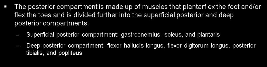
52 The Posterior Compartment of the Lower Leg

53 The Posterior Compartment of the Lower Leg (cont.) Gastrocnemius

54 The Lateral Compartment of the Lower Leg The lateral compartment is made up of muscles that plantarflex and evert the foot, including the peroneus longus and peroneus brevis.
55 The Endocrine System The endocrine system, which is made up of various glands throughout the body, is responsible for regulating bodily activities through the production of hormones. The principal glands are as follows: Pituitary Thyroid Parathyroids Adrenals Paradrenals Gonads

56 Major Endocrine Glands and Their Hormones
57 Summary To design safe and effective programs and group fitness classes, fitness professionals must have working knowledge of human anatomy. Understanding the terminology and major systems will provide a foundation for successfully working with clients or class participants to achieve health and fitness goals. This session covered: Anatomical terminology Structural levels of the body The cardiovascular, respiratory, digestive, skeletal, neuromuscular, muscular, and endocrine systems Planes of motion Upper- and lower-extremity and trunk muscles Muscle fiber types
ACE s Essentials of Exercise Science for Fitness Professionals. Chapter 1: Human Anatomy
 ACE s Essentials of Exercise Science for Fitness Professionals Chapter 1: Human Anatomy Learning Objectives This chapter covers the seven physiological systems of the human body that all fitness professionals
Temporalis Elevates & retracts mandible. Masseter Elevates mandible. Sternocleidomastoid Neck flexion. Trapezius Elevates & depresses shoulders
 Anterior Posterior Temporalis Elevates & retracts mandible Masseter Elevates mandible Sternocleidomastoid Neck flexion Trapezius Elevates & depresses shoulders Masseter Elevates mandible Temporalis Elevates
Types of Muscle: Skeletal- muscle involved in movement of the skeleton. Striated, has alternating bands of light and dark due to overlapping
 Types of Muscle: Skeletal- muscle involved in movement of the skeleton. Striated, has alternating bands of light and dark due to overlapping filaments within the muscle cell. Skeletal muscle can be consciously
Chapter 9. The Muscular System
 1 Chapter 9 The Muscular System 2 Introduction Skeletal muscles: movement in environment Smooth muscles: intestines, ureters, veins and arteries Cardiac muscle: pumps blood through heart and blood vessels
Cadaver Muscular System Practice Practical
 Cadaver Muscular System Practice Practical Station 1 Station 1 1. Specific structure 1. Rectus sheath 2. Red line 2. Linea alba Station 2 Station 2 3. Red muscle 1. Rectus abdominis 4. Red muscle actions
Certified Personal Trainer Re-Certification Manual
 Certified Personal Trainer Re-Certification Manual Section II 1 Anatomy & Physiology Terms Anatomy and physiology are closely related fields of study: anatomy is the study of form, and physiology is the
Muscles of the Cat. N Deltoid MUSCLES OF THE CHEST. Pectoralis major. (This muscle is superior to Pectoralis minor) MUSCLES OF THE CHEST
 MUSCLES OF THE CHEST") MUSCLES OF THE CHEST Pectoralis major (This muscle is superior to Pectoralis minor) 1. MUSCLES OF THE CHEST Pectoralis minor (This muscle is inferior to Pectoralis major) 2. MUSCLES OF THE ARM Deltoid
MUSCLES OF THE CHEST Pectoralis major (This muscle is superior to Pectoralis minor) 1. MUSCLES OF THE CHEST Pectoralis minor (This muscle is inferior to Pectoralis major) 2. MUSCLES OF THE ARM Deltoid
or Everything you ever wanted to know about Muscles, but were afraid to ask!!!
 The Muscular System or Everything you ever wanted to know about Muscles, but were afraid to ask!!! Did you know that? - more than 50% of body weight is muscle! - And muscle is made up of proteins and water
Exercise Science Section 3: The Muscular System
 Exercise Science Section 3: The Muscular System An Introduction to Health and Physical Education Ted Temertzoglou Paul Challen ISBN 1-55077-132-9 Major Functions of Muscles Movement Includes: breathing,
Muscle stations Answers
 Muscle Unit Muscle stations Answers A: What #is: C = 3 F = 5 E = 6 D = 1 B =4 A =2 B 5. superior 6. Inferior 4. anterior C: 1. What # is a,b,c,d 2. What muscle group #1? Quads 3. What muscle is #5? Gastrocnemius
Human Anatomy Lab #7: Muscles of the Cadaver
 Human Anatomy Lab #7: Muscles of the Cadaver Table of Contents: Expected Learning Outcomes.... 1 Introduction...... 1 Identifying Muscles on Yourself.... 2 Muscles of the Anterior Trunk and Arm.. 2 Muscles
Applied Human Biology for Exercise and Fitness Level 3 J/615/3220 MOCK PAPER
 Applied Human Biology for Exercise and Fitness Level 3 J/615/3220 MOCK PAPER There are 40 questions within this paper. To achieve a pass you will need to score 28 out of 40 marks. All questions are multiple
Location Terms. Anterior and posterior. Proximal and Distal The term proximal (Latin proximus; nearest) describes where the appendage joins the body.
 describes where the appendage joins the body.") HUMAN ANAT OMY Location Terms Anterior and posterior In human anatomical usage, anterior refers to the front of the individual. Similarly, posterior refers to the back of the subject. In standard anatomical
HUMAN ANAT OMY Location Terms Anterior and posterior In human anatomical usage, anterior refers to the front of the individual. Similarly, posterior refers to the back of the subject. In standard anatomical
The Muscular System. Myology the study of muscles
 The Muscular System Myology the study of muscles Functions of muscles: 1. Movement 2. Stability /support posture 3. Heat production 85% of our body heat 4. Communication 5. Constriction of organs and vessels
Unit 7: Skeletal and muscular systems
 Unit 7: Skeletal and muscular systems 1. The locomotor system 2. The skeletal system 2.1. The human skeleton 2.2. Bones 2.3. Joints 2.4. Tendons and ligaments 3. The muscular system 3.1. Muscles of the
Muscular System. IB Sports, exercise and health science 1.2
 Muscular System IB Sports, exercise and health science 1.2 Characteristics Common to Contractility-ability to shorten the muscles length Extensibility-ability to lengthen the muscles length Elasticity-muscle
Muscle. Dr. Carmen E. Rexach Anatomy 35 Mt San Antonio College
 Muscle Dr. Carmen E. Rexach Anatomy 35 Mt San Antonio College Functions Movements of bones and soft body parts Movements of fluids through a tube (blood, digestive) Functions Maintain posture Support soft
The Muscular System PART C. PowerPoint Lecture Slide Presentation by Patty Bostwick-Taylor, Florence-Darlington Technical College
 PowerPoint Lecture Slide Presentation by Patty Bostwick-Taylor, Florence-Darlington Technical College The Muscular System 6 PART C Five Golden Rules of Skeletal Muscle Activity Table 6.2 Muscles and Body
Human Anatomy and Physiology I Laboratory
 Human Anatomy and Physiology I Laboratory Gross Anatomy of the Muscular System (Two weeks) 1 This lab involves study of the laboratory exercise Gross Anatomy of the Muscular System. Complete the Review
Mock Paper Level 2 Anatomy and Physiology for Exercise. Unit Reference Number H/600/9013
 MULTIPLE CHOICE QUESTION PAPER Paper number MPAPEH2.01 Please insert this reference number in the appropriate boxes on your candidate answer sheet Title Time allocation 60 minutes Mock Paper Level 2 Anatomy
2/4/2018. Identify the two reasons why muscle cells may go through muscle fatigue. Ch.7 Review. Sternocleidomastoid.
 Ch.7 Review Identify the two reasons why muscle cells may go through muscle fatigue Temporalis Depressor anguli oris Sternocleidomastoid Tibialis anterior 1 Gluteus medius Deltoid Adducts & rotates scapula
Chiropractic Technician Class
 Chiropractic Technician Class Presentation By: Dr. Kay Miller. The Role of Exercise as it Relates to Our Musculoskeletal System Introduction to the topic and Preliminary Physical exam Musculoskeletal anatomy:
Level 2 Mock Paper Anatomy and Physiology For Exercise. Unit Accreditation Number H/600/9013
 MULTIPLE CHOICE QUESTION PAPER Paper number MPAPEH Please insert this reference number in the appropriate boxes on your candidate answer sheet Title Time allocation 60 minutes Level 2 Mock Paper Anatomy
Test Bank for The Human Body in Health and Illness 4th Edition by Herlihy
 Test Bank for The Human Body in Health and Illness 4th Edition by Herlihy Chapter 9: Muscular System Test Bank MULTIPLE CHOICE 1. Which of the following muscles is described as striated and involuntary?
Anatomy and Physiology for Exercise and Health Level 3 A/600/9051 Mock Paper March 1 st 2015 August 31 st 2015
 Anatomy and Physiology for Exercise and Health Level 3 A/600/9051 Mock Paper March 1 st 2015 August 31 st 2015 There are 40 questions within this paper. To achieve a pass you will need to score 28 out
5 Specification Content
 5 Specification Content These specifications are set out in the form of teaching modules. Each teaching module is assessed by its associated unit of assessment. 5.1 Module 2562: The Application of Physiological
A. All movements require muscle which are organs using chemical energy to contract.
 Ch 8 Muscles Introduction: A. All movements require muscle which are organs using chemical energy to contract. B. The three types of muscle in the body are skeletal, smooth, and cardiac muscle. C. This
Epicranius (frontal belly) Zygomaticus minor. Zygomaticus major Buccinator
 Zygomaticus minor. Zygomaticus major Buccinator") Epicranius (frontal belly) Zygomaticus minor Zygomaticus major Buccinator Masseter Digastric (posterior belly) Stylohyoid Sternocleidomastoid Trapezius Scalenus Omohyoid (inferior belly) Orbicularis oris
Epicranius (frontal belly) Zygomaticus minor Zygomaticus major Buccinator Masseter Digastric (posterior belly) Stylohyoid Sternocleidomastoid Trapezius Scalenus Omohyoid (inferior belly) Orbicularis oris
Level 2 Anatomy and Physiology Internal Practice Paper
 Level 2 Anatomy and Physiology Internal Practice Paper Time allocated: 60 minutes. 30 questions, multiple choice answers. Select A,B,C or D only select one answer. You are required to achieve 22 correct
CHAPTER 1: 1.1 Muscular skeletal system. Question - text book page 16. Question - text book page 20 QUESTIONS AND ANSWERS. Answers
 QUESTIONS AND ANSWERS CHAPTER 1: 1.1 Muscular skeletal system Question - text book page 16 Using the information on pages 12 to 14 above, complete the table below. joint joint type articulating bones associated
Level 2 Anatomy and Physiology for Exercise and Fitness Instructors (K/616/7823) - Sample Assessment Student: XXXXXX Sample 3
 - Sample Assessment Student: XXXXXX Sample 3") MULTIPLE CHOICE QUESTION PAPER Paper number: SAMPLE 3 Please ensure that this paper number is referenced on your candidate answer sheet Title: Student: XXXXXX Sample 3 Special Instructions: Level 2 Anatomy
MULTIPLE CHOICE QUESTION PAPER Paper number: SAMPLE 3 Please ensure that this paper number is referenced on your candidate answer sheet Title: Student: XXXXXX Sample 3 Special Instructions: Level 2 Anatomy
The Human Muscular System Required reading before beginning this lab: Saladin, KS: Human Anatomy 5th ed (2017) Chapters 10, 11, 12 INTRODUCTION
 Chapters 10, 11, 12 INTRODUCTION") Biology 322: Human Anatomy The Human Muscular System Required reading before beginning this lab: Saladin, KS: Human Anatomy 5 th ed (2017) Chapters 10, 11, 12 INTRODUCTION We will use a number of lab periods
Biology 322: Human Anatomy The Human Muscular System Required reading before beginning this lab: Saladin, KS: Human Anatomy 5 th ed (2017) Chapters 10, 11, 12 INTRODUCTION We will use a number of lab periods
Exercise Science Section 3: The Muscular System
 Exercise Science Section 3: The Muscular System An Introduction to Health and Physical Education Ted Temertzoglou Paul Challen ISBN 1-55077-132-9 Major Functions of Muscles Movement v Includes: breathing,
Due in Lab weeks because of Thanksgiving Prelab #10. Homework #8. Both sides! Both sides!
 Lab 8 MUSCLES Due in Lab 10 2 weeks because of Thanksgiving Prelab #10 Both sides! Homework #8 Both sides! Refer to Muscles 22-23 Naming of muscles Origin Site of muscle attachment that doesn t move during
Energy for Muscle Contractions: Direct phosphorylation. Creatine phosphate loses a phosphate to ADP to create ATP
 Energy for Muscle Contractions: Direct phosphorylation Aerobic respiration Anaerobic respiration (lactic acid fermentation) Creatine phosphate loses a phosphate to ADP to create ATP Requires oxygen to
3/27/2012. Muscle Classification: Functional Groups. Interactions of Skeletal Muscles. Naming Skeletal Muscles. Naming Skeletal Muscles
 Interactions of Skeletal Muscles Skeletal muscles work together or in opposition Muscles only pull (never push) As muscles shorten, the insertion generally moves toward the origin Whatever a muscle (or
Chapter 3: Applied Kinesiology. ACE Personal Trainer Manual Third Edition
 Chapter 3: Applied Kinesiology ACE Personal Trainer Manual Third Edition Introduction Kinesiology is the study of the body s infinite number of movements, positions, and postures and is grounded in the
Human Anatomy Unit 2 MUSCULAR SYSTEM
 Human Anatomy Unit 2 MUSCULAR SYSTEM In Anatomy Today Functions Movements of bones and soft body parts Movements of fluids through a tube (blood, digestive) Functions Maintain posture Support soft organs
Unit 1: Human body: combination I - IV
 Unit 1: Human body: combination I - IV Study online at quizlet.com/_1kzmm2 1. alveoli 6. bronchioles microscopic air sacs in the lung where diffusion of the respiratory gases, oxygen and carbon dioxide
The Human Body. Lesson Goal. Lesson Objectives 9/10/2012. Provide a brief overview of body systems, anatomy, physiology, and topographic anatomy
 The Human Body Lesson Goal Provide a brief overview of body systems, anatomy, physiology, and topographic anatomy Medial Lateral Proximal Distal Superior Inferior Anterior Lesson Objectives Explain the
Name this muscle. Name this muscle
 this muscle this muscle Pectoralis Major Pectoralis Minor Serratus anterior Pectoralis minor Serratus anterior this muscle Deltoid: The major abductor of the upper limb this muscle this muscle this muscle
OBJECTIVES. Unit 7:5 PROPERTIES OR CHARACTERISTICS OF MUSCLES. Introduction. 3 Kinds of Muscles. 3 Kinds of Muscles 4/17/2018 MUSCULAR SYSTEM
 OBJECTIVES Unit 7:5 MUSCULAR SYSTEM Compare the three main kinds of muscles by describing the action of each Differentiate between voluntary and involuntary muscles List at least three functions of muscles
In which arm muscle are intramuscular injections most often given? (not in text)
") AP1 Lab 9 - Muscles of the Arms and Legs Locate the following muscles on the models and on yourself. Recall anatomical position. Directional terms such as anterior, posterior, lateral, etc. all assume
AP1 Lab 9 - Muscles of the Arms and Legs Locate the following muscles on the models and on yourself. Recall anatomical position. Directional terms such as anterior, posterior, lateral, etc. all assume
Match the types of muscle tissues with the words and phrases. 1) Skeletal 2) Smooth 3) Cardiac 2 Walls of blood vessels. 2 Walls of digestive tract
 Skeletal 2) Smooth 3) Cardiac 2 Walls of blood vessels. 2 Walls of digestive tract") S T U D Y G U I D E. Types of Muscle Tissues Match the types of muscle tissues with the words and phrases. ) Skeletal ) Smooth ) Cardiac, Striated Walls of blood vessels, Single nucleus Heart muscle, Involuntary
S T U D Y G U I D E. Types of Muscle Tissues Match the types of muscle tissues with the words and phrases. ) Skeletal ) Smooth ) Cardiac, Striated Walls of blood vessels, Single nucleus Heart muscle, Involuntary
Biology 2401 Muscles List for CPC models
 Biology 2401 List for CPC models Italicized muscles are dissect and similar in the cat = Dissect and note the differences in human and cat Major of the Human Head Facial Expression Epicranius frontalis
Level 2 Anatomy and Physiology for Exercise and Fitness Instructors (K/616/7823) - Sample Assessment Student: XXXXXX Sample 4
 - Sample Assessment Student: XXXXXX Sample 4") MULTIPLE CHOICE QUESTION PAPER Paper number: SAMPLE 4 Please ensure that this paper number is referenced on your candidate answer sheet Title: Student: XXXXXX Sample 4 Special Instructions: Level 2 Anatomy
MULTIPLE CHOICE QUESTION PAPER Paper number: SAMPLE 4 Please ensure that this paper number is referenced on your candidate answer sheet Title: Student: XXXXXX Sample 4 Special Instructions: Level 2 Anatomy
Chapter 10: Muscular System: Gross Anatomy
 Chapter 10: Muscular System: Gross Anatomy I. General Principles A. General Terminology 1. Tendons attach 2. What is an aponeurosis? 3. The points of muscle attachment are called & 4. How is the "origin"
Unit 4: The Muscular System REVIEW GUIDE
 NPHS Anatomy & Physiology Questions to answer: 1) List the three functions of the muscular system. Unit 4: The Muscular System REVIEW GUIDE 2) What are the four characteristics of muscle tissue? Briefly
Synergist Muscles. Shoulder (glenohumeral joint) Flexion Deltoid (anterior fibers) Pectoralis major (upper fibers) Biceps Brachii Coracobrachialis
 Flexion Deltoid (anterior fibers) Pectoralis major (upper fibers) Biceps Brachii Coracobrachialis") Synergist Muscles Dr Gene Desepoli DrGeneLMT@gmail.com Shoulder (glenohumeral joint) Deltoid (anterior fibers) Pectoralis major (upper fibers) Biceps Brachii Coracobrachialis Deltoid (posterior fibers)
Synergist Muscles Dr Gene Desepoli DrGeneLMT@gmail.com Shoulder (glenohumeral joint) Deltoid (anterior fibers) Pectoralis major (upper fibers) Biceps Brachii Coracobrachialis Deltoid (posterior fibers)
LEARN - INSPIRE - SUCCEED
 Anatomy and Physiology Workbook LEARN - INSPIRE - SUCCEED Label The Skeletal System Fibula Lumbar vertebrae Patella Sternum Ilium Femur Scapula Phalanges Sacrum Ischium Tarsals Cranium Clavicle Pubis Ribs
EXERCISE PHOTOS, TIPS AND INSTRUCTIONS
 Page 1 of 21 EXERCISE PHOTOS, TIPS AND INSTRUCTIONS Page 2. Squat Page 12. Crab Walks Page 3. Single Leg Squat Page 13. Bench Press Page 4. Split Squat Page 14. Bench Pull Page 5. Deadlift Page 15. Shoulder
VCE PHYSICAL EDUCATION WORKBOOK UNIT 1 BODIES IN MOTION NAME:
 VCE PHYSICAL EDUCATION WORKBOOK UNIT 1 BODIES IN MOTION NAME: SKELETAL SYSTEM List the 5 functions of the skeletal system and complete the following table. FUNCTION DESCRIPTION Label the following features
CHAPTER 4: The musculo-skeletal system. Practice questions - text book pages QUESTIONS AND ANSWERS. Answers
 CHAPTER 4: The musculo-skeletal system Practice questions - text book pages 64-66 1) A prime mover of hip flexion is the: a. rectus femoris. b. Iliopsoas. c. vastus muscles. d. gluteus maximus. b. Key
VCE PHYSICAL EDUCATION WORKBOOK UNIT 1 BODIES IN MOTION NAME:
 VCE PHYSICAL EDUCATION WORKBOOK UNIT 1 BODIES IN MOTION NAME: SKELETAL SYSTEM List the 5 functions of the skeletal system and complete the following table. FUNCTION DESCRIPTION Label the following features
REVISION BOOKLET. The Body Systems
 REVISION BOOKLET The Body Systems GCSE PE 2016 Skeletal System Functions of the skeleton Joints for movement Muscle attachment Protection of vital organs Red and white blood cell production platelets Storage
2º ESO - PE Workbook - IES Joan Miró Physical Education Department THE MUSCULAR SYSTEM
 THE MUSCULAR SYSTEM The muscular system is one of 10 organ systems in the human body. The human body has more than 650 muscles, which make up half of a person's body weight. Without muscles, we would not
Unit 6: The Muscular System
 Unit 6: The Muscular System I. The Muscular System A. Muscles are responsible for all types of body movement B. Three basic muscle types are found in the body 1. Skeletal muscle 2. Cardiac muscle 3. Smooth
Anatomy and physiology for sport
 Anatomy and physiology for sport UV21527 D/502/5474 Learner name: VRQ Learner number: VTCT is the specialist awarding body for the Hairdressing, Beauty Therapy, Complementary Therapy, Hospitality and Catering
Anatomy and Physiology for Exercise Level 2
 Anatomy and Physiology for Exercise Level 2 H/600/9013 Mock Paper There are 30 questions within this paper To achieve a pass you will need to score 21 out of 30 marks All questions are multiple choice
The Muscular System The Muscular System Muscles are responsible for all types of body movement Three basic muscle types are found in the body
 The Muscular System The Muscular System Muscles are responsible for all types of body movement Three basic muscle types are found in the body Skeletal muscle Cardiac muscle Smooth muscle Characteristics
Monday, November 13, 2017 A & P 2401
 Monday, November 13, 2017 A & P 2401 Today you will complete the following handouts. Study the last part of the handout for this will be on your quiz, which will be on Wednesday. It is titled steps of
Lectures Muscular System 10-1
 Lectures 12-14 Muscular System 10-1 Properties of Muscle Ability of a muscle to shorten with force Capacity of muscle to respond to a stimulus Muscle can be stretched to its normal resting length and beyond
Muscles of the Hip 1. Tensor Fasciae Latae O: iliac crest I: lateral femoral condyle Action: abducts the thigh Nerve: gluteal nerve
 Muscles of the Hip 1. Tensor Fasciae Latae O: iliac crest I: lateral femoral condyle Action: abducts the thigh Nerve: gluteal nerve 2. Gluteus Maximus O: ilium I: femur Action: abduct the thigh Nerve:
Level 3 Diploma in Personal Training (Practitioner) - Sample Assessment Materials
 - Sample Assessment Materials") MULTIPLE CHOICE QUESTION PAPER Paper number: SAMPLE 5 Please ensure that this paper number is referenced on your candidate answer sheet Title: Student: XXXXXX Sample 5 Special Instructions: Level 3 Diploma
MULTIPLE CHOICE QUESTION PAPER Paper number: SAMPLE 5 Please ensure that this paper number is referenced on your candidate answer sheet Title: Student: XXXXXX Sample 5 Special Instructions: Level 3 Diploma
Anatomy. Anatomy deals with the structure of the human body, and includes a precise language on body positions and relationships between body parts.
 Anatomy deals with the structure of the human body, and includes a precise language on body positions and relationships between body parts. Proper instruction on safe and efficient exercise technique requires
Personal Training Certificate. Anatomy and Physiology Mock Paper
 Candidate Name Date Personal Training Certificate Anatomy and Physiology Mock Paper Instructions: Make sure your name is in the box at the top, followed by the date you did the test Tick or highlight your
10/30/2014 APPEARANCE
 APPEARANCE Striated: has a striped appearance due to the thickness of the protein fibers Smooth: protein fibers (which are arranged the same in striated muscle) is not as thick so you cannot see the pattern
Human Muscles (Anterior View) Model 3-44
 Model 3-44") Human Muscles (Anterior View) Model 3-44 Temporalis Frontalis Orbicularis Occuli Orbicularis Oris Masseter Sternocleidomastoid Orbicularis Occuli Human Muscles (Anterior View) Model 3-65 Temporalis Masseter
Human Muscles (Anterior View) Model 3-44 Temporalis Frontalis Orbicularis Occuli Orbicularis Oris Masseter Sternocleidomastoid Orbicularis Occuli Human Muscles (Anterior View) Model 3-65 Temporalis Masseter
National Fitness Leadership Alliance
 Exercise Theory s November 2005 National Fitness Leadership Alliance Exercise Theory s National Fitness Leadership Alliance (NFLA) 1 Exercise Theory s November 2005 National Fitness Leadership Alliance
The Muscular System Lab Power Point
 The Muscular System Lab Power Point Myoneural Junction Sarcoplasm Nucleus Myofibrils Sarcomere (black line to black line) Sarcolemma Myoneural space Nucleus Endomysium Motor Neuron Muscles of Facial Expression
Head & Neck The muscle names are followed by the chapter number
 Head & Neck The muscle names are followed by the chapter number. Splenius capitis (9) 2. Occipitalis (2) Temporalis () 3. Temporalis () 4. Semispinalis capitis (9) Facial / Scalp (2) 5. Temporalis () Facial
Prime movers provide the major force for producing a specific movement Antagonists oppose or reverse a particular movement Synergists
 Dr. Gary Mumaugh Prime movers provide the major force for producing a specific movement Antagonists oppose or reverse a particular movement Synergists Add force to a movement Reduce undesirable or unnecessary
Scapula Spine Lateral edge of clavicle. Medial border Scapula. Medial border of Scapula, between superior angle and root of spine. Scapula.
 Muscle attachments and actions answer sheet Muscle Origins insertions Movements Joints crossed Trapezius Base of skull Spinous process of C7 Thoracic Spine Lateral edge of clavicle Elevation Retraction
The Muscular System. Chapter 10 Part C. PowerPoint Lecture Slides prepared by Karen Dunbar Kareiva Ivy Tech Community College
 Chapter 10 Part C The Muscular System Annie Leibovitz/Contact Press Images PowerPoint Lecture Slides prepared by Karen Dunbar Kareiva Ivy Tech Community College Table 10.9: Muscles Crossing the Shoulder
11/15/2018. Temporalis Elevates & retracts mandible. Masseter = Prime mover of jaw closure. Levator scapulae Supraspinatus Clavicle.
 Due in Lab 10 Lab 8 MUSCLES 2 weeks because of Thanksgiving Prelab #10 Both sides! Homework #8 Both sides! Refer to Muscles 22-23 Examples of Origin & Insertion Naming of muscles Origin Site of muscle
Compiled and Designed by: Sport Dimensions - 2 -
 SOCCER TRAINING While all reasonable care has been taken during the preparation of this edition, neither the publisher, nor the authors can accept responsibility for any consequences arising from the use
10/4/18. Muscular System. 1 Copyright 2016 by Elsevier Inc. All rights reserved. Introduction. Anatomy. Physiology. Skeletal Muscle Anatomy
 Introduction Muscular System Chapter 20 Shortening or lengthening of a muscle results from changes in relative positions of one small part of a muscle cell to another To understand contraction, we will
INTRODUCTION. Objectives
 Objectives Functional Anatomy for Fitness Professionals focuses on functional anatomy, with an emphasis on weight training. Through the emphasis on biomechanics, neurology, and muscle physiology, participants
Unit 6 - The Muscular System 1
 Unit 6 - The Muscular System 1 I. Unit 6: The Muscular System A. The Muscular System 1. Muscles are responsible for all types of body movement 2. Three basic muscle types are found in the body a) Skeletal
National Fitness Leadership Alliance
 National Fitness Leadership Alliance Exercise Theory Prerequisite Knowledge Base 2015 National Fitness Leadership Alliance Exercise Theory Prerequisite Knowledge Base These competencies are intended as
Muscle Anatomy Review Chart
 Muscle Anatomy Review Chart BACK Superficial (5) Trapezius Transverse cervical a. Latissimus dorsi Thoracodorsal a. Rhomboideus major Dorsal scapular a. Rhomboideus minor Levator scapulae Intermediate
Chapter 6- The Muscular System
 Chapter 6- The Muscular System I. The muscular system A. Muscles are responsible for all types of body movement B. Three basic muscle types are found in the body 1. Skeletal muscle 2. Cardiac muscle 3.
BLUE SKY SCHOOL OF PROFESSIONAL MASSAGE AND THERAPEUTIC BODYWORK. Musculoskeletal Anatomy & Kinesiology MUSCLES, MOVEMENTS & BIOMECHANICS
 BLUE SKY SCHOOL OF PROFESSIONAL MASSAGE AND THERAPEUTIC BODYWORK Musculoskeletal Anatomy & Kinesiology MUSCLES, MOVEMENTS & BIOMECHANICS MSAK101-I Session 7 Learning Objectives: 1. List the three types
PHYSICAL EDUCATION. 4º E.S.O. 2nd TERM. The skeletal and muscular systems.
 PHYSICAL EDUCATION 4º E.S.O. 2nd TERM. The skeletal and muscular systems. PARTS OF THE BODY Head Torso / Trunk Dorsal: Back Ventral: Thorax y Abdomen Extremities Superior: Arm Forearm Hand Joint: Shoulder
Naming Skeletal Muscles
 Naming Skeletal Muscles Direction of Muscle Fibers Action Location Origin & Insertion Skeletal Muscle Size Shape Number Of Origins Direction of Muscle Fibers Relative to the Midline RECTUS = parallel to
MOCK PAPER Level 3 Anatomy and Physiology For Exercise and Health. Unit Reference Number A/600/9051
 MULTIPLE CHOICE QUESTION PAPER Paper number APEH 3.01 Please insert this reference number in the appropriate boxes on your candidate answer sheet Title Time allocation 60 minutes MOCK PAPER Level 3 Anatomy
7/10/18. Introduction. Muscular System. Anatomy. Physiology. Skeletal Muscle Anatomy. Muscle Fiber
 Introduction Muscular System Chapter 20 Shortening or lengthening of a muscle results from changes in relative positions of one small part of a muscle cell to another To understand contraction, we will
How Muscles are Classified Muscles The three main types of muscles are,, and
 7.5 Muscles: Designed for Motion How Muscles are Classified Muscles The three main types of muscles are,, and o Muscles Muscles that are generally under control (usually ) o Muscles Muscles that not under
YMCA Awards reserves the right to seek legal remedies for any such infringement.
 The content of this document remains the intellectual property of, and is copyright to London Central YMCA (Trading as YMCA Awards). No part of these materials may be reproduced, stored, copied, edited
The Muscular System. - composed of mostly skeletal muscle tissue, nervous tissue, blood and connective tissue
 The Muscular System Every action the body takes utilizes a muscular activity. Some of the muscles of the body are under voluntary control (skeletal muscles), and by using these muscle, you are able to
7 The Muscular System
 C h a p t e r 7 The Muscular System PowerPoint Lecture Slides prepared by Jason LaPres Lone Star College North Harris An Introduction to Muscle Tissue Muscle Tissue A primary tissue type, divided into
MOCK Level 3 Anatomy and Physiology for Exercise and Health
 MULTIPLE CHOICE QUESTION PAPER Paper number APEH 3.0 Please insert this reference number in the appropriate boxes on your candidate answer sheet Title Time allocation 50 minutes MOCK Level 3 Anatomy and
Muscles Unit TEST and Final Exam Study Guide May 2017
 Muscles Unit TEST and Final Exam Study Guide May 2017 Part 1 of final exam is pictures, see bottom of the study guide Part 2 of the final exam is only going to cover muscles unit. If you do this study
The Massage Routine. Start with your client lying face down - Prone Position. Clean YOUR HANDS and CLIENTS FEET using antibacterial wipes
 The Massage Routine Start with your client lying face down - Prone Position Clean YOUR HANDS and CLIENTS FEET using antibacterial wipes!!!! GROUNDING FOR 3 BREATHS TUNE YOUR BREATHING WITH THE CLIENTS!!!
Introduction to Human Body Systems
 The Human Organism: Introduction to Human Body Systems By Deanne Erdmann, MS Levels of Organization in the Body Cells Tissues Epithelial, connective, muscular, nervous Organs Examples include stomach,
Chapter 6 part 2. Skeletal Muscles of the Body
 Chapter 6 part 2 Skeletal Muscles of the Body Basic Principles 600 + muscles in the human body (you are required to learn 45, lucky kids)! Skeletal Muscles pull on bones Origin of a muscle = point of attachment
Three types of muscles
 The Muscular System Three types of muscles Smooth Cardiac Skeletal This chapter focuses on skeletal muscle walls of the viscera (organs), blood vessels, bronchioles Smooth muscle INVOLUNTARY muscle NONSTRIATED
Applied anatomy and physiology: definitions of key terms
 Applied anatomy and physiology: definitions of key terms See pages 5 46 These are the key terms from Chapter 1. Try cutting them out and then matching the key terms with their definitions, or asking friends
The Muscular System home study course
 The Muscular System home study course harmony house holistic therapy treatment centre and training academy www.harmony-house.org 1 Copyright 2010 by Mark and Katy Rogers All rights reserved. No part of