Acquired pediatric esophageal diseases Imaging approaches and findings. M. Mearadji International Foundation for Pediatric Imaging Aid
|
|
|
- Claude Booth
- 6 years ago
- Views:
Transcription
1
2 Acquired pediatric esophageal diseases Imaging approaches and findings M. Mearadji International Foundation for Pediatric Imaging Aid
3 Acquired pediatric esophageal diseases The clinical signs of acquired esophageal lesions are uncharacteristic with a long or short patient history An immediate diagnostic approach is indicated in even slightly suspicious cases
4 Acquired esophageal diseases in childhood 1. Gastro esophageal reflux disease 2. Foreign body ingestion 3. Esophageal trauma and perforation 4. Achalasia 5. Caustic ingestion 6. Infective esophagitis 7. Esophageal varices
5 Non-infective esophagitis (reflux esophagitis) Reflux esophagitis as a non infective inflammation is a common complication of severe gastro-esophageal reflux disease Recurrent vomiting with hematomesis is a usual finding Additional symptoms are failure to thrive and recurrent respiratory infections Patients with psychomotoric retardation are more affected Reflux esophagitis may lead to strictures
6 Radiological findings of reflux esophagitis Dysmotility Thickening of mucosal folds Peptic ulceration Strictures Eosinophilic esophagitis as an atopic condition mimicking the reflux esophagitis


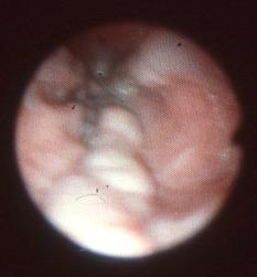
7 A B C Gastro-esophageal reflux disease with reflux esophagitis A. Hemorrhagic esophagitis endoscopical finding B. Sliding hernia with esophagitis confirmed by endoscopy C. Esophagitis detected on CT
8 Esophagitis complicated with strictures and ulcerations
9 Foreign body ingestion The peak incidence is between 6 months and 6 years The most frequent presenting symptoms are dysphagia, (drooling, retching) and vomiting Absence of symptoms does not exclude presence of foreign body in children Ingested foreign bodies range from cheap toys to costly jewels Complications: perforation and abscess formation
10 Radiological findings of esophageal foreign body ingestion Cervicothoraco-abdominal and lateral chest film X-ray film reveals all metal objects and less frequently glass and fishbones Other non-radio-opaque objects such as plastic toys should be visualized with contrast medium as filling defects Treatment: endoscopical or fluoroscopical (with extraction with Foley or magnetic catheter)


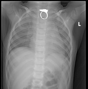
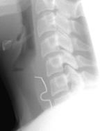
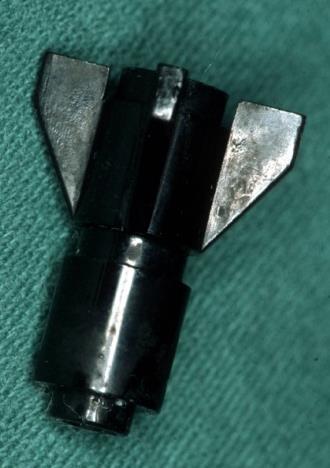
11 Several objects in esophagus 11
12 Esophageal trauma and perforation Mostly iatrogenic The incidence is increasing as more diagnostic and therapeutic endoscopy are performed Perforation occurs as a complication of stricture dilatation (4-6 per 1000 cases) Other etiologies are foreign body, caustic damage, infection and penetrating trauma Iatrogenic perforation of esophagus and pharynx due to malposition of nasogastric tube in neonatal nursery is a rare complication
13 Radiological finding and diagnostic procedures by esophageal perforation AP and lateral chestfilm is the first diagnostic procedure Findings include, pneumomediastinum, pneumothorax, hydropneumothorax, subcutaneous emphysema and pleural effusion Contrast studies with non-ionic media are needed to look for location and extention of esophageal perforation CT is only indicated, when positive clinical signs with negative esophagogram An abnormal position of nasogastric tube in neonates, outside the esophagus in mediastinum or in pharynx with or without pneumomediastinum is suspicious for perforation


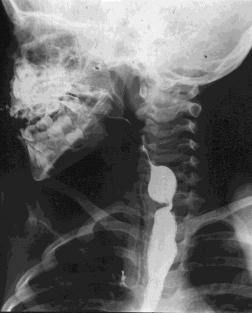
14 Para esophageal perforation caused by feeding tube Edema of the upper esophagus by strangulation Iatrogenic perforation during stretching of esphageal stenosis


15 Two cases of iatrogenic perforation of esophagus with pneumomediastinum during insertion of a nasogastric tube
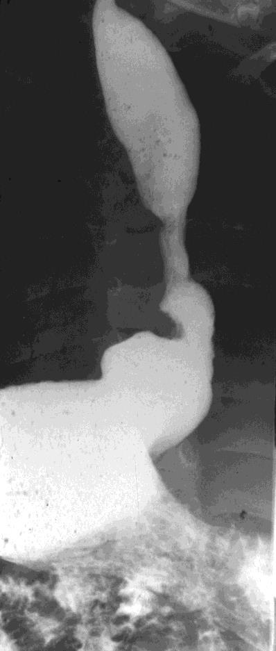
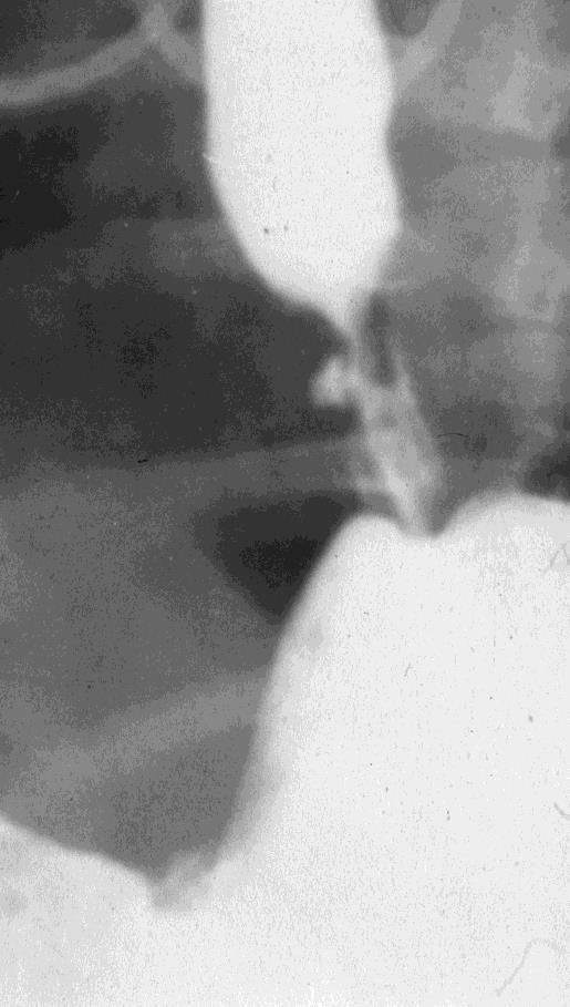
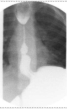
16 Achalasia Rare in children 5% of the affected population with achalasia are children Most affected children are above the age of 5 years, but it is not uncommon to observe the disease in younger children Characterized by defective relaxation of cardia and absence of esophageal peristalsis The clinical signs in older children are dysphagia, chest pain, vomiting of undigested food and poor weight gain In infancy the symptoms are similar to gastroesophageal reflux with regurgitation an recurrent pneumonia
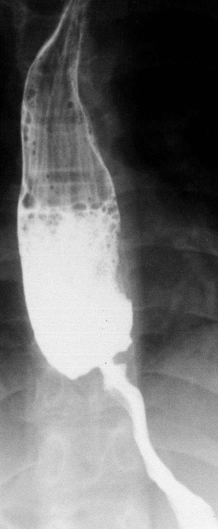
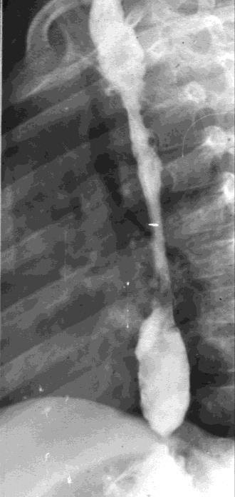
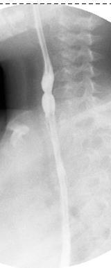
17 Radiological findings of achalasia The chest radiograph may reveal a dilated esophagus with an air fluid level with or without trachea displacement Barium study of esophagus shows a dilated esophagus that tapers smoothly to a birdsbeak Normal peristaltic activity above the aortic arch with abnormal motility in distal part of esophagus Retention of contrast media in esophagus 10 minutes or later after ingestion


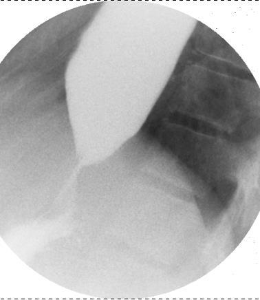
18 Two cases of achalasia Note the fluid level on the chestfilm and delayed esophageal passage direct 1 minute 10 minutes
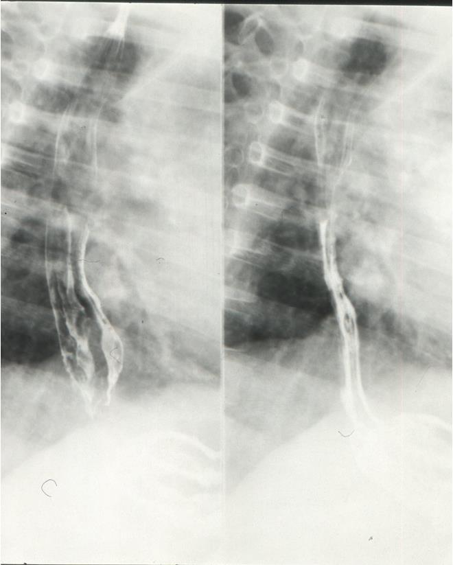
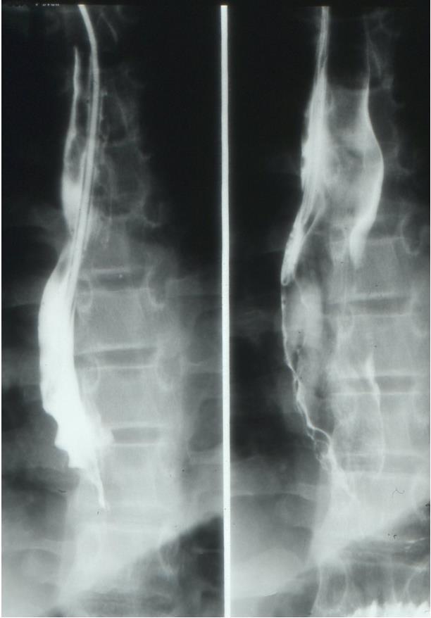
19 Caustic ingestion Most common in children less than 6 years of age Injury depends on physical, characteristic and quantity of the agent ingested and the duration of mucosal contact Caustic ingestion with acidic compounds affects usually the stomach, whereas alkali ingestion causes esophagitis Swelling of epiglottis indicates that the caustic agent has reached the esophagus Most damages occur in the upper and middle esophagus To reduce the risk of perforation an endoscopical evaluation is recommended 48 hours after such incident
20 Radiological procedures and findings for caustic ingestion Initial evaluation by neck film and thoraco-abdominal film Endoscopy is considered to be the diagnostic method of choice in the acute phase, to assess the extent and severity of injury An additional early radiological examination with nonionic contrast media is indicated in case of perforation CT is also indicated if suspicion for perforation is high A barium study should be routinely performed 3 weeks after corrosive injury The presence, number, length and location of esophageal strictures should be visualized for an adequate treatment


21 Four cases with caustic esophagitis and severe strictures
22 Infective esophagitis A rare complication of some severe diseases Usually by opportunistic organisms in children with low immunity (AIDS or leukemic patients) The most common infective agent is candida albicans, but also viral agents (CMV and herpes simplex) Clinical symptoms are stomatitis, ingestion problems and vomiting The radiological finding are similar to reflux esophagitis but more severe and acute with ulceration, strictures and perforation

23 A case of infective esophagitis by a leukemic patient (monoliasis)
24 Esophageal varices (EV) Common finding in severe portal hypertension Located usually in the distal and third of the esophagus, but may extend higher to the level of the azygos vein Symptoms of EV: upper GI bleeding Clinical signs of hepatic diseases are mostly present Endoscopy is the diagnostic and therapeutic method of choice, especially in acute cases
25 Imaging and findings of EV On esophagogram varices can be detected only if the EV are filled with blood Better visualized during residual filling of esophagus Periodic increase in size and shape may be seen Hypotonicity and poor peristalsis can be an additional sign of EV on esophagogram CTA is an adequate modality in visualization of portal system, especially in imaging of collateral portal circulation

26 Three cases of esophageal varices with endoscopic findings
27 Conclusions Imaging procedures of congenital and acquired esophageal lesions need accurate patient history and clinical information Contrast studies and endoscopy have still priority in evaluation of esophageal diseases US, CT and MRI are additional modalities in more complicated abnormalities Contrast studies should be performed with non-ionic media, especially in postoperative period and neonatal age
28 Conclusions Contrast media has to be avoided in diagnostic of esophageal atresia because of the risk of aspiration Application of contrast media by feeding tube is useful to reduce the risk of aspiration, especially in tracheal fistula and leakage Gastro-esophageal reflux is a frequent finding in infancy and childhood Esophageal imaging has to be focused not only on anatomic changes, but also on functional disorders
Esophageal Perforation
 Esophageal Perforation Dr. Carmine Simone Thoracic Surgeon, Division of General Surgery Head, Division of Critical Care May 15, 2006 Overview Case presentation Radiology Pre-operative management Operative
Oesophageal Disorders
 Oesophageal Disorders Anatomy Upper sphincter Oesophageal body Diaphragm Lower sphincter Gastric Cardia Symptoms Of Oesophageal Disorders Dysphagia Odynophagia Heartburn Atypical Chest Pain Regurgitation
An Overview on Pediatric Esophageal Disorders. Annamaria Staiano Department of Translational Medical Sciences University of Naples Federico II
 An Overview on Pediatric Esophageal Disorders Annamaria Staiano Department of Translational Medical Sciences University of Naples Federico II Case report F.C. 3 year old boy Preterm born from emergency
Esophageal injuries. Pre-test /11/10. 新光急診張志華醫師 Facebook.com/jack119. O What is the most common cause of esophageal injuries?
 Esophageal injuries 新光急診張志華醫師 Facebook.com/jack119 Pre-test 1 O What is the most common cause of esophageal injuries? A. Traffic accidents B. Gunshot wounds C. Iatrogenic 1 Pre-test 2 O Which contrast
Esophageal injuries. 新光急診張志華醫師 Facebook.com/jack119
 Esophageal injuries 新光急診張志華醫師 Facebook.com/jack119 Pre-test 1 What is the most common cause of esophageal injuries? A. Traffic accidents B. Gunshot wounds C. Iatrogenic Pre-test 2 Which contrast agent
Suspected Foreign Body Ingestion
 Teresa Liang Suspected Foreign Body Ingestion 1. General Presentation Background: Of more than 100,000 cases of foreign body ingestion reported each year in the United States, 80% occur in children, with
Gastroesophageal Reflux Disease in Infants and Children
 Gastroesophageal Reflux Disease in Infants and Children 4 Marzo 2017 Drssa Chiara Leoni Drssa Valentina Giorgio pediatriagastro@gmail.com valentinagiorgio1@gmail.com Definitions: GER GER is the passage
Foreign Body Management
 Foreign Body Management NYSGE Fellows Summer Course Susana Gonzalez, MD Assistant Professor of Medicine 1 Objectives Timing of endoscopy When? Anatomic location Where? High risk objects What? Choosing
Manar Hajeer. Hashem Dujaily. Nasser AlDoghmi
 1 Manar Hajeer Hashem Dujaily Nasser AlDoghmi Diseases of The Esophagus Esophagus A hollow, highly distensible muscular tube. Clarification: Tube: it is the connection between epiglottis and stomach through
Gastrointestinal Disorders. Disorders of the Esophagus 3/7/2013. Congenital Abnormalities. Achalasia. Not an easy repair. Types
 Gastrointestinal Disorders Congenital Abnormalities Disorders of the Esophagus Types Stenosis Atresia Fistula Newborn aspirates while feeding. Pneumonia Not an easy repair Achalasia Lack of relaxation
Departement of Surgery Faculty of Medicine University Sumatera Utara
 SSS EESOPHAGEAL HPOSAGEAL DISORDERS IN SURGICAL PERSPECTIVE Departement of Surgery Faculty of Medicine University Sumatera Utara CONTENT 1. Esophageal Atresia 2. Achalasia 3. Esophageal Rupture 4. Tumor
GIT RADIOLOGY. Water-soluble contrast media (e.g. gastrograffin) are the other available agents.which doesn t cause inflammatory peritonitis..
 are the other available agents.which doesn t cause inflammatory peritonitis..") GIT RADIOLOGY Imaging techniques-general principles: Contrast examinations: Barium sulphate is the best contrast for GIT (with good mucosal coating & excellent opacification & being inert); but is contraindicated
GIT RADIOLOGY Imaging techniques-general principles: Contrast examinations: Barium sulphate is the best contrast for GIT (with good mucosal coating & excellent opacification & being inert); but is contraindicated
9/18/2015. Disclosures. Objectives. Dysphagia Sherri Ekobena PA-C. I have no relevant financial interests to disclose I have no conflicts of interest
 Dysphagia Sherri Ekobena PA-C Disclosures I have no relevant financial interests to disclose I have no conflicts of interest Objectives Define what dysphagia is Define types of dysphagia Define studies
INTRODUCTION TO UPPER ENDOSCOPY
 INTRODUCTION TO UPPER ENDOSCOPY Satish Nagula, MD Associate Professor of Medicine Icahn School of Medicine at Mount Sinai NYSGE First Year Fellows Course July 14, 2018 Early endoscopes 1805: Bozzini Lichtleiter
pthaigastro.org Caustic injury The 5 th Pediatric GI Days Pediatric GI & Liver Emergency : Current Practical Management
 The 5 th Pediatric GI Days Pediatric GI & Liver Emergency : Current Practical Management Caustic injury Phisek Yimyaem Pediatric Department, Khon Kaen Regional Hospital 18 July 2013 Outlines Introduction
Endoscopic Treatment of Luminal Perforations and Leaks
 Endoscopic Treatment of Luminal Perforations and Leaks Ali A. Siddiqui, MD Professor of Medicine Director of Interventional Endoscopy Jefferson Medical College Philadelphia, PA When Do You Suspect a Luminal
Chapter 14: Training in Radiology. DDSEP Chapter 1: Question 12
 DDSEP Chapter 1: Question 12 A 52-year-old white male presents for evaluation of sudden onset of abdominal pain and shoulder pain. His past medical history is notable for a history of coronary artery disease,
Surgical aspects of dysphagia
 Dysphagia Why is dysphagia important? Surgery Surgical aspects of dysphagia Adrian P. Ireland aireland@eircom.net Academic RCSI Department of Surgery, Beaumont Hospital Why important Definitons Swallowing
Anatomy: From cricoid cartilage to diaphragm 25 Cms. 4 portions: Cervical 5 cms. Thoracic 25 cms. Abdominal 2 cms. Blood supply Lymphatic spread
 Esophagus Anatomy: From cricoid cartilage to diaphragm 25 Cms. 4 portions: Cervical 5 cms. Thoracic 25 cms. Abdominal 2 cms. Blood supply Lymphatic spread Upper 2/3 Cephalad Lower 1/3 Caudad Physiology:
Dysphagia after EA repair. Disclosure. Learning objectives 9/17/2013
 Dysphagia after EA repair Christophe Faure, M.D. Professor of Pediatrics, Division of Pediatric Gastroenterology, Sainte-Justine University Health Center, Université de Montréal, Montréal, QC, Canada christophe.faure@umontreal.ca
Combined Experience of Two European Centers
 Minimally Invasive Surgery for Achalasia: Combined Experience of Two European Centers Garzi A, Valla JS*, Molinaro F, Amato G, Messina M. Unit of Pediatric Surgery, University of Siena (Italy) *Lenval
INVESTIGATIONS OF GASTROINTESTINAL DISEAS
 INVESTIGATIONS OF GASTROINTESTINAL DISEAS Lecture 1 and 2 دز اسماعيل داود فرع الطب كلية طب الموصل Radiological tests of structure (imaging) Plain X-ray: May shows soft tissue outlines like liver, spleen,
Patient Presenting with Dysphagia
 Patient Presenting with Dysphagia Radiology Elective Presentation Mansur Ghani 5/18/2018 S L I D E 0 Patient Presentation 86 y/o female with a past medical history of DM type II, diabetic neuropathy, and
Peptic ulcer disease Disorders of the esophagus
 Peptic ulcer disease Disorders of the esophagus Peptic ulcer disease Burning epigastric pain Exacerbated by fasting Improved with meals Ulcer: disruption of mucosal integrity >5 mm in size, with depth
A Multidisciplinary Approach to Esophageal Dysphagia: Role of the SLP. Darlene Graner, M.A., CCC-SLP, BRS-S Sharon Burton, M.D.
 A Multidisciplinary Approach to Esophageal Dysphagia: Role of the SLP Darlene Graner, M.A., CCC-SLP, BRS-S Sharon Burton, M.D. What is the role of the SLP? Historically SLPs the preferred providers for
Incidental discovery of oesophageal-gastric pathologies on chest X-ray.
 Incidental discovery of oesophageal-gastric pathologies on chest X-ray. Poster No.: C-0839 Congress: ECR 2012 Type: Educational Exhibit Authors: P. Giusti, M. Marchetti, U. tani, E. Fruzzetti, P. Bemi,
SUMPh N. Testemitanu Radiology and Medical imaging department PEDIATRIC IMAGING. M. Crivceanschii, assistant professor
 SUMPh N. Testemitanu Radiology and Medical imaging department PEDIATRIC IMAGING M. Crivceanschii, assistant professor GOALS AND OBJECTIVES to be aware of the role of modern diagnostic imaging modalities
(a) (b) Plate 16.1 Esophageal tear after passage of the echoendoscope.
 (b) Plate 16.1 Esophageal tear after passage of the echoendoscope.") Plate 16.1 Esophageal tear after passage of the echoendoscope. Plate 11.1 Prophylactic pancreatic duct stents. 3-Fr, 4-cm long single-pigtail stent. 5-Fr, 3-cm long flanged stent. Plate 16.2 Esophageal
Plate 16.1 Esophageal tear after passage of the echoendoscope. Plate 11.1 Prophylactic pancreatic duct stents. 3-Fr, 4-cm long single-pigtail stent. 5-Fr, 3-cm long flanged stent. Plate 16.2 Esophageal
GASTROINTESTINAL TRACT
 GASTROINTESTINAL TRACT ESOPHAGUS Clinical manifestations: 1-Dysphagia (difficulty in swallowing), which is attributed either to deranged esophageal motor function or to narrowing or obstruction of the
THORACIC SURGERY: Dysphagia. Dr. Robert Zeldin Dr. John Dickie Dr. Carmine Simone. Thoracic Surgery Toronto East General Hospital
 THORACIC SURGERY: Dysphagia Dr. Robert Zeldin Dr. John Dickie Dr. Carmine Simone Thoracic Surgery Toronto East General Hospital Objectives Definitions Common causes Investigations Treatment options Anatomy
Oesophagus Esophagus. Symptoms of esophageal disease: Surgical Anatomy.
 Esophagus Surgical Anatomy. The esophagus is a muscular tube 25 cm long occupying the posterior mediastinum and extending from the cricopharyngeal sphincter to the cardia of the stomach 2 cm of this tube
GASTROINTESTINAL TRACT
 GASTROINTESTINAL TRACT A 40 yr old man complains of difficulty of swallowing & a tendency to regurgitate his food--------- YOUR DIAGNOSIS IS-------- ESOPHAGUS Clinical manifestations: 1-Dysphagia (difficulty
EGD. John M. Wo, M.D. University of Louisville July 3, 2008
 EGD John M. Wo, M.D. University of Louisville July 3, 2008 Different Ways to do an EGD Which scope? Pediatric, regular, jumbo EGD endoscope or pediatric colonoscope Transnasal vs. transoral insertion Sedation
Back to Basics: What Imaging Test should I order? Jeanne G. Hill, M.D. Pediatric Radiology Medical University of South Carolina
 Back to Basics: What Imaging Test should I order? Jeanne G. Hill, M.D. Pediatric Radiology Medical University of South Carolina Disclosure Neither I nor any member of my immediate family has a relevant
01/26/2010 GENERAL SURGERY ABSITE ANATOMY ANATOMY. Yvonne M. Carter, MD Georgetown University Medical Center. Layers. mucosa. squamous epithelium
 GENERAL SURGERY ABSITE REVIEW: ESOPHAGUS Yvonne M. Carter, MD Georgetown University Medical Center ANATOMY Layers mucosa muscle squamous epithelium columnar epithelium (distal 2cm) inner = circular outer
DR. SAAD AL-MUHAYAWI, M.D., FRCSC. ORL Head & Neck Surgery
 TRAUMA IN ORL DR. SAAD AL-MUHAYAWI, M.D., FRCSC Associate Professor & Consultant ORL Head & Neck Surgery TYPES OF TRAUMA EAR & TEMPORAL BONE TRAUMA NOSE & FACIAL BONES TRAUMA LARYNGEAL TRAUMA NECK TRAUMA
Safe Answers For The American Board of Surgery Certifying Exam & Recertifying Exam
 Safe Answers For The American Board of Surgery Certifying Exam & Recertifying Exam By Sarmad Aji, MD., FACS. A comprehensive review of the most commonly asked questions on the American Board of Surgery
The Foreign Body. Neil H. Stollman, MD, FACG
 The Foreign Body Neil H. Stollman, MD, FACG Associate Clinical Professor of Medicine University of California San Francisco Chief, Division of Gastroenterology Alta Bates Summit Medical Center, Oakland,
Historical perspective
 Raj Santharam, MD GI Associates, LLC Clinical Assistant Professor of Medicine Medical College of Wisconsin Historical perspective FFS first widespread use in the early 1970 s Expansion of therapeutic techniques
Gastroenterology. Certification Examination Blueprint. Purpose of the exam
 Gastroenterology Certification Examination Blueprint Purpose of the exam The exam is designed to evaluate the knowledge, diagnostic reasoning, and clinical judgment skills expected of the certified gastroenterologist
4/16/2017. Learning Objectives. Interpretation of the Chest Radiograph. Components. Production of the Radiograph. Density & Appearance
 Interpretation of the Arthur Jones, EdD, RRT Learning Objectives Identify technical defects in chest radiographs Identify common radiographic abnormalities This Presentation is Approved for 1 CRCE Credit
Children are not small adults Children are Not Small Adults Anatomic considerations Pliable bony & cartilagenous structures - Significant thoracic inj
 PEDIATRIC CHEST TRAUMA Children are not small adults Role of imaging Spectrum of injury Children are not small adults Children are Not Small Adults Anatomic considerations Pliable bony & cartilagenous
Caustic Esophageal Injury. Aliu Sanni, MD SUNY Downstate Medical Center March 21, 2013
 Caustic Esophageal Injury Aliu Sanni, MD SUNY Downstate Medical Center March 21, 2013 Case presentation 3F with no PMH presented to outside facility after drinking unmarked bottle containing oven cleaner
Gastroesophageal Reflux Disease, Paraesophageal Hernias &
 530.81 553.3 & 530.00 43289, 43659 1043432842, MD Assistant Clinical Professor of Surgery, UH JABSOM Associate General Surgery Program Director Director of Minimally Invasive & Bariatric Surgery Programs
Inflammation of the Esophagus (Esophagitis) Basics
 Basics") Inflammation of the Esophagus (Esophagitis) Basics OVERVIEW Inflammation of the esophagus typically involves the tubular area of the esophagus itself (known as the esophageal body ) and the muscular area
Inflammation of the Esophagus (Esophagitis) Basics OVERVIEW Inflammation of the esophagus typically involves the tubular area of the esophagus itself (known as the esophageal body ) and the muscular area
Learning Radiology: Recognizing the Basics. Text with Student Consult Online Access Code
 Learning Radiology: Recognizing the Basics. Text with Student Consult Online Access Code Herring, W ISBN-13: 9780323074445 Table of Contents 1. Recognizing Anything The "colorful" world of radiology A
Radiology of the abdomen Lecture -1-
 Radiology of the abdomen Lecture -1- Objectives To know radiology modalities used in abdomen imaging mainly GI tract. To know advantages and disadvantages of each modality. To know indications and contraindications
Pediatric ER Half-day Rounds October 12, 2011 Dr. Karen Bailey
 Pediatric ER Half-day Rounds October 12, 2011 Dr. Karen Bailey Objectives to identify various enteral and vascular access lines what do they look like? indications & contraindications proper placement
Spontaneous Esophageal Perforation Presenting as a Right- Sided Pleural Effusion: A Case Report
 Case Report 2013 NRITLD, National Research Institute of Tuberculosis and Lung Disease, Iran ISSN: 1735-0344 TANAFFOS Spontaneous Esophageal Perforation Presenting as a Right- Sided Pleural Effusion: A
 P R E S E N T S Dr. Mufa T. Ghadiali is skilled in all aspects of General Surgery. His General Surgery Services include: General Surgery Advanced Laparoscopic Surgery Surgical Oncology Gastrointestinal
EGD Data Collection Form
 Sociodemographic Information Type Zip Code Gender Height (in inches) Race Ethnicity Inpatient Outpatient Male Female Birth Date Weight (in pounds) American Indian (Native American) or Alaska Native Asian
Recurrent Retropharyngeal Abscess with Esophageal Perforation due to Chopstick Injury
 Recurrent Retropharyngeal Abscess with Esophageal Perforation due to Chopstick Injury Hye Kyung Chang, Jung-Tak Oh, Seung Hoon Choi, Seok Joo Han Division of Pediatric Surgery, Department of Surgery, Yonsei
Lung sequestration and Scimitar syndrome
 Lung sequestration and Scimitar syndrome Imaging approaches M. Mearadji International Foundation for Pediatric Imaging Aid Rotterdam, The Netherlands Pulmonary sequestration Pulmonary sequestration (PS)
Treating Achalasia. When to consider surgery and New options for therapy
 Treating Achalasia When to consider surgery and New options for therapy James B. Wooldridge,Jr., MD Ochsner Medical Center Senior Staff Surgeon General, Laparoscopic, and Bariatric Surgery Disclosures
Information Technology Solutions
 2016 2014 CPT Esophagoscopy Changes - Gastroenterology CPT Changes Information Technology Solutions ASGE LOGO AND INFO Esophagogastroduodenoscopy CPT Codes 43235-43270 The American Society for Gastrointestinal
Incidental Esophageal Findings on Chest CT. Amira Hussien, MD, Elliot Fishman, MD, Bouchra Younes, MD, Ahmed Hatw. Johns Hopkins Medical Institution
 Incidental Esophageal Findings on Chest CT Amira Hussien, MD, Elliot Fishman, MD, ouchra Younes, MD, Ahmed Hatw. Johns Hopkins Medical Institution I have nothing to disclose. DISCLOSURE INTRODUCTION Although
ESOPHAGEAL PERFORATION. Anju Sidhu MD University of Louisville Gastroenterology, Hepatology, and Nutrition January 24, 2013
 ESOPHAGEAL PERFORATION Anju Sidhu MD University of Louisville Gastroenterology, Hepatology, and Nutrition January 24, 2013 OUTLINE Risk factors Diagnosis Management GOALS Make sure you don t miss it If
Managing Foreign Objects in the GI Tract Tips & Tricks
 Managing Foreign Objects in the GI Tract Tips & Tricks STEPHEN LANDRENEAU, MD Take home o Perform emergent endoscopy in cases of: Esophageal obstruction due to food bolus impaction or foreign object. Disk
GASTRO-OESOPHAGEAL REFLUX DR RONALDA DELACY
 GASTRO-OESOPHAGEAL REFLUX DR RONALDA DELACY DEFINITIONS GERD -Involuntary, effortless passage of gastric contents into the oesophagus +/-ejected from the mouth resulting in troublesome symptoms or complications
34th Annual Toronto Thoracic Surgery Refresher Course
 34th Annual Toronto Thoracic Surgery Refresher Course TREATMENT OPTIONS FOR ACHALASIA Dr. Carmine Simone Director, Intensive Care Unit Head, Division of Critical Care Departments of Medicine and Surgery
Diaphragmatic Hernia Presenting With Intrathoracic Perforation
 ISPUB.COM The Internet Journal of Surgery Volume 2 Number 1 Diaphragmatic Hernia Presenting With Intrathoracic Perforation A ERDOGAN Citation A ERDOGAN.. The Internet Journal of Surgery. 2000 Volume 2
Interpreting thoracic x-ray of the supine immobile patient: Syllabus
 Interpreting thoracic x-ray of the supine immobile patient: Syllabus Johannes Godt Dep. of Radiology and Nuclear Medicine Oslo University Hospital Ullevål NORDTER 2017, Helsinki Content - Why bedside chest
Causes of pleural effusion and its imaging approach in pediatrics. M. Mearadji International Foundation for Pediatric Imaging Aid
 Causes of pleural effusion and its imaging approach in pediatrics M. Mearadji International Foundation for Pediatric Imaging Aid Pleural fluid A tiny amount of fluid in the pleural cavity is physiological.
Congenital esophageal stenosis diagnosed in an infant at 9 month of age
 Savino et al. Italian Journal of Pediatrics (2015) 41:72 DOI 10.1186/s13052-015-0182-y ITALIAN JOURNAL OF PEDIATRICS CASE REPORT Congenital esophageal stenosis diagnosed in an infant at 9 month of age
The forgotten Upper gastrointestinal series. When and how I do it?
 The forgotten Upper gastrointestinal series. When and how I do it? Poster No.: C-0617 Congress: ECR 2015 Type: Educational Exhibit Authors: W. Mnari, K. Bouslama, M. Maatouk, A. Zrig, B. Hmida, R. Salem,
GASTROENTEROLOGY Maintenance of Certification (MOC) Examination Blueprint
 Examination Blueprint") GASTROENTEROLOGY Maintenance of Certification (MOC) Examination Blueprint ABIM invites diplomates to help develop the Gastroenterology MOC exam blueprint Based on feedback from physicians that MOC assessments
GASTROENTEROLOGY Maintenance of Certification (MOC) Examination Blueprint ABIM invites diplomates to help develop the Gastroenterology MOC exam blueprint Based on feedback from physicians that MOC assessments
Use of Magill Forceps to Remove Foreign Bodies in Children
 THIEME Original Article e91 Use of Magill Forceps to Remove Foreign Bodies in Children Murat Oncel, MD 1 Guven Sadi Sunam, MD 1 Cagdas Elsurer, MD 2 Huseyin Yildiran, MD 1 1 Department of Thoracic Surgery,
Swallowed Foreign Body
 Swallowed Foreign Body Page 1 of 5 INTRODUCTION Swallowed foreign bodies can be divided into five distinct groups: 1 Small blunt objects 2 Large blunt objects 3 Potentially poisonous objects 4 Sharp objects
Plain abdomen The standard films are supine & erect AP views (alternative to erect, lateral decubitus film is used in ill patients).
.") Plain abdomen The standard films are supine & erect AP views (alternative to erect, lateral decubitus film is used in ill patients). The stomach can be readily identified by its location, gastric rugae
Plain abdomen The standard films are supine & erect AP views (alternative to erect, lateral decubitus film is used in ill patients). The stomach can be readily identified by its location, gastric rugae
Eosinophilic Esophagitis (EoE)
") Eosinophilic Esophagitis (EoE) 01.06.2016 EoE: immune-mediated disorder food or environmental antigens => Th2 inflammatory response. Key cytokines: IL-4, IL-5, and IL-13 stimulate the production of eotaxin-3
Eosinophilic Esophagitis (EoE) 01.06.2016 EoE: immune-mediated disorder food or environmental antigens => Th2 inflammatory response. Key cytokines: IL-4, IL-5, and IL-13 stimulate the production of eotaxin-3
Esophageal conditions such as obstruction,
 Imaging of esophageal emergencies Stephen Currie, MD; Christine O. Menias, MD; and Vincent Mellnick, MD Esophageal conditions such as obstruction, perforation, inflammation and infection are common and
Hiatal Hernias and Barrett s esophagus. Dr Sajida Ahad Mercy General Surgery
 Hiatal Hernias and Barrett s esophagus Dr Sajida Ahad Mercy General Surgery Objectives Identify the use of different diagnostic modalities for hiatal hernias List the different types of hiatal hernias
Tools of the Gastroenterologist: Introduction to GI Endoscopy
 Tools of the Gastroenterologist: Introduction to GI Endoscopy Objectives Endoscopy Upper endoscopy Colonoscopy Endoscopic retrograde cholangiopancreatography (ERCP) Endoscopic ultrasound (EUS) Endoscopic
TRACHEOSTOMY. Tracheostomy means creation an artificial opening in the trachea with tracheostomy tube insertion
 TRACHEOSTOMY Definition Tracheostomy means creation an artificial opening in the trachea with tracheostomy tube insertion Indications for tracheostomy 1-upper airway obstruction with stridor, air hunger,
Gastroesophageal Reflux in Children
 Gastroesophageal Reflux in Children Prof. A. K. Patwari MD, DCH, MNAMS, FIAP, FAMS Department of Paediatrics HIMSR & HAHC Hospital, New Delhi GER in Children GER : Merely a descriptive term Regurgitation
Dysphagia. Conflicts of Interest
 Dysphagia Bob Kizer MD Assistant Professor of Medicine Creighton University School of Medicine August 25, 2018 Conflicts of Interest None 1 Which patient does not need an EGD as the first test? 1. 50 year
Lines and tubes. 1 Nasogastric tubes Endotracheal tubes Central lines Permanent pacemakers Chest drains...
 Lines and tubes 1 Nasogastric tubes... 15 2 Endotracheal tubes.... 19 3 Central lines... 21 4 Permanent pacemakers.... 25 5 Chest drains... 30 This page intentionally left blank 1 Nasogastric tubes Background
Aliu Sanni MD SUNY Downstate Medical Center August 16, 2012
 Aliu Sanni MD SUNY Downstate Medical Center August 16, 2012 Case Presentation 60yr old AAF with PMH of CAD s/p PCI 1983, CVA, GERD, HTN presented with retrosternal chest pain on 06/12 Associated dysphagia
Esophagus: Spectrum of pathologies on Barium Swallow
 Esophagus: Spectrum of pathologies on Barium Swallow Poster No.: C-1426 Congress: ECR 2013 Type: Authors: Keywords: DOI: Educational Exhibit E. Dhamija 1, D. Chandan 1, D. Srivastava 2 ; 1 New Delhi/IN,
Abdominal Imaging: Luminal organs. Rowland Illing MA BMBCh DM FLS MRCS(Eng) FRCR
 FRCR") Abdominal Imaging: Luminal organs Rowland Illing MA BMBCh DM FLS MRCS(Eng) FRCR Aims Reference text & resources Management of a patient Imaging what and when to use What to ask and how to describe Segments
Abdominal Imaging: Luminal organs Rowland Illing MA BMBCh DM FLS MRCS(Eng) FRCR Aims Reference text & resources Management of a patient Imaging what and when to use What to ask and how to describe Segments
Lung- and airway emergencies
 Lung- and airway emergencies Charlotte de Lange,MD,PhD Pediatric Radiology unit, Oslo University Hospital, Norway 5th Nordic course - Emergency Radiology Oslo 18-21.5.2015 clange@ous-hf.no How come pediatric
Paraoesophageal Hernia
 Paraoesophageal Hernia Grand Round Adam Cichowitz Surgical Registrar Paraoesophageal Hernia Type of hiatal hernia Transdiaphragmatic migration of abdominal content gastric fundus gastric body pylorus colon
POSTOPERATIVE CONGENITAL ESOPHAGEAL ATRESIA COMPLICATIONS: A REVIEW
 CHILDREN S HOSPITAL II POSTOPERATIVE CONGENITAL ESOPHAGEAL ATRESIA COMPLICATIONS: A REVIEW Dr. Nguyen Thuy Hanh Ngan Neonatal Department CONTENTS 1. Background 2. Classification 3. Management 4. Complications
Pneumomediastinum and pneumoretroperitoneum PNEUMOMEDIASTINUM AND PNEUMORETROPERITONEUM ASSOCAITED WITH PERFORATED ESOPHAGEAL CANCER
 167 PNEUMOMEDIASTINUM AND PNEUMORETROPERITONEUM ASSOCAITED WITH PERFORATED ESOPHAGEAL CANCER Devadoss CW 1 *, Singla S 2, Mysorekar V 1, Dang M 1, Kenkare M 1, Harish K 3 1. Department of Pathology, M.S.
Clinical Study Congenital Paraesophageal Hernia with Intrathoracic Gastric Volvolus in Two Sisters
 International Scholarly Research Network ISRN Surgery Volume 2011, Article ID 856568, 5 pages doi:10.5402/2011/856568 Clinical Study Congenital Paraesophageal Hernia with Intrathoracic Gastric Volvolus
Dysphagia. A Problem Swallowing Foods or Liquids
 Dysphagia A Problem Swallowing Foods or Liquids What Is Dysphagia? If you have a problem swallowing foods or liquids, you may have dysphagia. It has a number of causes. Your doctor can find out what is
Pediatric Gastroenterology Referral Guidelines
 Suggested Pre-Referral Workup This is a general suggestion of possible testing to confirm a suspected diagnosis. Although referrals will be accepted without the suggested work up being complete, to ensure
AGA SECTION. Gastroenterology 2016;150:
 Gastroenterology 2016;150:1026 1030 April 2016 AGA Section 1027 Procedural intervention (3) Upper endoscopy indications 3 6 Non-response of symptoms to a 4 8 week empiric trial of twice-daily PPI Troublesome
Radiology of GI system diseases
 GI Cycle - Lecture 12 436 Teams Radiology of GI system diseases Objectives 1. 2. 3. To know common GIT Pathologies presentation. To understand step wise approach in requesting GIT Radiology Investigations.
Sreeni Jonnalagadda, MD., FASGE Professor of Medicine, UMKC Director of Interventional Endoscopy Saint Luke s Hospital, Kansas City
 Sreeni Jonnalagadda, MD., FASGE Professor of Medicine, UMKC Director of Interventional Endoscopy Saint Luke s Hospital, Kansas City Peptic stricture Shtki Schatzki s ring Esophageal cancer Radiation therapy
Radiology. Gastrointestinal. Transient Intraluminal Diverticulum of the Esophagus: A Significant Flow Artifact. Farooq P. Agha
 Gastrointest Radiol 9:9%103 (1984) Gastrointestinal Radiology 9 Springer-Verlag 1984 Transient Intraluminal Diverticulum of the Esophagus: A Significant Flow Artifact Farooq P. Agha Department of Radiology,
Myogenic Control. Esophageal Motility. Enteric Nervous System. Alimentary Tract Motility. Determinants of GI Tract Motility.
 Myogenic Control Esophageal Motility David Markowitz, MD Columbia University, College of Physicians and Surgeons Basic Electrical Rythym: intrinsic rhythmic fluctuation of smooth muscle membrane potential
Esophageal Motility. Alimentary Tract Motility
 Esophageal Motility David Markowitz, MD Columbia University, College of Physicians and Surgeons Alimentary Tract Motility Propulsion Movement of food and endogenous secretions Mixing Allows for greater
Foreign Body Ingested
 Princess Margaret Hospital for Children PAEDIATRIC ACUTE CARE GUIDELINE Foreign Body Ingested Scope (Staff): Scope (Area): All Emergency Department Clinicians Emergency Department This document should
Morbidity and mortality of oesophageal perforation
 Thorax (1972), 27, 353. Morbidity and mortality of oesophageal perforation M. R. B. KEIGHLEY, R. W. GIRDWOOD, G. H. WOOLER, and M. I. IONESCU Department of Cardiothoracic Surgery, The General Infirmary
Gastroesophageal reflux disease Principles of GERD treatment Treatment of reflux diseases GERD
 Esophagus Anatomy/Physiology Gastroesophageal reflux disease Principles of GERD treatment Treatment of reflux diseases GERD Manometry Question 50 years old female with chest pain and dysphagia. Manometry
A CURIOUS CASE OF HYPERTENSIVE LES. Erez Hasnis Department of Gastroenterology Rambam Health Care Campus
 A CURIOUS CASE OF HYPERTENSIVE LES Erez Hasnis Department of Gastroenterology Rambam Health Care Campus CASE DESCRIPTION 63yo, F, single, attending nurse. PMH includes T2DM (Sitagliptin/Metformin), Hyperlipidemia
David Markowitz, MD. Physicians and Surgeons
 Esophageal Motility David Markowitz, MD Columbia University, College of Columbia University, College of Physicians and Surgeons Alimentary Tract Motility Propulsion Movement of food and endogenous secretions
Tracheoesophageal Fistula and Esophageal Atresia
 Patient and Family Education Tracheoesophageal Fistula and Esophageal Atresia What is tracheoesophageal fistula? The word fistula means abnormal connection. Tracheoesophageal fistula (TEF) is a condition