The Physician as Medical Illustrator
|
|
|
- Blake Day
- 5 years ago
- Views:
Transcription
1 The Physician as Medical Illustrator Francois Luks Arlet Kurkchubasche Division of Pediatric Surgery Wednesday, December 9, 2015 Week 5 A good picture is worth a 1,000 bad ones

2 How to illustrate an operation Choose the correct orientation Be consistent or clear
3 How to illustrate an operation Choose the correct orientation Choose the key moments Thieme Verlag
4 How to illustrate an operation Can you reduce the number of figures?

5 How to illustrate an operation Can you reduce the number of figures? Herniated fat Linea alba Fascial defect
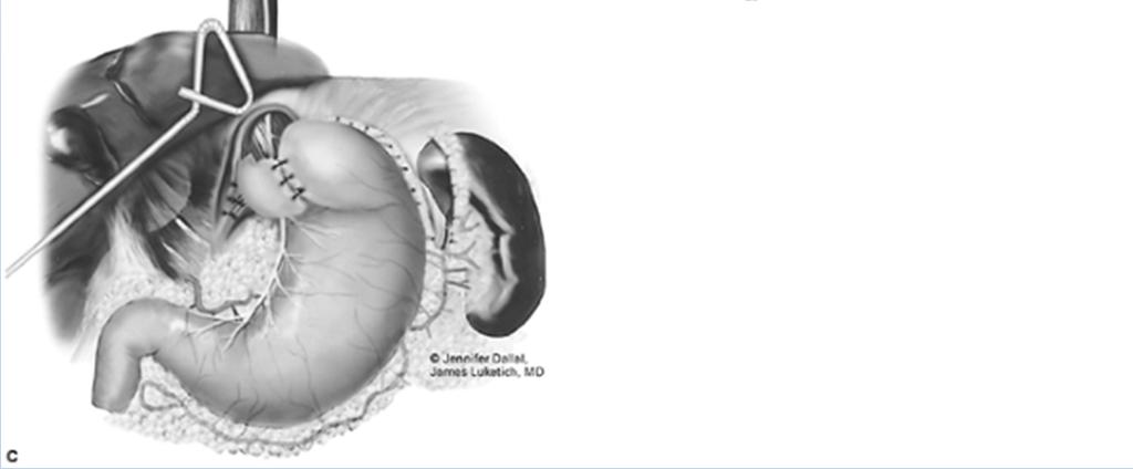
6 Show incomplete maneuvers: This shows the before, during and after Resect the duplication cyst, including the common serosa, exposing the muscularis of the stomach
7 Show incomplete maneuvers: This shows the before, during and after After placing a first piece of tape on the right of the mouth, a second piece starts on the left cheek, comes across over above the upper lip and is secured circumferentially around the ET tube
8 Show incomplete maneuvers: This shows the before, during and after The clamp is partially open, showing the suture being grasped The scalpel is partially through the vein

9 Show what was removed: This shows the before, during and after The proximal vena cava is tapered by excising a corner, and suturing it close The distal vessel is fish mouthed The diameters now match, and can be anastomosed


10 Figure and Organ Drawing Use symbols and other tricks Arrows Icons (scissors, blade) Insets Labels
11 Number the steps Narrative
12 Number the steps Narrative
13 Successive steps explained in a single image 1. Pinch the tube upstream (top right) 2. Squeeze the fluid downstream (arrow, bottom)
14 Successive steps explained in a single image 1. Palpate the symphysis pubis 2. Angle the syringe slightly toward the pubis 3. Insert the needle into the bladder
15 Successive steps explained in a single image 1. Stabilize the pylorus with the left index finger in the duodenal end and the left thumb in the gastric end 2. Spread the muscularis to expose the mucosa
16 Successive steps explained in a single image 1. Grasp the incarcerated bowel loops with the index finger and thumb of the left hand 2. Push the hernia further down first, 3. Then push it back into the canal
17 Successive steps explained in a single image 1. Insert the needle into the artery 2. Once there is blood return, 3. Advance the guidewire into the vessel

18 Successive steps explained in a single image 1. Cut the right ureter above the obstruction 2. Make a longitudinal incision in contralateral ureter 3. Swing the proximal right ureter across the midline 4. Anastomose in a end to side fashion
19 How did they do? Narrative
20 Fetal tracheal occlusion to treat lung hypoplasia Congenital diaphragmatic hernia: liver, intestines in the thoracic cavity, through a hole in the diaphragm Abdominal viscera compress the lung The lung fails to develop properly Occluding the trachea in utero traps lung fluid Increased lung fluid causes some alveolar stretch Stretch causes accelerated lung growth Lung hypoplasia is reversed by term
21 Fetal tracheal occlusion to treat lung hypoplasia

22 Fetal tracheal occlusion to treat lung hypoplasia Image reads top to bottom Arrow helps explain the dynamic Diaphragmatic hole suggested by a few bowel loops Text reads top to bottom as well

23 Fetal tracheal occlusion to treat lung hypoplasia
24 Fetal tracheal occlusion to treat lung hypoplasia Top image shows how it s done Key elements: scope, ultrasound, head and neck Insets show balloon inflated, then detached Orientation is the same insets are close ups


25 Fetal tracheal occlusion to treat lung hypoplasia

26 Fetal tracheal occlusion to treat lung hypoplasia Top image shows how it s done Bottom images are close ups, but different orientation unclear which body part Left inset redundant?
27 Fetal tracheal occlusion to treat lung hypoplasia
 Close up is on head,")
28 Fetal tracheal occlusion to treat lung hypoplasia 4 images seem essentially the same Shows progression (scope in, balloon inflated, balloon detached, balloon retrieved) Close up is on head, not on trachea
29 Gastroesophageal reflux: The abdominal portion of the esophagus is too short, or non existent The esophageal hiatus is too wide (hiatal hernia) Increased gastric pressure causes vomiting Fundoplication: Narrow the hiatus Wrap gastric fundus around the esophagus Adds turtle neck to the lower esophageal sphincter

30 Fundoplication for gastroesophageal reflux 11 images too much details, too confusing

31 Fundoplication for gastroesophageal reflux 3 images too similar?


32 Fundoplication for gastroesophageal reflux 3 images the essence of the operation 1. Free the esophagus and place sutures in the diaphragm 2. Pass the fundus behind the esophagus, place sutures 3. Complete the wrap


33 Fundoplication for gastroesophageal reflux Only 2 images clear, but less detailed

34 Fundoplication for gastroesophageal reflux One image doesn t show the approach Clear idea of what the operation achieves
35 Intestinal malrotation and volvulus The posterior attachment of the mesentery normally runs in a line from the duodenum to the cecum: midline upper abdomen to right lower quadrant In malrotation, the duodenum doesn t describe a C loop, but runs straight down; all the small bowel is to the right of midline, the colon to the left Duodenum and cecum are next too each other: the posterior attachment of the mesentery is now a single point, rather than a line This makes it more prone to twisting: volvulus
 Malrotation and volvulus")
36 Intestinal malrotation and volvulus Normal rotation (left) Malrotation and volvulus (right)
37 Intestinal malrotation and volvulus

38 Intestinal malrotation and volvulus

39 Intestinal malrotation and volvulus
40 Intestinal malrotation and volvulus

41 Intestinal malrotation and volvulus
42 Assignment # 5 Illustrate one of these three operations Using only 3 4 figures Laparoscopic appendectomy Laparoscopic cholecystectomy Laparoscopic right adrenalectomy
Medical Illustration PLME 0400
 Introduction to Medical Illustration PLME 0400 October 17 From sketch to narrative From Sketch to Sketch: Point of view Degree of detail Visible and invisble parts Landmarks : Plan your moves The IKEA
Placing PEG and Jejunostomy Tubes in Dogs and Cats
 Placing PEG and Jejunostomy Tubes in Dogs and Cats I. Gastrostomy tube A. Percutaneous Endoscopic Gastrostomy (PEG) tube placement Supplies for PEG tube placement: Supplies and equipment for general anesthesia
Step 1: Salivary Structures
 (Slide1) Step 1: Salivary Structures Remove the skin, fat and connective fascia to view the salivary glands and ducts. The submaxillary salivary gland is just behind the masseter muscle and pretty easy
 P R E S E N T S Dr. Mufa T. Ghadiali is skilled in all aspects of General Surgery. His General Surgery Services include: General Surgery Advanced Laparoscopic Surgery Surgical Oncology Gastrointestinal
Manual on Preparation of Tissue for Neonatal Skills Course. Version 1.0_2018. Prepared by: Haitham Dagash. MBBS, FRCSEd (Paed)
") Manual on Preparation of Tissue for Neonatal Skills Course Version 1.0_2018 Prepared by: Haitham Dagash MBBS, FRCSEd (Paed) University of Leicester and Leicester Infirmary Hospital Prof.Kokila Lakhoo PhD,FRCS(ENG+EDIN),FCS(SA),FCS(SA:PAED),MRCPCH,MBCHB
Manual on Preparation of Tissue for Neonatal Skills Course Version 1.0_2018 Prepared by: Haitham Dagash MBBS, FRCSEd (Paed) University of Leicester and Leicester Infirmary Hospital Prof.Kokila Lakhoo PhD,FRCS(ENG+EDIN),FCS(SA),FCS(SA:PAED),MRCPCH,MBCHB
Laparoscopic Salpingectomy for Ectopic Pregnancy Simulation
 Preparation Simulators to be used 1. Laparoscopic box trainers will be used 2. Laparoscopic Maryland graspers, laparoscopic endoshears and a locking grasper will be available for each participant 3. Premade
GASTROINTESTINAL SYSTEM
 GASTROINTESTINAL SYSTEM Topographic Anatomy of the Abdomen Surface Landmarks Xiphoid process T9/T10 Inferior costal margin L2/L3 Iliac Crest L4 level ASIS L5/S1 level Pubic symphysis level of greater trochanter
The Digestive System
 The Digestive System Identify the Structure and Function. Mesentery of the Large Intestine The mesentery functions to connect the visceral organs to the abdominal wall. Identify the Structure. Nasal Cavity
Biology Human Anatomy Abdominal and Pelvic Cavities
 Biology 351 - Human Anatomy Abdominal and Pelvic Cavities You must answer all questions on this exam. Because statistics demonstrate that, on average, between 2-5 questions on every 100-point exam are
Dissection: The Fetal Pig
 Lab Exercise Dissection: The Fetal Pig Objectives - To learn some of anatomical structures of the fetal pig. - To be able to make contrasts and comparisons of structures between different animal phyla
This lab activity is aligned with Visible Body s Human Anatomy Atlas app. Learn more at visiblebody.com/professors
 1 This lab activity is aligned with Visible Body s Human Anatomy Atlas app. Learn more at visiblebody.com/professors 2 A. Digestive System Overview To Start: Go to the Views menu and scroll down to the
Hiatal Hernias and Barrett s esophagus. Dr Sajida Ahad Mercy General Surgery
 Hiatal Hernias and Barrett s esophagus Dr Sajida Ahad Mercy General Surgery Objectives Identify the use of different diagnostic modalities for hiatal hernias List the different types of hiatal hernias
PRODUCTS FOR THE DIFFICULT AIRWAY. Courtesy of Cook Critical Care
 PRODUCTS FOR THE DIFFICULT AIRWAY Courtesy of Cook Critical Care EMERGENCY CRICOTHYROTOMY Thyroid Cartilage Access Site Cricoid Cartilage Identify the cricothyroid membrane between the cricoid and thyroid
Facing Surgery for GERD (Gastroesophageal
 Facing Surgery for GERD (Gastroesophageal Reflux Disease)? Learn about minimally invasive da Vinci Surgery The Condition GERD, Hiatal Hernia Gastroesophageal reflux disease or GERD occurs when stomach
Hirschprung s. Meconium plug R/S >1 R/S <1
 NEONATAL ABDOMINAL EMERGENCIES LOW OBSTRUCTION HIGH OBSTRUCTION INTESTINAL OBSTRUCTION High obstruction - proximal to mid-ileumileum Few dilated, air filled bowel loops Complete obstruction diagnosed by
Inguinal Hernia. Hernia Awareness Month. What is a Hernia? Common Hernia Types
 Hernia Awareness Month What is a Hernia? A hernia occurs when an organ pushes through an opening in the muscle or tissue that holds it in place. For example, the intestines may break through a weakened
Jhia Anjela D. Rivera 1 1. BS Biology, Department of Biology, College of Science, Polytechnic University of the Philippines
 DIGESTIVE SYSTEM Jhia Anjela D. Rivera 1 1 BS Biology, Department of Biology, College of Science, Polytechnic University of the Philippines DIGESTIVE SYSTEM Consists of the digestive tract (gastrointestinal
Ponsky * PEG Safety System - "Pull" Bard * PEG Safety System - "Guidewire" Information for Use
 Ponsky * PEG Safety System - "Pull" Bard * PEG Safety System - "Guidewire" Information for Use Rx only Single patient use DEHP-Free This product and package do not contain natural rubber latex STERILE
Combined Collis-Nissen Reconstruction. of the esophagogastric junction at. Mark B. Orringer, M.D., and Herbert Sloan, M.D.
 Combined Collis-Nissen Reconstruction of the Esophagogastric Junction Mark B. Orringer, M.D., and Herbert Sloan, M.D. ABSTRACT Recent reports have indicated that combined Collis-Belsey reconstruction of
Development of pancreas and Small Intestine. ANATOMY DEPARTMENT DR.SANAA AL-AlSHAARAWY DR.ESSAM Eldin Salama
 Development of pancreas and Small Intestine ANATOMY DEPARTMENT DR.SANAA AL-AlSHAARAWY DR.ESSAM Eldin Salama OBJECTIVES At the end of the lecture, the students should be able to : Describe the development
- Tamara Wahbeh. - Fareed Khdair. 0 P a g e
 -1 - Tamara Wahbeh - - Fareed Khdair 0 P a g e GI Embryology Note: I included everything in the records and slides; anything in the slide not included in this sheet was not mentioned by the doctor during
Block 3: DISSECTION 2 CELIAC TRUNK, JEJUNUM/ILEUM, LARGE INTESTINE, DUODENUM, PANCREAS, PORTAL VEIN; MOBILIZATION OF THE LIVER
 1 Block 3: DISSECTION 2 CELIAC TRUNK, JEJUNUM/ILEUM, LARGE INTESTINE, DUODENUM, PANCREAS, PORTAL VEIN; MOBILIZATION OF THE LIVER Attempt to complete as much as you can of the dissection explained in the
Dissection Lab Manuals: Required Content
 Dissection Lab Manuals: Required Content 1. Introduction a. Basic terminology (directions) b. External features of the cat c. Adaptations to predatory niche d. How to skin a cat e. How to make the incisions
Initial placement 20FR Guidewire PEG kit REORDER NO:
 Initial placement 20FR Guidewire PEG kit REORDER NO: 00710802 INSTRUCTIONS FOR USE 1 of 5 These products have been manufactured not to include latex. Intended Use: The Initial placement 20FR Guidewire
Cholecystectomy. Sarah Forsyth
 Cholecystectomy Sarah Forsyth History of Cholecystectomy First open cholecystectomy 1882 by Carl Langenbuch in Germany First lap cholecystectomy 1987, Philip Mouret (Gynaecologist) in Lyon, France 1990,
LECTURE 11 & 12: ABDOMINAL VISCERA ABDOMINAL CONTENTS DIVISION. The location of abdominal viscera is divided into 4 quadrants:
 LECTURE 11 & 12: ABDOMINAL VISCERA ABDOMINAL CONTENTS DIVISION The location of abdominal viscera is divided into 4 quadrants: - horizontal line across the umbilicus divides the upper quadrants from the
Managing Foreign Objects in the GI Tract Tips & Tricks
 Managing Foreign Objects in the GI Tract Tips & Tricks STEPHEN LANDRENEAU, MD Take home o Perform emergent endoscopy in cases of: Esophageal obstruction due to food bolus impaction or foreign object. Disk
Lab 9 Abdomen MUSCLES
 Lab 9 Abdomen MUSCLES External abdominal oblique continuous with the external intercostal muscle; its fibers point in a caudal direction as it moves anteriorly until it inserts on the linea alba via its
Limited en bloc Resection of the Gastroesophageal Junction with Isoperistaltic Jejunal Interposition
 22 Limited en bloc Resection of the Gastroesophageal Junction with Isoperistaltic Jejunal Interposition J.R. Izbicki, W.T. Knoefel, D. C. Broering ] Indications Severe dysplasia in the distal esophagus
Aus Artificial Uretheral Sphincter Port System
 NORFOLK VET PRODUCTS the AUS for the long-term relief of incontinence in dogs and cats making life easier for pets Speciality Medical Devices For The Veterinary Community the Aus Artificial Uretheral Sphincter
Topics for discussion. Pediatric General Surgery. Physiology. Surgical Newborns. Neonatal Intestinal Obstruction
 Topics for discussion Pediatric General Surgery Professor General & Thoracic Surgery What makes Pediatric Surgery unique? Neonatal intestinal obstruction Abdominal wall defects Inguinal hernias Appendicitis
Name: Fetal Pig Dissection Internal Anatomy
 In this lab exercise you will open the thoracic cavities and abdominal pelvic of a fetal pig and identify its major organs. Remember you are dissecting not butchering. The goal is for you to identify all
BELLWORK DEFINE: PERISTALSIS CHYME RUGAE Remember the structures of the digestive system 1
 BELLWORK DEFINE: PERISTALSIS CHYME RUGAE 2.07 Remember the structures of the digestive system 1 STANDARD 8) Outline basic concepts of normal structure and function of all body systems, and explain how
What is Laparoscopy All About?
 Disclaimer This movie is an educational resource only and should not be used to manage surgical health. All decisions about the management of Laparoscopy must be made in conjunction with your Physician
Infantile Hypertrophic Pyloric Stenosis
 A Sonographic walk-through: Infantile Hypertrophic Pyloric Stenosis Tara K. Cielma, RDMS, RDCS, RVT, RT(S) Anjum N. Bandarkar, MD, Adebunmi O. Adeyiga, MD, Diagnostic Imaging and Radiology, Children s
Hernias Umbilical Hernia When to See a Surgeon? What Are Symptoms of an Umbilical Hernia? How is Repair Performed?
 Hernias Umbilical Hernia An umbilical hernia occurs when part of the intestine protrudes through the umbilical opening in the abdominal muscles. Umbilical hernias are common and typically harmless. They
Lab 8: Digestive System
 BIOL 221 A&P II Lab 8: Digestive System Become familiar with the gross anatomy of the digestive system (Exercise 38) using the models, Fig. 38.1 (Activity 1), and the rat. Recognize and know the functions
Home FAQ Archives ABP Topics NeoReviews.org My Bookmarks CME Information Help. Print this Page Add to my Bookmarks Page 3 of 10
 Welcome Kristin Ingstrup [ Logout ] SEARCH Home FAQ Archives ABP Topics NeoReviews.org My Bookmarks CME Information Help Overview Editorial Board My Learning Plan January February March May June July August
Fareed Khdair, MD Assistant Professor Chief, Section of Pediatric Gastroenterology, Hepatology, and Nutrition University of Jordan School of Medicine
 Fareed Khdair, MD Assistant Professor Chief, Section of Pediatric Gastroenterology, Hepatology, and Nutrition University of Jordan School of Medicine Outline Lecture one : Gut formation Foregut: esophagus,
In the name ofgod. Abdomen 3. Dr. Zahiri
 In the name ofgod Abdomen 3 Dr. Zahiri Peritoneum Peritoneum It is the serous membrane(a type of loose connective tissue and is covered by mesothelium) that lines the abdominal cavity. Extensions of the
Fetal Pig Dissection Day 2 Circulatory and Respiratory Systems
 Name: Date: Period: Fetal Pig Dissection Day 2 Circulatory and Respiratory Systems Dissection Roles (choose a different role from Day 1): Recorder reads directions out loud to group and records answers
A novel plain abdominal radiograph sign to diagnose malrotation with volvulus
 A novel plain abdominal radiograph sign to diagnose malrotation with volvulus Nataraja RM 1, Mahomed AA 1* 1. Department of Paediatric Surgery, Royal Alexandra Hospital for Sick Children, Brighton,UK *
Minimally Invasive Esophagectomy
 American Association of Thoracic Surgery (AATS) 95 th Annual Meeting Seattle, WA April 29, 2015 General Thoracic Masters of Surgery Video Session Minimally Invasive Esophagectomy James D. Luketich MD,
The abdominal Esophagus, Stomach and the Duodenum. Prof. Oluwadiya KS
 The abdominal Esophagus, Stomach and the Duodenum Prof. Oluwadiya KS www.oluwadiya.com Viscera of the abdomen Abdominal esophagus: Terminal part of the esophagus The stomach Intestines: Small and Large
Respiratory & Digestive Organs of the Head and Neck, Human;
 Name Date Lab Exercise 5: Lab Exercise 6: Lab Exercise 7: Lab Exercise 8: Respiratory & Digestive Organs of the Head and Neck, Human; Histology of the Respiratory System Digestive System Models, Human
Digestive System 7/15/2015. Outline Digestive System. Digestive System
 Digestive System Biology 105 Lecture 18 Chapter 15 Outline Digestive System I. Functions II. Layers of the GI tract III. Major parts: mouth, pharynx, esophagus, stomach, small intestine, large intestine,
Honors Biology: Rat Dissection ONLINE ASSIGNMENT
 Name: Honors Biology: Rat Dissection ONLINE ASSIGNMENT You and your group members will use the Honors Biology WIKI to create an online dissection manual. The point of this assignment is to illustrate what
Pathology of Intestinal Obstruction. Dr. M. Madhavan, MBBS., MD., MIAC, Professor of Pathology Saveetha Medical College
 Pathology of Intestinal Obstruction Dr. M. Madhavan, MBBS., MD., MIAC, Professor of Pathology Saveetha Medical College Pathology of Intestinal Obstruction Objectives list the causes of intestinal obstruction
RECONSTRUCTION OF THE CARDIA AND FUNDUS OF THE STOMACH
 Thorax (1956), 11, 275. RECONSTRUCTION OF THE CARDIA AND FUNDUS OF THE STOMACH BY From tile United Leeds Hospitals (RECEIVED FOR PUBLICATION SEPTEMBER 15, 1956) This is a preliminary report describing
Nissen Fundoplication
 Nissen Fundoplication By Donna Weldon Nissen fundoplication is a surgical procedure used to treat gastroesophageal reflux disease, or GERD, and hiatus hernias. For GERD, is it usually performed when medical
AATS Focus on Thoracic Surgery: Minimally Invasive Esophagectomy: Are We Still Getting Better in 2017?
 AATS Focus on Thoracic Surgery: Mastering Surgical Innovation Las Vegas, NV October 28, 2017 Session VIII: Video Session Minimally Invasive Esophagectomy: Are We Still Getting Better in 2017? James D.
Digestive System. In one end and out the other.
 Digestive System In one end and out the other. Overview Every cell in the body needs nourishment, yet most cells cannot leave their position in the body and travel to a food source, so the food must be
ISUOG Basic Training. Examining Fetal Anatomy from Longitudinal Sections Titia Cohen-Overbeek, The Netherlands
 ISUOG Basic Training Examining Fetal Anatomy from Longitudinal Sections Titia Cohen-Overbeek, The Netherlands Learning objectives 2 & 3 At the end of the lecture you will be able to: describe how to obtain
Midgut. Over its entire length the midgut is supplied by the superior mesenteric artery
 Gi Embryology 3 Midgut the midgut is suspended from the dorsal abdominal wall by a short mesentery and communicates with the yolk sac by way of the vitelline duct or yolk stalk Over its entire length the
Cystotomy Laboratory Simulation
 Kelli Braun MD, Robert Stager MD, Chadburn Ray MD, Bunja Rungruang MD Medical College of Georgia at Augusta University Note: This model can be used for Open Cystotomy Repair or Laparoscopic Cystotomy Repair.
Initial placement 24FR Pull PEG kit REORDER NO:
 Initial placement 24FR Pull PEG kit REORDER NO: 00710805 INSTRUCTIONS FOR USE 1 of 5 These products have been manufactured not to include latex. Intended Use: The Initial placement 24FR Pull PEG kit is
Biology Human Anatomy Abdominal and Pelvic Cavities
 Biology 351 - Human Anatomy Abdominal and Pelvic Cavities Please place your name and I.D. number on the back of the last page of this exam. You must answer all questions on this exam. Because statistics
Duodenum retroperitoneal
 Duodenum retroperitoneal C shaped Initial region out of stomach into small intestine RETROperitoneal viscus Superior 1 st part duodenal cap ; moves upwards and backwards to lie on the R crura medial to
Hernia. emoryhealthcare.org
 Hernia Have you noticed a bulge or pain in your abdominal wall or groin? If so you may have a hernia. You may be in the process of confirming this diagnosis with your Primary Care Physician or already
Basic Training. ISUOG Basic Training The 20 Planes Approach to the Routine Mid Trimester Scan
 ISUOG The 20 Planes Approach to the Routine Mid Trimester Scan Learning objective At the end of the lecture you will be able to: Explain how to perform a structured routine examination, including measurements,
Volvulus of the Gastrointestinal Tract: x-ray and CT imaging
 Volvulus of the Gastrointestinal Tract: x-ray and CT imaging Poster No.: C-0076 Congress: ECR 2013 Type: Educational Exhibit Authors: E. Papadaki, S. Paschalidou, S. GIANNOU ; Rethymno, CR/ 1 2 2 3 1 3
BIO 139 ANATOMY AND PHYSIOLOGY II
 BIO 139 ANATOMY AND PHYSIOLOGY II THE DIGESTIVE SYSTEM LAB ANALOGY PAGES 248-265 MARY CATHERINE FLATH, Ph.D. DIGESTIVE ORGANS ALIMENTARY CANAL MOUTH PHARYNX ESOPHAGUS STOMACH SMALL INTESTINE LARGE INTESTINE
Neonatal intestinal obstruction: how to make etiological diagnosis?
 Neonatal intestinal obstruction: how to make etiological diagnosis? Poster No.: C-1414 Congress: ECR 2013 Type: Educational Exhibit Authors: W. Mnari, M. Zguidi, A. Zrig, M. Maatouk, B. Hmida, R. Salem,
Achalasia is a primary esophageal motility disorder of unknown
 Laparoscopic Heller Myotomy for Achalasia Andrew Pierre, MD, MSc Achalasia is a primary esophageal motility disorder of unknown etiology. Pathologically, it is characterized by loss of ganglion cells in
Transfemoral Amputation
 Transfemoral Amputation Pre-Op: 42 year old male who sustained severe injuries in a motorcycle accident. Note: he is a previous renal transplant recipient and is on immunosuppressive treatments. His injuries
BIO 139 ANATOMY AND PHYSIOLOGY II. THE DIGESTIVE SYSTEM LAB ANALOGY PAGES MARY CATHERINE FLATH, Ph.D.
 BIO 139 ANATOMY AND PHYSIOLOGY II THE DIGESTIVE SYSTEM LAB ANALOGY PAGES 248-265 MARY CATHERINE FLATH, Ph.D. DIGESTIVE ORGANS ALIMENTARY CANAL MOUTH PHARYNX ESOPHAGUS STOMACH SMALL INTESTINE LARGE INTESTINE
Neonatal intestinal obstruction: how to make etiological diagnosis?
 Neonatal intestinal obstruction: how to make etiological diagnosis? Poster No.: C-1414 Congress: ECR 2013 Type: Educational Exhibit Authors: W. MNARI, M. Zguidi, A. Zrig, M. MAATOUK, B. Hmida, R. Salem,
The Foregut. At first the esophagus is short. but with descent of the heart and lungs it lengthens rapidly
 GI embryology 2 The Foregut At first the esophagus is short but with descent of the heart and lungs it lengthens rapidly The muscular coat, which is formed by surrounding splanchnic mesenchyme, is striated
Gastro-Oesophageal Reflux Disease Information Sheet
 Gastro-Oesophageal Reflux Disease Information Sheet Gastro-Oesophageal Reflux Disease This sheet gives you information about Gastro-Oesophageal Reflux Disease & Fundoplication Surgery What is gastro-oesophageal
Fetal Pigs and You BIO 171 WEEK 10
 Fetal Pigs and You BIO 171 WEEK 10 The Domestic Pig: Sus scrofa Kingdom: Animalia Phylum: Chordata Class: Mammalia - Skin covered in hair or fur; Milk-producing glands (mammary glands) in the female to
A rare case of intestinal obstruction due to internal hernia. Dr. Jayanth 3 rd year PG Dept. Of General Surgery
 A rare case of intestinal obstruction due to internal hernia Dr. Jayanth 3 rd year PG Dept. Of General Surgery One of the common cause of acute abdomen May lead to high morbidity and mortality if not treated
The Digestive System. Chapter 25
 The Digestive System Chapter 25 Introduction Structure of the digestive system A tube that extends from mouth to anus Accessory organs are attached Functions include Ingestion Movement Digestion Absorption
Frog Dissection SNC2P Grade 10 Science Applied Biology Tissues, Organs and Systems of Living Things
 Frog Dissection SNC2P Grade 10 Science Applied Biology Tissues, Organs and Systems of Living Things Purpose To identify and examine the external and internal structures of the frog and compare them with
Fetal Pig Dissection:
 Fetal Pig Dissection: REMEMBER: Dissection involves disassembling and observing something to determine its internal structure and develop an understanding of the relationship of those structures to function.
HIATAL HERNIA, DIVERTICULITIS AND VARICOSE VEINS: Three Peas In A Pod By Evan Almeda READ ONLINE
 HIATAL HERNIA, DIVERTICULITIS AND VARICOSE VEINS: Three Peas In A Pod By Evan Almeda READ ONLINE What is a hiatal hernia? Unlike ventral hernias, which protrude through the abdominal wall, The good news
Lab Monitor Images Dissection of the Abdominal Vasculature + Lower Digestive System
 Lab Monitor Images Dissection of the Abdominal Vasculature + Lower Digestive System Stomach & Duodenum Frontal (AP) View Nasogastric tube 2 1 3 4 Stomach Pylorus Duodenum 1 Duodenum 2 Duodenum 3 Duodenum
The Digestive System. Chapter 16. Introduction. Overview of Digestive System. Histological Organization. Movement and Mixing of Digestive Materials
 The Digestive System Chapter 16 Introduction Structure of the digestive system A tube that extends from mouth to anus Accessory organs are attached Functions include Ingestion Movement Digestion Absorption
Tools of the Gastroenterologist: Introduction to GI Endoscopy
 Tools of the Gastroenterologist: Introduction to GI Endoscopy Objectives Endoscopy Upper endoscopy Colonoscopy Endoscopic retrograde cholangiopancreatography (ERCP) Endoscopic ultrasound (EUS) Endoscopic
The Digestive System Laboratory
 The Digestive System Laboratory 1 The Digestive Tract The alimentary canal is a continuous tube stretching from the mouth to the anus. Liver Gallbladder Small intestine Anus Parotid, sublingual, and submaxillary
Airway/Breathing. Chapter 5
 Airway/Breathing Chapter 5 Airway/Breathing Introduction Skillful, rapid assessment and management of airway and ventilation are critical to preventing morbidity and mortality. Airway compromise can occur
Geoffrey Axiak M.Sc. Nursing (Manch.), B.Sc. Nursing, P.G. Dip. Nutrition & Dietetics Clinical Nutrition Practice Nurse
, B.Sc. Nursing, P.G. Dip. Nutrition & Dietetics Clinical Nutrition Practice Nurse") The Percutaneous Endoscopic Gastrostomy Geoffrey Axiak M.Sc. Nursing (Manch.), B.Sc. Nursing, P.G. Dip. Nutrition & Dietetics Clinical Nutrition Practice Nurse What is a P.E.G.? Percutaneous Endoscopic
The Percutaneous Endoscopic Gastrostomy Geoffrey Axiak M.Sc. Nursing (Manch.), B.Sc. Nursing, P.G. Dip. Nutrition & Dietetics Clinical Nutrition Practice Nurse What is a P.E.G.? Percutaneous Endoscopic
Anatomy: Know Your Abdomen
 Anatomy: Know Your Abdomen Glossary Abdomen - part of the body below the thorax (chest cavity); separated by the diaphragm. Anterior - towards the front of the body. For example, the umbilicus is anterior
Flouroscopy CT. Principal Modality (2): Case Report # [] Date accepted: 14 May 2015
![Flouroscopy CT. Principal Modality (2): Case Report # [] Date accepted: 14 May 2015](/thumbs/85/92263701.jpg "Flouroscopy CT. Principal Modality (2): Case Report # [] Date accepted: 14 May 2015") Radiological Category: Emergency Principal Modality (1): Principal Modality (2): Flouroscopy CT Case Report # [] Submitted by: Haider Virani, M.D. Faculty reviewer: Naga Chinapuvvula, MD Date accepted:
Radiological Category: Emergency Principal Modality (1): Principal Modality (2): Flouroscopy CT Case Report # [] Submitted by: Haider Virani, M.D. Faculty reviewer: Naga Chinapuvvula, MD Date accepted:
OVARIES URETER FALLOPIAN TUBES BLADDER UROGENITAL OPENINGS (BOTH SEXES) PENIS VAGINA UTERUS
 PENIS VAGINA UTERUS") URETER OVARIES FALLOPIAN TUBES BLADDER UROGENITAL OPENINGS (BOTH SEXES) PENIS VAGINA UTERUS REPRODUCTIVE PRODUCE FEMALE HORMONES EXCRETORY FROM KIDNEY TO BLADDER EXCRETORY STORES URINE REPRODUCTIVE TRANSPORTS
URETER OVARIES FALLOPIAN TUBES BLADDER UROGENITAL OPENINGS (BOTH SEXES) PENIS VAGINA UTERUS REPRODUCTIVE PRODUCE FEMALE HORMONES EXCRETORY FROM KIDNEY TO BLADDER EXCRETORY STORES URINE REPRODUCTIVE TRANSPORTS
Traditionally, surgical antireflux therapy has been
 Laparoscopic Fundoplication Mary Maish, MD and Jeffrey A. Hagen, MD Traditionally, surgical antireflux therapy has been reserved for patients with complicated gastroesophageal reflux disease. The introduction
The Digestive System. Chapter
 The Digestive System Chapter 15.1 Functions: mechanical and chemical breakdown of food *absorption of nutrients Consists of alimentary canal and accessory organs Wall of the Alimentary Canal 15.2 Characteristics
HUMAN BODY. Contents:
 HUMAN BODY HUMAN BODY Contents: 1. Main objectives. 2. Useful websites. 3. Word wall cards. 4. Vocabulary cards. 5. Glossary. 6. Key question cards. 7. Prior learning diagram. 8. Hands on activity. 9.
PATIENT INFORMATION FROM YOUR SURGEON & SAGES Laparoscopic Anti-Reflux (GERD) Surgery
 Surgery") Patient Information published on: 03/2004 by the Society of American Gastrointestinal and Endoscopic Surgeons (SAGES) PATIENT INFORMATION FROM YOUR SURGEON & SAGES Laparoscopic Anti-Reflux (GERD) Surgery
Patient Information published on: 03/2004 by the Society of American Gastrointestinal and Endoscopic Surgeons (SAGES) PATIENT INFORMATION FROM YOUR SURGEON & SAGES Laparoscopic Anti-Reflux (GERD) Surgery
Echo PS Positioning System with Ventralight ST Mesh or Composix L/P Mesh
 Ventralight ST Mesh with Echo PS Positioning System Catalog Number Shape Mesh Size 5955450 Circle 4.5" (11.4 cm) 5955460 Ellipse 4" x 6" (10.2 cm x 15.2 cm) 5955600 Circle 6" (15.2 cm) 5955680 Ellipse
INTRA-THORACIC AND INTRA-ABDO-MINAL PERFORATION OF THE COLON IN TRAUMATIC DIAPHRAGMATIC
 INTRA-THORACIC AND INTRA-ABDO-MINAL PERFORATION OF THE COLON IN TRAUMATIC DIAPHRAGMATIC Pages with reference to book, From 14 To 16 S. Amjad Hussain, Chinda Suriyapa, Karl Grubaugh ( Depts. of Surger and
Vomiting in children: The good coordination between radiologists and pediatricians is the key to success
 Vomiting in children: The good coordination between radiologists and pediatricians is the key to success C. Santos Montón 1, M. T. Garzon Guiteria 2, A. Hortal Benito-Sendín 1, K. El Karzazi 1, P. Sanchez
UNDERSTANDING X-RAYS: ABDOMINAL IMAGING THE ABDOMEN
 UNDERSTANDING X-RAYS: ABDOMINAL IMAGING THE ABDOMEN Radiology Enterprises radiologyenterprises@gmail.com www.radiologyenterprises.com STOMACH AND SMALL BOWEL STOMACH AND SMALL BOWEL Swallowed air is a
2015 General Surgery Survival Guide
 2015 General Surgery Survival Guide Chapter 10: Hernia Repair Know What to Look for When Coding Hernia Repair Reporting hernia repair can be tricky. But if you know what to look for then half the work
Urinary 1 Checklist Gross Anatomy of the Urinary System
 Urinary 1 Checklist Gross Anatomy of the Urinary System Urinary system Kidneys Parietal peritoneum Retroperitoneal Renal fascia The urinary system consists of two kidneys, two ureters, the urinary bladder,
Anatomical Considerations for Lab Practical II
 Anatomical Considerations for Lab Practical II For each of the following please be prepared to provide: Identification System Organ(s) or ducts to Function(s) location which it is attached Use your lecture
The peritoneum. Prof. Oluwadiya KS, MBBS, FMCS(Orthop) Website:
 Website:") The peritoneum Prof. Oluwadiya KS, MBBS, FMCS(Orthop) Website: http://oluwadiya.com The peritoneum Serous membrane that lines the abdominopelvic cavity and invests the viscera The largest serous membrane
The peritoneum Prof. Oluwadiya KS, MBBS, FMCS(Orthop) Website: http://oluwadiya.com The peritoneum Serous membrane that lines the abdominopelvic cavity and invests the viscera The largest serous membrane
Exploring Anatomy: the Human Abdomen
 Exploring Anatomy: the Human Abdomen PERITONEUM AND PERITONEAL CAVITY PERITONEUM The peritoneum is a thin serous membrane that lines the abdominal cavity and covers, in variable amounts, the viscera within
Two main groups Alimentary canal continuous coiled hollow tube Accessory digestive organs
 Digestion Breakdown of ingested food Absorption of nutrients into the blood Metabolism Production of cellular energy (ATP) Constructive and degradative cellular activities Two main groups Alimentary canal
Small Bowel and Colon Surgery
 Small Bowel and Colon Surgery Why Do I Need a Small Bowel Resection? A variety of conditions can damage your small bowel. In severe cases, your doctor may recommend removing part of your small bowel. Conditions
Early View Article: Online published version of an accepted article before publication in the final form.
 Early View Article: Online published version of an accepted article before publication in the final form. Journal Name: Journal of Case Reports and Images in Surgery Type of Article: Case Report Title: