Anatomy of the orbit. Lay-out. Imaging technique. 3 x 3. brief overview of the basic anatomy of the orbit and its structures
|
|
|
- Rudolph Kennedy
- 5 years ago
- Views:
Transcription


 - surface ectoderm (lens) -")
")

1 Anatomy of the orbit Prof. Pia C Sundgren MD, PhD Department of Diagnostic Radiology, Clinical Sciences, Lund University, Sweden Lay-out brief overview of the basic anatomy of the orbit and its structures the orbit is a complicated structure due to its embryological composition high number of entities, and diseases due to its composition of ectoderm, surface ectoderm and mesoderm Recommend you to read for more details 3 x 3 3 layers: - neuroectoderm (retina, iris, optic nerve) - surface ectoderm (lens) - mesoderm (vascular structures, sclera, choroid) 3 spaces: - pre-septal extraconal - post-septal intraconal 3 motor nerves: - occulomotor (III) - trochlear (IV) - abducens (VI) Imaging technique CT and / or MR IOM plane thin slices axial and coronal projections CT: soft tissue and bone windows MR: T1 pre and post, T2, STIR, fat suppression, DWI (?)

 lacrimal bone maxilla Lateral wall greater wing of the")
 Frontal nerve")
 Inferior division of the oculomotor nerve (CN III) Abducens nerve ( CN VI)")


2 Superior orbital fissure cranial nerves (CN) III, IV, and VI lacrimal nerve frontal nerve nasociliary nerve orbital branch of middle meningeal artery recurrent branch of lacrimal artery superior orbital vein superior ophthalmic vein Roof frontal bone lesser wing of the sphenoid bone Floor maxilla zygomatic bone Medial wall ethmoid bone (lamina papyracea) lacrimal bone maxilla Lateral wall greater wing of the sphenoid bone zygomatic bone Mnemonic of the order in which the nerves passing through the Sup. orbital fissure Lazy French tarts sit naked in anticipation Lacrimal nerve (branch of CN V1) Frontal nerve (branch of CN V1) Trochlear nerve (CN IV) Superior division of the oculomotor nerve (CN III) Nascociliary nerve (branch of the CN V1) Inferior division of the oculomotor nerve (CN III) Abducens nerve ( CN VI) Courtesy Dr C Romanovski Orbital fissures and optic canal Superior orbital fissure: lesser and greater wing of the sphenoid bone Inferior orbital fissure: greater wing of sphenoid the maxilla and palatine bone Optic canal: at the apex of the orbit in the sphenoid bone Inferior orbital fissure infraorbital nerve zygomatic nerve parasympathetics to lacrimal gland infraorbital artery infraorbital vein inferior ophthalmic vein branch to pterygoid plexus


3 Infraorbital sulcus and foramen The infraorbital sulcus (SIO) crosses the floor of the orbit and carries infraorbital artery infraorbital vein infraorbital nerve from the inferior orbital fissure to the infraorbital foramen NOTE this is a route of spread for infection or maxillary tumors to the orbit and the skull base Optic canal Annulus of Zinn optic nerve ophthalmic artery central retinal vein dural sheath Calcifications of optic nerve Courtesy Dr A.I Ranchod, SA Optic nerve cranial nerve II transmits visual information from the retina to the brain composed of retinal ganglion cell axons and support cells optic n. glioma optic n. meningioma


 Episcleral")


4 septum - preseptal - postseptal intraconal extraconal preseptal soft tissue/edema Septum - med.palpebral lig. retrobulbar space - clinical term Courtesy M Annertz/Lund Mnemonic intraconal lesions of the orbita Mel met Rita mending hems on poor Charlies grave Melanoma Meningeoma pseudotumor Chemosis of the eye swelling of the tissue that lines the eyelids and surface of the eye (conjunctiva) Episcleral infiltration Metastasis Hemangioma cellulitis Retinoblastoma Optic glioma Graves disease Preseptal infiltration Courtesy Dr P Maly, Malmoe Postseptal extraconal Postseptal intraconal


5 Scleritis chronic, painful, inflammatory disease characterized by edema and cellular infiltration of the scleral and episcleral tissues (outermost coat of the eye) Buphthalmos and coloboma Buphthalmos enlargement of the globe due to increased intraocular pressure choroid lens anterior chamber secondary to obstruction of the Schlemm canal isolated / associated - Sturge Weber -NF type 1 - cobblestone lissencephaly sclera retina Cryptophthalmos A rare congenital anomaly in which the skin is continuous over the eyeball, with absence of eyelids. Eye might be normal or abnormal. Courtesy Dr A.I Ranchod, SA Retinal detachment occurs when the intraretinal space persists Courtesy Dr. S Andronikou, SA


")


6 Buphthalmos and coloboma Coloboma congenital fissures in the globe due to incomplete closure of the embryonic optic fissure 0.5 to 0.7 per 10,000 births iris coloboma: defect in anterior portion of the embryonic fissure cleft in the iris pigment epithelium retinochoroidal coloboma: arise from the wall of the globe optic nerve coloboma: insertion of the optic disc Retinoblastoma Clinical findings leukoaria (white pupillary reflex) cat eye reflex due to replacement of vitreous humor by a white mass reduced vision, eye pain, strabismus Courtesy Dr A Rossi, IT Staphyloma Persistent hyperplastic primary vitreous protrusion of the uveal tissue through a thinning of the sclera - weak point in the eyeball severe axial myopia, more common posteriorly anteriorly more common after infection or trauma M Tsatsos and T Eke Eye (2007) 21, Courtesy Dr A.I Ranchod, SA


 Extra ocular muscles develop in situ input from")


 Sup.")
7 Bulb laceration Action of the extra-ocular muscles Muscle Action Cranial Nerve Inf. rectus depresses, turns medially III Inf. oblique elevates turns laterally III Sup. obligue depresses, turns laterally IV (n. trochlearis) Extra ocular muscles develop in situ input from respective cranial nerve (III, IV,VI) 1 month gestation fully developed at 6 months gestation Annulus of Zinn a fibrous ring formed by the common origin of the 4 rectus muscles Thyroid ophthalmopathy m. rectus inferior > med >sup > lat Action of the extra-ocular muscles The major nerves Cranial nerves motor III oculomotor n IV trochlear n VI abducens n II optic nerve Muscle Action Cranial Nerve Lat. rectus moves eye laterally VI ( n.abducens) Med. rectus moves eye medially III (n. oculomotorius) Sup. rectus elevate, turns it medially III Cranial - sensory nerves V trigeminal n ophthalmic n V 1 (the upper eyelid, the conjunctiva and cornea of the eye) and its branches


8 The ophthalmic artery first branch of the a. int car. The ophthalmic artery enters the orbit on the inferolateral side of the optic nerve and gives off the central retinal artery and posterior ciliary arteries near the orbital apex Involvement of the ophthalmic artery in giant cell arteritis Copyright 2016 British Society for Rheumatology Ophthalmic nerve The orbital veins branches nasociliary nerve frontal nerve lacrimal nerve NOTE the superior ophthalmic vein is a communication between facial vein and cavernous sinus Danger Zone Kumar JB et al EyeNet Magazine May 2015


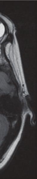
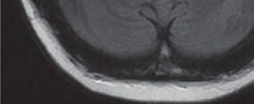
9 Thank you T1 weighted, without fat suppression, showing bilateral enlarged superior ophthalmic veins in carotid cavernous stula Ophthamology.standford.edu Lund University / Faculty of Medicine / Inst. Clinical Sciences / Radiology PC / ECNR Sundgren, Dubrovnik Spine / Oct 2018 Infect ASSR 12 Orbital phlebograhy showing left-sided venous fistula Courtesy Dr. P Maly, Malmoe Take home message complicated anatomy and embryology high number of possible diseases For correct diagnosis important to differentiate between the preseptal and postseptal space important to differentiate between extraconal and intraconal
The orbit-1. Dr. Heba Kalbouneh Assistant Professor of Anatomy and Histology
 The orbit-1 Dr. Heba Kalbouneh Assistant Professor of Anatomy and Histology Orbital plate of frontal bone Orbital plate of ethmoid bone Lesser wing of sphenoid Greater wing of sphenoid Lacrimal bone Orbital
1 Eyelids. Lacrimal Apparatus. Orbital Region. 3 The Orbit. The Eye
 1 1 Eyelids Orbital Region 2 Lacrimal Apparatus 3 The Orbit 4 The Eye 2 Eyelids The eyelids protect the eye from injury and excessive light by their closure. The upper eyelid is larger and more mobile
Bony orbit Roof The orbital plate of the frontal bone Lateral wall: the zygomatic bone and the greater wing of the sphenoid
 Bony orbit Roof: Formed by: The orbital plate of the frontal bone, which separates the orbital cavity from the anterior cranial fossa and the frontal lobe of the cerebral hemisphere Lateral wall: Formed
The Orbit. The Orbit OCULAR ANATOMY AND DISSECTION 9/25/2014. The eye is a 23 mm organ...how difficult can this be? Openings in the orbit
 The eye is a 23 mm organ...how difficult can this be? OCULAR ANATOMY AND DISSECTION JEFFREY M. GAMBLE, OD COLUMBIA EYE CONSULTANTS OPTOMETRY & UNIVERSITY OF MISSOURI DEPARTMENT OF OPHTHALMOLOGY CLINICAL
The sebaceous glands (glands of Zeis) open directly into the eyelash follicles, ciliary glands (glands of Moll) are modified sweat glands that open
 open directly into the eyelash follicles, ciliary glands (glands of Moll) are modified sweat glands that open") The Orbital Region The orbits are a pair of bony cavities that contain the eyeballs; their associated muscles, nerves, vessels, and fat; and most of the lacrimal apparatus upper eyelid is larger and more
The Orbital Region The orbits are a pair of bony cavities that contain the eyeballs; their associated muscles, nerves, vessels, and fat; and most of the lacrimal apparatus upper eyelid is larger and more
Unit VIII Problem 8 Anatomy: Orbit and Eyeball
 Unit VIII Problem 8 Anatomy: Orbit and Eyeball - The bony orbit: it is protecting our eyeball and resembling a pyramid: With a base directed: anterolaterally. And an apex directed: posteromedially. Notes:
213: HUMAN FUNCTIONAL ANATOMY: PRACTICAL CLASS 12 Cranial cavity, eye and orbit
 213: HUMAN FUNCTIONAL ANATOMY: PRACTICAL CLASS 12 Cranial cavity, eye and orbit OSTEOLOGY Identify the bones which comprise the walls of the orbit: maxilla, zygomatic, ethmoid, lachrymal, frontal, and
Major Anatomic Components of the Orbit
 Major Anatomic Components of the Orbit 1. Osseous Framework 2. Globe 3. Optic nerve and sheath 4. Extraocular muscles Bony Orbit Seven Bones Frontal bone Zygomatic bone Maxillary bone Ethmoid bone Sphenoid
MAXILLA, ORBIT & PTERYGOPALATINE FOSSA. Neophytos C Demetriades MD, DDS, MSc Associate professor European University of Cyprus School of Medicine
 MAXILLA, ORBIT & PTERYGOPALATINE FOSSA Neophytos C Demetriades MD, DDS, MSc Associate professor European University of Cyprus School of Medicine Maxilla MAXILLA Superior, middle, and inferior meatus Frontal
GNK485 The eye and related structures. Prof MC Bosman 2012
 GNK485 The eye and related structures Prof MC Bosman 2012 Surface anatomy Bony orbit Eyeball and Lacrimal apparatus Extra-ocular muscles Movements of the eye Innervation Arterial supply and venous drainage
Maxilla, ORBIT and infratemporal fossa. Neophytos C Demetriades MD, DDS, MSc Associate professor European University of Cyprus School of Medicine
 Maxilla, ORBIT and infratemporal fossa Neophytos C Demetriades MD, DDS, MSc Associate professor European University of Cyprus School of Medicine MAXILLA Superior, middle, and inferior meatus Frontal sinus
The orbit-2. Dr. Heba Kalbouneh Assistant Professor of Anatomy and Histology
 The orbit-2 Dr. Heba Kalbouneh Assistant Professor of Anatomy and Histology Eyelids The eyelids (act like the curtains) protect the eye from injury and excessive light by their closure The upper eyelid
mistake ;slides in bold but you still have to go back to our slides to see the figure, tables and some scheme
 Khozama jehad : I am doing my best and I am sorry for any unintended mistake ;slides in bold but you still have to go back to our slides to see the figure, tables and some scheme The Orbit, Orbital Contents
Sense of Vision. Chapter 8. The Eye and Vision. The Eye Orbit. Eyebrows, Eyelids, Eyelashes. Accessory Organs 5/3/2016.
 Sense of Vision Chapter 8 Special Senses The Eye and Vision 70 percent of all sensory receptors are in the eyes Each eye has over 1 million nerve fibers Protection for the eye Most of the eye is enclosed
REVIEW OF HEAD AND NECK CRANIAL NERVES AND EVERYTHING ELSE
 REVIEW OF HEAD AND NECK CRANIAL NERVES AND EVERYTHING ELSE OLFACTORY NERVE CN I ANTERIOR CRANIAL FOSSA CRISTA GALLI OF ETHMOID OLFACTORY FORAMINA IN CRIBIFORM PLATE OF ETHMOID BONE CN I OLFACTORY NERVE
Bony orbit. Lateral wall: Formed by : the zygomatic bone and the greater wing of the sphenoid
 Bony orbit Roof: Formed by: The orbital plate of the frontal bone, which separates the orbital cavity from the anterior cranial fossa and the frontal lobe of the cerebral hemisphere Lateral wall: Formed
THE SPECIAL SENSES. Introduction Vision
 THE SPECIAL SENSES Introduction Vision RECEPTORS Structures designed to respond to stimuli Variable complexity RECEPTORS: GENERAL PROPERTIES Transducers Receptor Potential Generator Potential RECEPTORS
Anatomic Relations Summary. Done by: Sohayyla Yasin Dababseh
 Anatomic Relations Summary Done by: Sohayyla Yasin Dababseh Anatomic Relations Lecture 1 Part-1 - The medial wall of the nose is the septum. - The vestibule lies directly inside the nostrils (Nares). -
Omran Saeed. Luma Taweel. Mohammad Almohtaseb. 1 P a g e
 2 Omran Saeed Luma Taweel Mohammad Almohtaseb 1 P a g e I didn t include all the photos in this sheet in order to keep it as small as possible so if you need more clarification please refer to slides In
Ocular Anatomy for the Paraoptometric
 Ocular Anatomy for the Paraoptometric Minnesota Optometric Association Paraoptometric CE Friday September 30, 2016 Lindsay A. Sicks, OD, FAAO Assistant Professor, Illinois College of Optometry lsicks@ico.edu
PTERYGOPALATINE FOSSA
 PTERYGOPALATINE FOSSA Outline Anatomical Structure and Boundaries Foramina and Communications with other spaces and cavities Contents Pterygopalatine Ganglion Especial emphasis on certain arteries and
Dr.Ban I.S. head & neck anatomy 2 nd y جامعة تكريت كلية طب االسنان مادة التشريح املرحلة الثانية أ.م.د. بان امساعيل صديق 6102/6102
 جامعة تكريت كلية طب االسنان مادة التشريح املرحلة الثانية أ.م.د. بان امساعيل صديق 6102/6102 Pterygopalatine fossa: The pterygopalatine fossa is a cone-shaped depression, It is located between the maxilla,
Trigeminal Nerve Worksheets, Distributions Page 1
 Trigeminal Nerve Worksheet #1 Distribution by Nerve Dr. Darren Hoffmann Dental Gross Anatomy, Spring 2013 We have drawn out each of the branches of CN V in lecture and you have an idea now for their basic
Dr. Sami Zaqout, IUG Medical School
 The skull The skull is composed of several separate bones united at immobile joints called sutures. Exceptions? Frontal bone Occipital bone Vault Cranium Sphenoid bone Zygomatic bones Base Ethmoid bone
Temporal fossa Infratemporal fossa Pterygopalatine fossa Terminal branches of external carotid artery Pterygoid venous plexus
 Outline of content Temporal fossa Infratemporal fossa Pterygopalatine fossa Terminal branches of external carotid artery Pterygoid venous plexus Boundary Content Communication Mandibular division of trigeminal
Introduction to Local Anesthesia and Review of Anatomy
 5-Sep Introduction and Anatomy Review 12-Sep Neurophysiology and Pain 19-Sep Physiology and Pharmacology part 1 26-Sep Physiology and Pharmacology part 2 Introduction to Local Anesthesia and Review of
C. Douglas Phillips MD FACR Director of Head and Neck Imaging Weill Cornell Medical College/NewYork-Presbyterian Hospital
 C. Douglas Phillips MD FACR Director of Head and Neck Imaging Weill Cornell Medical College/NewYork-Presbyterian Hospital Disclosures Neither I nor any family members have any pertinent financial relations
The Special Senses: Part A
 PowerPoint Lecture Slides prepared by Janice Meeking, Mount Royal College CHAPTER 15 The Special Senses: Part A Warm Up What is the function of the eyeball? List any structures of the eyeball that you
Mohammad Hisham Al-Mohtaseb. Lina Mansour. Reyad Jabiri. 0 P a g e
 2 Mohammad Hisham Al-Mohtaseb Lina Mansour Reyad Jabiri 0 P a g e This is only correction for the last year sheet according to our record. If you already studied this sheet just read the yellow notes which
This lab activity is aligned with Visible Body s Human Anatomy Atlas app.
 1 This lab activity is aligned with Visible Body s Human Anatomy Atlas app. Learn more at visiblebody.com/professors We've split our Cranial Nerves lab activity into two parts. Part 1 is pre-lab exercises
Brain and spinal nerve. By: shirin Kashfi
 Brain and spinal nerve By: shirin Kashfi Nervous system: central nervous system (CNS) peripheral nervous system (PNS) Brain (cranial) nerves Spinal nerves Ganglions (dorsal root ganglions, sympathetic
Cranial Cavity REFERENCES: OBJECTIVES OSTEOLOGY. Stephen A. Gudas, PT, PhD
 Stephen A. Gudas, PT, PhD Cranial Cavity REFERENCES: Moore and Agur, Essential Clinical Anatomy (ECA), 3rd ed., pp. 496 498; 500 507; 512 514 Grant s Atlas 12 th ed., Figs 7.6; 7.19 7.30. Grant s Dissector
Chapter 7: Head & Neck
 Chapter 7: Head & Neck Osteology I. Overview A. Skull The cranium is composed of irregularly shaped bones that are fused together at unique joints called sutures The skull provides durable protection from
Cranial nerves.
 Cranial nerves eaglezhyxzy@163.com Key Points of Learning Name Components Passing through Peripheral distribution Central connection Function Cranial nerves Ⅰ olfactory Ⅱ optic Ⅲ occulomotor Ⅳ trochlear
Temporal region. temporal & infratemporal fossae. Zhou Hong Ying Dept. of Anatomy
 Temporal region temporal & infratemporal fossae Zhou Hong Ying Dept. of Anatomy Temporal region is divided by zygomatic arch into temporal & infratemporal fossae. Temporal Fossa Infratemporal fossa Temporal
Trigeminal Nerve (V)
") Trigeminal Nerve (V) Lecture Objectives Discuss briefly how the face is developed. Follow up the course of trigeminal nerve from its point of central connections, exit and down to its target areas. Describe
Trigeminal Nerve (V) Lecture Objectives Discuss briefly how the face is developed. Follow up the course of trigeminal nerve from its point of central connections, exit and down to its target areas. Describe
*in general the blood supply of the nose comes from branches of the internal and external carotid arteries.
 In the previous lecture we talked about the anatomy of the nasal cavity, today we will talk about its blood supply, venous drainage, innervations, and finally about the paranasal sinuses. When we describe
Pediatric Ocular Sonography
 Pediatric Ocular Sonography Cicero J Torres A Silva, MD Associate Professor of Radiology 2016 SPR Pediatric Ultrasound Course Yale University School of Medicine None Disclosures Objectives of Presentation
Infratemporal fossa: Tikrit University college of Dentistry Dr.Ban I.S. head & neck Anatomy 2 nd y.
 Infratemporal fossa: This is a space lying beneath the base of the skull between the lateral wall of the pharynx and the ramus of the mandible. It is also referred to as the parapharyngeal or lateral pharyngeal
Nasal region. cartilages: septal cartilage (l); lateral nasal cartilage (2); greater alar cartilages (2); lesser alar cartilages (?
; lateral nasal cartilage (2); greater alar cartilages (2); lesser alar cartilages (?") Nasal region skull bones: nasal and frontal processes of maxilla cartilages: septal cartilage (l); lateral nasal cartilage (2); greater alar cartilages (2); lesser alar cartilages (?) 1 Nasal cavity Roof
Nasal region skull bones: nasal and frontal processes of maxilla cartilages: septal cartilage (l); lateral nasal cartilage (2); greater alar cartilages (2); lesser alar cartilages (?) 1 Nasal cavity Roof
Anatomy images for MSS practical exam- 2019
 Anatomy images for MSS practical exam- 2019 Ilium Ischium Pubis Acetabulaum Iliac crest Iliac tubercle ASIS (muscle and ligament attached) AIIS (muscle attached) PSIS PIIS Ischial spine Ischial tuberosity
Parotid Gland, Temporomandibular Joint and Infratemporal Fossa
 M1 - Anatomy Parotid Gland, Temporomandibular Joint and Infratemporal Fossa Jeff Dupree Sanger 9-057 jldupree@vcu.edu Parotid gland: wraps around the mandible positioned between the mandible and the sphenoid
o A cushion of fat surrounds most of the eye
 Name Period SPECIAL SENSES The Senses of touch o Temperature o Pressure o Pain o Smell o Taste o Sight o Hearing o Equilibrium The Eye and Vision are in the eyes has over a o Most of the eye is enclosed
Skull-2. Norma Basalis Interna. Dr. Heba Kalbouneh Assistant Professor of Anatomy and Histology
 Skull-2 Norma Basalis Interna Dr. Heba Kalbouneh Assistant Professor of Anatomy and Histology Norma basalis interna Base of the skull- superior view The interior of the base of the skull is divided into
Tracing the Cranial Nerves Osteologically
 CN I II III IV V 1 Supra-orbital ethmoidal nn. Ext. nasal V 2 Tracing the Cranial Nerves Osteologically Nucleus of Origin Olfactory tracts of frontal lobe of cerebrum Optic tracts from optic chiasma and
INTRODUCTION: ****************************************************************************************************
 BIOLOGY 211: HUMAN ANATOMY & PHYSIOLOGY **************************************************************************************************** EYES AND VISION ****************************************************************************************************
C h a p t e r PowerPoint Lecture Slides prepared by Jason LaPres North Harris College Houston, Texas
 C h a p t e r 15 The Nervous System: The Brain and Cranial Nerves PowerPoint Lecture Slides prepared by Jason LaPres North Harris College Houston, Texas Copyright 2009 Pearson Education, Inc., publishing
Muscles of the Eyeball (Extra Ocular Muscles) Prof. Dr. Imran Qureshi
 Prof. Dr. Imran Qureshi") Muscles of the Eyeball (Extra Ocular Muscles) Prof. Dr. Imran Qureshi There are six extrinsic muscles of the eyeball, namely the (S), Medial (M), (I), & Lateral (L) recti, and (SO) and (IO) Obliques. In
Muscles of the Eyeball (Extra Ocular Muscles) Prof. Dr. Imran Qureshi There are six extrinsic muscles of the eyeball, namely the (S), Medial (M), (I), & Lateral (L) recti, and (SO) and (IO) Obliques. In
Bony orbit. Sup. Med. Inf. Lat. frontal bone. frontal process of maxilla. zygomatic process of maxilla zygomatic bone
 Orbit 解剖學科鄭授德 本教材之圖片取自於 1. Gray s Anatomy for Students, 3rd ed., 2015, by Drake, Vogl, and Mitchell 2. Clinically Oriented Anatomy, 7th ed., 2014, by Moore, Dalley, and Agur 3. Anatomy, an Essential Textbook,
Lecture 10 Orbit and control of eye movements
 Lecture 10 Orbit and control of eye movements Overview of structures in the orbit (Moore pp 899, Netter Plate 1) The orbit contains the eye, from which the optic nerve exits into the cranial cavity optic
Bisection of Head & Nasal Cavity 頭部對切以及鼻腔. 解剖學科馮琮涵副教授 分機
 Bisection of Head & Nasal Cavity 頭部對切以及鼻腔 解剖學科馮琮涵副教授 分機 3250 E-mail: thfong@tmu.edu.tw Outline: The structure of nose The concha and meatus in nasal cavity The openings of paranasal sinuses Canals, foramens
Trigeminal Nerve Anatomy. Dr. Mohamed Rahil Ali
 Trigeminal Nerve Anatomy Dr. Mohamed Rahil Ali Trigeminal nerve Largest cranial nerve Mixed nerve Small motor root and large sensory root Motor root Nucleus of motor root present in the pons and medulla
Anatomy for ophthalmic anaesthesia
 British Journal of Anaesthesia 1995; 75: 80 87 REVIEW ARTICLES Anatomy for ophthalmic anaesthesia R. W. JOHNSON Study of the anatomy of the orbit, its contents and surrounding structures allows the anaesthetist
Biology 323 Human Anatomy for Biology Majors Week 10; Lecture 1; Tuesday Dr. Stuart S. Sumida. Cranial Nerves and Soft Tissues of the Skull
 Biology 323 Human Anatomy for Biology Majors Week 10; Lecture 1; Tuesday Dr. Stuart S. Sumida Cranial Nerves and Soft Tissues of the Skull FOREBRAIN MIDBRAIN HINDBRAIN Forebrain: Cerebrum Perception,
Orbital facia. Periororbital facia Orbital septum Bulbar facia Muscular facia
 Anatomy Orbital facia Periororbital facia Orbital septum Bulbar facia Muscular facia Physiology of symptoms 1) Proptosis ( exophthalmos) Pseudoproptosis Axial Non axial Pulsating Positional Intermittent
Pictorial review of extraconal and osseous orbital pathology - what can be found 'around' the orbits?
 Pictorial review of extraconal and osseous orbital pathology - what can be found 'around' the orbits? Poster No.: C-2011 Congress: ECR 2013 Type: Educational Exhibit Authors: M. Meissnitzer, T. Meissnitzer,
TRANSVERSE SECTION PLANE Scalp 2. Cranium. 13. Superior sagittal sinus
 TRANSVERSE SECTION PLANE 1 1. Scalp 2. Cranium 3. Superior sagittal sinus 4. Dura mater 5. Falx cerebri 6. Frontal lobes of the cerebrum 7. Middle meningeal artery 8. Cortex, grey matter 9. Cerebral vessels
Rashed Al-Jomard. Alanood Bostanji
 Anatomy #2 The Orbit & Cranial Nerve III, IV, VI Rashed AlJomard Alanood Bostanji 1 P a g e The Orbit & Cranial nerves III,IV&VI ** Some notes about the last lec & first MM : Lens :: "just clarify for
Special Senses: The Eye
 Unit 4 Special Senses: The Eye ESSENTIALS OF HUMAN ANATOMY & PHYSIOLOGY The Senses General senses of touch Temperature Pressure Pain Special senses Smell Taste Sight Hearing Equilibrium The Eye and Vision
A Case of Carotid-Cavernous Fistula
 A Case of Carotid-Cavernous Fistula By : Mohamed Elkhawaga 2 nd Year Resident of Ophthalmology Alexandria University A 19 year old male patient came to our outpatient clinic, complaining of : -Severe conjunctival
Human Anatomy and Physiology - Problem Drill 07: The Skeletal System Axial Skeleton
 Human Anatomy and Physiology - Problem Drill 07: The Skeletal System Axial Skeleton Question No. 1 of 10 Which of the following statements about the axial skeleton is correct? Question #01 A. The axial
Cavernous sinus thrombosis: Departmental guidelines
 Michele Long Division of Otorhinolaryngology Faculty of Health Sciences Tygerberg Campus, University of Stellenbosch Cavernous sinus thrombosis: Departmental guidelines Anatomy- cavernous sinus 2cm in
Skull-2. Norma Basalis Interna Norma Basalis Externa. Dr. Heba Kalbouneh Associate Professor of Anatomy and Histology
 Skull-2 Norma Basalis Interna Norma Basalis Externa Dr. Heba Kalbouneh Associate Professor of Anatomy and Histology Norma basalis interna Base of the skull- superior view The interior of the base of the
Special Senses: Vision
 ighapmlre24pg223_230 5/12/04 2:27 PM Page 223 impos03 302:bjighapmL:ighapmLrevshts:layouts: NAME LAB TIME/DATE Special Senses: Vision REVIEW SHEET exercise 24 Anatomy of the Eye 1. Name five accessory
Vision I. Steven McLoon Department of Neuroscience University of Minnesota
 Vision I Steven McLoon Department of Neuroscience University of Minnesota 1 Eye Cornea Sclera Conjunctiva 2 Eye The conjunctiva lines the inner surface of the eyelids and outer surface of the sclera. 3
Patterns of orbital disorders
 ISSN: 2250-0359 Volume 4 Issue 3.5 2014 Patterns of orbital disorders Balasubramanian Thiagarajan Stanley Medical College Abstract: This article discusses various patterns of presentations of orbital lesions.
Nose & Mouth OUTLINE. Nose. - Nasal Cavity & Its Walls. - Paranasal Sinuses. - Neurovascular Structures. Mouth. - Oral Cavity & Its Contents
 Dept. of Human Anatomy, Si Chuan University Zhou hongying eaglezhyxzy@163.com Nose & Mouth OUTLINE Nose - Nasal Cavity & Its Walls - Paranasal Sinuses - Neurovascular Structures Mouth - Oral Cavity & Its
Vision is the most dominant sense, about 70% of all sensory receptors in the body are in the eyes Accessory Structures of the eye : Eyelashes :
 Sight By Jess Kapp Vision is the most dominant sense, about 70% of all sensory receptors in the body are in the eyes Accessory Structures of the eye : Eyelashes : Protect eye from debris and bacteria Eyebrows
Embryology of the Ophthalmic Artery: a Revived Concept
 www.centauro.it Interventional Neuroradiology 15: 363-368, 2009 Letter to the Editor Embryology of the Ophthalmic Artery: a Revived Concept M. KOMIYAMA Department of Neurosurgery, Osaka City General Hospital;
Is there a role of CT in the evaluation of Proptosis
 International Journal of scientific research and management (IJSRM) Volume 3 Issue 4 Pages 2662-2666 2015 \ Website: www.ijsrm.in ISSN (e): 2321-3418 Is there a role of CT in the evaluation of Proptosis
1 Anatomy of the Eyeball
 1 Anatomy of the Eyeball 1 Anatomy of the Eyeball ANATOMY Eyeball Tunics of Eyeball Segments of the Eye Blood Supply of Eyeball Blood Supply of Retina Blood Supply of Uvea Nerve Supply of Eyeball DEVELOPMENT
Chapter 7 Part A The Skeleton
 Chapter 7 Part A The Skeleton Why This Matters Understanding the anatomy of the skeleton enables you to anticipate problems such as pelvic dimensions that may affect labor and delivery The Skeleton The
02/03/2014. Average Length: 23mm (Infant ~16mm) Approximately the size of a quarter Volume: ~5mL
 Approximately the size of a quarter Volume: ~5mL") Identify the anatomy of the eye. Explain the basic physiology of the parts of the eye. Briefly discuss various surgeries related to different parts of the anatomy. Average Length: 23mm (Infant ~16mm) Approximately
Identify the anatomy of the eye. Explain the basic physiology of the parts of the eye. Briefly discuss various surgeries related to different parts of the anatomy. Average Length: 23mm (Infant ~16mm) Approximately
Around The Globe in 60 Minutes
 Around The Globe in 60 Minutes Around the GLOBE in Sixty Minutes Basic Ocular Anatomy, Examination, and Diagnostic Techniques Introduction Focusing on canine and feline ocular anatomy and basic examination
The Eye. The Orbit. The EYE What a Trip!!! - The Anterior Segment 5/12/2015. Jill J Luebbert, CPOT, ABOC
 The EYE What a Trip!!! - The Anterior Segment Jill J Luebbert, CPOT, ABOC The Eye The Orbit Bony socket containing the eye and most of its accessory organs consisting of 7 bones 1 The Seven Bones of the
Unit 18: Cranial Cavity and Contents
 Unit 18: Cranial Cavity and Contents Dissection Instructions: The calvaria is to be removed without damage to the dura mater which is attached to the inner surface of the calvaria. Cut through the outer
Anatomy and Physiology. Bones, Sutures, Teeth, Processes and Foramina of the Human Skull
 Anatomy and Physiology Chapter 6 DRO Bones, Sutures, Teeth, Processes and Foramina of the Human Skull Name: Period: Bones of the Human Skull Bones of the Cranium: Frontal bone: forms the forehead and the
Bones of the Skull Lateral View
 Bones of the Skull Lateral View Frontal Bone Parietal Bone Occipital Bone Temporal Bone Sphenoid Bone Pterion Sutures of the Skull Lateral View Coronal Suture Lambdoid Suture Squamous Suture Sutures of
Anatomy of. External NOSE. By Dr Farooq Aman Ullah Khan PMC
 Anatomy of External NOSE By Dr Farooq Aman Ullah Khan PMC 24 th Nov. 2017 The External Nose Descriptions of the nose always begin with that part of it which is covered by the skin, i.e., the EXPOSED PART
Eye Movements. Geometry of the Orbit. Extraocular Muscles
 Eye Movements Geometry of the Orbit The eye (oculus) is located in the anterior aspect of the orbit: the equator of the eye (defined by a coronal plane passing through its middle) lies at the margin of
The Skull and Temporomandibular joint II Prof. Abdulameer Al-Nuaimi. E. mail:
 The Skull and Temporomandibular joint II Prof. Abdulameer Al-Nuaimi E-mail: a.al-nuaimi@sheffield.ac.uk E. mail: abdulameerh@yahoo.com Temporal fossa The temporal fossa is a depression on the temporal
 This article was originally published in the Encyclopedia of the Eye, published by Elsevier, and the attached copy is provided by Elsevier for the author's benefit and for the benefit of the author's institution,
The dura is sensitive to stretching, which produces the sensation of headache.
 Dural Nerve Supply Branches of the trigeminal, vagus, and first three cervical nerves and branches from the sympathetic system pass to the dura. Numerous sensory endings are in the dura. The dura is sensitive
Trigeminal nerve. Slide in bold and please go back to see the pictures, if I skipped any part of record that because it wasn t clear to me
 Trigeminal nerve Slide in bold and please go back to see the pictures, if I skipped any part of record that because it wasn t clear to me Hala nsour 2/26/2018 P a g e 1 this lecture contain two topics
4/22/16. Eye. External Anatomy of Eye. Accessory Structures. Bio 40B Dr. Kandula
 Eye Bio 40B Dr. Kandula External Anatomy of Eye Accessory Structures l Eyebrows l Levator Palpebrae Superioris - opens eye l Eyelashes l Ciliary glands modified sweat glands l Small sebaceous glands l
Imaging Orbit/Periorbital Injury
 Imaging Orbit/Periorbital Injury 9 th Nordic Trauma Radiology Course 2016 Stuart E. Mirvis, M.D., FACR Department of Radiology University of Maryland School of Medicine Fireworks Topics to Cover Struts
Structure Location Function
 Frontal Bone Cranium forms the forehead and roof of the orbits Occipital Bone Cranium forms posterior and inferior portions of the cranium Temporal Bone Cranium inferior to the parietal bone forms the
Sensory system. Dr. Carmen E. Rexach Anatomy 35 Mt San Antonio College
 Sensory system Dr. Carmen E. Rexach Anatomy 35 Mt San Antonio College Sensory receptors Detect stimuli Classified by structure Origin Distribution Modality Structural Classification naked nerve endings
Chapter 14: Nervous System Guided Notes (A-day)
") Chapter 14: Nervous System Guided Notes (A-day) Nervous System Overview Major Function: Control the body's and. Divided into the Nervous System (CNS=Brain and Spinal Cord) and the Nervous System (PNS=Cranial
Chapter 14: Nervous System Guided Notes (A-day) Nervous System Overview Major Function: Control the body's and. Divided into the Nervous System (CNS=Brain and Spinal Cord) and the Nervous System (PNS=Cranial
THE EYE: RETINA AND GLOBE
 Neuroanatomy Suzanne Stensaas February 24, 2011, 10:00-12:00 p.m. Reading: Waxman Ch. 15. Your histology and gross anatomy books should be useful. Reading: Histology of the Eye from any histology book
3-Deep fascia: is absent (except over the parotid gland & buccopharngeal fascia covering the buccinator muscle)
") The Face 1-Skin of the Face The skin of the face is: Elastic Vascular (bleed profusely however heal rapidly) Rich in sweat and sebaceous glands (can cause acne in adults) It is connected to the underlying
The Face 1-Skin of the Face The skin of the face is: Elastic Vascular (bleed profusely however heal rapidly) Rich in sweat and sebaceous glands (can cause acne in adults) It is connected to the underlying
HEAD AND NECK ANATOMY PRACTICE QUESTIONS
 HEAD AND NECK ANATOMY PRACTICE QUESTIONS 1. A patient complains that he has lost sensation on his face and that the skin of his face feels numb. The physician tests tactile acuity by touching the forehead
University of Palestine. Midterm Exam 2013/2014 Total Grade:
 Course No: DNTS2208 Course Title: Head and Neck Anatomy Date: 09/11/2013 No. of Questions: (50) Time: 1hour Using Calculator (No) University of Palestine Midterm Exam 2013/2014 Total Grade: Instructor
Ocular Anatomy & Physiology. Learning Objectives: Let s get oriented first. 3 Major Layers (Tunics) of EYE. Topics to be covered: FIBROUS TUNIC
 of EYE. Topics to be covered: FIBROUS TUNIC") Lecturer: Ocular Anatomy & Physiology M. Patrick COLEMAN, ABOC, COT Kerrville, TX Learning Objectives: 1. Correctly identify ocular structures around or within the eye 2. List the key functions of various
Lecturer: Ocular Anatomy & Physiology M. Patrick COLEMAN, ABOC, COT Kerrville, TX Learning Objectives: 1. Correctly identify ocular structures around or within the eye 2. List the key functions of various
ACTIVITIES. Complete Diagrams PNS 18 and 19 Complete PNS 23 Worksheet 3 #1 only Complete PNS 24 Practice Quiz
 ACTIVITIES Complete Diagrams PNS 18 and 19 Complete PNS 23 Worksheet 3 #1 only Complete PNS 24 Practice Quiz THE SPECIAL SENSES Introduction Vision RECEPTORS Structures designed to respond to stimuli Variable
Orbit Deformities in Craniofacial Neurofibromatosis Type 1
 AJNR Am J Neuroradiol 24:1678 1682, September 2003 Orbit Deformities in Craniofacial Neurofibromatosis Type 1 Claude Jacquemin, Thomas M. Bosley, and Helena Svedberg BACKGROUND AND PURPOSE: The possible