By : Prof Saeed Abuel Makarem & Dr.Sanaa Alshaarawi
|
|
|
- Gwendoline Barker
- 5 years ago
- Views:
Transcription

1 By : Prof Saeed Abuel Makarem & Dr.Sanaa Alshaarawi

2 OBJECTIVES By the end of the lecture, students shouldbe able to: List the nuclei of the deep origin of the trigeminal and facial nerves in the brain stem. Describe the type and site of each nucleus. Describe the superficial attachment of trigeminal and facial nerves to the brain stem. Describe the main course and distribution of trigeminal and facial nerves in the face. Describe the main motor & sensory manifestation in case of lesion of the trigeminal & facial nerves.

3 TRIGEMINAL NERVE Type: Mixed (sensory & motor). Fibers: 1. General somatic afferent: Carrying general sensations from face. 2. Special visceral efferent: Supplying muscles developed from the 1 st pharyngeal arch, (8 muscles).
 3")
4 TRIGEMINAL NERVE NUCLEI(Deep origin) 3 sensory + 1 Motor
 sensory nucleus (pons): receives touch fibers from face & scalp 3.")
5 Four nuclei: (3 sensory + 1 Motor). General somatic afferent: 1. Mesencephalic nucleus (midbrain &pons): receives proprioceptive fibers from muscles of mastication. 2. Principal (main) sensory nucleus (pons): receives touch fibers from face & scalp 3. Spinal nucleus (pons, medulla & upper 2-3 cervical segments of spinal cord): receives pain & temperature sensations from face & scalp. Special visceral efferent: 4. Motor nucleus (pons): supplies: Four Muscles of mastication (temporalis, masseter, medial & lateral pterygoid). Other four muscles (Anterior belly of digastric, mylohyoid, tensor palati & tensor tympani). TRIGEMINAL NERVE NUCLEI

6 TRIGEMINAL GANGLION Site: Occupies a depression in the middle cranial fossa. Importance: Contains cell bodies : 1. Whose dendrites carry sensations from the face. 2. Whose axons form the sensory root of trigeminal nerve.
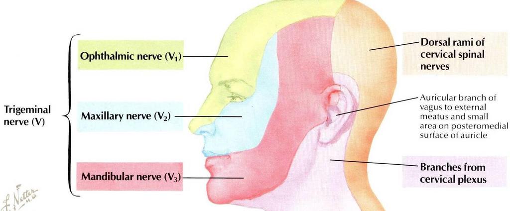
: 1. Ophthalmic. 2. Maxillary. 3. Mandibular.")
7 TRIGEMINAL NERVE Emerges from the middle of the ventral surface of the pons by 2 roots (Large Lateral sensory root & small medial motor root). Divides into 3 divisions (dendrites of trigeminal ganglion): 1. Ophthalmic. 2. Maxillary. 3. Mandibular. Axons of cells of motor nucleus join only the mandibular division.
8

9 OPHTHALMIC (PURE SENSORY) Divides into 3 branches: Frontal, Lacrimal & Nasociliary which pass through superior orbital fissure to the orbit Frontal: supplies skin of face & scalp. 2. Lacrimal: supplies skin of face & lacrimal gland. 3. Nasociliary: supplies skin of face, nasal cavity & eyeball.

. 1.")
10 MAXILLARY (PURE SENSORY) Supplies: 1. Upper teeth, gums & maxillary air sinus (posterior, middle & anterior superior alveolar nerves). 1. Face: (zygomaticofacial & infraorbital nerves).
 4. Auriculotemporal: supplies auricle, temple, parotid gland & TMJ.")
11 MANDIBULAR (MIXED) SENSORY BRANCHES: 1. Lingual: receives General sensations from anterior 2/3 the of tongue. 2. Inferior alveolar: supplies Lower teeth, gums & face. 3. Buccal: supplies Face (cheek on upper jaw) 4. Auriculotemporal: supplies auricle, temple, parotid gland & TMJ. MOTOR BRANCHES: to 8 muscles (4 muscles of mastication & other 4 muscles)
 This condition is characterized by recurring episodes of intense stabbing excruciating pain radiating from the angle of the jaw along a branches of the trigeminal nerve.")
12 Trigeminal Neuralgia Compression, degeneration or inflammation of the 5 th cranial nerve may result in a condition called trigeminal neuralgia or tic douloureux (spasmodic contraction of the muscles in the face) This condition is characterized by recurring episodes of intense stabbing excruciating pain radiating from the angle of the jaw along a branches of the trigeminal nerve. Usually involves maxillary & mandibular branches, rarely in the ophthalmic division.

13 FACIAL NERVE Type: Mixed ( Motor, special sensory, parasympathetic). Fibers: 1. Special visceral afferent: carrying taste sensation from anterior 2/3 of the tongue. 2. Special visceral efferent: supplying muscles developed from the 2 nd pharyngeal arch. 3. General visceral efferent: supplying parasympathetic secretory fibers to submandibular, sublingual, lacrimal, nasal & palatine glands. Parasymp. taste sensation motor
14 FACIAL NERVE NUCLEI 3 Nuclei : Special visceral afferent: (nucleus solitarius): receives taste from the anterior 2/3 of tongue. Special visceral efferent: motor nucleus of facial nerve: supplies: muscles of face, posterior belly of digastric, stylohyoid, platysma, stapedius, and occipitofrontalis. General visceral efferent: superior salivatory nucleus: sends preganglionic parasympathetic secretory fibers to sublingual, submandibular, lacrimal, nasal & palatine glands
15 COURSE OF FACIAL NERVE Emerges from the cerebellopontine angle by 2 roots: 1. Medial motor root: contains motor fibers. 2. Lateral root (nervous intermedius): contains parasympathetic & taste fibers.


16 COURSE OF FACIAL NERVE Passes through internal auditory meatus to inner ear where it runs in facial canal. Emerges from the stylomastoid foramen & enters the parotid gland where it ends.
17 BRANCHES OF FACIAL NERVE In facial canal: 1. Greater petrosal nerve: carries preganglionic parasympathetic fibers to lacrimal, nasal & palatine glands. 2. Chorda tympani: carries: a) preganglionic parasympathetic fibers to submandibular & sublingual glands. b) taste fibers from anterior 2/3 of tongue. 3. Nerve to stapedius. control the amplitude of sound waves from the external environment to the inner ear. N.B.: Geniculate ganglion: contains cell bodies of neurones ; its fibres carrying taste sensations from anterior 2/3 of tongue; ending in solitary nucleus in M.O. Lies in internal acoustic meatus


18 BRANCHES OF FACIAL NERVE Just as it emerges from the stylomastoid foramen it gives: 1. Posterior auricular: to occipitofrontalis muscle. 2. Muscular branches to posterior belly of digastric & stylohyoid. Inside parotid gland: gives 5 terminal motor branches: Temporal, Zygomatic, Buccal, Mandibular & Cervical. To the muscles of the face.
.")
19 Bell s Palsy Damage of the facial nerve results in paralysis of muscles of facial expressions : Facial (Bell s) palsy; lower motor neuron lesion (whole face affected) NB. In upper motor neuron lesion (upper face is intact). Face is distorted: Drooping of lower eyelid, Sagging of mouth angle, Dribbling of saliva, Loss of facial expressions, Loss of chewing, Loss of blowing, Loss of sucking, Unable to show teeth or close the eye on that side.
20 THANK YOU & BEST LUCK
21 SUMMARY Both trigeminal & facial nerves are mixed. Nuclei of trigeminal nerve are found in midbrain, pons & medulla. They are of the general somatic afferent & special visceral efferent types. The trigeminal nerve emerges from the pons and divides into: ophthalmic, maxillary & mandibular divisions that receive sensory supply from the face (with an exception of a small area over ramus of mandible by great auricular nerve C2,3). All motor fibers are included in the mandibular division & supply muscles of mastication.

22 SUMMARY Nuclei of facial nerve are found in pons. They are of the special visceral afferent & efferent types, as well as general visceral efferent type. The facial nerve emerges from the cerebellopontine angle, gives motor fibers to muscles of facial expression, secretory fibers to submandibular, sublingual, lacrimal, nasal & palatine glands & receives taste fibers from anterior 2/3 of tongue.
23 Lower Motor Neuron Lesion Results from injury of facial nerve fibres in internal acoustic meatus;in the middle ear;in the facial canal or in parotid gland. Manifested by complete paralysis of facial muscles on the same side of lesion. For the Students Upper Motor Neuron Lesion This occurs after injury to the pyramidal tract (corticonuclear) above facial nucleus... Leads to paralysis of facial muscles of lower ½ of face of opposite side but the upper ½ of the face intact because : Ms.of lower ½ of face receive pyramidal fibres from opposite cerebral cortex only, While Ms..of upper ½ of face receive pyramidal fibres from both cerebral hemispheres (Bilateral represented).
24 TEST YOUR SELF! Stimulation of which of the following nerves could lead to salivation and lacrimation?: a) Facial. b) Glossopharyngeal. c) Trigeminal. d) Vagus. Lesion of mandibular nerve may result in: a) Loss of sensation of skin over the nose. b) Loss of lacrimation. c) Loss of sensory supply of upper teeth. d) Loss of general sensations of anterior 2/3 of tongue.
Functional components
 Facial Nerve VII cranial nerve Emerges from Pons Two roots Functional components: 1. GSA (general somatic afferent) 2. SA (Somatic afferent) 3. GVE (general visceral efferent) 4. BE (Special visceral/branchial
Lec [8]: Mandibular nerve:
![Lec [8]: Mandibular nerve:](/thumbs/94/121295776.jpg "Lec [8]: Mandibular nerve:") Lec [8]: Mandibular nerve: The mandibular branch from the trigeminal ganglion lies in the middle cranial fossa lateral to the cavernous sinus. With the motor root of the trigeminal nerve [motor roots lies
Lec [8]: Mandibular nerve: The mandibular branch from the trigeminal ganglion lies in the middle cranial fossa lateral to the cavernous sinus. With the motor root of the trigeminal nerve [motor roots lies
Cranial Nerve VII - Facial Nerve. The facial nerve has 3 main components with distinct functions
 Cranial Nerve VII - Facial Nerve The facial nerve has 3 main components with distinct functions Somatic motor efferent Supplies the muscles of facial expression; posterior belly of digastric muscle; stylohyoid,
Laith Sorour. Facial nerve (vii):
:") Laith Sorour Cranial nerves 7 & 8 Hello, there are edited slides please go back to them to see pictures, they are not that much important in this lecture but still, and yes slides are included :p Let s
Laith Sorour Cranial nerves 7 & 8 Hello, there are edited slides please go back to them to see pictures, they are not that much important in this lecture but still, and yes slides are included :p Let s
Trigeminal Nerve (V)
") Trigeminal Nerve (V) Lecture Objectives Discuss briefly how the face is developed. Follow up the course of trigeminal nerve from its point of central connections, exit and down to its target areas. Describe
Trigeminal Nerve (V) Lecture Objectives Discuss briefly how the face is developed. Follow up the course of trigeminal nerve from its point of central connections, exit and down to its target areas. Describe
Parotid Gland, Temporomandibular Joint and Infratemporal Fossa
 M1 - Anatomy Parotid Gland, Temporomandibular Joint and Infratemporal Fossa Jeff Dupree Sanger 9-057 jldupree@vcu.edu Parotid gland: wraps around the mandible positioned between the mandible and the sphenoid
Cranial nerves.
 Cranial nerves eaglezhyxzy@163.com Key Points of Learning Name Components Passing through Peripheral distribution Central connection Function Cranial nerves Ⅰ olfactory Ⅱ optic Ⅲ occulomotor Ⅳ trochlear
Temporal region. temporal & infratemporal fossae. Zhou Hong Ying Dept. of Anatomy
 Temporal region temporal & infratemporal fossae Zhou Hong Ying Dept. of Anatomy Temporal region is divided by zygomatic arch into temporal & infratemporal fossae. Temporal Fossa Infratemporal fossa Temporal
Cranial Nerve VII & VIII
 Cranial Nerve VII & VIII Lecture Objectives Follow up the course of facial nerve from its point of central connections, exit and down to its target areas. Follow up the central connections of the facial
Dr.Ban I.S. head & neck anatomy 2 nd y. جامعة تكريت كلية طب االسنان املرحلة الثانية أ.م.د. بان امساعيل صديق 6102/6102
 جامعة تكريت كلية طب االسنان التشريح مادة املرحلة الثانية أ.م.د. بان امساعيل صديق 6102/6102 Parotid region The part of the face in front of the ear and below the zygomatic arch is the parotid region. The
Trigeminal Nerve Worksheets, Distributions Page 1
 Trigeminal Nerve Worksheet #1 Distribution by Nerve Dr. Darren Hoffmann Dental Gross Anatomy, Spring 2013 We have drawn out each of the branches of CN V in lecture and you have an idea now for their basic
Trigeminal Nerve Anatomy. Dr. Mohamed Rahil Ali
 Trigeminal Nerve Anatomy Dr. Mohamed Rahil Ali Trigeminal nerve Largest cranial nerve Mixed nerve Small motor root and large sensory root Motor root Nucleus of motor root present in the pons and medulla
INTRODUCTION: ANATOMY UNDERLYING CLINICAL TESTS OF CRANIAL NERVES
 INTRODUCTION: ANATOMY UNDERLYING CLINICAL TESTS OF CRANIAL NERVES CRANIAL NERVE I - OLFACTORY I - OLFACTORY NERVE - SMELL TEST: SMELL ODORS (note: not ammonia; pain in nasal cavity CN5 DAMAGE: LOSS OF
PTERYGOPALATINE FOSSA
 PTERYGOPALATINE FOSSA Outline Anatomical Structure and Boundaries Foramina and Communications with other spaces and cavities Contents Pterygopalatine Ganglion Especial emphasis on certain arteries and
The Seventh Cranial Nerve The Facial By Prof. Dr. Muhammad Imran Qureshi
 The Seventh Cranial Nerve The Facial By Prof. Dr. Muhammad Imran Qureshi Functional Components: SVE: Fibers originate from nucleus of facial nerve, and supply facial muscles GVE: Fibers derived from superior
Introduction to Head and Neck Anatomy
 Introduction to Head and Neck Anatomy Nervous Tissue Controls and integrates all body activities within limits that maintain life Three basic functions 1. sensing changes with sensory receptors 2. interpreting
For the following questions, indicate the letter that corresponds to the SINGLE MOST APPROPRIATE ANSWER
 GROSS ANATOMY EXAMINATION May 15, 2000 For the following questions, indicate the letter that corresponds to the SINGLE MOST APPROPRIATE ANSWER 1. Pain associated with an infection limited to the middle
Temporal fossa Infratemporal fossa Pterygopalatine fossa Terminal branches of external carotid artery Pterygoid venous plexus
 Outline of content Temporal fossa Infratemporal fossa Pterygopalatine fossa Terminal branches of external carotid artery Pterygoid venous plexus Boundary Content Communication Mandibular division of trigeminal
human anatomy 2016 lecture fifteen Dr meethak ali ahmed neurosurgeon
 Cranial Nerves Organization of the Cranial Nerves The cranial nerves are named as follows: I. Olfactory II. Optic III. Oculomotor IV. Trochlear V. Trigeminal VI. Abducent VII. Facial VIII. Vestibulocochlear
function - sensory & postganglionic sympathetic [communication from the internal carotid plexus in the cavernous sinus] innervation of the mucosa of
![function - sensory & postganglionic sympathetic [communication from the internal carotid plexus in the cavernous sinus] innervation of the mucosa of](/thumbs/74/71276096.jpg "function - sensory & postganglionic sympathetic [communication from the internal carotid plexus in the cavernous sinus] innervation of the mucosa of") Nerves I. Cranial nerves A. Olfactory (CN I) 1. Olfactory bulb 2. Olfactory tract B. Optic n. (CNII) function - carries visual sensory information from the neural retina to the diencephalon & midbrain
Nerves I. Cranial nerves A. Olfactory (CN I) 1. Olfactory bulb 2. Olfactory tract B. Optic n. (CNII) function - carries visual sensory information from the neural retina to the diencephalon & midbrain
3-Deep fascia: is absent (except over the parotid gland & buccopharngeal fascia covering the buccinator muscle)
") The Face 1-Skin of the Face The skin of the face is: Elastic Vascular (bleed profusely however heal rapidly) Rich in sweat and sebaceous glands (can cause acne in adults) It is connected to the underlying
The Face 1-Skin of the Face The skin of the face is: Elastic Vascular (bleed profusely however heal rapidly) Rich in sweat and sebaceous glands (can cause acne in adults) It is connected to the underlying
Parotid Gland. Parotid Gland. Largest of 3 paired salivary glands (submandibular; sublingual) Ramus of Mandible. Medial pterygoid.
 Ramus of Mandible. Medial pterygoid.") Parotid region Parotid Gland Largest of 3 paired salivary glands (submandibular; sublingual) Ramus of Mandible Medial pterygoid Cross section of mandible Masseter D S SCM Parotid Gland Mastoid Process
Parotid region Parotid Gland Largest of 3 paired salivary glands (submandibular; sublingual) Ramus of Mandible Medial pterygoid Cross section of mandible Masseter D S SCM Parotid Gland Mastoid Process
CN I Olfactory. CN II Optic. CN III Oculomotor. Special Sensory Efferent fibers to Olfactory Bulb. Cribiform Plate of Ethmoid
 CN I Olfactory Efferent fibers to Olfactory Bulb Olfactory Tract Olfactory Bulb Cribiform Plate of Ethmoid Anosmia Loss of sense of smell Uncinate Fits olfactory hallucinations To Olfactory Epithelium
Brainstem and Cranial Nerves II. Nerves covered in other lectures. A reminder about embryology. Prof. Stuart Bunt
 Brainstem and Cranial Nerves II Prof. Stuart Bunt Nerves covered in other lectures 1 Olfactory 2 Optic 3,4,6 Extraocular eye muscles 8 Vestibulo-cochlear 5 Motor and Sensory to the face and muscles of
Anatomy of the Trigeminal Nerve
 19 Anatomy of the Trigeminal Nerve.1 Introduction 0. The Central Part of the Trigeminal Nerve 1..1 Origin 1.. Trigeminal Nuclei.3 The Peripheral Part of the Trigeminal Nerve 4.3.1 Ophthalmic Nerve 4.3.
Trigeminal nerve. Slide in bold and please go back to see the pictures, if I skipped any part of record that because it wasn t clear to me
 Trigeminal nerve Slide in bold and please go back to see the pictures, if I skipped any part of record that because it wasn t clear to me Hala nsour 2/26/2018 P a g e 1 this lecture contain two topics
Infratemporal fossa: Tikrit University college of Dentistry Dr.Ban I.S. head & neck Anatomy 2 nd y.
 Infratemporal fossa: This is a space lying beneath the base of the skull between the lateral wall of the pharynx and the ramus of the mandible. It is also referred to as the parapharyngeal or lateral pharyngeal
Biology 323 Human Anatomy for Biology Majors Week 10; Lecture 1; Tuesday Dr. Stuart S. Sumida. Cranial Nerves and Soft Tissues of the Skull
 Biology 323 Human Anatomy for Biology Majors Week 10; Lecture 1; Tuesday Dr. Stuart S. Sumida Cranial Nerves and Soft Tissues of the Skull FOREBRAIN MIDBRAIN HINDBRAIN Forebrain: Cerebrum Perception,
Omran Saeed. Luma Taweel. Mohammad Almohtaseb. 1 P a g e
 2 Omran Saeed Luma Taweel Mohammad Almohtaseb 1 P a g e I didn t include all the photos in this sheet in order to keep it as small as possible so if you need more clarification please refer to slides In
Tracing the Cranial Nerves Osteologically
 CN I II III IV V 1 Supra-orbital ethmoidal nn. Ext. nasal V 2 Tracing the Cranial Nerves Osteologically Nucleus of Origin Olfactory tracts of frontal lobe of cerebrum Optic tracts from optic chiasma and
PERIPHERAL NERVOUS SYSTEM
 CHAPTER 13 PERIPHERAL NERVOUS SYSTEM Functional division of nervous system = afferent info to the CNS ascending spinal cord = efferent info from CNS descending spinal cord somatic skin, muscles visceral
Brain and spinal nerve. By: shirin Kashfi
 Brain and spinal nerve By: shirin Kashfi Nervous system: central nervous system (CNS) peripheral nervous system (PNS) Brain (cranial) nerves Spinal nerves Ganglions (dorsal root ganglions, sympathetic
SCHOOL OF ANATOMICAL SCIENCES Mock Run Questions. 4 May 2012
 SCHOOL OF ANATOMICAL SCIENCES Mock Run Questions 4 May 2012 1. With regard to the muscles of the neck: a. the platysma muscle is supplied by the accessory nerve. b. the stylohyoid muscle is supplied by
Cranial nerve Dept. of Anatomy Zhou Hong Ying
 Cranial nerve Dept. of Anatomy Zhou Hong Ying Key Points of Learning Name Components Passing through Peripheral distribution Central connection Function Cranial nerve Ⅰ olfactory Ⅱ optic Ⅲ occulomotor
CRANIAL NERVES. Dr. Amani A. Elfaki Associate Professor Department of Anatomy
 CRANIAL NERVES Dr. Amani A. Elfaki Associate Professor Department of Anatomy LEARNING OBJECTIVES Named the cranial nerves Identify the funcunal component of each cranial nerve Identify the effect of each
General Sensory Pathways of the Face Area, Taste Pathways and Hearing Pathways
 General Sensory Pathways of the Face Area, Taste Pathways and Hearing Pathways Lecture Objectives Describe pathways for general sensations (pain, temperature, touch and proprioception) from the face area.
Dr. Sami Zaqout Faculty of Medicine IUG
 Auricle External Ear External auditory meatus The Ear Middle Ear (Tympanic Cavity) Auditory ossicles Internal Ear (Labyrinth) Bony labyrinth Membranous labyrinth External Ear Auricle External auditory
Face. Definition: The area between the two ears and from the chin to the eye brows. The muscles of the face
 Face Definition: The area between the two ears and from the chin to the eye brows. The muscles of the face The muscle of facial expression (include the muscle of the face and the scalp). All are derived
Mohammad Hisham Al-Mohtaseb. Lina Mansour. Reyad Jabiri. 0 P a g e
 2 Mohammad Hisham Al-Mohtaseb Lina Mansour Reyad Jabiri 0 P a g e This is only correction for the last year sheet according to our record. If you already studied this sheet just read the yellow notes which
C h a p t e r PowerPoint Lecture Slides prepared by Jason LaPres North Harris College Houston, Texas
 C h a p t e r 15 The Nervous System: The Brain and Cranial Nerves PowerPoint Lecture Slides prepared by Jason LaPres North Harris College Houston, Texas Copyright 2009 Pearson Education, Inc., publishing
Face and Scalp 解剖學科鄭授德
 Face and Scalp 解剖學科鄭授德 本教材之圖片取自於 1 Gray s Anatomy for Students, 3rd ed, 2015, by Drake, Vogl, and Mitchell 2 Clinically Oriented Anatomy, 7th ed, 2014, by Moore, Dalley, and Agur 3 Clinically Oriented
Veins of the Face and the Neck
 Veins of the Face and the Neck Facial Vein The facial vein is formed at the medial angle of the eye by the union of the supraorbital and supratrochlear veins. connected through the ophthalmic veins with
Introduction to Local Anesthesia and Review of Anatomy
 5-Sep Introduction and Anatomy Review 12-Sep Neurophysiology and Pain 19-Sep Physiology and Pharmacology part 1 26-Sep Physiology and Pharmacology part 2 Introduction to Local Anesthesia and Review of
CN modalities Sensory: SSA (Vision) Mixed: GSE, proprioceptive. Mixed: GSE, proprioceptive
 Mixed: GSE, proprioceptive. Mixed: GSE, proprioceptive") CN 2 3 4 6 modalities Sensory: SSA (Vision) course Rods and cones of the retina bipolar neurons gangli on cells Optic nerve optic foramen Optic chiasm Optic tracts Sup colliculi LGN Optic radiation cortex
CN 2 3 4 6 modalities Sensory: SSA (Vision) course Rods and cones of the retina bipolar neurons gangli on cells Optic nerve optic foramen Optic chiasm Optic tracts Sup colliculi LGN Optic radiation cortex
Unit VIII Problem 9 Anatomy of The Ear
 Unit VIII Problem 9 Anatomy of The Ear - The ear is an organ with 2 functions: Hearing. Maintenance of equilibrium/balance. - The ear is divided into 3 parts: External ear. Middle ear (which is also known
Unit VIII Problem 3 Neuroanatomy: Brain Stem, Cranial Nerves and Scalp
 Unit VIII Problem 3 Neuroanatomy: Brain Stem, Cranial Nerves and Scalp - Brain stem: It is connected to the cerebellum and cerebral hemispheres. Rostral end of brain stem: diencephalon is the area which
Lecture 07. Lymphatic's of Head & Neck. By: Dr Farooq Amanullah Khan PMC
 Lecture 07 Lymphatic's of Head & Neck By: Dr Farooq Amanullah Khan PMC Dated: 28.11.2017 Lymphatic Vessels Of the 800 lymph nodes in the human body, 300 are in the Head & neck region. The lymphatic vessels
Tikrit University collage of dentistry Dr.Ban I.S. head & neck anatomy 2 nd y. Lec [5] / Temporal fossa :
![Tikrit University collage of dentistry Dr.Ban I.S. head & neck anatomy 2 nd y. Lec [5] / Temporal fossa :](/thumbs/88/115294566.jpg "Tikrit University collage of dentistry Dr.Ban I.S. head & neck anatomy 2 nd y. Lec [5] / Temporal fossa :") Lec [5] / Temporal fossa : Borders of the Temporal Fossa: Superior: Superior temporal line. Inferior: gap between zygomatic arch and infratemporal crest of sphenoid bone. Anterior: Frontal process of the
Lec [5] / Temporal fossa : Borders of the Temporal Fossa: Superior: Superior temporal line. Inferior: gap between zygomatic arch and infratemporal crest of sphenoid bone. Anterior: Frontal process of the
Learning Outcomes. The Carotid 20/02/2013. Scalp, Face, Parotid. Layers of the Scalp. The Parotid Gland. The Scalp. The Carotid The Facial Artery
 Learning Outcomes The Scalp Layers of the Scalp Bleeding from the Scalp The Carotid The Facial Artery Major Muscles of the Face and Jaw(s) Muscles of Mastication Muscles of Facial Expression The Parotid
Cranial Nerves. Steven McLoon Department of Neuroscience University of Minnesota
 Cranial Nerves Steven McLoon Department of Neuroscience University of Minnesota 1 Course News Change in Lab Sequence Week of Oct 2 Lab 5 Week of Oct 9 Lab 4 2 Sensory and Motor Systems Sensory Systems:
V1-ophthalmic. V2-maxillary. V3-mandibular. motor
 4. Trigeminal Nerve I. Objectives:. Understand the types of sensory information transmitted by the trigeminal system.. Describe the major peripheral divisions of the trigeminal nerve and how they innervate
Maxilla, ORBIT and infratemporal fossa. Neophytos C Demetriades MD, DDS, MSc Associate professor European University of Cyprus School of Medicine
 Maxilla, ORBIT and infratemporal fossa Neophytos C Demetriades MD, DDS, MSc Associate professor European University of Cyprus School of Medicine MAXILLA Superior, middle, and inferior meatus Frontal sinus
Organization of The Nervous System PROF. MOUSAED ALFAYEZ & DR. SANAA ALSHAARAWY
 Organization of The Nervous System PROF. MOUSAED ALFAYEZ & DR. SANAA ALSHAARAWY Objectives At the end of the lecture, the students should be able to: List the parts of the nervous system. List the function
Doctor Osama Asa ad Khader. Mohammad Alsalem
 6 Doctor 2015 Osama Asa ad Khader Mohammad Alsalem A quick revision for the spinal cord blood supply: Arterial Blood supply of spinal cord The spinal cord got its arterial supply by two ways: Longitudinal
Anatomic Relations Summary. Done by: Sohayyla Yasin Dababseh
 Anatomic Relations Summary Done by: Sohayyla Yasin Dababseh Anatomic Relations Lecture 1 Part-1 - The medial wall of the nose is the septum. - The vestibule lies directly inside the nostrils (Nares). -
lecture #2 Done by : Tyma'a Al-zaben
 lecture #2 Done by : Tyma'a Al-zaben ** Hello SERTONIN! note:: the slide included within the sheet but make sure back to slide for pictures in the previous lecture we talk about ascending tract and its
Organization of The Nervous System PROF. SAEED ABUEL MAKAREM
 Organization of The Nervous System PROF. SAEED ABUEL MAKAREM Objectives By the end of the lecture, you should be able to: List the parts of the nervous system. List the function of the nervous system.
Oral cavity : consist of two parts: the oral vestibule and the oral cavity proper. Oral vestibule : is slit like space between.
 Oral cavity Oral cavity : consist of two parts: the oral vestibule and the oral cavity proper Oral vestibule : is slit like space between the teeth, buccal gingiva, lips, and cheeks 1 Oral cavity Oral
University of Palestine. Final Exam 1 st Semester 2014/2015 Total Grade: 60
 Question One: MCQ: 1- The coronal suture joins the a) frontal and parietal bones. b) left and right parietal bones. c) parietal and occipital bones. d) parietal, squamous temporal and greater wing of the
The Skull and Temporomandibular joint II Prof. Abdulameer Al-Nuaimi. E. mail:
 The Skull and Temporomandibular joint II Prof. Abdulameer Al-Nuaimi E-mail: a.al-nuaimi@sheffield.ac.uk E. mail: abdulameerh@yahoo.com Temporal fossa The temporal fossa is a depression on the temporal
Bony orbit Roof The orbital plate of the frontal bone Lateral wall: the zygomatic bone and the greater wing of the sphenoid
 Bony orbit Roof: Formed by: The orbital plate of the frontal bone, which separates the orbital cavity from the anterior cranial fossa and the frontal lobe of the cerebral hemisphere Lateral wall: Formed
Anatomy and Physiology. Bones, Sutures, Teeth, Processes and Foramina of the Human Skull
 Anatomy and Physiology Chapter 6 DRO Bones, Sutures, Teeth, Processes and Foramina of the Human Skull Name: Period: Bones of the Human Skull Bones of the Cranium: Frontal bone: forms the forehead and the
Structure Location Function
 Frontal Bone Cranium forms the forehead and roof of the orbits Occipital Bone Cranium forms posterior and inferior portions of the cranium Temporal Bone Cranium inferior to the parietal bone forms the
Major Anatomic Components of the Orbit
 Major Anatomic Components of the Orbit 1. Osseous Framework 2. Globe 3. Optic nerve and sheath 4. Extraocular muscles Bony Orbit Seven Bones Frontal bone Zygomatic bone Maxillary bone Ethmoid bone Sphenoid
Lab 16: PNS: Nerves and Autonomic NS Hamilton Answers to Pre- Lab Assignments
 Lab 16: PNS: Nerves and Autonomic NS Hamilton Answers to Pre- Lab Assignments Pre-Lab Activity 1: 1. a. olfactory nerve b. optic nerve c. oculomotor nerve d. abducens nerve e. trochlear nerve f. trigeminal
Nose & Mouth OUTLINE. Nose. - Nasal Cavity & Its Walls. - Paranasal Sinuses. - Neurovascular Structures. Mouth. - Oral Cavity & Its Contents
 Dept. of Human Anatomy, Si Chuan University Zhou hongying eaglezhyxzy@163.com Nose & Mouth OUTLINE Nose - Nasal Cavity & Its Walls - Paranasal Sinuses - Neurovascular Structures Mouth - Oral Cavity & Its
Perineural Tumor Spread (PNS) Perineural Tumor Spread (PNS) PNS Anatomic Considerations. Perineural Tumor Spread-Imaging
 Perineural Tumor Spread (PNS) PNS Anatomic Considerations. Perineural Tumor Spread-Imaging") Imaging of Perineural Tumor Spread in Head and Neck Cancer Lawrence E. Ginsberg, MD Departments of Diagnostic Radiology and Head and Neck Surgery University of Texas M.D. Anderson Cancer Center Houston,
Imaging of Perineural Tumor Spread in Head and Neck Cancer Lawrence E. Ginsberg, MD Departments of Diagnostic Radiology and Head and Neck Surgery University of Texas M.D. Anderson Cancer Center Houston,
The sebaceous glands (glands of Zeis) open directly into the eyelash follicles, ciliary glands (glands of Moll) are modified sweat glands that open
 open directly into the eyelash follicles, ciliary glands (glands of Moll) are modified sweat glands that open") The Orbital Region The orbits are a pair of bony cavities that contain the eyeballs; their associated muscles, nerves, vessels, and fat; and most of the lacrimal apparatus upper eyelid is larger and more
The Orbital Region The orbits are a pair of bony cavities that contain the eyeballs; their associated muscles, nerves, vessels, and fat; and most of the lacrimal apparatus upper eyelid is larger and more
Dr.Ban I.S. head & neck anatomy 2 nd y جامعة تكريت كلية طب االسنان مادة التشريح املرحلة الثانية أ.م.د. بان امساعيل صديق 6102/6102
 جامعة تكريت كلية طب االسنان مادة التشريح املرحلة الثانية أ.م.د. بان امساعيل صديق 6102/6102 Pterygopalatine fossa: The pterygopalatine fossa is a cone-shaped depression, It is located between the maxilla,
04 Development of the Face and Neck. Development of the Face Development of the neck
 04 Development of the Face and Neck Development of the Face Development of the neck Development of the face Overview of facial development The fourth week ~ the twelfth week of prenatal development Between
Bones of the skull & face
 Bones of the skull & face Cranium= brain case or helmet Copyright The McGraw-Hill Companies, Inc. Permission required for reproduction or display. The cranium is composed of eight bones : frontal Occipital
Cranial Nerves and Spinal Cord Flashcards
 1. Name the cranial nerves and their Roman numeral. 2. What is Cranial Nerve I called, and what does it 3. Scientists who are trying to find a way to make neurons divide to heal nerve injuries often study
THE BRAINSTEM. Raymond S. Price, MD University of Pennsylvania
 THE BRAINSTEM Raymond S. Price, MD University of Pennsylvania Overview of Brainstem Functions The brainstem serves numerous crucial neurologic functions. The most clinically relevant functions include:
-Ibrahim Al-Naser. -Dr Al- Muhtaseb. 1 P a g e
 -1 -Ibrahim Al-Naser - -Dr Al- Muhtaseb 1 P a g e The Digestive System The doctor started the lecture by talking about the class rules. The GI system is an organ system, it is divided into: The Alimentary
REVIEW/PREVIEW OF HEAD AND NECK ANATOMY FOR ENT EXAM
 REVIEW/PREVIEW OF HEAD AND NECK ANATOMY FOR ENT EXAM - 2017 PALPATE CAROTID ARTERY: AT LEVEL OF CAROTID BIFURCATION VERTEBRAL LEVEL C4 Sternocleidomastoid Muscle INTERNAL CAROTID EXTERNAL CAROTID COMMON
MAXILLA, ORBIT & PTERYGOPALATINE FOSSA. Neophytos C Demetriades MD, DDS, MSc Associate professor European University of Cyprus School of Medicine
 MAXILLA, ORBIT & PTERYGOPALATINE FOSSA Neophytos C Demetriades MD, DDS, MSc Associate professor European University of Cyprus School of Medicine Maxilla MAXILLA Superior, middle, and inferior meatus Frontal
Omar Sami. Aseel Abdeen. Muhammad Al-Salem. 1 P a g e
 Omar Sami Aseel Abdeen Muhammad Al-Salem 1 P a g e Using only section 2 record, I wrote this sheet; as the video is not ready yet. Despite pointing the structures, I ve tried to include all the scientific
Cranial Nerve VIII (The Vestibulo-Cochlear Nerve)
") Cranial Nerve VIII (The Vestibulo-Cochlear Nerve) Please view our Editing File before studying this lecture to check for any changes. Color Code Important Doctors Notes Notes/Extra explanation Objectives
Cranial Nerve VIII (The Vestibulo-Cochlear Nerve) Please view our Editing File before studying this lecture to check for any changes. Color Code Important Doctors Notes Notes/Extra explanation Objectives
3. The Jaw and Related Structures
 Overview and objectives of this dissection 3. The Jaw and Related Structures The goal of this dissection is to observe the muscles of jaw raising. You will also have the opportunity to observe several
The Ear The ear consists of : 1-THE EXTERNAL EAR 2-THE MIDDLE EAR, OR TYMPANIC CAVITY 3-THE INTERNAL EAR, OR LABYRINTH 1-THE EXTERNAL EAR.
 The Ear The ear consists of : 1-THE EXTERNAL EAR 2-THE MIDDLE EAR, OR TYMPANIC CAVITY 3-THE INTERNAL EAR, OR LABYRINTH 1-THE EXTERNAL EAR Made of A-AURICLE B-EXTERNAL AUDITORY MEATUS A-AURICLE It consists
Imaging of facial paralysis
 Imaging of facial paralysis Poster No.: C-2151 Congress: ECR 2013 Type: Educational Exhibit Authors: N. Martinez Molina, L. Aleman Romero, L. A. Sanchez Alonso, A. Puerta Sales, V. Garcia Medina; Murcia/ES
Faculty of Dental Medicine and Surgery. Sem 4 Cranial Nerves Dr. Abbas Garib Alla
 Faculty of Dental Medicine and Surgery Sem 4 Cranial Nerves Dr. Abbas Garib Alla Cranial Nerves I through XII FUNCTIPONAL CLSSIFICATION OF THE CN parasympathetic nerves 1973 PHARYNGEAL ARCHES nerves 1975
Dr. Sami Zaqout, IUG Medical School
 The skull The skull is composed of several separate bones united at immobile joints called sutures. Exceptions? Frontal bone Occipital bone Vault Cranium Sphenoid bone Zygomatic bones Base Ethmoid bone
CHAPTER 13. FACIAL NERVE PARALYSIS
 CHAPTER 13. FACIAL NERVE PARALYSIS Introduction Facial nerve paralysis, whilst not a disease of the ear itself, commonly arises within the ear due to its anatomical course, and often as a result of ear
Tympanic Bulla Temporal Bone. Digastric Muscle. Masseter Muscle
 Superior view Hyoid Bone The hyoid bone does not articulate with any other bones. It is held in place by ligaments to the styloid process of the temporal bone and the thyroid cartilage of the larynx. It
By Dr. Saeed Vohra & Dr. Sanaa Alshaarawy
 By Dr. Saeed Vohra & Dr. Sanaa Alshaarawy 1 By the end of the lecture, students will be able to : Distinguish the internal structure of the components of the brain stem in different levels and the specific
Head and Face Anatomy
 Head and Face Anatomy Epicranial region The Scalp The soft tissue that covers the vault of skull. Extends from supraorbital margin to superior nuchal line. Layers of the scalp S C A L P = skin = connective
Chapter 14: Nervous System Guided Notes (A-day)
") Chapter 14: Nervous System Guided Notes (A-day) Nervous System Overview Major Function: Control the body's and. Divided into the Nervous System (CNS=Brain and Spinal Cord) and the Nervous System (PNS=Cranial
Chapter 14: Nervous System Guided Notes (A-day) Nervous System Overview Major Function: Control the body's and. Divided into the Nervous System (CNS=Brain and Spinal Cord) and the Nervous System (PNS=Cranial
Cranial Nerves IX-X (Glossopharyngeal & Vagus Nerves)
") Cranial Nerves IX-X (Glossopharyngeal & Vagus Nerves) Please view our Editing File before studying this lecture to check for any changes. Color Code Important Doctors Notes Notes/Extra explanation Objectives
Cranial Nerves IX-X (Glossopharyngeal & Vagus Nerves) Please view our Editing File before studying this lecture to check for any changes. Color Code Important Doctors Notes Notes/Extra explanation Objectives
ParasymPathetic Nervous system. Done by : Zaid Al-Ghnaneem
 ParasymPathetic Nervous system Done by : Zaid Al-Ghnaneem In this lecture we are going to discuss Parasympathetic, in the last lecture we took sympathetic and one of the objectives of last lecture was
FACE CN V & VII PAROTID GLAND. Jacek Baj, MD, PhD Department of Human Anatomy
 FACE CN V & VII PAROTID GLAND Jacek Baj, MD, PhD Department of Human Anatomy THE FACE THE SCALP The scalp is composed of five layers: Skin ConnecAve Assue Aponeurosis Loose connecave Assue Pericranium
Bisection of Head & Nasal Cavity 頭部對切以及鼻腔. 解剖學科馮琮涵副教授 分機
 Bisection of Head & Nasal Cavity 頭部對切以及鼻腔 解剖學科馮琮涵副教授 分機 3250 E-mail: thfong@tmu.edu.tw Outline: The structure of nose The concha and meatus in nasal cavity The openings of paranasal sinuses Canals, foramens
Basic Anatomy and Physiology of the Lips and Oral Cavity. Dr. Faghih
 Basic Anatomy and Physiology of the Lips and Oral Cavity Dr. Faghih It is divided into seven specific subsites : 1. Lips 2. dentoalveolar ridges 3. oral tongue 4. retromolar trigone 5. floor of mouth 6.
Brainstem. Telencephalon Diencephalon Cerebellum Brain stem
 Brainstem Brainstem 脑 脊髓 Brainstem Telencephalon Diencephalon Cerebellum Brain stem Ventral view Lateral view 10 pairs of the cranial nerves are attached to the brain stem The brainstem Midbrain Pons Medulla
Peripheral Nervous System
 Peripheral Nervous System Sensory Receptors Motor Endings Cranial Nerves The Four Plexuses Extremities Review of Reflexes Fast, preprogrammed, inborn, automatic responses Occur in the CNS at the spinal
b. The groove between the two crests is called 2. The neural folds move toward each other & the fuse to create a
 Chapter 13: Brain and Cranial Nerves I. Development of the CNS A. The CNS begins as a flat plate called the B. The process proceeds as: 1. The lateral sides of the become elevated as waves called a. The