Bony orbit. Sup. Med. Inf. Lat. frontal bone. frontal process of maxilla. zygomatic process of maxilla zygomatic bone
|
|
|
- Nora Cole
- 5 years ago
- Views:
Transcription
1 Orbit 解剖學科鄭授德 本教材之圖片取自於 1. Gray s Anatomy for Students, 3rd ed., 2015, by Drake, Vogl, and Mitchell 2. Clinically Oriented Anatomy, 7th ed., 2014, by Moore, Dalley, and Agur 3. Anatomy, an Essential Textbook, 2013, by Giloroy, Voll, and Wesker 4. Clemente Anatomy, A Regional Atlas of the Human Body, 1997, 4th ed 5. Gray s Anatomy, the Anatomical Basis of Clinical Practice, 2008, 40th ed.
2 Bony orbit Sup. frontal bone Med. frontal process of maxilla Inf. zygomatic process of maxilla zygomatic bone Lat. frontal processes of zygomatic bone zygomatic processes of frontal bone
3 Roof Frontal and sphenoid bones (lesser wing); anterior cranial fossa is above the roof Anteromedially frontal sinus, trochlear fovea Anterolaterally lacrimal fossa
4 Medial wall By maxilla, lacrimal, ethmoid, and sphenoid bones Ethmoid air cells, frontoethmoidal suture, anterior and posterior ethmoidal foramina (for ant. and post. ethmoidal nn. and vessels) Lacrimal bone Lacrimal groove (with posterior lacrimal crest of lacrimal bone, and anterior lacrimal crest of maxilla) Optic canal on the sphenoid bone


5 Floor also the roof of maxillary sinus, contains maxilla, zygomatic, and palatine bones Inferior orbital fissure separates the inferior wall from lateral wall. Lateral wall zygomatic bone and greater wing of sphenoid bone
6 Eyelids Palpebral fissure between upper and lower eyelids 1. Skin and subcutaneous tissue ST may accumulate blood under injury 2. Orbicularis oculi 3. Orbital septum Extension of periosteum in upper and lower eyelids Attaches to the tendon of levator palpebrae superioris m. and tarsus of lower eyelid 4. Tarsus and levator palpebrae superioris 5. Conjunctiva reflects onto sclera as superior and inferior conjunctival fornices. Conjunctival sac is formed when eyelids close.


7 Orbicularis oculi innervated by CN VII 1. Palpebral part: in eyelids Medial palpebral ligament to ant. lacrimal crest Lateral palpebral ligament to zygomaitc bone 2. Orbital part: around orbit 3. Lacrimal part: medial end, attaches to post. lacrimal crest drains tears

8 Tarsus Superior tarsus and inferior tarsus attach through medial palpebral ligament to anterior lacrimal crest of maxilla attach through lateral palpebral ligament to zygomatic bone Tarsal glands embed in the tarsal plates, and open to the free margin of eyelids
9 Levator palpebrae superioris Levator palpebrae superioris muscle O: post. part of the roof of orbit, sup. to optic foramen I: ant. surface of superior tarsus F: raises upper eyelid N: CN III Superior tarsal muscle O: inferior surface of levator palpebrae superioris I: upper edge of superior tarsus F: assists to maintain upper eyelid elevation N: sympathetic fibers from superior cervical ganglion Loss function of either of these two muscles causes drooping of upper eyelid (ptosis)
10 Glands Sebaceous glands and sweat glands associated with eyelash blockage and inflammation cause stye ( 麥粒腫 針眼 ) on the edge of eyelids Tarsal glands opening on the edge of eyelids blockage and inflammation cause chalazion ( 霰ㄒㄧㄢˋ 粒腫 ) on the inner surface of eyelids

11 Vessels 1. Ophthalmic a. Supratrochlear a. Supra-orbital a. Lacrimal a. Dorsal nasal a. 2. Facial a. Angular a. 3. Superficial temporal a. Transverse facial a. 4. Brr. of superficial temporal a. Ext. venous drainage with associated aa. Int. venous drainage to ophthalmic vv. Lymph drainage to parotid primarily, and (medial corner of orbit to) submandibular nodes
 CN III levator palpebrae superioris (complete ptosis) Sympathetic ff. superior tarsal m.")
12 Innervation Sensory: Supra-orbital, supratrochlear, infratrochlear, lacrimal brr. of CN V 1 Infra-orbital br. of CN V 2 Motor: CN VII palpebral part of orbicularis oculi (unable to close eyelid tightly) CN III levator palpebrae superioris (complete ptosis) Sympathetic ff. superior tarsal m. (partial ptosis) Horner s syndrome Lesion on sympathetic trunk in neck pupillary constriction( 縮瞳 ) partial ptosis( 垂瞼 ) absence of sweating( 乾 ) vasodilation( 結膜紅 ) endophthalmos( 眼球下陷 )

13 Lacrimal apparatus Lacrimal glands and ducts: Orbital part: in lacrimal fossa of frontal bone Palpebral part: inferior to levator palpebrae superioris Lacrimal lake medial end of conjunctival sac Lacrimal punctum opening of lacrimal canaliculus Lacrimal canaliculi drain to lacrimal sac Lacrimal sac between medial palpebral ligament and lacrimal part of orbicularis oculi m. Nasolacrimal duct drain to inferior meatus of nasal cavity
14 Innervation 1. Sensory innervation Lacrimal br. of CN V1 2. Secretomotor (parasympathetic) innervation Preganglionic parasympathetic ff. in CN VII > Greater petrosal n. > N. of pterygoid canal > Pterygopalatine gang. (synapse) > Postganglionic ff. > Maxillary n. > Zygomatic n. > Zygomaticotemporal n. > Lacrimal n. > Lacrimal gland
15 Innervation 3. Sympathetic innervation Superior cervical gang. > Postgang. sympathetic ff. > Plexus surrounding int. carotid a. > Deep petrosal n. > N. of pterygoid canal > Pass through pterygopalatine ganglion > same as postganglionic parasympathetic ff. > > > Lacrimal gland
16 Fissures and foramina Optic canal Between body and lesser wing of sphenoid bone passed by optic n. (CN II) and ophthalmic a. Superior orbital fissure Between roof and lateral wall of orbit passed by CN III (superior and inferior brr.), CN IV, CN VI, and CN V 1 (lacrimal, frontal, and nasociliary brr. of ophthalmic n.)

17 Fissures and foramina Inferior orbital fissure Between lateral wall and floor Connects orbit with pterygopalatine fossa passed by maxillary n. and zygomatic br., and vv. to pterygoid venous plexus Connects orbit with infratemporal fossa Connects orbit with temporal fossa Infra-orbital foramen Infra-orbital groove on the floor connects to infra-orbital canal anteriorly, and opens on the face at infra-orbital f. Passed by infra-orbital n. (br. of CN V 2 )


18 Other openings on medial wall Anterior and posterior ethmoidal foramina passed by ant. and post. ethmoidal nn. and vessels, to ethmoid bone Nasoacrimal canal passed by nasolacrimal duct, leads to inferior nasal meatus Lacrimal gland Optic canal Ethmoidal foramina Nasolacrimal duct

19 Periosteum lining the orbit; extends as orbital septa Thickening in posterior end around optic canal as common tendinous ring (origin of four rectus mm.) Periorbita

20 Fascial sheath of the eyeball Enclose eyeball, attaches to: sclera at the point of optic nerve entering eyeball, sclera near the edge of cornea, fascial sheath of muscles The lower part of fascial sheath is suspensory ligament.

21 Check ligaments of the medial and lateral rectus muscles Thickening of investing sheath covering med. and lat. recti Medial check ligament attaches to the point immediately posterior to posterior lacrimal crest. Lateral check ligament attaches to the orbital tubercle of zygomatic bone.

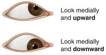
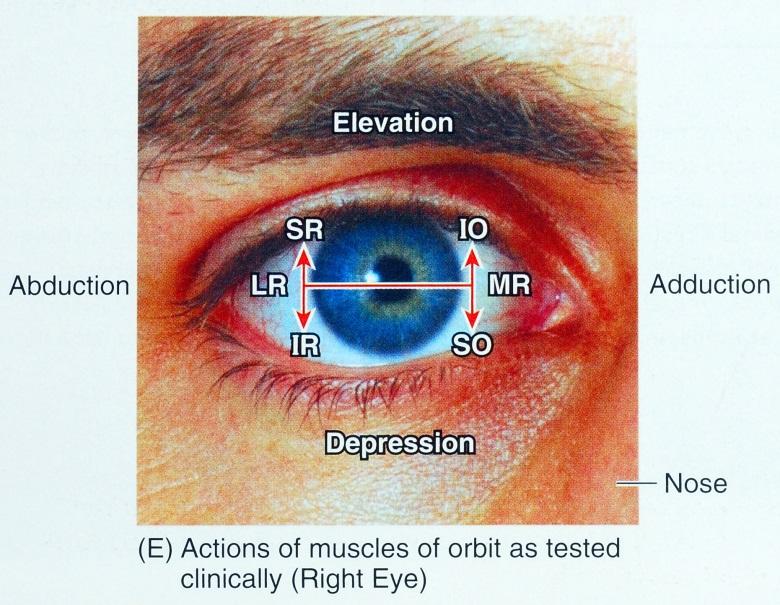
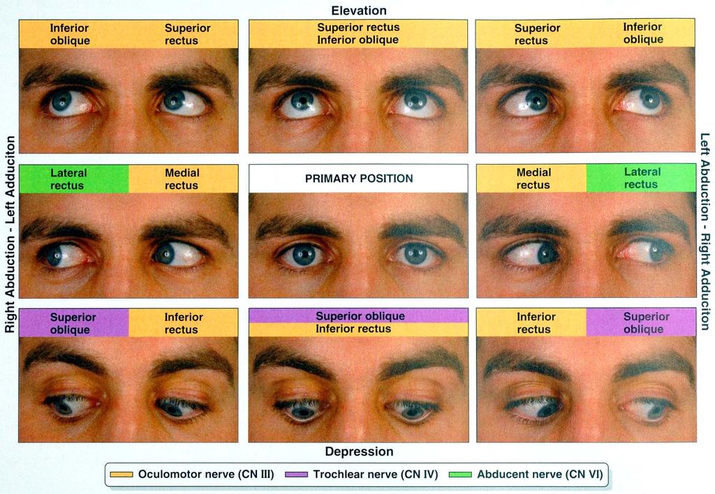
22 Extrinsic muscles Extra-ocular mm. for moving eyeball or raising upper eyelid Elevation moving pupil superiorly Depression moving pupil inferiorly Abduction moving pupil laterally Adduction moving pupil medially Internal rotation rotating upper part of pupil medially External rotation rotating upper part of pupil laterally

23 Levator palpebrae superioris Function: raises upper eyelid; innervation: CN III Superior tarsal muscle (smooth muscle) by sympathetic ff. Origin: inferior surface of levator palpebrae superioris Function: assists to maintain upper eyelid elevation
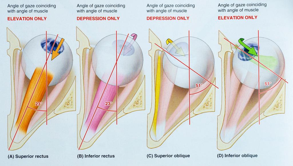
24 Rectus muscles Superior and inferior rectus muscles Function: Sup. rectus elevates, adducts, and internal rotates Inf. rectus depresses, adducts, and external rotates Innervation: Sup. rectus sup. br. of CN III Inf. rectus inf. br. of CN III Medial and lateral rectus muscles Function: Med. rectus adducts Lat. rectus abducts Innervation: Med. rectus inf. br. of CN III Lat. rectus CN VI


25 Oblique muscles Superior oblique Origin: body of sphenoid bone It runs along medial border of roof of orbit, passes through fibrocartilaginous pully (trochlea). The muscle runs deep to superior rectus muscle Inserts to outer posterior quadrant of eyeball Function: turn pupil down and out Innervation: CN IV

26 Oblique muscles Inferior oblique Origin: medial side of floor of orbit, posterior to orbital rim Insertion: outer posterior quadrant of eyeball Function: turn pupil up and out Innervation: inferior br. of CN III

27 SR IO IR SO
28
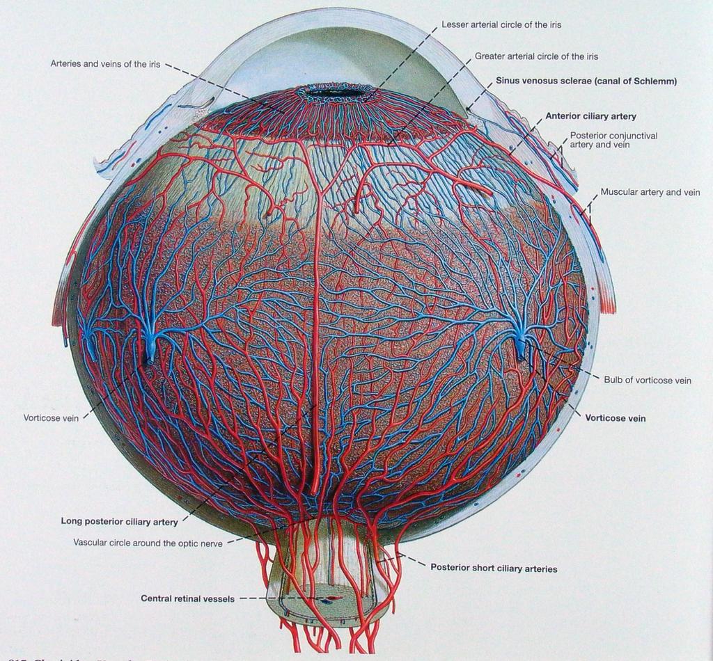
29 Internal carotid a. gives off ophthalmic a. Arteries through optic canal into orbit gives off: Lacrimal a. lateral side of obit, to supply lacrimal gland, anterior ciliary br. to eyeball, lateral eyelid Central retinal a. enters the center of optic n. to retina; its branches can be seen with a ophthalmoscope; occlusion leads to blindness Long and short posterior ciliary aa. pierce sclera to supply inside of eyeball Muscular aa. supply extrinsic mm. Supra-orbital a. passes through supra-orbital f., to supply forehead and scalp to vertex
30 Posterior ethmoidal a. through posterior ethmoidal f. to supply ethmoidal air cells and nasal cavity Anterior ethmoidal a. through anterior ethmoidal f.; gives off anterior meningeal br.; enters nasal cavity to supply septum and lateral wall; ends as dorsal nasal a. (ext. nasal a.) Medial palpebral aa. supply medial area of upper and lower eyelids Dorsal nasal a. supply upper surface of nose Supratrochlear a. supplies forehead

31 Veins 1. Supra-orbital v. and angular v. > Superior ophthalmic v. > through superior orbital fissure > Cavernous sinus 2. Inferior ophthalmic v. > joining with superior ophthalmic v. through superior orbital fissure > Cavernous sinus (infection can be spread from outside to cranial cavity) through inferior orbital fissure > Pterygoid plexus of veins 1
32 1. Optic nerve [II] 2. Oculomotor nerve [III] 3. Trochlear nerve [IV] 4. Abducent nerve [VI] 5. Postganglionic sympathetic fibers 6. Ophthalmic nerve [V1] 6-1. Lacrimal nerve 6-2. Frontal nerve 6-3. Nasociliary nerve 7. Ciliary ganglion 7-1. Parasympathetic root 7-2. Sensory root 7-3. Sympathetic root Innervation
33 Optic nerve [II] Extension of brain; surrounded by meninges and subarachnoid space High intracranial pressure increases the pressure in subarachnoid space, and causes edema of optic disc (papilledema). Through optic canal with ophthalmic a.
34 Oculomotor nerve [III] Anterior surface of brainstem, between midbrain and pons > in the lateral wall of cavernous sinus > divides into sup. and inf. brr. > enter orbit through superior orbital fissure >
![Oculomotor nerve [III] Sup. br. > 1. superior rectus m. 2. levator palpebrae superioris m. Inf. br. > 1. medial rectus 2.](/docs-images/88/117436014/images/35-0.jpg "inferior rectus 3. inferior oblique 4. br. to ciliary ganglion (with pregang. parasym. ff.)")
35 Oculomotor nerve [III] Sup. br. > 1. superior rectus m. 2. levator palpebrae superioris m. Inf. br. > 1. medial rectus 2. inferior rectus 3. inferior oblique 4. br. to ciliary ganglion (with pregang. parasym. ff.)
![Trochlear nerve [IV] From posterior surface of midbrain > enters the edge of tentorium cerebelli > in the lateral wall of](/docs-images/88/117436014/images/36-0.jpg "cavernous sinus (below CN III) > through sup.")
36 Trochlear nerve [IV] From posterior surface of midbrain > enters the edge of tentorium cerebelli > in the lateral wall of cavernous sinus (below CN III) > through sup. orbital fissure (above common tendinous ring) > upper border of superior oblique
37 Abducent nerve [VI] Anterior surface of brain stem between pons and medulla > through cavernous sinus (lateral to int. carotid a.) > through superior orbital fissure > through common tendinous ring > lateral rectus m.

38 Moore, 7th ed., Fig. I-45, p.60

39 Sympathetic fibers T1 (upper thoracic segments) > Preganglionic ff. > Spinal nn. > White rami > thru T1 sym. gang. > Sym. trunk > Superior cervical ganglion (synapse) >
40 Sympathetic fibers Superior cervical ganglion (synapse) > Postganglionic ff. > along internal carotid a. and brr. > Ophthalmic a. > Orbit > Pass through ciliary ganglion (without synapse) > Short ciliary nn. > Eyeball > Dilator pupillae m. Long ciliary nn. > Eyeball > Dilator pupillae m.
41 Ophthalmic nerve [V1] Pure sensory n. from orbit, face, scalp Trigeminal ganglion > in the lateral wall of cavernous sinus (inferior to CN IV & III) > divides into 3 brr.: 1. Lacrimal n. > above common tendinous ring > through superior orbital fissure 2. Frontal n. > above common tendinous ring > through superior orbital fissure 3. Nasociliary n. > through common tendinous ring > through superior orbital fissure

42 Lacrimal n. Along upper border of lat. rectus > receives a br. from zygomaticotemporal n. (carries postgang. parasym. & sym. ff.) > Lacrimal gland (by CN VII) Conjunctiva Lateral upper eyelid
43 Frontal n. Between levator palpebrae superioris and periorbita > divides into 2 brr.: 1. Supratrochlear n. > above trochlea > Conjunctiva Skin on upper eyelid Lower medial forehead 2. Supra-orbital n. > through supra-orbital notch > Upper eyelid Conjunctiva Forehead to midscalp
44 Nasociliary n. Deep in orbit > between sup. and inf. brr. of CN III > gives off communicating br. with ciliary gang. (sensory root) > medial wall of orbit > gives off brr.: 1. Long ciliary nn. > eyeball (with sensory and sym. ff.) 2. Posterior ethmoidal n. > through posterior ethmoidal foramen > posterior ethmoidal air cells sphenoid sinus 3. Infratrochlear n. > medial part of upper and lower eyelids lacrimal sac skin of the upper nose 4. Anterior ethmoidal n. > through anterior ethmoidal foramen > anterior cranial fossa nasal cavity skin of the lower nose
45 Ciliary ganglion Parasympathetic ganglion of CN III; associated with nasociliary n. of CN V1; located at the lateral side of optic n. Synapse for pre- and postgang. parasym. ff.; traversed by postgang. sym. ff. It receives three roots: 1. Parasympathetic root 2. Sensory root 3. Sympathetic root 2 1 3
46 Parasympathetic root CN III > gives off parasym. root (carries pregang. parasym. ff.) > synapse in ciliary ganglion > gives off postgang. parasym. ff. > short ciliary nn. > eyeball > Sphincter pupillae m. (constrict pupil) Ciliary m. (accommodation of lens) Sensory root Trigeminal gang. > CN V 1 > Nasociliary n. > sensory root pass through ganglion > short ciliary nn. > Sensory innervation of eyeball Sympathetic root Superior cervical ganglion > gives off postgang. sym. ff. > along plexus on int. carotid a. > though common tendinous ring > gives off a br. to join ophthalmic n. > from nasociliary n. gives off a sym. root > traverse ciliary gang. > short ciliary nn. > eyeball > Dilator pupillae m.
47 Eyeball Globe-shaped, bulges anteriorly as transparent cornea (1/6 surface area) Light passes from ant. to post.: 1. Cornea > 2. Anterior chamber > 3. Iris, pupil > 4. Posterior chamber, lens > 5. Vitreous chamber > 6. Retina
48 Anterior and posterior chambers Anterior chamber between cornea and iris Pupil central opening of iris Posterior chamber between iris and lens Aqueous humor fills the posterior chambers, and flows through pupil into anterior chamber, and is absorbed into scleral venous sinus. Glaucoma amount of intraocular fluid increases, and consequently the intra-ocular pressure.
49 Lens and vitreous humor Biconvex transparent disc cataract is caused by the opacity of lens Vitreous body (vitreous humor) gelatinous substance in posterior segment (postrenal chamber)

50 Walls of the eyeball 1. Outer fibrous layer: cornea and sclera 2. Middle vascular layer: choroid, ciliary body and iris 3. Inner layer: retina, and internal covering of iris and ciliary body

51 Arterial supply 1. Short posterior ciliary aa. from ophthalmic a to pierce sclera and enter choroid layer 2. Long posterior ciliary aa. lateral and medial to optic n., run in choroids and anastomose with anterior ciliary aa. 3. Anterior ciliary aa. from muscular brr.; anastomose with long posterior ciliary aa. in choroid layer 4. Central retinal a. traverse the optic n. and enters eyeball at optic disc Venous drainage Four large veins (vorticose veins) enter superior and inferior ophthalmic vv. Central retinal v. runs with a.
52
53 1-1. Sclera 1. Fibrous layer of the eyeball Dense connective tissue; pierced by optic n. and attached by mm.; covering lateral and posterior of 5/6 eyeball surface Fascial sheath covering outer surface of sclera from entrance of optic n. to corneoscleral junction Inner surface of sclera loosely attaches to choroid vascular layer Cornea Transparent; anterior 1/6 of eyeball surface
54 2. Vascular layer of the eyeball 2-1. Choroid Thin, highly vascular pigmented layer firmly attaches to retina loosely attaches to sclera 2-2. Ciliary body between choroid and iris Ciliary m. smooth muscle ff., arranged longitudinally, circularly, and radially; innervated by CN III Contraction of the ciliary m. shrinks the ciliary body ring. Ciliary processes projections toward lens Zonular fibers extending from ciliary processes, attach to lens. Collectively form suspensory ligament of lens 2-3. Iris Circular structure with central opening, pupil; smooth mm. including: Sphincter pupillae m. innervated by parasym. ff., constricts the pupil Dilator pupillae m. innervated by sym. ff., dilates the pupil
55 Retina 3. Inner layer of the eyeball 3-1. Nonvisual part covers internal surface of ciliary body and iris Junction irregular ora serrata 3-2. Optic part sensitive to light Pigmented layer outer layer, attaches to choroids Neural layer attaches to pigmented layer around optic n. and at ora serrata
 Macula lutea with central depression, fovea centralis, thinner retina, and higher")
56 Optic disc optic n. leaves the eyeball there; the central a. enters eyeball there; without light receptor there (blind spot) Macula lutea with central depression, fovea centralis, thinner retina, and higher sensitivity with more cones (sensitive to color) and fewer rods (functioning in dim light)
Bony orbit Roof The orbital plate of the frontal bone Lateral wall: the zygomatic bone and the greater wing of the sphenoid
 Bony orbit Roof: Formed by: The orbital plate of the frontal bone, which separates the orbital cavity from the anterior cranial fossa and the frontal lobe of the cerebral hemisphere Lateral wall: Formed
The orbit-2. Dr. Heba Kalbouneh Assistant Professor of Anatomy and Histology
 The orbit-2 Dr. Heba Kalbouneh Assistant Professor of Anatomy and Histology Eyelids The eyelids (act like the curtains) protect the eye from injury and excessive light by their closure The upper eyelid
1 Eyelids. Lacrimal Apparatus. Orbital Region. 3 The Orbit. The Eye
 1 1 Eyelids Orbital Region 2 Lacrimal Apparatus 3 The Orbit 4 The Eye 2 Eyelids The eyelids protect the eye from injury and excessive light by their closure. The upper eyelid is larger and more mobile
The sebaceous glands (glands of Zeis) open directly into the eyelash follicles, ciliary glands (glands of Moll) are modified sweat glands that open
 open directly into the eyelash follicles, ciliary glands (glands of Moll) are modified sweat glands that open") The Orbital Region The orbits are a pair of bony cavities that contain the eyeballs; their associated muscles, nerves, vessels, and fat; and most of the lacrimal apparatus upper eyelid is larger and more
The Orbital Region The orbits are a pair of bony cavities that contain the eyeballs; their associated muscles, nerves, vessels, and fat; and most of the lacrimal apparatus upper eyelid is larger and more
The orbit-1. Dr. Heba Kalbouneh Assistant Professor of Anatomy and Histology
 The orbit-1 Dr. Heba Kalbouneh Assistant Professor of Anatomy and Histology Orbital plate of frontal bone Orbital plate of ethmoid bone Lesser wing of sphenoid Greater wing of sphenoid Lacrimal bone Orbital
Unit VIII Problem 8 Anatomy: Orbit and Eyeball
 Unit VIII Problem 8 Anatomy: Orbit and Eyeball - The bony orbit: it is protecting our eyeball and resembling a pyramid: With a base directed: anterolaterally. And an apex directed: posteromedially. Notes:
Bony orbit. Lateral wall: Formed by : the zygomatic bone and the greater wing of the sphenoid
 Bony orbit Roof: Formed by: The orbital plate of the frontal bone, which separates the orbital cavity from the anterior cranial fossa and the frontal lobe of the cerebral hemisphere Lateral wall: Formed
GNK485 The eye and related structures. Prof MC Bosman 2012
 GNK485 The eye and related structures Prof MC Bosman 2012 Surface anatomy Bony orbit Eyeball and Lacrimal apparatus Extra-ocular muscles Movements of the eye Innervation Arterial supply and venous drainage
Maxilla, ORBIT and infratemporal fossa. Neophytos C Demetriades MD, DDS, MSc Associate professor European University of Cyprus School of Medicine
 Maxilla, ORBIT and infratemporal fossa Neophytos C Demetriades MD, DDS, MSc Associate professor European University of Cyprus School of Medicine MAXILLA Superior, middle, and inferior meatus Frontal sinus
213: HUMAN FUNCTIONAL ANATOMY: PRACTICAL CLASS 12 Cranial cavity, eye and orbit
 213: HUMAN FUNCTIONAL ANATOMY: PRACTICAL CLASS 12 Cranial cavity, eye and orbit OSTEOLOGY Identify the bones which comprise the walls of the orbit: maxilla, zygomatic, ethmoid, lachrymal, frontal, and
MAXILLA, ORBIT & PTERYGOPALATINE FOSSA. Neophytos C Demetriades MD, DDS, MSc Associate professor European University of Cyprus School of Medicine
 MAXILLA, ORBIT & PTERYGOPALATINE FOSSA Neophytos C Demetriades MD, DDS, MSc Associate professor European University of Cyprus School of Medicine Maxilla MAXILLA Superior, middle, and inferior meatus Frontal
The Special Senses: Part A
 PowerPoint Lecture Slides prepared by Janice Meeking, Mount Royal College CHAPTER 15 The Special Senses: Part A Warm Up What is the function of the eyeball? List any structures of the eyeball that you
Ocular Anatomy for the Paraoptometric
 Ocular Anatomy for the Paraoptometric Minnesota Optometric Association Paraoptometric CE Friday September 30, 2016 Lindsay A. Sicks, OD, FAAO Assistant Professor, Illinois College of Optometry lsicks@ico.edu
4/22/16. Eye. External Anatomy of Eye. Accessory Structures. Bio 40B Dr. Kandula
 Eye Bio 40B Dr. Kandula External Anatomy of Eye Accessory Structures l Eyebrows l Levator Palpebrae Superioris - opens eye l Eyelashes l Ciliary glands modified sweat glands l Small sebaceous glands l
REVIEW OF HEAD AND NECK CRANIAL NERVES AND EVERYTHING ELSE
 REVIEW OF HEAD AND NECK CRANIAL NERVES AND EVERYTHING ELSE OLFACTORY NERVE CN I ANTERIOR CRANIAL FOSSA CRISTA GALLI OF ETHMOID OLFACTORY FORAMINA IN CRIBIFORM PLATE OF ETHMOID BONE CN I OLFACTORY NERVE
Face and Scalp 解剖學科鄭授德
 Face and Scalp 解剖學科鄭授德 本教材之圖片取自於 1 Gray s Anatomy for Students, 3rd ed, 2015, by Drake, Vogl, and Mitchell 2 Clinically Oriented Anatomy, 7th ed, 2014, by Moore, Dalley, and Agur 3 Clinically Oriented
mistake ;slides in bold but you still have to go back to our slides to see the figure, tables and some scheme
 Khozama jehad : I am doing my best and I am sorry for any unintended mistake ;slides in bold but you still have to go back to our slides to see the figure, tables and some scheme The Orbit, Orbital Contents
Sense of Vision. Chapter 8. The Eye and Vision. The Eye Orbit. Eyebrows, Eyelids, Eyelashes. Accessory Organs 5/3/2016.
 Sense of Vision Chapter 8 Special Senses The Eye and Vision 70 percent of all sensory receptors are in the eyes Each eye has over 1 million nerve fibers Protection for the eye Most of the eye is enclosed
THE SPECIAL SENSES. Introduction Vision
 THE SPECIAL SENSES Introduction Vision RECEPTORS Structures designed to respond to stimuli Variable complexity RECEPTORS: GENERAL PROPERTIES Transducers Receptor Potential Generator Potential RECEPTORS
The Orbit. The Orbit OCULAR ANATOMY AND DISSECTION 9/25/2014. The eye is a 23 mm organ...how difficult can this be? Openings in the orbit
 The eye is a 23 mm organ...how difficult can this be? OCULAR ANATOMY AND DISSECTION JEFFREY M. GAMBLE, OD COLUMBIA EYE CONSULTANTS OPTOMETRY & UNIVERSITY OF MISSOURI DEPARTMENT OF OPHTHALMOLOGY CLINICAL
PTERYGOPALATINE FOSSA
 PTERYGOPALATINE FOSSA Outline Anatomical Structure and Boundaries Foramina and Communications with other spaces and cavities Contents Pterygopalatine Ganglion Especial emphasis on certain arteries and
Lecture 10 Orbit and control of eye movements
 Lecture 10 Orbit and control of eye movements Overview of structures in the orbit (Moore pp 899, Netter Plate 1) The orbit contains the eye, from which the optic nerve exits into the cranial cavity optic
Face. Definition: The area between the two ears and from the chin to the eye brows. The muscles of the face
 Face Definition: The area between the two ears and from the chin to the eye brows. The muscles of the face The muscle of facial expression (include the muscle of the face and the scalp). All are derived
Omran Saeed. Luma Taweel. Mohammad Almohtaseb. 1 P a g e
 2 Omran Saeed Luma Taweel Mohammad Almohtaseb 1 P a g e I didn t include all the photos in this sheet in order to keep it as small as possible so if you need more clarification please refer to slides In
Chapter(2):the lid page (1) THE LID
:the lid page (1) THE LID") Chapter(2):the lid page (1) THE LID Anatomy of the lid: * Check movie anatomy of the lid model The eyelids are two movable muco-cutaneous folds which protect the eye on closure. The are joined temporary
Chapter(2):the lid page (1) THE LID Anatomy of the lid: * Check movie anatomy of the lid model The eyelids are two movable muco-cutaneous folds which protect the eye on closure. The are joined temporary
Mohammad Hisham Al-Mohtaseb. Lina Mansour. Reyad Jabiri. 0 P a g e
 2 Mohammad Hisham Al-Mohtaseb Lina Mansour Reyad Jabiri 0 P a g e This is only correction for the last year sheet according to our record. If you already studied this sheet just read the yellow notes which
Eye Movements. Geometry of the Orbit. Extraocular Muscles
 Eye Movements Geometry of the Orbit The eye (oculus) is located in the anterior aspect of the orbit: the equator of the eye (defined by a coronal plane passing through its middle) lies at the margin of
Special Senses: The Eye
 Unit 4 Special Senses: The Eye ESSENTIALS OF HUMAN ANATOMY & PHYSIOLOGY The Senses General senses of touch Temperature Pressure Pain Special senses Smell Taste Sight Hearing Equilibrium The Eye and Vision
3-Deep fascia: is absent (except over the parotid gland & buccopharngeal fascia covering the buccinator muscle)
") The Face 1-Skin of the Face The skin of the face is: Elastic Vascular (bleed profusely however heal rapidly) Rich in sweat and sebaceous glands (can cause acne in adults) It is connected to the underlying
The Face 1-Skin of the Face The skin of the face is: Elastic Vascular (bleed profusely however heal rapidly) Rich in sweat and sebaceous glands (can cause acne in adults) It is connected to the underlying
Trigeminal Nerve Worksheets, Distributions Page 1
 Trigeminal Nerve Worksheet #1 Distribution by Nerve Dr. Darren Hoffmann Dental Gross Anatomy, Spring 2013 We have drawn out each of the branches of CN V in lecture and you have an idea now for their basic
Temporal fossa Infratemporal fossa Pterygopalatine fossa Terminal branches of external carotid artery Pterygoid venous plexus
 Outline of content Temporal fossa Infratemporal fossa Pterygopalatine fossa Terminal branches of external carotid artery Pterygoid venous plexus Boundary Content Communication Mandibular division of trigeminal
Parotid Gland, Temporomandibular Joint and Infratemporal Fossa
 M1 - Anatomy Parotid Gland, Temporomandibular Joint and Infratemporal Fossa Jeff Dupree Sanger 9-057 jldupree@vcu.edu Parotid gland: wraps around the mandible positioned between the mandible and the sphenoid
The Eye. The Orbit. The EYE What a Trip!!! - The Anterior Segment 5/12/2015. Jill J Luebbert, CPOT, ABOC
 The EYE What a Trip!!! - The Anterior Segment Jill J Luebbert, CPOT, ABOC The Eye The Orbit Bony socket containing the eye and most of its accessory organs consisting of 7 bones 1 The Seven Bones of the
Temporal region. temporal & infratemporal fossae. Zhou Hong Ying Dept. of Anatomy
 Temporal region temporal & infratemporal fossae Zhou Hong Ying Dept. of Anatomy Temporal region is divided by zygomatic arch into temporal & infratemporal fossae. Temporal Fossa Infratemporal fossa Temporal
Special Senses PART A
 8 Special Senses PART A PowerPoint Lecture Slide Presentation by Jerry L. Cook, Sam Houston University ESSENTIALS OF HUMAN ANATOMY & PHYSIOLOGY EIGHTH EDITION ELAINE N. MARIEB The Senses General senses
Dr.Ban I.S. head & neck anatomy 2 nd y جامعة تكريت كلية طب االسنان مادة التشريح املرحلة الثانية أ.م.د. بان امساعيل صديق 6102/6102
 جامعة تكريت كلية طب االسنان مادة التشريح املرحلة الثانية أ.م.د. بان امساعيل صديق 6102/6102 Pterygopalatine fossa: The pterygopalatine fossa is a cone-shaped depression, It is located between the maxilla,
Bisection of Head & Nasal Cavity 頭部對切以及鼻腔. 解剖學科馮琮涵副教授 分機
 Bisection of Head & Nasal Cavity 頭部對切以及鼻腔 解剖學科馮琮涵副教授 分機 3250 E-mail: thfong@tmu.edu.tw Outline: The structure of nose The concha and meatus in nasal cavity The openings of paranasal sinuses Canals, foramens
Tikrit University collage of dentistry Dr.Ban I.S. head & neck anatomy 2 nd y. Lec [5] / Temporal fossa :
![Tikrit University collage of dentistry Dr.Ban I.S. head & neck anatomy 2 nd y. Lec [5] / Temporal fossa :](/thumbs/88/115294566.jpg "Tikrit University collage of dentistry Dr.Ban I.S. head & neck anatomy 2 nd y. Lec [5] / Temporal fossa :") Lec [5] / Temporal fossa : Borders of the Temporal Fossa: Superior: Superior temporal line. Inferior: gap between zygomatic arch and infratemporal crest of sphenoid bone. Anterior: Frontal process of the
Lec [5] / Temporal fossa : Borders of the Temporal Fossa: Superior: Superior temporal line. Inferior: gap between zygomatic arch and infratemporal crest of sphenoid bone. Anterior: Frontal process of the
Anatomic Relations Summary. Done by: Sohayyla Yasin Dababseh
 Anatomic Relations Summary Done by: Sohayyla Yasin Dababseh Anatomic Relations Lecture 1 Part-1 - The medial wall of the nose is the septum. - The vestibule lies directly inside the nostrils (Nares). -
Cranial nerves.
 Cranial nerves eaglezhyxzy@163.com Key Points of Learning Name Components Passing through Peripheral distribution Central connection Function Cranial nerves Ⅰ olfactory Ⅱ optic Ⅲ occulomotor Ⅳ trochlear
Vision I. Steven McLoon Department of Neuroscience University of Minnesota
 Vision I Steven McLoon Department of Neuroscience University of Minnesota 1 Eye Cornea Sclera Conjunctiva 2 Eye The conjunctiva lines the inner surface of the eyelids and outer surface of the sclera. 3
Infratemporal fossa: Tikrit University college of Dentistry Dr.Ban I.S. head & neck Anatomy 2 nd y.
 Infratemporal fossa: This is a space lying beneath the base of the skull between the lateral wall of the pharynx and the ramus of the mandible. It is also referred to as the parapharyngeal or lateral pharyngeal
Anatomy images for MSS practical exam- 2019
 Anatomy images for MSS practical exam- 2019 Ilium Ischium Pubis Acetabulaum Iliac crest Iliac tubercle ASIS (muscle and ligament attached) AIIS (muscle attached) PSIS PIIS Ischial spine Ischial tuberosity
INTRODUCTION: ****************************************************************************************************
 BIOLOGY 211: HUMAN ANATOMY & PHYSIOLOGY **************************************************************************************************** EYES AND VISION ****************************************************************************************************
Introduction to Head and Neck Anatomy
 Introduction to Head and Neck Anatomy Nervous Tissue Controls and integrates all body activities within limits that maintain life Three basic functions 1. sensing changes with sensory receptors 2. interpreting
Major Anatomic Components of the Orbit
 Major Anatomic Components of the Orbit 1. Osseous Framework 2. Globe 3. Optic nerve and sheath 4. Extraocular muscles Bony Orbit Seven Bones Frontal bone Zygomatic bone Maxillary bone Ethmoid bone Sphenoid
Cranial Cavity REFERENCES: OBJECTIVES OSTEOLOGY. Stephen A. Gudas, PT, PhD
 Stephen A. Gudas, PT, PhD Cranial Cavity REFERENCES: Moore and Agur, Essential Clinical Anatomy (ECA), 3rd ed., pp. 496 498; 500 507; 512 514 Grant s Atlas 12 th ed., Figs 7.6; 7.19 7.30. Grant s Dissector
HEAD AND NECK ANATOMY PRACTICE QUESTIONS
 HEAD AND NECK ANATOMY PRACTICE QUESTIONS 1. A patient complains that he has lost sensation on his face and that the skin of his face feels numb. The physician tests tactile acuity by touching the forehead
Muscles of the Eyeball (Extra Ocular Muscles) Prof. Dr. Imran Qureshi
 Prof. Dr. Imran Qureshi") Muscles of the Eyeball (Extra Ocular Muscles) Prof. Dr. Imran Qureshi There are six extrinsic muscles of the eyeball, namely the (S), Medial (M), (I), & Lateral (L) recti, and (SO) and (IO) Obliques. In
Muscles of the Eyeball (Extra Ocular Muscles) Prof. Dr. Imran Qureshi There are six extrinsic muscles of the eyeball, namely the (S), Medial (M), (I), & Lateral (L) recti, and (SO) and (IO) Obliques. In
Tikrit University College of Dentistry Dr.Ban I.S. head & neck anatomy 2 nd y.
 Lec [3]/The scalp The scalp extends from the supraorbital margins anteriorly to the nuchal lines at the back of the skull and down to the temporal lines at the sides. The forehead, from eyebrows to hairline,
Introduction to Local Anesthesia and Review of Anatomy
 5-Sep Introduction and Anatomy Review 12-Sep Neurophysiology and Pain 19-Sep Physiology and Pharmacology part 1 26-Sep Physiology and Pharmacology part 2 Introduction to Local Anesthesia and Review of
Taste buds Gustatory cells extend taste hairs through a narrow taste pore
 The Special Senses Objectives Describe the sensory organs of smell, and olfaction. Identify the accessory and internal structures of the eye, and explain their function. Explain how light stimulates the
Head and Face Anatomy
 Head and Face Anatomy Epicranial region The Scalp The soft tissue that covers the vault of skull. Extends from supraorbital margin to superior nuchal line. Layers of the scalp S C A L P = skin = connective
THE EYE: RETINA AND GLOBE
 Neuroanatomy Suzanne Stensaas February 24, 2011, 10:00-12:00 p.m. Reading: Waxman Ch. 15. Your histology and gross anatomy books should be useful. Reading: Histology of the Eye from any histology book
Tracing the Cranial Nerves Osteologically
 CN I II III IV V 1 Supra-orbital ethmoidal nn. Ext. nasal V 2 Tracing the Cranial Nerves Osteologically Nucleus of Origin Olfactory tracts of frontal lobe of cerebrum Optic tracts from optic chiasma and
ACTIVITIES. Complete Diagrams PNS 18 and 19 Complete PNS 23 Worksheet 3 #1 only Complete PNS 24 Practice Quiz
 ACTIVITIES Complete Diagrams PNS 18 and 19 Complete PNS 23 Worksheet 3 #1 only Complete PNS 24 Practice Quiz THE SPECIAL SENSES Introduction Vision RECEPTORS Structures designed to respond to stimuli Variable
Dr.Ban I.S. head & neck anatomy 2 nd y. جامعة تكريت كلية طب االسنان املرحلة الثانية أ.م.د. بان امساعيل صديق 6102/6102
 جامعة تكريت كلية طب االسنان التشريح مادة املرحلة الثانية أ.م.د. بان امساعيل صديق 6102/6102 Parotid region The part of the face in front of the ear and below the zygomatic arch is the parotid region. The
SPECIAL SENSES PART I: OLFACTION & GUSTATION
 SPECIAL SENSES PART I: OLFACTION & GUSTATION 5 Special Senses Olfaction Gustation Vision Equilibrium Hearing Olfactory Nerves Extend through cribriform plate into nasal cavity on both sides of nasal septum
The Nervous System: General and Special Senses Pearson Education, Inc.
 18 The Nervous System: General and Special Senses Introduction Sensory information arrives at the CNS Information is picked up by sensory receptors Sensory receptors are the interface between the nervous
02/03/2014. Average Length: 23mm (Infant ~16mm) Approximately the size of a quarter Volume: ~5mL
 Approximately the size of a quarter Volume: ~5mL") Identify the anatomy of the eye. Explain the basic physiology of the parts of the eye. Briefly discuss various surgeries related to different parts of the anatomy. Average Length: 23mm (Infant ~16mm) Approximately
Identify the anatomy of the eye. Explain the basic physiology of the parts of the eye. Briefly discuss various surgeries related to different parts of the anatomy. Average Length: 23mm (Infant ~16mm) Approximately
Cranial Nerve VII - Facial Nerve. The facial nerve has 3 main components with distinct functions
 Cranial Nerve VII - Facial Nerve The facial nerve has 3 main components with distinct functions Somatic motor efferent Supplies the muscles of facial expression; posterior belly of digastric muscle; stylohyoid,
Trigeminal Nerve (V)
") Trigeminal Nerve (V) Lecture Objectives Discuss briefly how the face is developed. Follow up the course of trigeminal nerve from its point of central connections, exit and down to its target areas. Describe
Trigeminal Nerve (V) Lecture Objectives Discuss briefly how the face is developed. Follow up the course of trigeminal nerve from its point of central connections, exit and down to its target areas. Describe
External Occipital Protuberance
 Osteology Exterior Skull Frontal Bone Glabella Superciliary Arch Supraorbital Notch/Foramen Nasion (junction w/ Nasal bone) Frontal/Metopic Suture (usually absent in adult, b/w ossification centers of
Ocular Anatomy & Physiology. Learning Objectives: Let s get oriented first. 3 Major Layers (Tunics) of EYE. Topics to be covered: FIBROUS TUNIC
 of EYE. Topics to be covered: FIBROUS TUNIC") Lecturer: Ocular Anatomy & Physiology M. Patrick COLEMAN, ABOC, COT Kerrville, TX Learning Objectives: 1. Correctly identify ocular structures around or within the eye 2. List the key functions of various
Lecturer: Ocular Anatomy & Physiology M. Patrick COLEMAN, ABOC, COT Kerrville, TX Learning Objectives: 1. Correctly identify ocular structures around or within the eye 2. List the key functions of various
Anatomy and Physiology. Bones, Sutures, Teeth, Processes and Foramina of the Human Skull
 Anatomy and Physiology Chapter 6 DRO Bones, Sutures, Teeth, Processes and Foramina of the Human Skull Name: Period: Bones of the Human Skull Bones of the Cranium: Frontal bone: forms the forehead and the
*in general the blood supply of the nose comes from branches of the internal and external carotid arteries.
 In the previous lecture we talked about the anatomy of the nasal cavity, today we will talk about its blood supply, venous drainage, innervations, and finally about the paranasal sinuses. When we describe
Anatomy of the orbit. Lay-out. Imaging technique. 3 x 3. brief overview of the basic anatomy of the orbit and its structures
 Anatomy of the orbit Prof. Pia C Sundgren MD, PhD Department of Diagnostic Radiology, Clinical Sciences, Lund University, Sweden Lay-out brief overview of the basic anatomy of the orbit and its structures
Histology of the Eye
 Histology of the Eye Objectives By the end of this lecture, the student should be able to describe: The general structure of the eye. The microscopic structure of:»cornea.»retina. EYE BULB Three coats
SCHOOL OF ANATOMICAL SCIENCES Mock Run Questions. 4 May 2012
 SCHOOL OF ANATOMICAL SCIENCES Mock Run Questions 4 May 2012 1. With regard to the muscles of the neck: a. the platysma muscle is supplied by the accessory nerve. b. the stylohyoid muscle is supplied by
Skull-2. Norma Basalis Interna Norma Basalis Externa. Dr. Heba Kalbouneh Associate Professor of Anatomy and Histology
 Skull-2 Norma Basalis Interna Norma Basalis Externa Dr. Heba Kalbouneh Associate Professor of Anatomy and Histology Norma basalis interna Base of the skull- superior view The interior of the base of the
o A cushion of fat surrounds most of the eye
 Name Period SPECIAL SENSES The Senses General senses of touch o Temperature o Pressure o Pain Special senses o Smell o Taste o Sight o Hearing o Equilibrium The Eye and Vision 70 percent of all sensory
Lec [8]: Mandibular nerve:
![Lec [8]: Mandibular nerve:](/thumbs/94/121295776.jpg "Lec [8]: Mandibular nerve:") Lec [8]: Mandibular nerve: The mandibular branch from the trigeminal ganglion lies in the middle cranial fossa lateral to the cavernous sinus. With the motor root of the trigeminal nerve [motor roots lies
Lec [8]: Mandibular nerve: The mandibular branch from the trigeminal ganglion lies in the middle cranial fossa lateral to the cavernous sinus. With the motor root of the trigeminal nerve [motor roots lies
Bones Ethmoid bone Inferior nasal concha Lacrimal bone Maxilla Nasal bone Palatine bone Vomer Zygomatic bone Mandible
 splanchnocranium - Consists of part of skull that is derived from branchial arches - The facial bones are the bones of the anterior and lower human skull Bones Ethmoid bone Inferior nasal concha Lacrimal
 This article was originally published in the Encyclopedia of the Eye, published by Elsevier, and the attached copy is provided by Elsevier for the author's benefit and for the benefit of the author's institution,
Special Senses: Vision
 ighapmlre24pg223_230 5/12/04 2:27 PM Page 223 impos03 302:bjighapmL:ighapmLrevshts:layouts: NAME LAB TIME/DATE Special Senses: Vision REVIEW SHEET exercise 24 Anatomy of the Eye 1. Name five accessory
CN I Olfactory. CN II Optic. CN III Oculomotor. Special Sensory Efferent fibers to Olfactory Bulb. Cribiform Plate of Ethmoid
 CN I Olfactory Efferent fibers to Olfactory Bulb Olfactory Tract Olfactory Bulb Cribiform Plate of Ethmoid Anosmia Loss of sense of smell Uncinate Fits olfactory hallucinations To Olfactory Epithelium
Trigeminal nerve. Slide in bold and please go back to see the pictures, if I skipped any part of record that because it wasn t clear to me
 Trigeminal nerve Slide in bold and please go back to see the pictures, if I skipped any part of record that because it wasn t clear to me Hala nsour 2/26/2018 P a g e 1 this lecture contain two topics
THIEME. Scalp and Superficial Temporal Region
 CHAPTER 2 Scalp and Superficial Temporal Region Scalp Learning Objectives At the end of the dissection of the scalp, you should be able to identify, understand and correlate the clinical aspects: Layers
Biology 323 Human Anatomy for Biology Majors Week 10; Lecture 1; Tuesday Dr. Stuart S. Sumida. Cranial Nerves and Soft Tissues of the Skull
 Biology 323 Human Anatomy for Biology Majors Week 10; Lecture 1; Tuesday Dr. Stuart S. Sumida Cranial Nerves and Soft Tissues of the Skull FOREBRAIN MIDBRAIN HINDBRAIN Forebrain: Cerebrum Perception,
Anatomy for ophthalmic anaesthesia
 British Journal of Anaesthesia 1995; 75: 80 87 REVIEW ARTICLES Anatomy for ophthalmic anaesthesia R. W. JOHNSON Study of the anatomy of the orbit, its contents and surrounding structures allows the anaesthetist
Dr. Sami Zaqout, IUG Medical School
 The skull The skull is composed of several separate bones united at immobile joints called sutures. Exceptions? Frontal bone Occipital bone Vault Cranium Sphenoid bone Zygomatic bones Base Ethmoid bone
Dr.Ban I.S. head & neck anatomy 2 nd y. جامعة تكريت كلية طب االسنان مادة التشريح املرحلة الثانية أ.م.د. بان امساعيل صديق 6102/6102
 جامعة تكريت كلية طب االسنان مادة التشريح املرحلة الثانية أ.م.د. بان امساعيل صديق 6102/6102 The scalp The scalp extends from the supraorbital margins anteriorly to the nuchal lines at the back of the skull
Nose & Mouth OUTLINE. Nose. - Nasal Cavity & Its Walls. - Paranasal Sinuses. - Neurovascular Structures. Mouth. - Oral Cavity & Its Contents
 Dept. of Human Anatomy, Si Chuan University Zhou hongying eaglezhyxzy@163.com Nose & Mouth OUTLINE Nose - Nasal Cavity & Its Walls - Paranasal Sinuses - Neurovascular Structures Mouth - Oral Cavity & Its
Superior View of the Skull (Norma Verticalis) Anteriorly the frontal bone articulates with the two parietal bones AT THE CORONAL SUTURE
 Anteriorly the frontal bone articulates with the two parietal bones AT THE CORONAL SUTURE") Superior View of the Skull (Norma Verticalis) Anteriorly the frontal bone articulates with the two parietal bones AT THE CORONAL SUTURE 1 The two parietal bones articulate in the midline AT THE SAGITTAL
Superior View of the Skull (Norma Verticalis) Anteriorly the frontal bone articulates with the two parietal bones AT THE CORONAL SUTURE 1 The two parietal bones articulate in the midline AT THE SAGITTAL
Rashed Al-Jomard. Alanood Bostanji
 Anatomy #2 The Orbit & Cranial Nerve III, IV, VI Rashed AlJomard Alanood Bostanji 1 P a g e The Orbit & Cranial nerves III,IV&VI ** Some notes about the last lec & first MM : Lens :: "just clarify for
The Senses. Chapter 10 7/8/11. Introduction
 Chapter 10 The Senses Introduction A. Sensory receptors detect changes in the environment and stimulate neurons to send nerve impulses to the brain. B. A sensation is formed based on the sensory input.
human anatomy 2016 lecture fifteen Dr meethak ali ahmed neurosurgeon
 Cranial Nerves Organization of the Cranial Nerves The cranial nerves are named as follows: I. Olfactory II. Optic III. Oculomotor IV. Trochlear V. Trigeminal VI. Abducent VII. Facial VIII. Vestibulocochlear
The Pharynx. Dr. Nabil Khouri MD. MSc, Ph.D
 The Pharynx Dr. Nabil Khouri MD. MSc, Ph.D Introduction The pharynx is the Musculo-fascial halfcylinder that links the oral and nasal cavities in the head to the larynx and esophagus in the neck Common
Veins of the Face and the Neck
 Veins of the Face and the Neck Facial Vein The facial vein is formed at the medial angle of the eye by the union of the supraorbital and supratrochlear veins. connected through the ophthalmic veins with
Brain ميهاربا لض اف دمح ا د The Meninges 1- Dura Mater of the Brain endosteal layer does not extend meningeal layer falx cerebri tentorium cerebelli
 .احمد د فاضل ابراهيم Lecture 15 Brain The Meninges Three protective membranes or meninges surround the brain in the skull: the dura mater, the arachnoid mater, and the pia mater 1- Dura Mater of the Brain
Essential questions. What are the structures of the sensory system? 3.03 Remember the structures of the sensory system 2
 Essential questions What are the structures of the sensory system? 3.03 Remember the structures of the sensory system 2 The Senses Eyes Sight Ears Hearing Nose Smell Tongue Taste Skin Touch 3.03 Remember
Parotid Gland. Parotid Gland. Largest of 3 paired salivary glands (submandibular; sublingual) Ramus of Mandible. Medial pterygoid.
 Ramus of Mandible. Medial pterygoid.") Parotid region Parotid Gland Largest of 3 paired salivary glands (submandibular; sublingual) Ramus of Mandible Medial pterygoid Cross section of mandible Masseter D S SCM Parotid Gland Mastoid Process
Parotid region Parotid Gland Largest of 3 paired salivary glands (submandibular; sublingual) Ramus of Mandible Medial pterygoid Cross section of mandible Masseter D S SCM Parotid Gland Mastoid Process
This lab activity is aligned with Visible Body s Human Anatomy Atlas app.
 1 This lab activity is aligned with Visible Body s Human Anatomy Atlas app. Learn more at visiblebody.com/professors We've split our Cranial Nerves lab activity into two parts. Part 1 is pre-lab exercises
Nasal region. cartilages: septal cartilage (l); lateral nasal cartilage (2); greater alar cartilages (2); lesser alar cartilages (?
; lateral nasal cartilage (2); greater alar cartilages (2); lesser alar cartilages (?") Nasal region skull bones: nasal and frontal processes of maxilla cartilages: septal cartilage (l); lateral nasal cartilage (2); greater alar cartilages (2); lesser alar cartilages (?) 1 Nasal cavity Roof
Nasal region skull bones: nasal and frontal processes of maxilla cartilages: septal cartilage (l); lateral nasal cartilage (2); greater alar cartilages (2); lesser alar cartilages (?) 1 Nasal cavity Roof
1 Anatomy of the Eyeball
 1 Anatomy of the Eyeball 1 Anatomy of the Eyeball ANATOMY Eyeball Tunics of Eyeball Segments of the Eye Blood Supply of Eyeball Blood Supply of Retina Blood Supply of Uvea Nerve Supply of Eyeball DEVELOPMENT