Maxilla, ORBIT and infratemporal fossa. Neophytos C Demetriades MD, DDS, MSc Associate professor European University of Cyprus School of Medicine
|
|
|
- Phyllis Gilmore
- 5 years ago
- Views:
Transcription
1 Maxilla, ORBIT and infratemporal fossa Neophytos C Demetriades MD, DDS, MSc Associate professor European University of Cyprus School of Medicine
2
3 MAXILLA
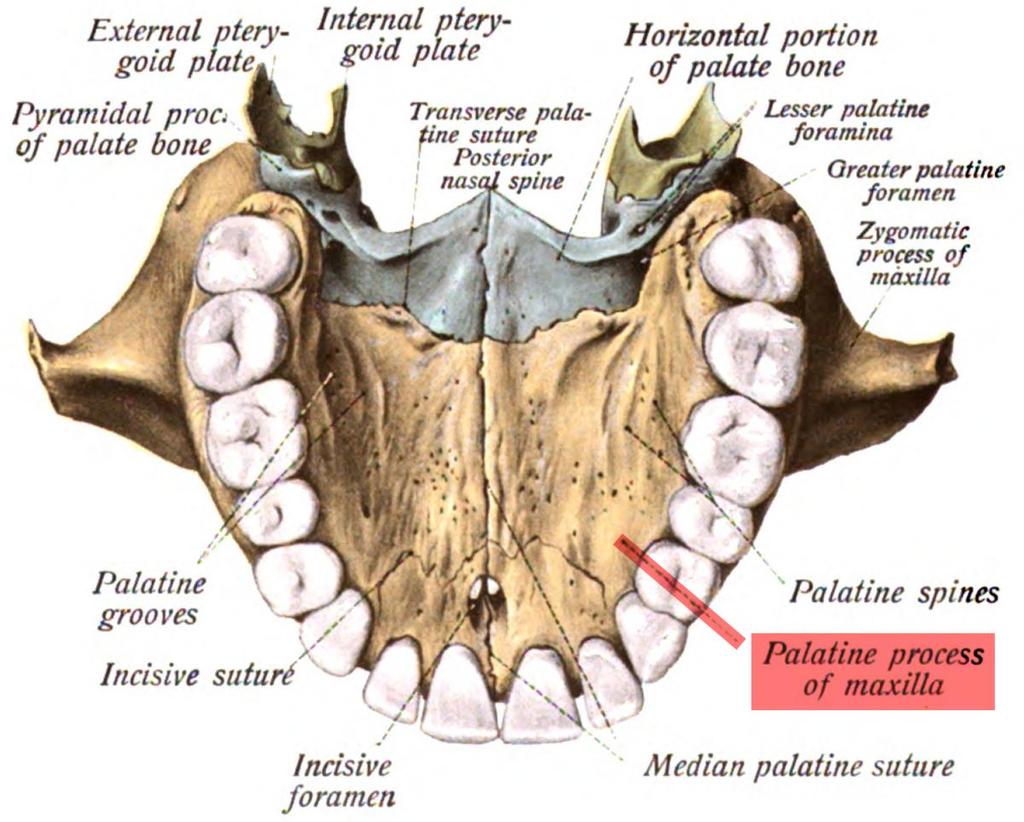
 Palatine bone (horizontal plate) Maxillary bone (palatine process) (a) Bones forming the left lateral wall of the nasal cavity (nasal septum removed) Anterior nasal spine")
4 Superior, middle, and inferior meatus Frontal sinus Superior nasal concha Middle nasal concha Inferior nasal concha Ethmoid bone Nasal bone Sphenoid bone Sphenoid sinus Pterygoid process Palatine bone (perpendicular plate) Palatine bone (horizontal plate) Maxillary bone (palatine process) (a) Bones forming the left lateral wall of the nasal cavity (nasal septum removed) Anterior nasal spine Figure 7.14a
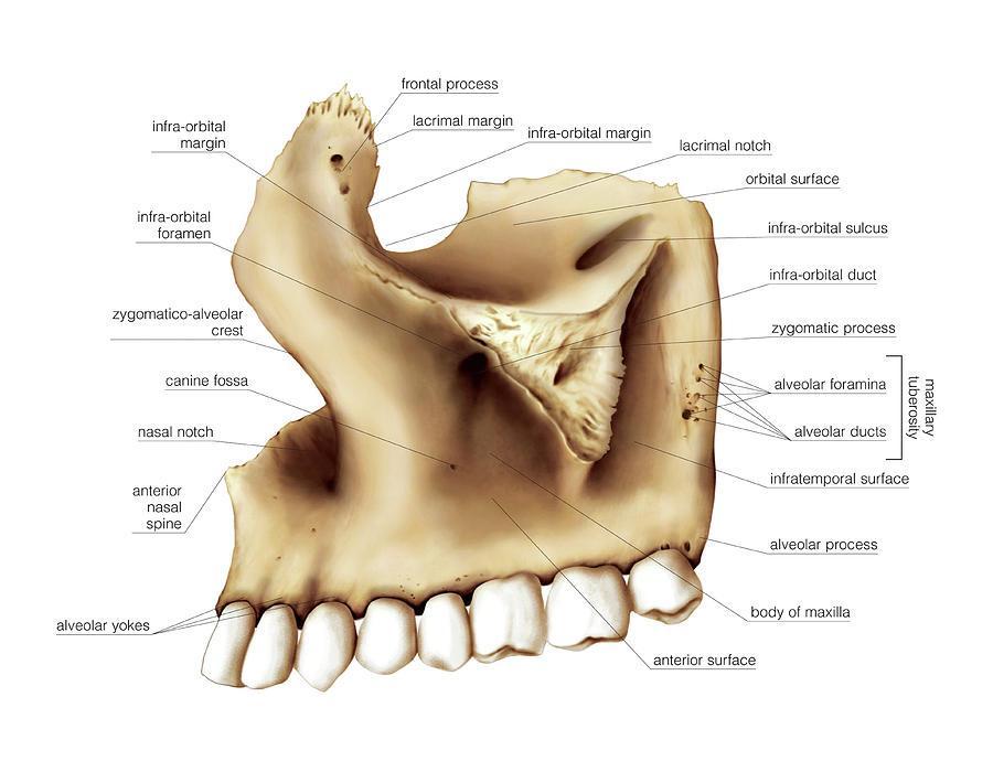
5 Maxilla
6 ORBIT SALIENT ANATOMICAL FEATURES 7 bones 4 walls 4 margins 4 important openings 6 contents 5 important relationships
7 Boundaries Roof: Is formed by orbital plate of frontal bone Lateral wall: Composed of zygomatic bone & greater wing of sphenoid bone Floor: Formed by the orbital plate of maxilla Medial wall: Frontal process of maxilla, Lacrimal bone, orbital plate of ethmoid & body of sphenoid bone
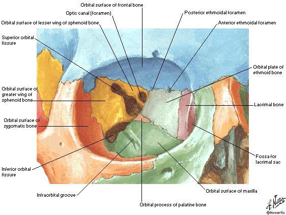
8
9 Adult orbital dimensions Entrance height 35 mm Entrance width 40 mm 35mm 45mm Medial wall length / depth Volume 45 mm 30 cc 45mm Distance from the back of the globe to the optic foramen 18 mm
10 Supraorbital Notch The supraorbital notch is situated on the superior orbital margin It transmits the supraorbital nerve and blood vessels
11
12 Infraorbital Groove & Canal Situated on the floor of the orbit in the orbital plate of the maxilla They transmit the infraorbital nerve (a continuation of the maxillary nerve) and blood vessels
13
14 Nasolacrimal Canal Located anteriorly on the medial wall It communicates with the inferior meatus of the nose It transmits the nasolacrimal duct
15
16 Inferior Orbital Fissure Located posteriorly between the maxilla and the greater wing of the sphenoid It communicates with the pterygopalatine fossa It transmits the maxillary nerve and its zygomatic branch, the inferior ophthalmic vein, and sympathetic nerves
17
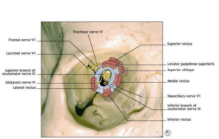
18 Superior Orbital Fissure Located posteriorly between the greater and lesser wings of the sphenoid It communicates with the middle cranial fossa It transmits the lacrimal nerve, the frontal nerve, the trochlear nerve, the oculomotor nerve (upper and lower divisions), the abducent nerve, the nasociliary nerve, and the superior ophthalmic vein
19
20 Optic Canal Located posteriorly in the lesser wing of the sphenoid It communicates with the middle cranial fossa It transmits the optic nerve and the ophthalmic artery
 Intracanalicular portion (located within the optic canal) Intracranial portion (ending in the optic")
21 Optic nerve The optic nerve may be divided into the following topographic areas: Intraocular portion of the optic nerve: optic disc, or nerve head; prelaminar; and laminar portions Intraorbital portion (located within the muscle cone) Intracanalicular portion (located within the optic canal) Intracranial portion (ending in the optic chiasm)
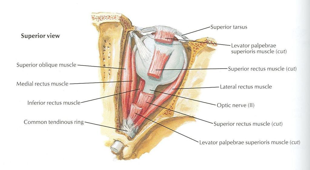
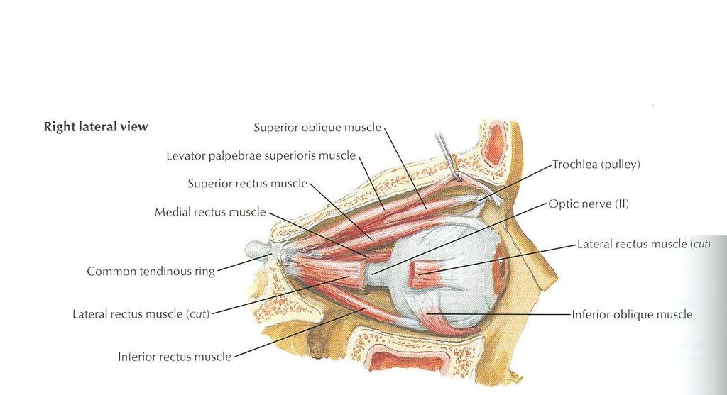
22 Muscles of Orbit Muscles of the orbit are the levator palpebrae superioris, the four recti and two oblique muscles
23 Levator Palpebrae Superioris Origin: Undersurface of lesser wing of sphenoid bone Insertion: Splits into 2 lamellae, superior into tarsal plate & skin of upper lid, inferior into upper margin of superior tarsal plate NS: Oculomotor nerve Action: Raises the upper lid
24
25 The Recti Arise from a fibrous ring called common tendinous ring Insertion: form the muscular cone that encloses the optic nerve, pierces the facial sheath of the eyeball, in the sclera about 6 mm behind the margin of cornea
26
27
28
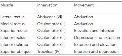
29 The Recti NS: Lateral by abducent, all others by oculomotor Action: lateral rectus rotates the eyeball so that the cornea looks laterally Medial rectus rotates the eyeball so that the cornea looks medially
30
31 The Recti Superior and inferior recti are inserted on the medial side of the vertical axis of the eyeball Superior rectus raises the cornea and rotates it medially Inferior rectus depresses the cornea and rotates it medially
32 Superior Oblique Origin: From body of the sphenoid bone Insertion: into the sclera beneath the superior rectus after passing through the fibrocartilaginous pulley NS: Trochlear nerve Action: Rotates the eyeball so that the cornea looks downward & laterally
33
34 Inferior Oblique Origin: Anterior part of floor of the orbit Insertion: In the sclera behind the coronal equator NS: Oculomotor Action: Rotates the eyeball so that the cornea looks upward and laterally
35 Motor Supply Lacrimal Nerve Frontal Nerve Trochlear Nerve Oculomotor Nerve Abducent Nerve
36 Motor Cranial Nerves Ⅲ Superior orbital fissure Ⅳ Ⅵ Ⅹ Jugular foramen Ⅻ Ⅺ Hypoglossal canal

37 Trochlear n. Oculomotor n. Abducent n.


38 Vascular Supply and Drainage of the Orbit The posterior ciliary vessels supply the whole uveal tract, the cilioretinal arteries, the sclera, the margin of the cornea, and the adjacent conjunctiva. The anterior ciliary arteries supply the rectus muscles.
39 Infratemporal Fossa Irregularly shaped space deep and inferior to the zygomatic arch, deep to the ramus of the mandible and posterior to the maxilla. Communicates with the temporal fossa through the interval between (deep to) the zygomatic arch and (superficial to) the cranial bones. Temporal fossa is superior to the zygomatic arch, The infratemporal fossa is inferior to the zygomatic arch.

40 Skeletal framework The walls of the pterygopalatine fossa are formed by: The anterior wall is formed by the posterior surface of the maxilla; The medial wall is formed by the lateral surface of the palatine bone; The posterior wall and roof are formed by parts of the sphenoid bone.
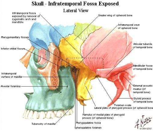
41
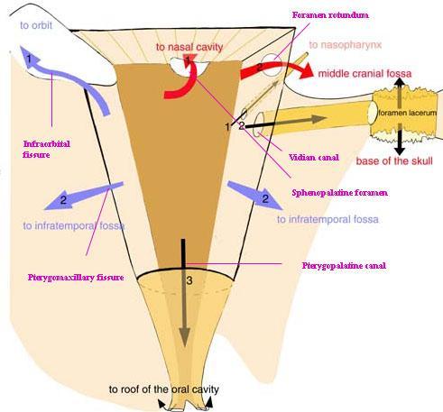
42 Gateways Seven foramina and fissures provide apertures through which structures enter and leave the pterygopalatine fossa 1. Foramen rotundum and pterygoid canal communicate with the middle cranial fossa 2. Palatovaginal canal opens onto the posterior wall and leads to the nasopharynx; 3. Palatine canal leads to the roof of the oral cavity (hard palate) and opens inferiorly; 4 Sphenopalatine foramen opens onto the lateral wall of the nasal cavity and is in the medial wall;
43 Gateways 5. Pterygomaxillary fissure between lateral aspect of the pterygopalatine fossa and the infratemporal fossa; 6. Inferior orbital fissure between the superior aspect of the fossa into the floor of the orbit

44
45
46 Contents 1. The maxillary nerve [V2] 2. Terminal part of the maxillary artery 3. Nerve of the pterygoid canal 4. The pterygopalatine ganglion 5. Veins and lymphatics also pass through the pterygopalatine fossa.
47 Pterygopalatine ganglion
![Nerve of the pterygoid canal Formed in the middle cranial fossa by the union of: 1. The greater petrosal nerve (a branch of the facial nerve [VII]); 2.](/docs-images/94/119580323/images/48-0.jpg "The deep petrosal nerve (a branch of the internal carotid plexus).")
48 Nerve of the pterygoid canal Formed in the middle cranial fossa by the union of: 1. The greater petrosal nerve (a branch of the facial nerve [VII]); 2. The deep petrosal nerve (a branch of the internal carotid plexus). Joins the pterygopalatine ganglion Carries mainly preganglionic parasympathetic (great petrosal) and postganglionic sympathetic (deep petrosal) fibers.

49 Pterygopalatine ganglion Largest of the four parasympathetic ganglia in the head Formed by the cell bodies neurons associated with: 1. Preganglionic parasympathetic fibers of the facial nerve carried by the greater petrosal nerve and the nerve of the pterygoid canal. 2.Sensory and ganglionic branches of the maxillary nerve 3.Postganglionic sympathetic fibers (deep petrosal)

50 Pterygopalatine ganglion These fibers form orbital, palatine, nasal, and pharyngeal branches, which leave the ganglion. Other fibers pass superiorly through the ganglionic branches of the maxillary nerve to enter the main trunk of the maxillary nerve And then distributed with the zygomatic, posterior superior alveolar, and infra-orbital nerves
51 Pterygopalatine ganglion
52 Orbital branches Pass through the inferior orbital fissure Supply of the orbital wall (periosteum) and lacrimal gland Supply the sphenoidal and ethmoidal sinuses.
53 Pharyngeal nerve Passes posteriorly from the pterygopalatine ganglion Leaves the fossa through the palatovaginal canal Supply the mucosa and glands of the nasopharynx.
54 Greater and lesser palatine nerves Pass through the palatine canal Enter the oral surface of the palate through the greater and lesser palatine foramina. Lesser palatine (Middle, Post, palatine) nerve passes posteriorly to supply the soft palate.

55 More anteriorly posterior superior alveolar nerves are given off. Pass through the pterygopalatine maxillary fissure into the infratemporal fossa.
 nerve passes forward on the roof of the oral cavity Innervate mucosa and glands of the hard palate and the adjacent gingiva, almost as far forward as the incisor teeth Also")
56 Greater and lesser palatine nerves The Greater palatine (Ant.palatine) nerve passes forward on the roof of the oral cavity Innervate mucosa and glands of the hard palate and the adjacent gingiva, almost as far forward as the incisor teeth Also supply the mucosa over the middle and lower part of the lateral wall of the nasal cavity Joins the long sphenopalatine nerve

57 Nasal nerves Seven in number Pass medially through the sphenopalatine foramen to enter the nasal cavity Short spheno-palatine (Post.Sup. Lateral nasal) supply the mucosa of the Post,Sup. quadrant of the nasal cavity. The Nasopalatine nerve (long Sphenopalatine) is the largest of the nasal nerves Passes anteriorly grooving down the nasal septum Through the incisive canal and fossa in the hard palate Supply mucosa, gingiva, and glands adjacent to the incisor teeth. Join the greater palatine nerve.
58 Maxillary Nerve
59 Maxillary nerve [V2] Purely sensory Originates from the trigeminal ganglion in the cranial cavity Exits the middle cranial fossa, and enters the pterygopalatine fossa through the foramen rotundum It terminates as the infra-orbital nerve through the inferior orbital fissure.
60 Maxillary nerve Branches: Meningeal (before it enters the Fossa) Two ganglionic branches pass through the pterygopalatine ganglion (Postganglionic parasympathetic fibers and sensory). Zygomatic nerve Posterior superior alveolar nerve Infra-orbital

61 Zygomatic nerve Originates directly from the maxillary nerve in the pterygopalatine fossa Enter the orbit through the inferior orbital fissure Divides into zygomaticotemporal and zygomaticofacial branches Zygomaticotemporal branch enter the temporal fossa and passes superficially to supply skin over the temple Carries postganglionic parasympathetic and sympathetic fibers and form a special autonomic nerve to join the lacrimal nerve The Zygomaticofacial branch opens on the anterolateral surface of the zygomatic bone, and supply the adjacent skin.
62 Posterior superior alveolar nerve Passes laterally out of the fossa through the pterygomaxillary fissure Enter the posterior surface of the maxilla approximately midway between the last molar tooth and the inferior orbital fissure Supplies the molar teeth and adjacent buccal gingivae contributes to the supply of the maxillary sinus
63 Infra-orbital nerve Anterior continuation of the maxillary nerve Leaves the pterygopalatine fossa through the inferior orbital fissure First in the infra-orbital groove in the floor of the orbit and then continues forward in the infra-orbital canal. While in the infra-orbital groove and canal, the infra-orbital nerve gives origin to middle and anterior superior alveolar nerves: They Join the superior alveolar plexus to supply the upper teeth Middle superior alveolar nerve also supplies the maxillary sinus Anterior superior alveolar nerve also gives origin to a small nasal branch

64 Infra-orbital nerve The infra-orbital nerve exits the infraorbital canal through the infra-orbital foramen Divides into nasal, palpebral, and superior labial branches Nasal branches supply skin over the lateral aspect of the external nose and part of the nasal septum; Palpebral branches supply skin of the lower eyelid; Superior labial branches supply skin over the cheek and upper lip, and the related oral mucosa.

65 1. infraorbital nerve 2. posterior superior alveolar nerve 3. pterygopalatine ganglion (parasympathetic) 4. greater palatine nerve 5. lesser palatine nerve cut 6. nasopalatine nerve 7. nerve of the pharyngeal canal
66 Maxillary Artery
")
67 Maxillary artery Major branch of the external carotid artery in the neck Originates adjacent to the neck of mandible Originates within the substance of the parotid gland Passes forward through the infratemporal fossa Enters the pterygopalatine fossa through the pterygomaxillary fissure (the third part)
 and the maxillary vein (below).")
68 First part The first part of the maxillary artery is the part between the neck of mandible (Lat.) and the sphenomandibular ligament (Med.) Also related to the auriculo.temporal nerve (above) and the maxillary vein (below). Gives origin to two major branches (the middle meningeal and inferior alveolar arteries) Smaller branches (deep auricular, anterior tympanic, and accessory meningeal);
69 Second part The second part of the maxillary artery the part related to the lateral pterygoid muscle Gives origin to deep temporal, masseteric, buccal, and pterygoid branches (muscles of mastication) Course with branches of the mandibular nerve
70 Terminal (3 rd ) part In the pterygopalatine fossa Anterior to the pterygopalatine ganglion Gives origin to branches that accompany branches of the maxillary nerve [V2] and the pterygopalatine ganglion. These branches supply much of the nasal cavity, the roof of the oral cavity, and all upper teeth. In addition, they contribute to the blood supply of the sinuses, oropharynx, and floor of the orbit.

71 Branches of the 3 rd part maxillary artery 1. The posterior superior alveolar, 2. Infra-orbital, 3. Greater palatine, 4. Pharyngeal, 5. Sphenopalatine arteries, 6.The artery of the pterygoid canal
72 Posterior superior alveolar artery Originates from the maxillary artery as it passes through the pterygomaxillary fissure Meets the posterior superior alveolar nerve, Accompanies it through the alveolar foramen on the infratemporal surface of the maxilla Supplies the molar and premolar teeth, adjacent gingiva, and the maxillary sinus.

73 Infra-orbital artery Passes forward with the infra-orbital nerve and leaves the pterygopalatine fossa through the inferior orbital fissure With the infra-orbital nerve, it lies in the infra-orbital groove and infra-orbital canal Emerges through the infra-orbital foramen to supply parts of the face. In the orbital canal gives : 1. Branches that contribute to the blood supply of structures near the floor of the orbit-the inferior rectus and inferior oblique muscles, and the lacrimal sac; 2. Anterior superior alveolar arteries, which supply the incisor and canine teeth and the maxillary sinus.
74 Greater palatine artery Travels posteriorly and leaves the pterygopalatine fossa through the palatovaginal canal with the pharyngeal nerve Supplies the posterior aspect of the roof of the nasal cavity, the sphenoidal sinus, and the pharyngotympanic tube.
75 Sphenopalatine artery The terminal branch of the maxillary artery Leaves the pterygopalatine fossa medially through the sphenopalatine foramen Accompanies the nasal nerves, giving off: 1. Posterior lateral nasal arteries, which supply the lateral wall of the nasal cavity and contribute to supply of the paranasal sinuses; 2. Posterior septal branches, which supply the nasal septum-the largest of these branches passes anteriorly down the septum to anastomose with the end of the greater palatine artery.

76 Maxillary artery

77 Veins Drain areas supplied by branches of the terminal part of the maxillary artery Generally travel with these branches back into the pterygopalatine fossa. The veins coalesce in the fossa and then pass laterally through the pterygomaxillary fissure to join the pterygoid plexus of veins in the infratemporal fossa The infra-orbital vein, drains the inferior aspect of the orbit, May pass directly into the infratemporal fossa, so bypassing the pterygopalatine fossa


78 Veins The veins pass through the pterygomaxillary fissure to join the pterygoid plexus of veins in the infratemporal fossa.
79 Thank you
MAXILLA, ORBIT & PTERYGOPALATINE FOSSA. Neophytos C Demetriades MD, DDS, MSc Associate professor European University of Cyprus School of Medicine
 MAXILLA, ORBIT & PTERYGOPALATINE FOSSA Neophytos C Demetriades MD, DDS, MSc Associate professor European University of Cyprus School of Medicine Maxilla MAXILLA Superior, middle, and inferior meatus Frontal
PTERYGOPALATINE FOSSA
 PTERYGOPALATINE FOSSA Outline Anatomical Structure and Boundaries Foramina and Communications with other spaces and cavities Contents Pterygopalatine Ganglion Especial emphasis on certain arteries and
Anatomic Relations Summary. Done by: Sohayyla Yasin Dababseh
 Anatomic Relations Summary Done by: Sohayyla Yasin Dababseh Anatomic Relations Lecture 1 Part-1 - The medial wall of the nose is the septum. - The vestibule lies directly inside the nostrils (Nares). -
Mohammad Hisham Al-Mohtaseb. Lina Mansour. Reyad Jabiri. 0 P a g e
 2 Mohammad Hisham Al-Mohtaseb Lina Mansour Reyad Jabiri 0 P a g e This is only correction for the last year sheet according to our record. If you already studied this sheet just read the yellow notes which
Omran Saeed. Luma Taweel. Mohammad Almohtaseb. 1 P a g e
 2 Omran Saeed Luma Taweel Mohammad Almohtaseb 1 P a g e I didn t include all the photos in this sheet in order to keep it as small as possible so if you need more clarification please refer to slides In
Bony orbit Roof The orbital plate of the frontal bone Lateral wall: the zygomatic bone and the greater wing of the sphenoid
 Bony orbit Roof: Formed by: The orbital plate of the frontal bone, which separates the orbital cavity from the anterior cranial fossa and the frontal lobe of the cerebral hemisphere Lateral wall: Formed
The sebaceous glands (glands of Zeis) open directly into the eyelash follicles, ciliary glands (glands of Moll) are modified sweat glands that open
 open directly into the eyelash follicles, ciliary glands (glands of Moll) are modified sweat glands that open") The Orbital Region The orbits are a pair of bony cavities that contain the eyeballs; their associated muscles, nerves, vessels, and fat; and most of the lacrimal apparatus upper eyelid is larger and more
The Orbital Region The orbits are a pair of bony cavities that contain the eyeballs; their associated muscles, nerves, vessels, and fat; and most of the lacrimal apparatus upper eyelid is larger and more
Dr.Ban I.S. head & neck anatomy 2 nd y جامعة تكريت كلية طب االسنان مادة التشريح املرحلة الثانية أ.م.د. بان امساعيل صديق 6102/6102
 جامعة تكريت كلية طب االسنان مادة التشريح املرحلة الثانية أ.م.د. بان امساعيل صديق 6102/6102 Pterygopalatine fossa: The pterygopalatine fossa is a cone-shaped depression, It is located between the maxilla,
Infratemporal fossa: Tikrit University college of Dentistry Dr.Ban I.S. head & neck Anatomy 2 nd y.
 Infratemporal fossa: This is a space lying beneath the base of the skull between the lateral wall of the pharynx and the ramus of the mandible. It is also referred to as the parapharyngeal or lateral pharyngeal
The orbit-1. Dr. Heba Kalbouneh Assistant Professor of Anatomy and Histology
 The orbit-1 Dr. Heba Kalbouneh Assistant Professor of Anatomy and Histology Orbital plate of frontal bone Orbital plate of ethmoid bone Lesser wing of sphenoid Greater wing of sphenoid Lacrimal bone Orbital
Trigeminal Nerve Worksheets, Distributions Page 1
 Trigeminal Nerve Worksheet #1 Distribution by Nerve Dr. Darren Hoffmann Dental Gross Anatomy, Spring 2013 We have drawn out each of the branches of CN V in lecture and you have an idea now for their basic
Temporal region. temporal & infratemporal fossae. Zhou Hong Ying Dept. of Anatomy
 Temporal region temporal & infratemporal fossae Zhou Hong Ying Dept. of Anatomy Temporal region is divided by zygomatic arch into temporal & infratemporal fossae. Temporal Fossa Infratemporal fossa Temporal
1 Eyelids. Lacrimal Apparatus. Orbital Region. 3 The Orbit. The Eye
 1 1 Eyelids Orbital Region 2 Lacrimal Apparatus 3 The Orbit 4 The Eye 2 Eyelids The eyelids protect the eye from injury and excessive light by their closure. The upper eyelid is larger and more mobile
Bisection of Head & Nasal Cavity 頭部對切以及鼻腔. 解剖學科馮琮涵副教授 分機
 Bisection of Head & Nasal Cavity 頭部對切以及鼻腔 解剖學科馮琮涵副教授 分機 3250 E-mail: thfong@tmu.edu.tw Outline: The structure of nose The concha and meatus in nasal cavity The openings of paranasal sinuses Canals, foramens
Parotid Gland, Temporomandibular Joint and Infratemporal Fossa
 M1 - Anatomy Parotid Gland, Temporomandibular Joint and Infratemporal Fossa Jeff Dupree Sanger 9-057 jldupree@vcu.edu Parotid gland: wraps around the mandible positioned between the mandible and the sphenoid
Temporal fossa Infratemporal fossa Pterygopalatine fossa Terminal branches of external carotid artery Pterygoid venous plexus
 Outline of content Temporal fossa Infratemporal fossa Pterygopalatine fossa Terminal branches of external carotid artery Pterygoid venous plexus Boundary Content Communication Mandibular division of trigeminal
The orbit-2. Dr. Heba Kalbouneh Assistant Professor of Anatomy and Histology
 The orbit-2 Dr. Heba Kalbouneh Assistant Professor of Anatomy and Histology Eyelids The eyelids (act like the curtains) protect the eye from injury and excessive light by their closure The upper eyelid
Dr. Sami Zaqout, IUG Medical School
 The skull The skull is composed of several separate bones united at immobile joints called sutures. Exceptions? Frontal bone Occipital bone Vault Cranium Sphenoid bone Zygomatic bones Base Ethmoid bone
Cranial nerves.
 Cranial nerves eaglezhyxzy@163.com Key Points of Learning Name Components Passing through Peripheral distribution Central connection Function Cranial nerves Ⅰ olfactory Ⅱ optic Ⅲ occulomotor Ⅳ trochlear
Trigeminal Nerve Anatomy. Dr. Mohamed Rahil Ali
 Trigeminal Nerve Anatomy Dr. Mohamed Rahil Ali Trigeminal nerve Largest cranial nerve Mixed nerve Small motor root and large sensory root Motor root Nucleus of motor root present in the pons and medulla
Trigeminal Nerve (V)
") Trigeminal Nerve (V) Lecture Objectives Discuss briefly how the face is developed. Follow up the course of trigeminal nerve from its point of central connections, exit and down to its target areas. Describe
Trigeminal Nerve (V) Lecture Objectives Discuss briefly how the face is developed. Follow up the course of trigeminal nerve from its point of central connections, exit and down to its target areas. Describe
Tikrit University collage of dentistry Dr.Ban I.S. head & neck anatomy 2 nd y. Lec [5] / Temporal fossa :
![Tikrit University collage of dentistry Dr.Ban I.S. head & neck anatomy 2 nd y. Lec [5] / Temporal fossa :](/thumbs/88/115294566.jpg "Tikrit University collage of dentistry Dr.Ban I.S. head & neck anatomy 2 nd y. Lec [5] / Temporal fossa :") Lec [5] / Temporal fossa : Borders of the Temporal Fossa: Superior: Superior temporal line. Inferior: gap between zygomatic arch and infratemporal crest of sphenoid bone. Anterior: Frontal process of the
Lec [5] / Temporal fossa : Borders of the Temporal Fossa: Superior: Superior temporal line. Inferior: gap between zygomatic arch and infratemporal crest of sphenoid bone. Anterior: Frontal process of the
Anatomy of the Trigeminal Nerve
 19 Anatomy of the Trigeminal Nerve.1 Introduction 0. The Central Part of the Trigeminal Nerve 1..1 Origin 1.. Trigeminal Nuclei.3 The Peripheral Part of the Trigeminal Nerve 4.3.1 Ophthalmic Nerve 4.3.
Introduction to Local Anesthesia and Review of Anatomy
 5-Sep Introduction and Anatomy Review 12-Sep Neurophysiology and Pain 19-Sep Physiology and Pharmacology part 1 26-Sep Physiology and Pharmacology part 2 Introduction to Local Anesthesia and Review of
Bones Ethmoid bone Inferior nasal concha Lacrimal bone Maxilla Nasal bone Palatine bone Vomer Zygomatic bone Mandible
 splanchnocranium - Consists of part of skull that is derived from branchial arches - The facial bones are the bones of the anterior and lower human skull Bones Ethmoid bone Inferior nasal concha Lacrimal
Face. Definition: The area between the two ears and from the chin to the eye brows. The muscles of the face
 Face Definition: The area between the two ears and from the chin to the eye brows. The muscles of the face The muscle of facial expression (include the muscle of the face and the scalp). All are derived
Parotid Gland. Parotid Gland. Largest of 3 paired salivary glands (submandibular; sublingual) Ramus of Mandible. Medial pterygoid.
 Ramus of Mandible. Medial pterygoid.") Parotid region Parotid Gland Largest of 3 paired salivary glands (submandibular; sublingual) Ramus of Mandible Medial pterygoid Cross section of mandible Masseter D S SCM Parotid Gland Mastoid Process
Parotid region Parotid Gland Largest of 3 paired salivary glands (submandibular; sublingual) Ramus of Mandible Medial pterygoid Cross section of mandible Masseter D S SCM Parotid Gland Mastoid Process
213: HUMAN FUNCTIONAL ANATOMY: PRACTICAL CLASS 12 Cranial cavity, eye and orbit
 213: HUMAN FUNCTIONAL ANATOMY: PRACTICAL CLASS 12 Cranial cavity, eye and orbit OSTEOLOGY Identify the bones which comprise the walls of the orbit: maxilla, zygomatic, ethmoid, lachrymal, frontal, and
Tracing the Cranial Nerves Osteologically
 CN I II III IV V 1 Supra-orbital ethmoidal nn. Ext. nasal V 2 Tracing the Cranial Nerves Osteologically Nucleus of Origin Olfactory tracts of frontal lobe of cerebrum Optic tracts from optic chiasma and
Dr.Ban I.S. head & neck anatomy 2 nd y. جامعة تكريت كلية طب االسنان املرحلة الثانية أ.م.د. بان امساعيل صديق 6102/6102
 جامعة تكريت كلية طب االسنان التشريح مادة املرحلة الثانية أ.م.د. بان امساعيل صديق 6102/6102 Parotid region The part of the face in front of the ear and below the zygomatic arch is the parotid region. The
3-Deep fascia: is absent (except over the parotid gland & buccopharngeal fascia covering the buccinator muscle)
") The Face 1-Skin of the Face The skin of the face is: Elastic Vascular (bleed profusely however heal rapidly) Rich in sweat and sebaceous glands (can cause acne in adults) It is connected to the underlying
The Face 1-Skin of the Face The skin of the face is: Elastic Vascular (bleed profusely however heal rapidly) Rich in sweat and sebaceous glands (can cause acne in adults) It is connected to the underlying
Nasal region. cartilages: septal cartilage (l); lateral nasal cartilage (2); greater alar cartilages (2); lesser alar cartilages (?
; lateral nasal cartilage (2); greater alar cartilages (2); lesser alar cartilages (?") Nasal region skull bones: nasal and frontal processes of maxilla cartilages: septal cartilage (l); lateral nasal cartilage (2); greater alar cartilages (2); lesser alar cartilages (?) 1 Nasal cavity Roof
Nasal region skull bones: nasal and frontal processes of maxilla cartilages: septal cartilage (l); lateral nasal cartilage (2); greater alar cartilages (2); lesser alar cartilages (?) 1 Nasal cavity Roof
Skull-2. Norma Basalis Interna Norma Basalis Externa. Dr. Heba Kalbouneh Associate Professor of Anatomy and Histology
 Skull-2 Norma Basalis Interna Norma Basalis Externa Dr. Heba Kalbouneh Associate Professor of Anatomy and Histology Norma basalis interna Base of the skull- superior view The interior of the base of the
Trigeminal nerve. Slide in bold and please go back to see the pictures, if I skipped any part of record that because it wasn t clear to me
 Trigeminal nerve Slide in bold and please go back to see the pictures, if I skipped any part of record that because it wasn t clear to me Hala nsour 2/26/2018 P a g e 1 this lecture contain two topics
Bony orbit. Lateral wall: Formed by : the zygomatic bone and the greater wing of the sphenoid
 Bony orbit Roof: Formed by: The orbital plate of the frontal bone, which separates the orbital cavity from the anterior cranial fossa and the frontal lobe of the cerebral hemisphere Lateral wall: Formed
*in general the blood supply of the nose comes from branches of the internal and external carotid arteries.
 In the previous lecture we talked about the anatomy of the nasal cavity, today we will talk about its blood supply, venous drainage, innervations, and finally about the paranasal sinuses. When we describe
Lec [8]: Mandibular nerve:
![Lec [8]: Mandibular nerve:](/thumbs/94/121295776.jpg "Lec [8]: Mandibular nerve:") Lec [8]: Mandibular nerve: The mandibular branch from the trigeminal ganglion lies in the middle cranial fossa lateral to the cavernous sinus. With the motor root of the trigeminal nerve [motor roots lies
Lec [8]: Mandibular nerve: The mandibular branch from the trigeminal ganglion lies in the middle cranial fossa lateral to the cavernous sinus. With the motor root of the trigeminal nerve [motor roots lies
Anatomy and Physiology. Bones, Sutures, Teeth, Processes and Foramina of the Human Skull
 Anatomy and Physiology Chapter 6 DRO Bones, Sutures, Teeth, Processes and Foramina of the Human Skull Name: Period: Bones of the Human Skull Bones of the Cranium: Frontal bone: forms the forehead and the
Bones of the skull & face
 Bones of the skull & face Cranium= brain case or helmet Copyright The McGraw-Hill Companies, Inc. Permission required for reproduction or display. The cranium is composed of eight bones : frontal Occipital
Nose & Mouth OUTLINE. Nose. - Nasal Cavity & Its Walls. - Paranasal Sinuses. - Neurovascular Structures. Mouth. - Oral Cavity & Its Contents
 Dept. of Human Anatomy, Si Chuan University Zhou hongying eaglezhyxzy@163.com Nose & Mouth OUTLINE Nose - Nasal Cavity & Its Walls - Paranasal Sinuses - Neurovascular Structures Mouth - Oral Cavity & Its
Major Anatomic Components of the Orbit
 Major Anatomic Components of the Orbit 1. Osseous Framework 2. Globe 3. Optic nerve and sheath 4. Extraocular muscles Bony Orbit Seven Bones Frontal bone Zygomatic bone Maxillary bone Ethmoid bone Sphenoid
Anatomy #1; Respiratory Nose and the Nasal Cavity December 1st, 2013
 Note #1: the doctor skipped some slides in the lecture. Those slides are not included in this sheet and so you will have to review the slides to study them. The reason they were not included is because
Veins of the Face and the Neck
 Veins of the Face and the Neck Facial Vein The facial vein is formed at the medial angle of the eye by the union of the supraorbital and supratrochlear veins. connected through the ophthalmic veins with
mistake ;slides in bold but you still have to go back to our slides to see the figure, tables and some scheme
 Khozama jehad : I am doing my best and I am sorry for any unintended mistake ;slides in bold but you still have to go back to our slides to see the figure, tables and some scheme The Orbit, Orbital Contents
Structure Location Function
 Frontal Bone Cranium forms the forehead and roof of the orbits Occipital Bone Cranium forms posterior and inferior portions of the cranium Temporal Bone Cranium inferior to the parietal bone forms the
Unit VIII Problem 8 Anatomy: Orbit and Eyeball
 Unit VIII Problem 8 Anatomy: Orbit and Eyeball - The bony orbit: it is protecting our eyeball and resembling a pyramid: With a base directed: anterolaterally. And an apex directed: posteromedially. Notes:
Chapter 7 Part A The Skeleton
 Chapter 7 Part A The Skeleton Why This Matters Understanding the anatomy of the skeleton enables you to anticipate problems such as pelvic dimensions that may affect labor and delivery The Skeleton The
The Skull and Temporomandibular joint II Prof. Abdulameer Al-Nuaimi. E. mail:
 The Skull and Temporomandibular joint II Prof. Abdulameer Al-Nuaimi E-mail: a.al-nuaimi@sheffield.ac.uk E. mail: abdulameerh@yahoo.com Temporal fossa The temporal fossa is a depression on the temporal
human anatomy 2016 lecture fifteen Dr meethak ali ahmed neurosurgeon
 Cranial Nerves Organization of the Cranial Nerves The cranial nerves are named as follows: I. Olfactory II. Optic III. Oculomotor IV. Trochlear V. Trigeminal VI. Abducent VII. Facial VIII. Vestibulocochlear
SCHOOL OF ANATOMICAL SCIENCES Mock Run Questions. 4 May 2012
 SCHOOL OF ANATOMICAL SCIENCES Mock Run Questions 4 May 2012 1. With regard to the muscles of the neck: a. the platysma muscle is supplied by the accessory nerve. b. the stylohyoid muscle is supplied by
University of Palestine. Midterm Exam 2013/2014 Total Grade:
 Course No: DNTS2208 Course Title: Head and Neck Anatomy Date: 09/11/2013 No. of Questions: (50) Time: 1hour Using Calculator (No) University of Palestine Midterm Exam 2013/2014 Total Grade: Instructor
function - sensory & postganglionic sympathetic [communication from the internal carotid plexus in the cavernous sinus] innervation of the mucosa of
![function - sensory & postganglionic sympathetic [communication from the internal carotid plexus in the cavernous sinus] innervation of the mucosa of](/thumbs/74/71276096.jpg "function - sensory & postganglionic sympathetic [communication from the internal carotid plexus in the cavernous sinus] innervation of the mucosa of") Nerves I. Cranial nerves A. Olfactory (CN I) 1. Olfactory bulb 2. Olfactory tract B. Optic n. (CNII) function - carries visual sensory information from the neural retina to the diencephalon & midbrain
Nerves I. Cranial nerves A. Olfactory (CN I) 1. Olfactory bulb 2. Olfactory tract B. Optic n. (CNII) function - carries visual sensory information from the neural retina to the diencephalon & midbrain
Chapter 7: Head & Neck
 Chapter 7: Head & Neck Osteology I. Overview A. Skull The cranium is composed of irregularly shaped bones that are fused together at unique joints called sutures The skull provides durable protection from
Dr.Noor Hashem Mohammad Lecture (5)
") Dr.Noor Hashem Mohammad Lecture (5) 2016-2017 If the mandible is discarded, the anterior part of this aspect of the skull is seen to be formed by the hard palate. The palatal processes of the maxillae
Dr.Noor Hashem Mohammad Lecture (5) 2016-2017 If the mandible is discarded, the anterior part of this aspect of the skull is seen to be formed by the hard palate. The palatal processes of the maxillae
CN I Olfactory. CN II Optic. CN III Oculomotor. Special Sensory Efferent fibers to Olfactory Bulb. Cribiform Plate of Ethmoid
 CN I Olfactory Efferent fibers to Olfactory Bulb Olfactory Tract Olfactory Bulb Cribiform Plate of Ethmoid Anosmia Loss of sense of smell Uncinate Fits olfactory hallucinations To Olfactory Epithelium
Introduction to Head and Neck Anatomy
 Introduction to Head and Neck Anatomy Nervous Tissue Controls and integrates all body activities within limits that maintain life Three basic functions 1. sensing changes with sensory receptors 2. interpreting
The Pharynx. Dr. Nabil Khouri MD. MSc, Ph.D
 The Pharynx Dr. Nabil Khouri MD. MSc, Ph.D Introduction The pharynx is the Musculo-fascial halfcylinder that links the oral and nasal cavities in the head to the larynx and esophagus in the neck Common
Skull basic structures. Neurocranium
 Assoc. Prof. Květuše Lovásová, M.V.D., PhD. Skull basic structures Skull consists of two groups of bones: neurocranium (bones forming the brain box) splanchnocranium (bones forming the facial skeleton)
Dr.Ban I.S. head & neck anatomy 2 nd y. جامعة تكريت كلية طب االسنان املرحلة الثانية
 جامعة تكريت كلية طب االسنان التشريح مادة املرحلة الثانية أ.م.د. بان امساعيل صديق 6102-6102 1 The Palate The palate forms the roof of the mouth and the floor of the nasal cavity. It is divided into two
University of Palestine. Midterm Exam 2013/2014 Total Grade:
 [ Course No: DNTS2208 Course Title: Head and Neck Anatomy Date: 17/11/1024 No. of Questions: (52) Time: 2hours Using Calculator (No) University of Palestine Midterm Exam 2013/2014 Total Grade: Instructor
Skeletal System: Skull.
 Skeletal System: Skull www.fisiokinesiterapia.biz Bones of the Skull SPLANCHNOCRANIUM Nasal (2) Maxilla (2) Lacrimal (2) Zygomatic (2) Palatine (2) Inferior concha (2) Vomer Mandible NEUROCRANIUM Frontal
Chapter(2):the lid page (1) THE LID
:the lid page (1) THE LID") Chapter(2):the lid page (1) THE LID Anatomy of the lid: * Check movie anatomy of the lid model The eyelids are two movable muco-cutaneous folds which protect the eye on closure. The are joined temporary
Chapter(2):the lid page (1) THE LID Anatomy of the lid: * Check movie anatomy of the lid model The eyelids are two movable muco-cutaneous folds which protect the eye on closure. The are joined temporary
Skull-2. Norma Basalis Interna. Dr. Heba Kalbouneh Assistant Professor of Anatomy and Histology
 Skull-2 Norma Basalis Interna Dr. Heba Kalbouneh Assistant Professor of Anatomy and Histology Norma basalis interna Base of the skull- superior view The interior of the base of the skull is divided into
Biology 218 Human Anatomy. Adapted from Martini Human Anatomy 7th ed. Chapter 6 The Skeletal System: Axial Division
 Adapted from Martini Human Anatomy 7th ed. Chapter 6 The Skeletal System: Axial Division Introduction The axial skeleton: Composed of bones along the central axis of the body Divided into three regions:
By : Prof Saeed Abuel Makarem & Dr.Sanaa Alshaarawi
 By : Prof Saeed Abuel Makarem & Dr.Sanaa Alshaarawi OBJECTIVES By the end of the lecture, students shouldbe able to: List the nuclei of the deep origin of the trigeminal and facial nerves in the brain
GNK485 The eye and related structures. Prof MC Bosman 2012
 GNK485 The eye and related structures Prof MC Bosman 2012 Surface anatomy Bony orbit Eyeball and Lacrimal apparatus Extra-ocular muscles Movements of the eye Innervation Arterial supply and venous drainage
EXAM NUMBER STRUCTURAL BASIS OF MEDICAL PRACTICE EXAMINATION 7 October 28, PART l. Answer in the space provided. (9 pts)
") STRUCTURAL BASIS OF MEDICAL PRACTICE EXAMINATION 7 October 28, 2005 PART l. Answer in the space provided. (9 pts) 1. Identify the structures. (3 pts) a. _Frontal Sinus b. _Lateral Posterior Inferior Nasal
STRUCTURAL BASIS OF MEDICAL PRACTICE EXAMINATION 7 October 28, 2005 PART l. Answer in the space provided. (9 pts) 1. Identify the structures. (3 pts) a. _Frontal Sinus b. _Lateral Posterior Inferior Nasal
SKULL / CRANIUM BONES OF THE NEUROCRANIUM (7) Occipital bone (1) Sphenoid bone (1) Temporal bone (2) Frontal bone (1) Parietal bone (2)
 Occipital bone (1) Sphenoid bone (1) Temporal bone (2) Frontal bone (1) Parietal bone (2)") Important! 1. Memorizing these pages only does not guarantee the succesfull passing of the midterm test or the semifinal exam. 2. The handout has not been supervised, and I can not guarantee, that these
Important! 1. Memorizing these pages only does not guarantee the succesfull passing of the midterm test or the semifinal exam. 2. The handout has not been supervised, and I can not guarantee, that these
REVIEW OF HEAD AND NECK CRANIAL NERVES AND EVERYTHING ELSE
 REVIEW OF HEAD AND NECK CRANIAL NERVES AND EVERYTHING ELSE OLFACTORY NERVE CN I ANTERIOR CRANIAL FOSSA CRISTA GALLI OF ETHMOID OLFACTORY FORAMINA IN CRIBIFORM PLATE OF ETHMOID BONE CN I OLFACTORY NERVE
Cranial Nerve VII - Facial Nerve. The facial nerve has 3 main components with distinct functions
 Cranial Nerve VII - Facial Nerve The facial nerve has 3 main components with distinct functions Somatic motor efferent Supplies the muscles of facial expression; posterior belly of digastric muscle; stylohyoid,
Bony orbit. Sup. Med. Inf. Lat. frontal bone. frontal process of maxilla. zygomatic process of maxilla zygomatic bone
 Orbit 解剖學科鄭授德 本教材之圖片取自於 1. Gray s Anatomy for Students, 3rd ed., 2015, by Drake, Vogl, and Mitchell 2. Clinically Oriented Anatomy, 7th ed., 2014, by Moore, Dalley, and Agur 3. Anatomy, an Essential Textbook,
Cranial nerve Dept. of Anatomy Zhou Hong Ying
 Cranial nerve Dept. of Anatomy Zhou Hong Ying Key Points of Learning Name Components Passing through Peripheral distribution Central connection Function Cranial nerve Ⅰ olfactory Ⅱ optic Ⅲ occulomotor
Anatomy images for MSS practical exam- 2019
 Anatomy images for MSS practical exam- 2019 Ilium Ischium Pubis Acetabulaum Iliac crest Iliac tubercle ASIS (muscle and ligament attached) AIIS (muscle attached) PSIS PIIS Ischial spine Ischial tuberosity
Prevertebral Region, Pharynx and Soft Palate
 Unit 20: Prevertebral Region, Pharynx and Soft Palate Dissection Instructions: Step1 Step 2 Step 1: Insert your fingers posterior to the sternocleidomastoid muscle, vagus nerve, internal jugular vein,
Head and Face Anatomy
 Head and Face Anatomy Epicranial region The Scalp The soft tissue that covers the vault of skull. Extends from supraorbital margin to superior nuchal line. Layers of the scalp S C A L P = skin = connective
Basic Anatomy and Physiology of the Lips and Oral Cavity. Dr. Faghih
 Basic Anatomy and Physiology of the Lips and Oral Cavity Dr. Faghih It is divided into seven specific subsites : 1. Lips 2. dentoalveolar ridges 3. oral tongue 4. retromolar trigone 5. floor of mouth 6.
C h a p t e r PowerPoint Lecture Slides prepared by Jason LaPres North Harris College Houston, Texas
 C h a p t e r 15 The Nervous System: The Brain and Cranial Nerves PowerPoint Lecture Slides prepared by Jason LaPres North Harris College Houston, Texas Copyright 2009 Pearson Education, Inc., publishing
The Orbit. The Orbit OCULAR ANATOMY AND DISSECTION 9/25/2014. The eye is a 23 mm organ...how difficult can this be? Openings in the orbit
 The eye is a 23 mm organ...how difficult can this be? OCULAR ANATOMY AND DISSECTION JEFFREY M. GAMBLE, OD COLUMBIA EYE CONSULTANTS OPTOMETRY & UNIVERSITY OF MISSOURI DEPARTMENT OF OPHTHALMOLOGY CLINICAL
Mandibular and Maxillary Anesthesia
 Mandibular and Maxillary Anesthesia Uses of the Conduction Technique JACK H. SELTSAM, D.D.S., M.D., Los Angeles THE ARMAMENTARIUM of a surgeon who operates on the head and neck should include the ability
Cranial Cavity REFERENCES: OBJECTIVES OSTEOLOGY. Stephen A. Gudas, PT, PhD
 Stephen A. Gudas, PT, PhD Cranial Cavity REFERENCES: Moore and Agur, Essential Clinical Anatomy (ECA), 3rd ed., pp. 496 498; 500 507; 512 514 Grant s Atlas 12 th ed., Figs 7.6; 7.19 7.30. Grant s Dissector
Biology 323 Human Anatomy for Biology Majors Week 10; Lecture 1; Tuesday Dr. Stuart S. Sumida. Cranial Nerves and Soft Tissues of the Skull
 Biology 323 Human Anatomy for Biology Majors Week 10; Lecture 1; Tuesday Dr. Stuart S. Sumida Cranial Nerves and Soft Tissues of the Skull FOREBRAIN MIDBRAIN HINDBRAIN Forebrain: Cerebrum Perception,
Tikrit University College of Dentistry Dr.Ban I.S. head & neck anatomy 2 nd y.
 Lec [3]/The scalp The scalp extends from the supraorbital margins anteriorly to the nuchal lines at the back of the skull and down to the temporal lines at the sides. The forehead, from eyebrows to hairline,
Upper arch. 1Prosthodontics. Dr.Bassam Ali Al-Turaihi. Basic anatomy & & landmark of denture & mouth
 1Prosthodontics Lecture 2 Dr.Bassam Ali Al-Turaihi Basic anatomy & & landmark of denture & mouth Upper arch Palatine process of maxilla: it form the anterior three quarter of the hard palate. Horizontal
The Ear The ear consists of : 1-THE EXTERNAL EAR 2-THE MIDDLE EAR, OR TYMPANIC CAVITY 3-THE INTERNAL EAR, OR LABYRINTH 1-THE EXTERNAL EAR.
 The Ear The ear consists of : 1-THE EXTERNAL EAR 2-THE MIDDLE EAR, OR TYMPANIC CAVITY 3-THE INTERNAL EAR, OR LABYRINTH 1-THE EXTERNAL EAR Made of A-AURICLE B-EXTERNAL AUDITORY MEATUS A-AURICLE It consists
Subdivided into Vestibule & Oral cavity proper
 Extends from the lips to the oropharyngeal isthmus The oropharyngeal isthmus: Is the junction of mouth and pharynx. Is bounded: Above by the soft palate and the palatoglossal folds Below by the dorsum
Eye Movements. Geometry of the Orbit. Extraocular Muscles
 Eye Movements Geometry of the Orbit The eye (oculus) is located in the anterior aspect of the orbit: the equator of the eye (defined by a coronal plane passing through its middle) lies at the margin of
Skeletal System -Axial System. Chapter 7 Part A
 Skeletal System -Axial System Chapter 7 Part A Skeleton Learn: Names of the s. Identify specific landmarks that allow: Bones to fit into each other, Organs to fit into the cavities, Muscles to attach,
Face and Scalp 解剖學科鄭授德
 Face and Scalp 解剖學科鄭授德 本教材之圖片取自於 1 Gray s Anatomy for Students, 3rd ed, 2015, by Drake, Vogl, and Mitchell 2 Clinically Oriented Anatomy, 7th ed, 2014, by Moore, Dalley, and Agur 3 Clinically Oriented
Chapter 35: Anatomy. Daniel O. Graney, Shan R. Baker. Nasal Pyramid. Bony pyramid
 Chapter 35: Anatomy Daniel O. Graney, Shan R. Baker Nasal Pyramid The nose is a pyramidal structure with its apex projecting anteriorly and its base attached to the facial skeleton. The superior part of
Learning Outcomes. The Carotid 20/02/2013. Scalp, Face, Parotid. Layers of the Scalp. The Parotid Gland. The Scalp. The Carotid The Facial Artery
 Learning Outcomes The Scalp Layers of the Scalp Bleeding from the Scalp The Carotid The Facial Artery Major Muscles of the Face and Jaw(s) Muscles of Mastication Muscles of Facial Expression The Parotid
University of Palestine. Final Exam 1 st Semester 2014/2015 Total Grade: 60
 Question One: MCQ: 1- The coronal suture joins the a) frontal and parietal bones. b) left and right parietal bones. c) parietal and occipital bones. d) parietal, squamous temporal and greater wing of the
Dr. Sami Zaqout Faculty of Medicine IUG
 Auricle External Ear External auditory meatus The Ear Middle Ear (Tympanic Cavity) Auditory ossicles Internal Ear (Labyrinth) Bony labyrinth Membranous labyrinth External Ear Auricle External auditory
Perineural Tumor Spread (PNS) Perineural Tumor Spread (PNS) PNS Anatomic Considerations. Perineural Tumor Spread-Imaging
 Perineural Tumor Spread (PNS) PNS Anatomic Considerations. Perineural Tumor Spread-Imaging") Imaging of Perineural Tumor Spread in Head and Neck Cancer Lawrence E. Ginsberg, MD Departments of Diagnostic Radiology and Head and Neck Surgery University of Texas M.D. Anderson Cancer Center Houston,
Imaging of Perineural Tumor Spread in Head and Neck Cancer Lawrence E. Ginsberg, MD Departments of Diagnostic Radiology and Head and Neck Surgery University of Texas M.D. Anderson Cancer Center Houston,
APPENDICULAR SKELETON 126 AXIAL SKELETON SKELETAL SYSTEM. Cranium. Skull. Face. Skull and associated bones. Auditory ossicles. Associated bones.
 SKELETAL SYSTEM 206 AXIAL SKELETON 80 APPENDICULAR SKELETON 26 Skull Skull and associated s 29 Cranium Face Auditory ossicles 8 4 6 Associated s Hyoid Thoracic cage 25 Sternum Ribs 24 Vertebrae 24 column
Lecture 10 Orbit and control of eye movements
 Lecture 10 Orbit and control of eye movements Overview of structures in the orbit (Moore pp 899, Netter Plate 1) The orbit contains the eye, from which the optic nerve exits into the cranial cavity optic
Human Anatomy and Physiology - Problem Drill 07: The Skeletal System Axial Skeleton
 Human Anatomy and Physiology - Problem Drill 07: The Skeletal System Axial Skeleton Question No. 1 of 10 Which of the following statements about the axial skeleton is correct? Question #01 A. The axial
OPEN ACCESS ATLAS OF OTOLARYNGOLOGY, HEAD & NECK OPERATIVE SURGERY
 OPEN ACCESS ATLAS OF OTOLARYNGOLOGY, HEAD & NECK OPERATIVE SURGERY INFERIOR MAXILLECTOMY Tumours of the hard palate and superior alveolus may be resected by inferior maxillectomy (Figure 1). A Le Fort