Dr.Noor Hashem Mohammad Lecture (5)
|
|
|
- Elfreda Turner
- 5 years ago
- Views:
Transcription


1 Dr.Noor Hashem Mohammad Lecture (5)
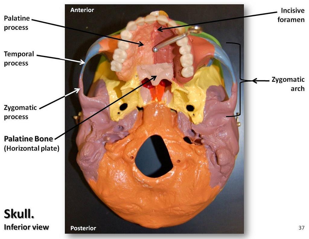
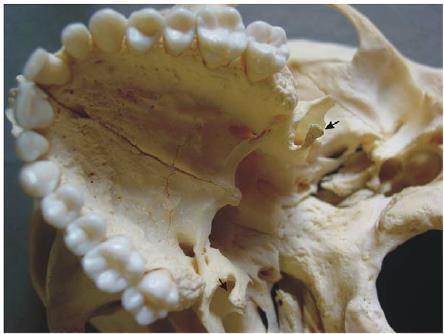
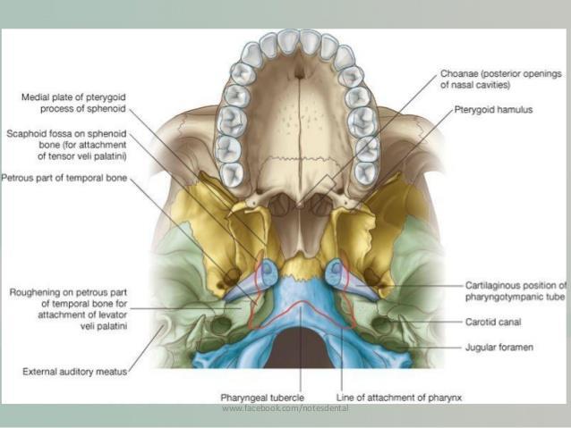
2 If the mandible is discarded, the anterior part of this aspect of the skull is seen to be formed by the hard palate. The palatal processes of the maxillae and the horizontal plates of the palatine bones can be identified. In the midline anteriorly is the incisive fossa and foramen. Posterolaterally are the greater and lesser palatine foramina.
3
4
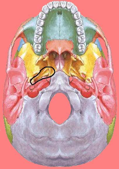
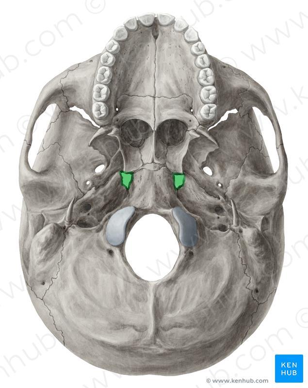
5 Above the posterior edge of the hard palate are the Choanae (posterior nasal apertures). These are separated from each other by the posterior margin of the vomer and are bounded laterally by the medial pterygoid plates of the sphenoid bone. The inferior end of the medial pterygoid plate is prolonged as a curved spike of bone, the pterygoid hamulus.
6
7
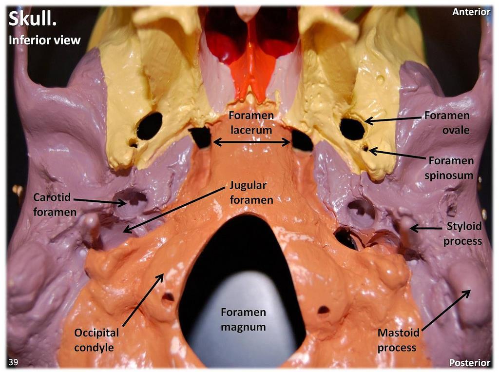
8 Posterolateral to the lateral pterygoid plate, the greater wing of the sphenoid is pierced by the large foramen ovale and the small foramen spinosum. Posterolateral to the foramen spinosum is the spine of the sphenoid.
9
10
11 Behind the spine of the sphenoid, in the interval between the greater wing of the sphenoid and the petrous part of the temporal bone, is a groove for the cartilaginous part of the auditory tube. The opening of the bony part of the tube can be identified.
12
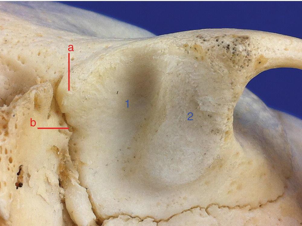
13 The mandibular fossa of the temporal bone and the articular tubercle form the upper articular surfaces for the temporomandibular joint. Separating the mandibular fossa from the tympanic plate posteriorly is the squamotympanic fissure, through the medial end of which the chorda tympani nerve exits from the tympanic cavity.

14
15 The styloid process of the temporal bone projects downward and forward from its inferior aspect. The opening of the carotid canal can be seen on the inferior surface of the petrous part of the temporal bone.
16
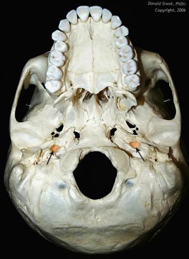
17 The medial end of the petrous part of the temporal bone is irregular and, together with the basilar part of the occipital bone and the greater wing of the sphenoid, forms the foramen lacerum. During life, the foramen lacerum is closed with fibrous tissue, and only a few small vessels pass through this foramen from the cavity of the skull to the exterior.
18
19 The tympanic plate, which forms part of the temporal bone, is C shaped on section and forms the bony part of the external auditory meatus.
20
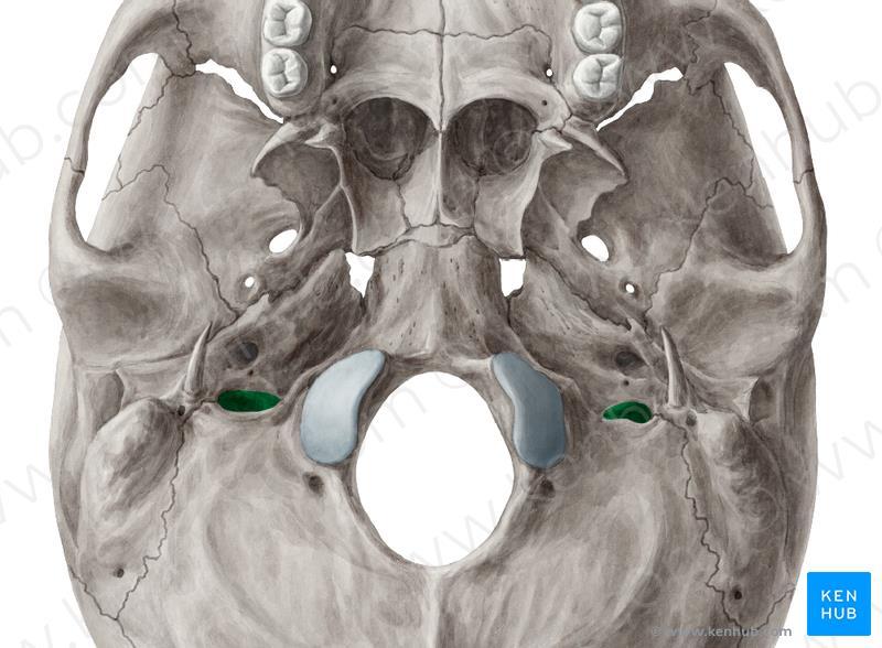
21 In the interval between the styloid and mastoid processes, the stylomastoid foramen can be seen. Medial to the styloid process, the petrous part of the temporal bone has a deep notch, which, together with a shallower notch on the occipital bone, forms the jugular foramen.
22

23
24 Behind the posterior apertures of the nose and in front of the foramen magnum are the sphenoid bone and the basilar part of the occipital bone. The pharyngeal tubercle is a small prominence on the undersurface of the basilar part of the occipital bone in the midline.

25
26
27 The occipital condyles articulate with the superior aspect of the lateral mass of the first cervical vertebra, the atlas. Superior to the occipital condyle is the hypoglossal canal for transmission of the hypoglossal nerve
28

29
30 Posterior to the foramen magnum in the midline is the external occipital protuberance. The superior nuchal lines should be identified as they curve laterally on each side.
31
32

33
Skull-2. Norma Basalis Interna Norma Basalis Externa. Dr. Heba Kalbouneh Associate Professor of Anatomy and Histology
 Skull-2 Norma Basalis Interna Norma Basalis Externa Dr. Heba Kalbouneh Associate Professor of Anatomy and Histology Norma basalis interna Base of the skull- superior view The interior of the base of the
Anatomy and Physiology. Bones, Sutures, Teeth, Processes and Foramina of the Human Skull
 Anatomy and Physiology Chapter 6 DRO Bones, Sutures, Teeth, Processes and Foramina of the Human Skull Name: Period: Bones of the Human Skull Bones of the Cranium: Frontal bone: forms the forehead and the
Bones of the skull & face
 Bones of the skull & face Cranium= brain case or helmet Copyright The McGraw-Hill Companies, Inc. Permission required for reproduction or display. The cranium is composed of eight bones : frontal Occipital
Structure Location Function
 Frontal Bone Cranium forms the forehead and roof of the orbits Occipital Bone Cranium forms posterior and inferior portions of the cranium Temporal Bone Cranium inferior to the parietal bone forms the
SKULL / CRANIUM BONES OF THE NEUROCRANIUM (7) Occipital bone (1) Sphenoid bone (1) Temporal bone (2) Frontal bone (1) Parietal bone (2)
 Occipital bone (1) Sphenoid bone (1) Temporal bone (2) Frontal bone (1) Parietal bone (2)") Important! 1. Memorizing these pages only does not guarantee the succesfull passing of the midterm test or the semifinal exam. 2. The handout has not been supervised, and I can not guarantee, that these
Important! 1. Memorizing these pages only does not guarantee the succesfull passing of the midterm test or the semifinal exam. 2. The handout has not been supervised, and I can not guarantee, that these
Introduction to Local Anesthesia and Review of Anatomy
 5-Sep Introduction and Anatomy Review 12-Sep Neurophysiology and Pain 19-Sep Physiology and Pharmacology part 1 26-Sep Physiology and Pharmacology part 2 Introduction to Local Anesthesia and Review of
Dr. Sami Zaqout, IUG Medical School
 The skull The skull is composed of several separate bones united at immobile joints called sutures. Exceptions? Frontal bone Occipital bone Vault Cranium Sphenoid bone Zygomatic bones Base Ethmoid bone
Chapter 7 Part A The Skeleton
 Chapter 7 Part A The Skeleton Why This Matters Understanding the anatomy of the skeleton enables you to anticipate problems such as pelvic dimensions that may affect labor and delivery The Skeleton The
Skull basic structures. Neurocranium
 Assoc. Prof. Květuše Lovásová, M.V.D., PhD. Skull basic structures Skull consists of two groups of bones: neurocranium (bones forming the brain box) splanchnocranium (bones forming the facial skeleton)
External Acoustic Meatus. Mastoid Process. Zygomatic Process. Temporal Bone
 Bone lab review 1. Frontal Bone 2. Supra-Orbital Foramen 3. Orbit (Orbital Cavity) 4. Superior Orbital Fissure 5. Inferior Orbital Fissure 6. Zygomatic Bone 7. Infra-Orbital Foramen 8. Maxilla 9. Mandible
Infratemporal fossa: Tikrit University college of Dentistry Dr.Ban I.S. head & neck Anatomy 2 nd y.
 Infratemporal fossa: This is a space lying beneath the base of the skull between the lateral wall of the pharynx and the ramus of the mandible. It is also referred to as the parapharyngeal or lateral pharyngeal
Biology 218 Human Anatomy. Adapted from Martini Human Anatomy 7th ed. Chapter 6 The Skeletal System: Axial Division
 Adapted from Martini Human Anatomy 7th ed. Chapter 6 The Skeletal System: Axial Division Introduction The axial skeleton: Composed of bones along the central axis of the body Divided into three regions:
Skeletal System: Skull.
 Skeletal System: Skull www.fisiokinesiterapia.biz Bones of the Skull SPLANCHNOCRANIUM Nasal (2) Maxilla (2) Lacrimal (2) Zygomatic (2) Palatine (2) Inferior concha (2) Vomer Mandible NEUROCRANIUM Frontal
APPENDICULAR SKELETON 126 AXIAL SKELETON SKELETAL SYSTEM. Cranium. Skull. Face. Skull and associated bones. Auditory ossicles. Associated bones.
 SKELETAL SYSTEM 206 AXIAL SKELETON 80 APPENDICULAR SKELETON 26 Skull Skull and associated s 29 Cranium Face Auditory ossicles 8 4 6 Associated s Hyoid Thoracic cage 25 Sternum Ribs 24 Vertebrae 24 column
View of a Skull, 1489 by Leonardo Da Vinci. Kaan Yücel M.D., Ph.D Tuesday
 View of a Skull, 1489 by Leonardo Da Vinci Kaan Yücel M.D., Ph.D. 26.11.2013 Tuesday 1.SKULL skeleton of the head cranium 22 bones excluding ossicles of the ear 1.SKULL Mandible Lower jaw bone Neurocranium
THE SKELETAL SYSTEM. Focus on the Skull
 THE SKELETAL SYSTEM Focus on the Skull Review Anatomical Terms Anterior/Posterior Dorsal/Ventral Medial/Lateral Superior/Inferior Bone Markings - Review Projections for attachment of muscles, ligaments
Skull-2. Norma Basalis Interna. Dr. Heba Kalbouneh Assistant Professor of Anatomy and Histology
 Skull-2 Norma Basalis Interna Dr. Heba Kalbouneh Assistant Professor of Anatomy and Histology Norma basalis interna Base of the skull- superior view The interior of the base of the skull is divided into
The Skull DANIL HAMMOUDI.MD
 The Skull DANIL HAMMOUDI.MD summary of bones/structures in Chapter 15 of the manual need tp be print as soon as possible http://www.mnsu.edu/emuseum/biology/humananatomy/skeletal/skul l/frontal/frontal.html
AXIAL SKELETON SKULL
 AXIAL SKELETON SKULL CRANIAL BONES (8 total flat bones w/ 2 paired) 1. Frontal forms forehead & upper portion of eyesocket (orbital) 2. Parietal paired bones; form superior & lateral walls of cranium 3.
Cranium Facial bones. Sternum Rib
 Figure 7.1 The human skeleton. Skull Thoracic cage (ribs and sternum) Cranium Facial bones Sternum Rib Bones of pectoral girdle Vertebral column Sacrum Vertebra Bones of pelvic girdle (a) Anterior view
Mohammad Hisham Al-Mohtaseb. Lina Mansour. Reyad Jabiri. 0 P a g e
 2 Mohammad Hisham Al-Mohtaseb Lina Mansour Reyad Jabiri 0 P a g e This is only correction for the last year sheet according to our record. If you already studied this sheet just read the yellow notes which
The Axial Skeleton. C h a p t e r. PowerPoint Lecture Slides prepared by Jason LaPres Lone Star College - North Harris
 C h a p t e r 7 The Axial Skeleton PowerPoint Lecture Slides prepared by Jason LaPres Lone Star College - North Harris Copyright 2009 Pearson Education, Inc., publishing as Pearson Benjamin Cummings An
Anatomy images for MSS practical exam- 2019
 Anatomy images for MSS practical exam- 2019 Ilium Ischium Pubis Acetabulaum Iliac crest Iliac tubercle ASIS (muscle and ligament attached) AIIS (muscle attached) PSIS PIIS Ischial spine Ischial tuberosity
University of Palestine. Midterm Exam 2013/2014 Total Grade:
 Course No: DNTS2208 Course Title: Head and Neck Anatomy Date: 09/11/2013 No. of Questions: (50) Time: 1hour Using Calculator (No) University of Palestine Midterm Exam 2013/2014 Total Grade: Instructor
YOU MUST BRING YOUR OWN GLOVES FOR THIS ACTIVITY.
 ACTIVITY 3: AXIAL SKELETON AND LONG BONE DISSECTION Objectives: 1) How to get ready: Read Chapter 7, McKinley et al., Human Anatomy, 5e. All text references are for this textbook. Learning the meanings
Anatomic Relations Summary. Done by: Sohayyla Yasin Dababseh
 Anatomic Relations Summary Done by: Sohayyla Yasin Dababseh Anatomic Relations Lecture 1 Part-1 - The medial wall of the nose is the septum. - The vestibule lies directly inside the nostrils (Nares). -
Chapter 7. Skeletal System
 Chapter 7 Skeletal System 1 Skull A. The skull is made up of 22 bones: 8 cranial bones, 13 facial bones, and the mandible. B. The Cranium encloses and protects the brain, provides attachments for muscles,
ACTIVITY 3: AXIAL SKELETON AND LONG BONE DISSECTION COW BONE DISSECTION
 ACTIVITY 3: AXIAL SKELETON AND LONG BONE DISSECTION Objectives: 1) How to get ready: Read Chapter 7, McKinley et al., Human Anatomy, 4e. All text references are for this textbook. Learning the meanings
Skeletal System -Axial System. Chapter 7 Part A
 Skeletal System -Axial System Chapter 7 Part A Skeleton Learn: Names of the s. Identify specific landmarks that allow: Bones to fit into each other, Organs to fit into the cavities, Muscles to attach,
Bones Ethmoid bone Inferior nasal concha Lacrimal bone Maxilla Nasal bone Palatine bone Vomer Zygomatic bone Mandible
 splanchnocranium - Consists of part of skull that is derived from branchial arches - The facial bones are the bones of the anterior and lower human skull Bones Ethmoid bone Inferior nasal concha Lacrimal
Parotid Gland, Temporomandibular Joint and Infratemporal Fossa
 M1 - Anatomy Parotid Gland, Temporomandibular Joint and Infratemporal Fossa Jeff Dupree Sanger 9-057 jldupree@vcu.edu Parotid gland: wraps around the mandible positioned between the mandible and the sphenoid
Bone Flashcards for 10a
 Bone Flashcards for 0a CLAVICLE (collar bone). Sternal extremity (end) flat end. Acromial extremity (end) rounded end. SCAPULA (shoulder blade). Right or left scapula?. Superior border (superior margin).
Gross Anatomy of the. TEMPORAL BONE, EXTERNAL EAR, and MIDDLE EAR
 Gross Anatomy of the TEMPORAL BONE, EXTERNAL EAR, and MIDDLE EAR M1 Gross and Developmental Anatomy 9:00 AM, December 11, 2008 Dr. Milton M. Sholley Professor of Anatomy and Neurobiology Assignment: Head
Trigeminal Nerve Worksheets, Distributions Page 1
 Trigeminal Nerve Worksheet #1 Distribution by Nerve Dr. Darren Hoffmann Dental Gross Anatomy, Spring 2013 We have drawn out each of the branches of CN V in lecture and you have an idea now for their basic
University of Palestine. Midterm Exam 2013/2014 Total Grade:
 [ Course No: DNTS2208 Course Title: Head and Neck Anatomy Date: 17/11/1024 No. of Questions: (52) Time: 2hours Using Calculator (No) University of Palestine Midterm Exam 2013/2014 Total Grade: Instructor
Axial skeleton bones and markings
 Axial skeleton bones and markings Skull Cranial bones Frontal x 1 Supraorbital foramen Occipital x 1 Foramen magnum Occipital condyles Superior nuchal line Inferior nuchal line Anterior cranial fossa External
Gross Anatomy of the. TEMPORAL BONE, EXTERNAL EAR, and MIDDLE EAR. Assignment: Head to Toe Temporomandibular Joint (TMJ)
") Gross Anatomy the TEMPORAL BONE, EXTERNAL EAR, and MIDDLE EAR M1 Gross and Developmental Anatomy 9:00 AM, December 11, 2008 Dr. Milton M. Sholley Pressor Anatomy and Neurobiology Assignment: Head to Toe
Gross Anatomy the TEMPORAL BONE, EXTERNAL EAR, and MIDDLE EAR M1 Gross and Developmental Anatomy 9:00 AM, December 11, 2008 Dr. Milton M. Sholley Pressor Anatomy and Neurobiology Assignment: Head to Toe
SKULL AS A WHOLE + ANTERIOR CRANIAL FOSSA
 SKULL AS A WHOLE + ANTERIOR CRANIAL FOSSA LEARNING OBJECTIVES At the end of this lecture, the student should be able to know: Parts of skeleton (axial and appendicular) Parts of skull Sutures of skull
PTERYGOPALATINE FOSSA
 PTERYGOPALATINE FOSSA Outline Anatomical Structure and Boundaries Foramina and Communications with other spaces and cavities Contents Pterygopalatine Ganglion Especial emphasis on certain arteries and
Omran Saeed. Luma Taweel. Mohammad Almohtaseb. 1 P a g e
 2 Omran Saeed Luma Taweel Mohammad Almohtaseb 1 P a g e I didn t include all the photos in this sheet in order to keep it as small as possible so if you need more clarification please refer to slides In
Crafton Hills College Human Anatomy & Physiology Axial Skeleton
 A. Major Divisions Crafton Hills College Human Anatomy & Physiology Axial keleton 1. Axial: Part of skeleton lies along long axis of body 2. Appendicular: Bones & features of the appendages B. AXIAL KELETON
The Skull and Temporomandibular joint II Prof. Abdulameer Al-Nuaimi. E. mail:
 The Skull and Temporomandibular joint II Prof. Abdulameer Al-Nuaimi E-mail: a.al-nuaimi@sheffield.ac.uk E. mail: abdulameerh@yahoo.com Temporal fossa The temporal fossa is a depression on the temporal
Temporal fossa Infratemporal fossa Pterygopalatine fossa Terminal branches of external carotid artery Pterygoid venous plexus
 Outline of content Temporal fossa Infratemporal fossa Pterygopalatine fossa Terminal branches of external carotid artery Pterygoid venous plexus Boundary Content Communication Mandibular division of trigeminal
Temporal region. temporal & infratemporal fossae. Zhou Hong Ying Dept. of Anatomy
 Temporal region temporal & infratemporal fossae Zhou Hong Ying Dept. of Anatomy Temporal region is divided by zygomatic arch into temporal & infratemporal fossae. Temporal Fossa Infratemporal fossa Temporal
The Pharynx. Dr. Nabil Khouri MD. MSc, Ph.D
 The Pharynx Dr. Nabil Khouri MD. MSc, Ph.D Introduction The pharynx is the Musculo-fascial halfcylinder that links the oral and nasal cavities in the head to the larynx and esophagus in the neck Common
Chapter 7: Skeletal System: Gross Anatomy
 Chapter 7: Skeletal System: Gross Anatomy I. General Considerations A. How many bones in an average adult skeleton? B. Anatomic features of bones are based on II. Axial Skeleton A. Skull 1. Functionally
Important Parts of Bones
 Important Parts of Bones For 2015 Know: Humerus (posterior) Clavical Femur (Anterior) Foot Hand Mandible Os Coxa Scapula Skull (Anterior, Inferior, Lateral) Sternum Humerus (posterior) A. olecranon fossa
Human Anatomy & Physiology I Dr. Sullivan Unit VIIIa The Axial Skeleton Chapter 8 (Sections )
") Human Anatomy & Physiology I Dr. Sullivan Unit VIIIa The Axial Skeleton Chapter 8 (Sections 8.1-8.3) I. Divisions of the skeletal system a) An adult human skeleton has 206 named bones b) Most are paired
Human Anatomy & Physiology I Dr. Sullivan Unit VIIIa The Axial Skeleton Chapter 8 (Sections 8.1-8.3) I. Divisions of the skeletal system a) An adult human skeleton has 206 named bones b) Most are paired
Dr.Ban I.S. head & neck anatomy 2 nd y جامعة تكريت كلية طب االسنان مادة التشريح املرحلة الثانية أ.م.د. بان امساعيل صديق 6102/6102
 جامعة تكريت كلية طب االسنان مادة التشريح املرحلة الثانية أ.م.د. بان امساعيل صديق 6102/6102 Pterygopalatine fossa: The pterygopalatine fossa is a cone-shaped depression, It is located between the maxilla,
o Diaphysis o Area where red marrow is found o Area where yellow marrow is found o Epiphyseal plate AXIAL SKELETON Skull
 64 Anatomy & Physiology Coloring Workbook 7. Figure 5-2A is a midlevel, cross-sectional view of the diaphysis of the femur. Label the membrane that lines the cavity and the membrane that covers the outside
3. The Jaw and Related Structures
 Overview and objectives of this dissection 3. The Jaw and Related Structures The goal of this dissection is to observe the muscles of jaw raising. You will also have the opportunity to observe several
Chapter 8A. The Skeletal System: The Axial Skeleton. The Skeletal System: The Axial Skeleton. Types of Bones. Types of Bones
 Chapter 8A The Skeletal System: The Axial Skeleton The Skeletal System: The Axial Skeleton 206 named bones Axial Skeleton 80 bones lie along longitudinal axis skull, hyoid, vertebrae, ribs, sternum, ear
Dr.Ban I.S. head & neck anatomy 2 nd y. جامعة تكريت كلية طب االسنان املرحلة الثانية
 جامعة تكريت كلية طب االسنان التشريح مادة املرحلة الثانية أ.م.د. بان امساعيل صديق 6102-6102 1 The Palate The palate forms the roof of the mouth and the floor of the nasal cavity. It is divided into two
Riverside Community College Anatomy & Physiology 2B SPRING 2012 EXAM #1-ABC (Nervous System)
") Riverside Community College Anatomy & Physiology 2B SPRING 2012 EXAM #1-ABC (Nervous System) Name: 1) This vertebra is an example of a(n). 1) A) thoracic B) axis C) atlas D) lumbar E) sacral 1 2) W hich
Riverside Community College Anatomy & Physiology 2B SPRING 2012 EXAM #1-ABC (Nervous System) Name: 1) This vertebra is an example of a(n). 1) A) thoracic B) axis C) atlas D) lumbar E) sacral 1 2) W hich
The Ear The ear consists of : 1-THE EXTERNAL EAR 2-THE MIDDLE EAR, OR TYMPANIC CAVITY 3-THE INTERNAL EAR, OR LABYRINTH 1-THE EXTERNAL EAR.
 The Ear The ear consists of : 1-THE EXTERNAL EAR 2-THE MIDDLE EAR, OR TYMPANIC CAVITY 3-THE INTERNAL EAR, OR LABYRINTH 1-THE EXTERNAL EAR Made of A-AURICLE B-EXTERNAL AUDITORY MEATUS A-AURICLE It consists
Dr. Sami Zaqout Faculty of Medicine IUG
 Auricle External Ear External auditory meatus The Ear Middle Ear (Tympanic Cavity) Auditory ossicles Internal Ear (Labyrinth) Bony labyrinth Membranous labyrinth External Ear Auricle External auditory
Human Anatomy and Physiology - Problem Drill 07: The Skeletal System Axial Skeleton
 Human Anatomy and Physiology - Problem Drill 07: The Skeletal System Axial Skeleton Question No. 1 of 10 Which of the following statements about the axial skeleton is correct? Question #01 A. The axial
Parotid Gland. Parotid Gland. Largest of 3 paired salivary glands (submandibular; sublingual) Ramus of Mandible. Medial pterygoid.
 Ramus of Mandible. Medial pterygoid.") Parotid region Parotid Gland Largest of 3 paired salivary glands (submandibular; sublingual) Ramus of Mandible Medial pterygoid Cross section of mandible Masseter D S SCM Parotid Gland Mastoid Process
Parotid region Parotid Gland Largest of 3 paired salivary glands (submandibular; sublingual) Ramus of Mandible Medial pterygoid Cross section of mandible Masseter D S SCM Parotid Gland Mastoid Process
Tracing the Cranial Nerves Osteologically
 CN I II III IV V 1 Supra-orbital ethmoidal nn. Ext. nasal V 2 Tracing the Cranial Nerves Osteologically Nucleus of Origin Olfactory tracts of frontal lobe of cerebrum Optic tracts from optic chiasma and
10/11/2009. What is wrong with this picture? BONE PICTURE EXAM MARS / IN WHAT PART OF THE BONE DO YOU FIND THIS TISSUE SLIDE?
 What is wrong with this picture? BONE PICTURE EXAM MARS 2008 1/ IN WHAT PART OF THE BONE DO YOU FIND THIS TISSUE SLIDE? 1 2/ IDENTIFY THIS TISSUE? 3/what is wrong in this picture? 2 4/ Identify these cells
Chapter 7: Head & Neck
 Chapter 7: Head & Neck Osteology I. Overview A. Skull The cranium is composed of irregularly shaped bones that are fused together at unique joints called sutures The skull provides durable protection from
Maxilla, ORBIT and infratemporal fossa. Neophytos C Demetriades MD, DDS, MSc Associate professor European University of Cyprus School of Medicine
 Maxilla, ORBIT and infratemporal fossa Neophytos C Demetriades MD, DDS, MSc Associate professor European University of Cyprus School of Medicine MAXILLA Superior, middle, and inferior meatus Frontal sinus
Spring Written By: J. E. Sutton. Contents: I. Overview of the Skeleton: II. Appendicular Skeleton III. Axial Skeleton IV.
 Spring 2012 Written By: J. E. Sutton Contents: I. Overview of the Skeleton: II. Appendicular Skeleton III. Axial Skeleton IV. Articulations Overview of the Skeleton: I. Orientation to Human Skeleton: a.
Biology 2401 The Skeletal System
 Biology 2401 The Skeletal System Purpose: The lab will describe the microscopic and gross anatomy of bone, identify bones of the body, and identify important bone markings. I. Overview of the Skeleton
Prevertebral Region, Pharynx and Soft Palate
 Unit 20: Prevertebral Region, Pharynx and Soft Palate Dissection Instructions: Step1 Step 2 Step 1: Insert your fingers posterior to the sternocleidomastoid muscle, vagus nerve, internal jugular vein,
VERTEBRAL COLUMN REFERENCES: TEST: CHAPTER 7 / LAB MANUAL 1: SK1-12
 VERTEBRAL COLUMN REFERENCES: TEST: CHAPTER 7 / LAB MANUAL 1: SK1-12 1. A vertebra consists of two main masses of bone: an anterior vertebral _?_ and a posterior vertebral or neural _?_. 2. In the articulated
Anatomy and Physiology II. Review Spine and Neck
 Anatomy and Physiology II Review Spine and Neck Spine regions How many cervical vertibrae are there? 7 The curvature is the cervical region posterior? Concave posterior How many thoracic? And curvature?
MAXILLA, ORBIT & PTERYGOPALATINE FOSSA. Neophytos C Demetriades MD, DDS, MSc Associate professor European University of Cyprus School of Medicine
 MAXILLA, ORBIT & PTERYGOPALATINE FOSSA Neophytos C Demetriades MD, DDS, MSc Associate professor European University of Cyprus School of Medicine Maxilla MAXILLA Superior, middle, and inferior meatus Frontal
Lec [8]: Mandibular nerve:
![Lec [8]: Mandibular nerve:](/thumbs/94/121295776.jpg "Lec [8]: Mandibular nerve:") Lec [8]: Mandibular nerve: The mandibular branch from the trigeminal ganglion lies in the middle cranial fossa lateral to the cavernous sinus. With the motor root of the trigeminal nerve [motor roots lies
Lec [8]: Mandibular nerve: The mandibular branch from the trigeminal ganglion lies in the middle cranial fossa lateral to the cavernous sinus. With the motor root of the trigeminal nerve [motor roots lies
locomotice system Plastinated specimensⅠ: Silicone specimens Regional specimens and organs
 locomotice system Plastinated specimensⅠ: Silicone specimens Regional specimens and organs Art-No. Name Description The locomotor system SL001 Two hundred pieces of plastinated bones (without six The bones
Major Anatomic Components of the Orbit
 Major Anatomic Components of the Orbit 1. Osseous Framework 2. Globe 3. Optic nerve and sheath 4. Extraocular muscles Bony Orbit Seven Bones Frontal bone Zygomatic bone Maxillary bone Ethmoid bone Sphenoid
TEST YOURSELF- Chapter 7
 TEST YOURSELF- Chapter 7 Cranial Bones 1. Give the name of the bone for each of the following markings. Some of the markings are found on more than one bone. List all that apply. Cranium a. Frontal squama:
Dr.Ban I.S. head & neck anatomy 2 nd y. جامعة تكريت كلية طب االسنان املرحلة الثانية أ.م.د. بان امساعيل صديق 6102/6102
 جامعة تكريت كلية طب االسنان التشريح مادة املرحلة الثانية أ.م.د. بان امساعيل صديق 6102/6102 Parotid region The part of the face in front of the ear and below the zygomatic arch is the parotid region. The
Cranial Nerve VII - Facial Nerve. The facial nerve has 3 main components with distinct functions
 Cranial Nerve VII - Facial Nerve The facial nerve has 3 main components with distinct functions Somatic motor efferent Supplies the muscles of facial expression; posterior belly of digastric muscle; stylohyoid,
the Skeletal System provided by Academic Web Services Grand Canyon University
 Anatomy Resource Center Study Guides the Skeletal System HEAD & NECK REGIONAL VIEW SKULL BONES CRANIUM FACE SKULL LANDMARKS ANTERIOR SIDE SUPERIOR/INFERIOR VERTEBRAL COLUMN VERTEBRAL REGIONS CERVICAL C1
Bones of the Skull Lateral View
 Bones of the Skull Lateral View Frontal Bone Parietal Bone Occipital Bone Temporal Bone Sphenoid Bone Pterion Sutures of the Skull Lateral View Coronal Suture Lambdoid Suture Squamous Suture Sutures of
Subdivided into Vestibule & Oral cavity proper
 Extends from the lips to the oropharyngeal isthmus The oropharyngeal isthmus: Is the junction of mouth and pharynx. Is bounded: Above by the soft palate and the palatoglossal folds Below by the dorsum
Perpendicular Plate Zygomatic Bone. Mental Foramen Mandible
 Glabella Frontal Middle Nasal Concha Nasal Lacrimal Perpendicular Plate Zygomatic Inferior Nasal Concha Maxilla Mental Mandible Skull (anterior view) Squamosal Suture Coronal Suture Frontal Parietal Nasal
The Ear. Dr. Heba Kalbouneh Assistant Professor of Anatomy and Histology
 The Ear Dr. Heba Kalbouneh Assistant Professor of Anatomy and Histology The Ear The ear consists of the external ear; the middle ear (tympanic cavity); and the internal ear (labyrinth), which contains
ANATOMY & PHYSIOLOGY I Laboratory Version B Name Section. REVIEW SHEET Exercise 10 Axial Skeleton
 ANATOMY & PHYSIOLOGY I Laboratory Version B Name Section REVIEW SHEET Exercise 10 Axial Skeleton 1 POINT EACH. THE SKULL MULTIPLE CHOICE 1. The major components of the axial skeleton include the 7. The
Inferior view of the skull showing foramina (Atlas of Human Anatomy, 5th edition, Plate 12)
") Section 1 Head and Neck Skull, Basal View Incisive foramen Choanae Foramen ovale Foramen lacerum Foramen spinosum Carotid canal Jugular fossa Mastoid process Inferior view of the skull showing foramina
Section 1 Head and Neck Skull, Basal View Incisive foramen Choanae Foramen ovale Foramen lacerum Foramen spinosum Carotid canal Jugular fossa Mastoid process Inferior view of the skull showing foramina
SCHOOL OF ANATOMICAL SCIENCES Mock Run Questions. 4 May 2012
 SCHOOL OF ANATOMICAL SCIENCES Mock Run Questions 4 May 2012 1. With regard to the muscles of the neck: a. the platysma muscle is supplied by the accessory nerve. b. the stylohyoid muscle is supplied by
Anatomy Skull and Spinal Cord
 1 Anatomy Skull and Spinal Cord 1. Skull The skull is anterior to the spinal column and is the bony structure that encases the brain. Its purpose is to protect the brain and allow attachments for the facial
BONE CHALLENGE DANIL HAMMOUDI.MD
 BONE CHALLENGE DANIL HAMMOUDI.MD Bone Basic functions? A. support B. protection C. movement assistance in D. RBC formation-hemopoiesis E. mineral homeostasis +importance of calcium F. energy supply -yellow
Head & Neck Radiology (I) Waseem Jerjes
 Waseem Jerjes") Head & Neck Radiology (I) Waseem Jerjes Lamboid Suture Frontal Sinus Left Orbit (roof) Left Supraorbital Margin Left Frontozygomatic Suture Left Superior Orbital Fissure Rt Petrous Ridge of Temporal Bone
Head & Neck Radiology (I) Waseem Jerjes Lamboid Suture Frontal Sinus Left Orbit (roof) Left Supraorbital Margin Left Frontozygomatic Suture Left Superior Orbital Fissure Rt Petrous Ridge of Temporal Bone
in compact bone, large vertical canals carrying blood vessels and nerves. in compact bone, large horizontal canals carrying blood vessels and nerves.
 Carl Christensen, PhD Skeletal System (Bones`) Bio. 2304 Human Anatomy 1. Identify a term for each of the following: shaft of a long bone ends of a long bone ossified remnant of the "growth plate" connective
Upper arch. 1Prosthodontics. Dr.Bassam Ali Al-Turaihi. Basic anatomy & & landmark of denture & mouth
 1Prosthodontics Lecture 2 Dr.Bassam Ali Al-Turaihi Basic anatomy & & landmark of denture & mouth Upper arch Palatine process of maxilla: it form the anterior three quarter of the hard palate. Horizontal
Skeletal System - Prelab 1
 Skeletal System - Prelab 1 1. Which bones contain the paranasal sinuses? What function do the sinuses serve? 2. What two areas are separated from each other by the hard palate? Name the two bones that
The sebaceous glands (glands of Zeis) open directly into the eyelash follicles, ciliary glands (glands of Moll) are modified sweat glands that open
 open directly into the eyelash follicles, ciliary glands (glands of Moll) are modified sweat glands that open") The Orbital Region The orbits are a pair of bony cavities that contain the eyeballs; their associated muscles, nerves, vessels, and fat; and most of the lacrimal apparatus upper eyelid is larger and more
The Orbital Region The orbits are a pair of bony cavities that contain the eyeballs; their associated muscles, nerves, vessels, and fat; and most of the lacrimal apparatus upper eyelid is larger and more
Overview of the Skeleton: Bone Markings
 Name Overview of the Skeleton: Bone Markings Match the terms in column B with the appropriate description in column A. Column A 1. sharp, slender process* 2. small rounded projection* 3. narrow ridge of
Cranial Cavity REFERENCES: OBJECTIVES OSTEOLOGY. Stephen A. Gudas, PT, PhD
 Stephen A. Gudas, PT, PhD Cranial Cavity REFERENCES: Moore and Agur, Essential Clinical Anatomy (ECA), 3rd ed., pp. 496 498; 500 507; 512 514 Grant s Atlas 12 th ed., Figs 7.6; 7.19 7.30. Grant s Dissector
Anatomy Lab: The skeletal system. Part I: Vertebrae and Thoracic cage
 ANA Lab: Bone 1 Anatomy Lab: The skeletal system Part I: Vertebrae and Thoracic cage Spine (Vertebrae) Body Vertebral arch Vertebral canal Pedicle Lamina Spinous process Transverse process Sup. articular
Anatomy Made Easy MSS
 Anatomy Made Easy MSS part #1 هذا الملف يشمل تفريغ المحاضرة الثانية لعون بدءا من الصفحة 11 وحتى األخير Done By :MohamedA. Diabat Edited by Awn Academic team The Axial Skeleton The axial skeleton consist
Skeletal system overview. Classification of Bones
 Skeletal system overview BIOL241 Lab #9 Classification of Bones Bone are identified by: shape internal tissues bone markings 1 1. Flat bones 2. Long bones 3. Short bones 4. Irregular bones 5. Sutural bones
Copy Right- Hongqi ZHANG-Department of Anatomy-Fudan University. Systemic Anatomy
 Systemic Anatomy Locomotor system - Part 2 Joint of trunk bones Cranial bones and their joints Dr. Hongqi Zhang ( 张红旗 ) Dept.of Anatomy, Histoembryology, Fudan.Univ. Email: zhanghq58@126.com Telephone:
Labs 9 and 10. Classification of Bones. Bone Shapes 1/05/13. Skeletal system overview. Bone are identified by:
 Labs 9 and 10 Skeletal system overview Classification of Bones Bone are identified by: shape internal tissues bone markings 1. Flat bones 2. Long bones 3. Short bones 4. Irregular bones 5. Sutural bones
Tikrit University collage of dentistry Dr.Ban I.S. head & neck anatomy 2 nd y. Lec [5] / Temporal fossa :
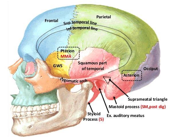
![Tikrit University collage of dentistry Dr.Ban I.S. head & neck anatomy 2 nd y. Lec [5] / Temporal fossa :](/thumbs/88/115294566.jpg "Tikrit University collage of dentistry Dr.Ban I.S. head & neck anatomy 2 nd y. Lec [5] / Temporal fossa :") Lec [5] / Temporal fossa : Borders of the Temporal Fossa: Superior: Superior temporal line. Inferior: gap between zygomatic arch and infratemporal crest of sphenoid bone. Anterior: Frontal process of the
Lec [5] / Temporal fossa : Borders of the Temporal Fossa: Superior: Superior temporal line. Inferior: gap between zygomatic arch and infratemporal crest of sphenoid bone. Anterior: Frontal process of the
Head BLUE BOX: Face and Scalp. BLUE BOX: Cranium. BLUE BOX: Cranial Cavity and Meninges. 820
 CHAPTER 7 Head OVERVIEW / 822 CRANIUM / 822 Facial Aspect of Cranium / 822 Lateral Aspect of Cranium / 827 TABLE 7.1. Craniometric Points of Cranium / 828 Occipital Aspect of Cranium / 828 Superior Aspect