Skeletal System: Skull.
|
|
|
- Josephine Clarke
- 5 years ago
- Views:
Transcription
1 Skeletal System: Skull
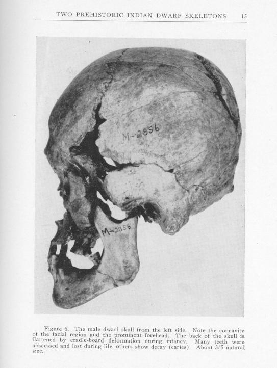
2 Bones of the Skull SPLANCHNOCRANIUM Nasal (2) Maxilla (2) Lacrimal (2) Zygomatic (2) Palatine (2) Inferior concha (2) Vomer Mandible NEUROCRANIUM Frontal Parietal (2) Temporal (2) Occipital Sphenoid Ethmoid EAR OSSICLES Stapes (2) Malleus (2) Incus (2) HYOID

3 2 Functional Areas Splanchnocranium face Neurocranium vault and cranial base

4 Development
5 General Developmental Organization Chondocranium (cranial base) cartilage models (enchochondral ossification) Neurocranial vault from mesenchyme (intramembranous) Viscerocranium from mesenchyme (pharyngeal arches) (intramembranous)


6 Neonatal Skull (neurocranial syndesmoses) Frontal Metopic suture Ant. fontanelle Post. fontanelle
7 795 WUSTL Med School 376 Korean War Dead
8 Functional Matrix Hypothesis Growth of skull bones depends almost entirely on surrounding soft tissues. Bone growth is secondary and compensatory to soft tissue growth
9 Achondroplasia
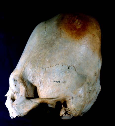
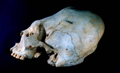
10 Artificial cranial modification


11 Craniosynostosis Crouzon s Syndrome (coronal synostosis) Apert s Syndrome (extensive synostoses)

 W.A.")
12 Trigonocephaly (premature metopic suture closure) W.A. Mozart s cranium
13 Anatomy of the Skull Individual bones Structures within bones Regions Intracranial fossae Temporal fossa, Infratemporal fossa, Pterygopalatine fossa Other structures and landmarks
 Posterior Cranial Fossa")
14 Intracranial Fossae ACF MCF PCF Anterior Cranial Fossa (frontal lobe) Middle Cranial Fossa (temporal lobe) Posterior Cranial Fossa (cerebellum) 9
15 FYI The following slides show pictures and drawings of the skull and individual bones with important landmarks identified. These aren t landmarks that you need to memorize now! You will in the future!

16 Frontal Temporal Sphenoid Lacrimal Zygomatic Superciliary Arch Supraorbital Notch Superior Orbital Fissure Frontal Process (Maxilla) Inferior Orbital Fissure Nasal Zygomatic Arch Maxilla Inferior Nasal Concha Mandible Anterior Nasal Spine Infraorbital Foramen Canine Fossa Mental Foramen Mental Eminence

17 Maxilla Incisive Fossa Greater Palatine Foramen Foramen Lacerum Palatine Foramen Ovale Zygomatic Foramen Spinosum Vomer Mandibular Fossa Sphenoid Temporal Styloid Process Mastoid Process External Auditory Meatus Carotid Canal Hypoglossal Canal Stylomastoid Foramen Occipital Foramen Magnum Jugular Foramen

18 Cribriform Plate Frontal Crista Galli Lesser Wing of Sphenoid Foramen Cecum Optic Foramen Optic Fissure Foramen Lacerum Greater Wing of Sphenoid Sphenoid Sella Turcica Carotid Canal Foramen Ovale Foramen Spinosum Temporal Jugular Foramen Hypoglossal Canal Occipital Foramen Magnum
19 Parietal Frontal Nasal Zygomatic Occipital Maxilla Temporal Sphenoid Mandible

20 Squamosal Suture Coronal Suture Zygomatic Process of the Temporal Bone Superciliary Arch External Auditory Meatus Temporal Process of the Zygomatic Bone Lamdoidal Suture Anterior Nasal Spine External Occipital Protuberance Mental Foramen Mastoid Process Mental Eminence Styloid Process Mandibular Condyle Gonial Angle Mandibular Notch Coronoid Process

21 Frontal process Zygomatic N L e p sphe Infraorbital margin Zygomaticofacial foramen Anterior nasal spine Infraorbital foramen Alveolar process Maxilla, Nasal, Lacrimal Zygomatic

22 Maxilla Palatine Incisive foramen Palatine process Horizontal plate Grtr. palatine for. Posterior nasal spine Lssr. palatine foramina


23 Lssr. wing Cribriform plate Sphenoid Anterior clinoid process Carotid grv. Grtr. wing Grv. optic chiasma Hypophyseal (pituitary) fossa Dorsum sellae Sella turcica Posterior clinoid process
24 Optic canal Superior orbital fissure ---lotsa, V 1 Foramen rotundum ---V 2 Foramen ovale ---V 3 Foramen spinosum ---Middle meningeal a. CN V, Trigeminal Nerve

25 Sphenoid Hamulus Pterygoid fossa Lateral pterygoid plate Vomer Medial pterygoid plate Scaphoid fossa Pterygoid canal Spine

26 Sphenoid Bone, Superior View (with parts visible from inside skull with skullcap removed) Lesser wing Anterior clinoid processes Pituitary fossa Optic canals Greater wing Posterior clinoid processes Dorsum sella Clivus Sella turcica
 Pterygoid canal Medial & lateral")
27 Sphenoid Bone, Anterior View Greater wings Superior orbital fissure Lesser wings Sphenoidal sinuses Pterygoid fossa Pterygoid processes Sphenoidal crest Sphenoidal rostrum (most A-I part of crest) Pterygoid canal Medial & lateral ptyerygoid plates

28 Sphenoid bone, Inferior view (with parts visible from outside of skull) Pterygoid fossa Pterygoid process Medial pterygoid plate Lateral pterygoid plate Infratemporal crests Pterygoid hamulus Foramen ovale Foramen spinosum

29 Infratemporal fossa Fr Pa Sp Te Temporal fossa Zy Mx Pterygomaxillary fissure Pterygopalatine fossa, to Sphenopalatine foramen

30 SUPERIOR Temporal Bone, Lateral view POSTERIOR Squamous portion ANTERIOR Supramastoid crest Suprameatal crest External auditory meatus INFERIOR Mastoid process Tympanic portion Postglenoid process Glenoid fossa Articular eminence Zygomatic process
 Zygomatic process Internal auditory meatus")
31 Temporal Bone, Medial view SUPERIOR POSTERIOR ANTERIOR Squamous portion Petrous portion Sigmoid sulcus (gray curve) Zygomatic process Internal auditory meatus INFERIOR

32 Temporal Bone, Inferior view Zygomatic process Articular eminence External auditory meatus Glenoid fossa Postglenoid process Styloid process Occipital groove Mastoid notch Mastoid process

33 Malleus Incus Stapes VII VIII IAM EAM Tympanic membrane

34 Mandible Coronoid process Mandibular foramen Lingula Mand. notch Head Neck Condylar process Ramus Mental protuberance Alveolar process Body Oblique line Angle Mental tubercle Mental foramen

35 Mylohyoid line Mylohyoid groove Rt. submandibular fossa Lt. digastric fossa Rt. mental spine
36 Joints in the Skull - Sutures


37 Fontanelles Bregmatic fontanelle Parietal Frontal Lambdoid fontanelle Anterolateral fontanelle Posterolateral fontanelle Sphenoid Occipital Temporal

38 Sinuses Void chambers Linked to nasal cavity Frontal Maxillary Ethmoid Sphenoid
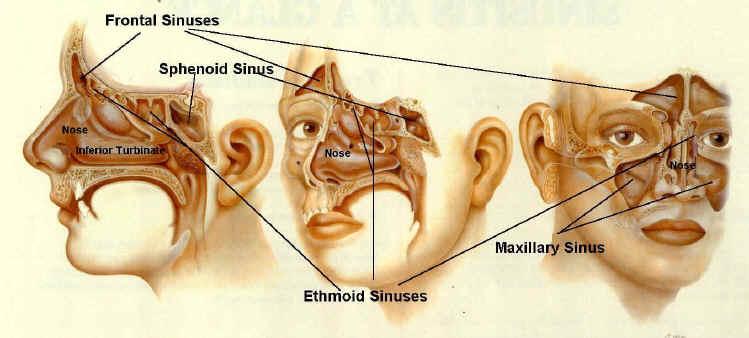
39 Sinuses Cross-section
Skull basic structures. Neurocranium
 Assoc. Prof. Květuše Lovásová, M.V.D., PhD. Skull basic structures Skull consists of two groups of bones: neurocranium (bones forming the brain box) splanchnocranium (bones forming the facial skeleton)
SKULL / CRANIUM BONES OF THE NEUROCRANIUM (7) Occipital bone (1) Sphenoid bone (1) Temporal bone (2) Frontal bone (1) Parietal bone (2)
 Occipital bone (1) Sphenoid bone (1) Temporal bone (2) Frontal bone (1) Parietal bone (2)") Important! 1. Memorizing these pages only does not guarantee the succesfull passing of the midterm test or the semifinal exam. 2. The handout has not been supervised, and I can not guarantee, that these
Important! 1. Memorizing these pages only does not guarantee the succesfull passing of the midterm test or the semifinal exam. 2. The handout has not been supervised, and I can not guarantee, that these
Skull-2. Norma Basalis Interna Norma Basalis Externa. Dr. Heba Kalbouneh Associate Professor of Anatomy and Histology
 Skull-2 Norma Basalis Interna Norma Basalis Externa Dr. Heba Kalbouneh Associate Professor of Anatomy and Histology Norma basalis interna Base of the skull- superior view The interior of the base of the
Bones of the skull & face
 Bones of the skull & face Cranium= brain case or helmet Copyright The McGraw-Hill Companies, Inc. Permission required for reproduction or display. The cranium is composed of eight bones : frontal Occipital
Introduction to Local Anesthesia and Review of Anatomy
 5-Sep Introduction and Anatomy Review 12-Sep Neurophysiology and Pain 19-Sep Physiology and Pharmacology part 1 26-Sep Physiology and Pharmacology part 2 Introduction to Local Anesthesia and Review of
APPENDICULAR SKELETON 126 AXIAL SKELETON SKELETAL SYSTEM. Cranium. Skull. Face. Skull and associated bones. Auditory ossicles. Associated bones.
 SKELETAL SYSTEM 206 AXIAL SKELETON 80 APPENDICULAR SKELETON 26 Skull Skull and associated s 29 Cranium Face Auditory ossicles 8 4 6 Associated s Hyoid Thoracic cage 25 Sternum Ribs 24 Vertebrae 24 column
Biology 218 Human Anatomy. Adapted from Martini Human Anatomy 7th ed. Chapter 6 The Skeletal System: Axial Division
 Adapted from Martini Human Anatomy 7th ed. Chapter 6 The Skeletal System: Axial Division Introduction The axial skeleton: Composed of bones along the central axis of the body Divided into three regions:
Chapter 7 Part A The Skeleton
 Chapter 7 Part A The Skeleton Why This Matters Understanding the anatomy of the skeleton enables you to anticipate problems such as pelvic dimensions that may affect labor and delivery The Skeleton The
Dr. Sami Zaqout, IUG Medical School
 The skull The skull is composed of several separate bones united at immobile joints called sutures. Exceptions? Frontal bone Occipital bone Vault Cranium Sphenoid bone Zygomatic bones Base Ethmoid bone
External Acoustic Meatus. Mastoid Process. Zygomatic Process. Temporal Bone
 Bone lab review 1. Frontal Bone 2. Supra-Orbital Foramen 3. Orbit (Orbital Cavity) 4. Superior Orbital Fissure 5. Inferior Orbital Fissure 6. Zygomatic Bone 7. Infra-Orbital Foramen 8. Maxilla 9. Mandible
Anatomy and Physiology. Bones, Sutures, Teeth, Processes and Foramina of the Human Skull
 Anatomy and Physiology Chapter 6 DRO Bones, Sutures, Teeth, Processes and Foramina of the Human Skull Name: Period: Bones of the Human Skull Bones of the Cranium: Frontal bone: forms the forehead and the
Skull-2. Norma Basalis Interna. Dr. Heba Kalbouneh Assistant Professor of Anatomy and Histology
 Skull-2 Norma Basalis Interna Dr. Heba Kalbouneh Assistant Professor of Anatomy and Histology Norma basalis interna Base of the skull- superior view The interior of the base of the skull is divided into
Cranium Facial bones. Sternum Rib
 Figure 7.1 The human skeleton. Skull Thoracic cage (ribs and sternum) Cranium Facial bones Sternum Rib Bones of pectoral girdle Vertebral column Sacrum Vertebra Bones of pelvic girdle (a) Anterior view
Anatomy images for MSS practical exam- 2019
 Anatomy images for MSS practical exam- 2019 Ilium Ischium Pubis Acetabulaum Iliac crest Iliac tubercle ASIS (muscle and ligament attached) AIIS (muscle attached) PSIS PIIS Ischial spine Ischial tuberosity
Structure Location Function
 Frontal Bone Cranium forms the forehead and roof of the orbits Occipital Bone Cranium forms posterior and inferior portions of the cranium Temporal Bone Cranium inferior to the parietal bone forms the
AXIAL SKELETON SKULL
 AXIAL SKELETON SKULL CRANIAL BONES (8 total flat bones w/ 2 paired) 1. Frontal forms forehead & upper portion of eyesocket (orbital) 2. Parietal paired bones; form superior & lateral walls of cranium 3.
Dr.Noor Hashem Mohammad Lecture (5)
") Dr.Noor Hashem Mohammad Lecture (5) 2016-2017 If the mandible is discarded, the anterior part of this aspect of the skull is seen to be formed by the hard palate. The palatal processes of the maxillae
Dr.Noor Hashem Mohammad Lecture (5) 2016-2017 If the mandible is discarded, the anterior part of this aspect of the skull is seen to be formed by the hard palate. The palatal processes of the maxillae
Bone Flashcards for 10a
 Bone Flashcards for 0a CLAVICLE (collar bone). Sternal extremity (end) flat end. Acromial extremity (end) rounded end. SCAPULA (shoulder blade). Right or left scapula?. Superior border (superior margin).
University of Palestine. Midterm Exam 2013/2014 Total Grade:
 [ Course No: DNTS2208 Course Title: Head and Neck Anatomy Date: 17/11/1024 No. of Questions: (52) Time: 2hours Using Calculator (No) University of Palestine Midterm Exam 2013/2014 Total Grade: Instructor
View of a Skull, 1489 by Leonardo Da Vinci. Kaan Yücel M.D., Ph.D Tuesday
 View of a Skull, 1489 by Leonardo Da Vinci Kaan Yücel M.D., Ph.D. 26.11.2013 Tuesday 1.SKULL skeleton of the head cranium 22 bones excluding ossicles of the ear 1.SKULL Mandible Lower jaw bone Neurocranium
The Skull DANIL HAMMOUDI.MD
 The Skull DANIL HAMMOUDI.MD summary of bones/structures in Chapter 15 of the manual need tp be print as soon as possible http://www.mnsu.edu/emuseum/biology/humananatomy/skeletal/skul l/frontal/frontal.html
Skeletal System -Axial System. Chapter 7 Part A
 Skeletal System -Axial System Chapter 7 Part A Skeleton Learn: Names of the s. Identify specific landmarks that allow: Bones to fit into each other, Organs to fit into the cavities, Muscles to attach,
University of Palestine. Midterm Exam 2013/2014 Total Grade:
 Course No: DNTS2208 Course Title: Head and Neck Anatomy Date: 09/11/2013 No. of Questions: (50) Time: 1hour Using Calculator (No) University of Palestine Midterm Exam 2013/2014 Total Grade: Instructor
Bones of the Skull Lateral View
 Bones of the Skull Lateral View Frontal Bone Parietal Bone Occipital Bone Temporal Bone Sphenoid Bone Pterion Sutures of the Skull Lateral View Coronal Suture Lambdoid Suture Squamous Suture Sutures of
YOU MUST BRING YOUR OWN GLOVES FOR THIS ACTIVITY.
 ACTIVITY 3: AXIAL SKELETON AND LONG BONE DISSECTION Objectives: 1) How to get ready: Read Chapter 7, McKinley et al., Human Anatomy, 5e. All text references are for this textbook. Learning the meanings
ACTIVITY 3: AXIAL SKELETON AND LONG BONE DISSECTION COW BONE DISSECTION
 ACTIVITY 3: AXIAL SKELETON AND LONG BONE DISSECTION Objectives: 1) How to get ready: Read Chapter 7, McKinley et al., Human Anatomy, 4e. All text references are for this textbook. Learning the meanings
THE SKELETAL SYSTEM. Focus on the Skull
 THE SKELETAL SYSTEM Focus on the Skull Review Anatomical Terms Anterior/Posterior Dorsal/Ventral Medial/Lateral Superior/Inferior Bone Markings - Review Projections for attachment of muscles, ligaments
Bones Ethmoid bone Inferior nasal concha Lacrimal bone Maxilla Nasal bone Palatine bone Vomer Zygomatic bone Mandible
 splanchnocranium - Consists of part of skull that is derived from branchial arches - The facial bones are the bones of the anterior and lower human skull Bones Ethmoid bone Inferior nasal concha Lacrimal
Axial skeleton bones and markings
 Axial skeleton bones and markings Skull Cranial bones Frontal x 1 Supraorbital foramen Occipital x 1 Foramen magnum Occipital condyles Superior nuchal line Inferior nuchal line Anterior cranial fossa External
Chapter 7: Head & Neck
 Chapter 7: Head & Neck Osteology I. Overview A. Skull The cranium is composed of irregularly shaped bones that are fused together at unique joints called sutures The skull provides durable protection from
SKULL AS A WHOLE + ANTERIOR CRANIAL FOSSA
 SKULL AS A WHOLE + ANTERIOR CRANIAL FOSSA LEARNING OBJECTIVES At the end of this lecture, the student should be able to know: Parts of skeleton (axial and appendicular) Parts of skull Sutures of skull
The Axial Skeleton. C h a p t e r. PowerPoint Lecture Slides prepared by Jason LaPres Lone Star College - North Harris
 C h a p t e r 7 The Axial Skeleton PowerPoint Lecture Slides prepared by Jason LaPres Lone Star College - North Harris Copyright 2009 Pearson Education, Inc., publishing as Pearson Benjamin Cummings An
Chapter 7. Skeletal System
 Chapter 7 Skeletal System 1 Skull A. The skull is made up of 22 bones: 8 cranial bones, 13 facial bones, and the mandible. B. The Cranium encloses and protects the brain, provides attachments for muscles,
Longitudinal fissure separates right and left hemispheres.
 L 10 A B O R A T O R Y Brain/Skull CEREBRAL CORTEX (telencephalon) Longitudinal fissure separates right and left hemispheres. Identify the following structures of the frontal lobe: lateral sulcus central
Anatomy Lab: The skeletal system. Part I: Vertebrae and Thoracic cage
 ANA Lab: Bone 1 Anatomy Lab: The skeletal system Part I: Vertebrae and Thoracic cage Spine (Vertebrae) Body Vertebral arch Vertebral canal Pedicle Lamina Spinous process Transverse process Sup. articular
PTERYGOPALATINE FOSSA
 PTERYGOPALATINE FOSSA Outline Anatomical Structure and Boundaries Foramina and Communications with other spaces and cavities Contents Pterygopalatine Ganglion Especial emphasis on certain arteries and
Parotid Gland. Parotid Gland. Largest of 3 paired salivary glands (submandibular; sublingual) Ramus of Mandible. Medial pterygoid.
 Ramus of Mandible. Medial pterygoid.") Parotid region Parotid Gland Largest of 3 paired salivary glands (submandibular; sublingual) Ramus of Mandible Medial pterygoid Cross section of mandible Masseter D S SCM Parotid Gland Mastoid Process
Parotid region Parotid Gland Largest of 3 paired salivary glands (submandibular; sublingual) Ramus of Mandible Medial pterygoid Cross section of mandible Masseter D S SCM Parotid Gland Mastoid Process
Temporal fossa Infratemporal fossa Pterygopalatine fossa Terminal branches of external carotid artery Pterygoid venous plexus
 Outline of content Temporal fossa Infratemporal fossa Pterygopalatine fossa Terminal branches of external carotid artery Pterygoid venous plexus Boundary Content Communication Mandibular division of trigeminal
10/11/2009. What is wrong with this picture? BONE PICTURE EXAM MARS / IN WHAT PART OF THE BONE DO YOU FIND THIS TISSUE SLIDE?
 What is wrong with this picture? BONE PICTURE EXAM MARS 2008 1/ IN WHAT PART OF THE BONE DO YOU FIND THIS TISSUE SLIDE? 1 2/ IDENTIFY THIS TISSUE? 3/what is wrong in this picture? 2 4/ Identify these cells
Anatomic Relations Summary. Done by: Sohayyla Yasin Dababseh
 Anatomic Relations Summary Done by: Sohayyla Yasin Dababseh Anatomic Relations Lecture 1 Part-1 - The medial wall of the nose is the septum. - The vestibule lies directly inside the nostrils (Nares). -
Chapter 7: Skeletal System: Gross Anatomy
 Chapter 7: Skeletal System: Gross Anatomy I. General Considerations A. How many bones in an average adult skeleton? B. Anatomic features of bones are based on II. Axial Skeleton A. Skull 1. Functionally
Infratemporal fossa: Tikrit University college of Dentistry Dr.Ban I.S. head & neck Anatomy 2 nd y.
 Infratemporal fossa: This is a space lying beneath the base of the skull between the lateral wall of the pharynx and the ramus of the mandible. It is also referred to as the parapharyngeal or lateral pharyngeal
Head & Neck Radiology (I) Waseem Jerjes
 Waseem Jerjes") Head & Neck Radiology (I) Waseem Jerjes Lamboid Suture Frontal Sinus Left Orbit (roof) Left Supraorbital Margin Left Frontozygomatic Suture Left Superior Orbital Fissure Rt Petrous Ridge of Temporal Bone
Head & Neck Radiology (I) Waseem Jerjes Lamboid Suture Frontal Sinus Left Orbit (roof) Left Supraorbital Margin Left Frontozygomatic Suture Left Superior Orbital Fissure Rt Petrous Ridge of Temporal Bone
Lab Unit One Flashcards
 CLAVICLE (collar bone). Sternal extremity (end) flat end. Acromial extremity (end) rounded end.. Conoid tubercle near round end SCAPULA (shoulder blade). Right or left scapula?. Superior border (superior
Spring Written By: J. E. Sutton. Contents: I. Overview of the Skeleton: II. Appendicular Skeleton III. Axial Skeleton IV.
 Spring 2012 Written By: J. E. Sutton Contents: I. Overview of the Skeleton: II. Appendicular Skeleton III. Axial Skeleton IV. Articulations Overview of the Skeleton: I. Orientation to Human Skeleton: a.
Parotid Gland, Temporomandibular Joint and Infratemporal Fossa
 M1 - Anatomy Parotid Gland, Temporomandibular Joint and Infratemporal Fossa Jeff Dupree Sanger 9-057 jldupree@vcu.edu Parotid gland: wraps around the mandible positioned between the mandible and the sphenoid
Major Anatomic Components of the Orbit
 Major Anatomic Components of the Orbit 1. Osseous Framework 2. Globe 3. Optic nerve and sheath 4. Extraocular muscles Bony Orbit Seven Bones Frontal bone Zygomatic bone Maxillary bone Ethmoid bone Sphenoid
Biology 2401 The Skeletal System
 Biology 2401 The Skeletal System Purpose: The lab will describe the microscopic and gross anatomy of bone, identify bones of the body, and identify important bone markings. I. Overview of the Skeleton
the Skeletal System provided by Academic Web Services Grand Canyon University
 Anatomy Resource Center Study Guides the Skeletal System HEAD & NECK REGIONAL VIEW SKULL BONES CRANIUM FACE SKULL LANDMARKS ANTERIOR SIDE SUPERIOR/INFERIOR VERTEBRAL COLUMN VERTEBRAL REGIONS CERVICAL C1
Tracing the Cranial Nerves Osteologically
 CN I II III IV V 1 Supra-orbital ethmoidal nn. Ext. nasal V 2 Tracing the Cranial Nerves Osteologically Nucleus of Origin Olfactory tracts of frontal lobe of cerebrum Optic tracts from optic chiasma and
Biol 353 Pre-Professional Human Anatomy Exam I Fall 2016 page 1 of 8
 Biol 353 Pre-Professional Human Anatomy Exam I Fall 2016 page 1 of 8 IMPORTANT INSTRUCTIONS: ANSWER ONLY 50 QUESTIONS. Do not answer more than 50 questions. If you answer more than 50 questions, then you
Omran Saeed. Luma Taweel. Mohammad Almohtaseb. 1 P a g e
 2 Omran Saeed Luma Taweel Mohammad Almohtaseb 1 P a g e I didn t include all the photos in this sheet in order to keep it as small as possible so if you need more clarification please refer to slides In
Perpendicular Plate Zygomatic Bone. Mental Foramen Mandible
 Glabella Frontal Middle Nasal Concha Nasal Lacrimal Perpendicular Plate Zygomatic Inferior Nasal Concha Maxilla Mental Mandible Skull (anterior view) Squamosal Suture Coronal Suture Frontal Parietal Nasal
Trigeminal Nerve Worksheets, Distributions Page 1
 Trigeminal Nerve Worksheet #1 Distribution by Nerve Dr. Darren Hoffmann Dental Gross Anatomy, Spring 2013 We have drawn out each of the branches of CN V in lecture and you have an idea now for their basic
Mohammad Hisham Al-Mohtaseb. Lina Mansour. Reyad Jabiri. 0 P a g e
 2 Mohammad Hisham Al-Mohtaseb Lina Mansour Reyad Jabiri 0 P a g e This is only correction for the last year sheet according to our record. If you already studied this sheet just read the yellow notes which
OUTLINE ANATOMY, RADIOGRAPHY,
 20 SKULL OUTLINE SUMMARY OF PROJECTIONS, 262 ANATOMY, 263 Skull, 263 Cranial bones, 267 Ear, 277 Facial bones, 278 Articulations of the skull, 281 SUMMARY OF ANATOMY, 282 SUMMARY OF PATHOLOGY, 284 EXPOSURE
Riverside Community College Anatomy & Physiology 2B SPRING 2012 EXAM #1-ABC (Nervous System)
") Riverside Community College Anatomy & Physiology 2B SPRING 2012 EXAM #1-ABC (Nervous System) Name: 1) This vertebra is an example of a(n). 1) A) thoracic B) axis C) atlas D) lumbar E) sacral 1 2) W hich
Riverside Community College Anatomy & Physiology 2B SPRING 2012 EXAM #1-ABC (Nervous System) Name: 1) This vertebra is an example of a(n). 1) A) thoracic B) axis C) atlas D) lumbar E) sacral 1 2) W hich
Anatomy and Physiology 1 Chapter 7 self quiz Pro, Dima Darwish,MD.
 Anatomy and Physiology 1 Chapter 7 self quiz Pro, Dima Darwish,MD. 1) How many bones make up the axial skeleton? A) 50 B) 60 C) 70 D) 80 E) 90 2) Which of the following is a function of the axial skeleton?
Bisection of Head & Nasal Cavity 頭部對切以及鼻腔. 解剖學科馮琮涵副教授 分機
 Bisection of Head & Nasal Cavity 頭部對切以及鼻腔 解剖學科馮琮涵副教授 分機 3250 E-mail: thfong@tmu.edu.tw Outline: The structure of nose The concha and meatus in nasal cavity The openings of paranasal sinuses Canals, foramens
Temporal region. temporal & infratemporal fossae. Zhou Hong Ying Dept. of Anatomy
 Temporal region temporal & infratemporal fossae Zhou Hong Ying Dept. of Anatomy Temporal region is divided by zygomatic arch into temporal & infratemporal fossae. Temporal Fossa Infratemporal fossa Temporal
Head BLUE BOX: Face and Scalp. BLUE BOX: Cranium. BLUE BOX: Cranial Cavity and Meninges. 820
 CHAPTER 7 Head OVERVIEW / 822 CRANIUM / 822 Facial Aspect of Cranium / 822 Lateral Aspect of Cranium / 827 TABLE 7.1. Craniometric Points of Cranium / 828 Occipital Aspect of Cranium / 828 Superior Aspect
Crafton Hills College Human Anatomy & Physiology Axial Skeleton
 A. Major Divisions Crafton Hills College Human Anatomy & Physiology Axial keleton 1. Axial: Part of skeleton lies along long axis of body 2. Appendicular: Bones & features of the appendages B. AXIAL KELETON
BONE CHALLENGE DANIL HAMMOUDI.MD
 BONE CHALLENGE DANIL HAMMOUDI.MD Bone Basic functions? A. support B. protection C. movement assistance in D. RBC formation-hemopoiesis E. mineral homeostasis +importance of calcium F. energy supply -yellow
Skeletal System - Prelab 1
 Skeletal System - Prelab 1 1. Which bones contain the paranasal sinuses? What function do the sinuses serve? 2. What two areas are separated from each other by the hard palate? Name the two bones that
Copy Right- Hongqi ZHANG-Department of Anatomy-Fudan University. Systemic Anatomy
 Systemic Anatomy Locomotor system - Part 2 Joint of trunk bones Cranial bones and their joints Dr. Hongqi Zhang ( 张红旗 ) Dept.of Anatomy, Histoembryology, Fudan.Univ. Email: zhanghq58@126.com Telephone:
Maxilla, ORBIT and infratemporal fossa. Neophytos C Demetriades MD, DDS, MSc Associate professor European University of Cyprus School of Medicine
 Maxilla, ORBIT and infratemporal fossa Neophytos C Demetriades MD, DDS, MSc Associate professor European University of Cyprus School of Medicine MAXILLA Superior, middle, and inferior meatus Frontal sinus
Gross Anatomy of the. TEMPORAL BONE, EXTERNAL EAR, and MIDDLE EAR
 Gross Anatomy of the TEMPORAL BONE, EXTERNAL EAR, and MIDDLE EAR M1 Gross and Developmental Anatomy 9:00 AM, December 11, 2008 Dr. Milton M. Sholley Professor of Anatomy and Neurobiology Assignment: Head
Chapter 8A. The Skeletal System: The Axial Skeleton. The Skeletal System: The Axial Skeleton. Types of Bones. Types of Bones
 Chapter 8A The Skeletal System: The Axial Skeleton The Skeletal System: The Axial Skeleton 206 named bones Axial Skeleton 80 bones lie along longitudinal axis skull, hyoid, vertebrae, ribs, sternum, ear
Cranial Cavity REFERENCES: OBJECTIVES OSTEOLOGY. Stephen A. Gudas, PT, PhD
 Stephen A. Gudas, PT, PhD Cranial Cavity REFERENCES: Moore and Agur, Essential Clinical Anatomy (ECA), 3rd ed., pp. 496 498; 500 507; 512 514 Grant s Atlas 12 th ed., Figs 7.6; 7.19 7.30. Grant s Dissector
Human Anatomy & Physiology I Dr. Sullivan Unit VIIIa The Axial Skeleton Chapter 8 (Sections )
") Human Anatomy & Physiology I Dr. Sullivan Unit VIIIa The Axial Skeleton Chapter 8 (Sections 8.1-8.3) I. Divisions of the skeletal system a) An adult human skeleton has 206 named bones b) Most are paired
Human Anatomy & Physiology I Dr. Sullivan Unit VIIIa The Axial Skeleton Chapter 8 (Sections 8.1-8.3) I. Divisions of the skeletal system a) An adult human skeleton has 206 named bones b) Most are paired
NEUROCRANIUM VISCEROCRANIUM VISCEROCRANIUM VISCEROCRANIUM
 LECTURE 4 SKULL NEUROCRANIUM VISCEROCRANIUM VISCEROCRANIUM VISCEROCRANIUM CRANIUM NEUROCRANIUM (protective case around brain) VISCEROCRANIUM (skeleton of face) NASOMAXILLARY COMPLEX MANDIBLE (DESMOCRANIUM)
Skull. Sphenoid and Ethmoid bones
 Skull. Sphenoid and Ethmoid bones PhD., Dr. David Lendvai Department of Anatomy, Histology and Embriology Semmelweis University, Faculty of Medicine 2018. Skeletal system Structure of the skull Border
Anatomy Skull and Spinal Cord
 1 Anatomy Skull and Spinal Cord 1. Skull The skull is anterior to the spinal column and is the bony structure that encases the brain. Its purpose is to protect the brain and allow attachments for the facial
Skeletal system overview. Classification of Bones
 Skeletal system overview BIOL241 Lab #9 Classification of Bones Bone are identified by: shape internal tissues bone markings 1 1. Flat bones 2. Long bones 3. Short bones 4. Irregular bones 5. Sutural bones
Labs 9 and 10. Classification of Bones. Bone Shapes 1/05/13. Skeletal system overview. Bone are identified by:
 Labs 9 and 10 Skeletal system overview Classification of Bones Bone are identified by: shape internal tissues bone markings 1. Flat bones 2. Long bones 3. Short bones 4. Irregular bones 5. Sutural bones
Biol 353 Pre-Professional Human Anatomy Exam I Fall 2017 p 1 of 8
 Biol 353 Pre-Professional Human Anatomy Exam I Fall 2017 p 1 of 8 IMPORTANT INSTRUCTIONS: ANSWER ONLY 50 QUESTIONS. Do not answer more than 50 questions. If you answer more than 50 questions, then you
Lab 6, 7, 8: Skeletal System
 107 Lab 6, 7, 8: Skeletal System Adult Skull Bony orbit (FLEZMS) Frontal bone supraorbital foramen frontal sinus Lacrimal bone Ethmoid bone perpendicular plate of ethmoid middle nasal conchae cribriform
Anatomy images for MSS practical exam- 2019
 Anatomy images for MSS practical exam- 2019 Ilium Ischium Pubis Acetabulaum Iliac crest Iliac tubercle ASIS (muscle and ligament attached) AIIS (muscle attached) PSIS PIIS Ischial spine Ischial tuberosity
Gross Anatomy of the. TEMPORAL BONE, EXTERNAL EAR, and MIDDLE EAR. Assignment: Head to Toe Temporomandibular Joint (TMJ)
") Gross Anatomy the TEMPORAL BONE, EXTERNAL EAR, and MIDDLE EAR M1 Gross and Developmental Anatomy 9:00 AM, December 11, 2008 Dr. Milton M. Sholley Pressor Anatomy and Neurobiology Assignment: Head to Toe
Gross Anatomy the TEMPORAL BONE, EXTERNAL EAR, and MIDDLE EAR M1 Gross and Developmental Anatomy 9:00 AM, December 11, 2008 Dr. Milton M. Sholley Pressor Anatomy and Neurobiology Assignment: Head to Toe
Human Anatomy and Physiology - Problem Drill 07: The Skeletal System Axial Skeleton
 Human Anatomy and Physiology - Problem Drill 07: The Skeletal System Axial Skeleton Question No. 1 of 10 Which of the following statements about the axial skeleton is correct? Question #01 A. The axial
Human Skeletal Anatomy
 C H P T E R Human Skeletal natomy Detail of Compact one CHPTER Figure - Skull: Face ONE. Frontal. Inferior concha. Lacrimal. Nasal. Parietal. Sphenoid. Temporal. Vomer. Zygomatic (malar). Maxilla FORMIN
Biol 353 Pre-Professional Human Anatomy Exam I Fall 2018 p 1 of 8
 Biol 353 Pre-Professional Human Anatomy Exam I Fall 2018 p 1 of 8 IMPORTANT INSTRUCTIONS: ANSWER ONLY 50 QUESTIONS. Do not answer more than 50 questions. If you answer more than 50 questions, then you
Unit 18: Cranial Cavity and Contents
 Unit 18: Cranial Cavity and Contents Dissection Instructions: The calvaria is to be removed without damage to the dura mater which is attached to the inner surface of the calvaria. Cut through the outer
MAXILLA, ORBIT & PTERYGOPALATINE FOSSA. Neophytos C Demetriades MD, DDS, MSc Associate professor European University of Cyprus School of Medicine
 MAXILLA, ORBIT & PTERYGOPALATINE FOSSA Neophytos C Demetriades MD, DDS, MSc Associate professor European University of Cyprus School of Medicine Maxilla MAXILLA Superior, middle, and inferior meatus Frontal
TEST YOURSELF- Chapter 7
 TEST YOURSELF- Chapter 7 Cranial Bones 1. Give the name of the bone for each of the following markings. Some of the markings are found on more than one bone. List all that apply. Cranium a. Frontal squama:
in compact bone, large vertical canals carrying blood vessels and nerves. in compact bone, large horizontal canals carrying blood vessels and nerves.
 Carl Christensen, PhD Skeletal System (Bones`) Bio. 2304 Human Anatomy 1. Identify a term for each of the following: shaft of a long bone ends of a long bone ossified remnant of the "growth plate" connective
Nervous & Skeletal Systems. Virtual Science University
 Nervous & Skeletal Systems Virtual Science University 1 Nervous & Skeletal Systems Texas TEK B.10(A) The student will interpret the function of systems in organisms (humans) including the nervous and skeletal
Exercise 10. The Axial Skeleton
 Exercise 10 The Axial Skeleton The Axial Skeleton Consists of the skeletal structures found along the midline of the body. Includes the skull, hyoid, vertebrae, ribs, sternum, and sacrum. The cartilages
The Skull and Temporomandibular joint II Prof. Abdulameer Al-Nuaimi. E. mail:
 The Skull and Temporomandibular joint II Prof. Abdulameer Al-Nuaimi E-mail: a.al-nuaimi@sheffield.ac.uk E. mail: abdulameerh@yahoo.com Temporal fossa The temporal fossa is a depression on the temporal
Anatomy Made Easy MSS
 Anatomy Made Easy MSS part #1 هذا الملف يشمل تفريغ المحاضرة الثانية لعون بدءا من الصفحة 11 وحتى األخير Done By :MohamedA. Diabat Edited by Awn Academic team The Axial Skeleton The axial skeleton consist
Labs 6, 7, 8: Skeletal System
 153 Labs 6, 7, 8: Skeletal System Unit 6: Skeletal System: Bone tissue, Bones and Joints (p. 105-152) Ex. 6-1: Histology of Osseous Tissue, p. 113 Model: Osteon Tiss Lamella Osteocyte Lacunae Canaliculi
Dr.Ban I.S. head & neck anatomy 2 nd y جامعة تكريت كلية طب االسنان مادة التشريح املرحلة الثانية أ.م.د. بان امساعيل صديق 6102/6102
 جامعة تكريت كلية طب االسنان مادة التشريح املرحلة الثانية أ.م.د. بان امساعيل صديق 6102/6102 Pterygopalatine fossa: The pterygopalatine fossa is a cone-shaped depression, It is located between the maxilla,
o Diaphysis o Area where red marrow is found o Area where yellow marrow is found o Epiphyseal plate AXIAL SKELETON Skull
 64 Anatomy & Physiology Coloring Workbook 7. Figure 5-2A is a midlevel, cross-sectional view of the diaphysis of the femur. Label the membrane that lines the cavity and the membrane that covers the outside
Chapter 07. Lecture Outline. See separate PowerPoint slides for all figures and tables pre-inserted into PowerPoint without notes and animations.
 Chapter 07 Lecture Outline See separate PowerPoint slides for all figures and tables pre-inserted into PowerPoint without notes and animations. 1-1 Chapter 7 Skeletal System Gross Anatomy 7.1 Skeletal
External Occipital Protuberance
 Osteology Exterior Skull Frontal Bone Glabella Superciliary Arch Supraorbital Notch/Foramen Nasion (junction w/ Nasal bone) Frontal/Metopic Suture (usually absent in adult, b/w ossification centers of
SKELETON FUNCTIONS OF BONE:
 SKELETON FUNCTIONS OF BONE: SKELETON: 1. Performs a mechanical function in forming the skeletal support of the body and in forming a leverage system whereby work and movement are possible. 2. Serves as
REFERENCE BONE SET: VERTEBRAE
 REFERENCE BONE SET: VERTEBRAE TYPICAL CERVICAL VERTEBRAE 1. Vertebral body (blue-green) 2. Pedicle (orange) 3. Lamina (blue) 4. Spinous process (yellow) 5. Superior articular process (light pink) 6. Joint
For educational and institutional use. This test bank is licensed for noncommercial, educational inhouse or online educational course use only in
 For educational and institutional use. This test bank is licensed for noncommercial, educational inhouse or online educational course use only in educational and corporate institutions. Any broadcast,
ANATOMY & PHYSIOLOGY I Laboratory Version B Name Section. REVIEW SHEET Exercise 10 Axial Skeleton
 ANATOMY & PHYSIOLOGY I Laboratory Version B Name Section REVIEW SHEET Exercise 10 Axial Skeleton 1 POINT EACH. THE SKULL MULTIPLE CHOICE 1. The major components of the axial skeleton include the 7. The