This lab activity is aligned with Visible Body s Human Anatomy Atlas app.
|
|
|
- Annis Madeline Jacobs
- 5 years ago
- Views:
Transcription
1 1
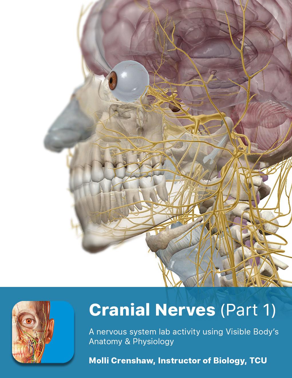
2 This lab activity is aligned with Visible Body s Human Anatomy Atlas app. Learn more at visiblebody.com/professors We've split our Cranial Nerves lab activity into two parts. Part 1 is pre-lab exercises as well as exercises that incorporate cranial nerves I-VI. Part 2 includes exercises covering cranial nerves VII-XII as well as post-lab exercises. 2
3 PRE-LAB EXERCISES A. In the main menu at the top of the screen, use the Views tab to search by body systems. Under Nervous System Views, select 2. Brain, and make the following observations: 1. Which two structures make up the central nervous system (CNS)? 2. This view demonstrates the location of bones that form the cranium. What important roles does the cranium play in regard to the brain? 3. Identify each of the bones that form the cranium. Hide each of these bones after you ve identified them: a. Frontal b. Parietal (left and right) c. Occipital d. Temporal (left and right) e. Sphenoid f. Zygomatic (left and right) g. Ethmoid 4. Rotate the image so that you are viewing the lateral surface of the left cerebral hemisphere. Select and identify each of the following regions of the brain: a. Frontal lobe of the cerebrum b. Precentral gyrus of the frontal cortex c. Postcentral gyrus of the parietal lobe d. Parietal lobe of the cerebrum e. Temporal lobe of the cerebrum f. Occipital lobe of the cerebrum g. Cerebellum h. Midbrain 3
4 i. Cerebral crus of the midbrain j. Superior colliculus of the midbrain k. Inferior colliculus of the midbrain l. Pons m. Medulla oblongata 5. While it is not visible from this view, what part of the nervous system would extend below the medulla oblongata? 6. Select any of the cranial nerves in this view. Then, in the content box, select the caret at the top left. The caret will open a breadcrumb trail with a hierarchy of structures. In this hierarchy, select "Cranial nerves" and use the definition to answer the following questions: a. How many pairs of cranial nerves are there in the human body? b. Cranial nerves are named for their connection to which part of the central nervous system? c. How many pairs of spinal nerves are there in the human body? B. In preparation for more extensive learning in lab, use Nervous System View 6. Cranial Nerves to identify the 12 pairs of cranial nerves. Be sure to use the Systems menu on the left side of the screen to deselect the skeletal, muscular, and digestive systems for the best view of the nerves. Use the space below to write out the scientific name of each nerve and a brief description of its location. 1. Cranial nerve 01 (I) 2. Cranial nerve 02 (II) 3. Cranial nerve 03 (III) 4. Cranial nerve 04 (IV) 5. Cranial nerve 05 (V) 6. Cranial nerve 06 (VI) 4
5 7. Cranial nerve 07 (VII) 8. Cranial nerve 08 (VIII) 9. Cranial nerve 09 (XI) 10. Cranial nerve 10 (X) 11. Cranial nerve 11 (XI) 12. Cranial nerve 12 (XII) 5
6 IN-LAB EXERCISES: Obtain a brain model or a preserved specimen. Use the following modules to guide your exploration of the brain and the cranial nerves. As you identify structures, use the book icon to access definitions so you can answer the questions below. You are responsible for the identification of all bolded terms. 1. Brain Parietal bone Frontal bone Occipital bone Ethmoid bone Sphenoid bone Zygomatic bone Temporal bone 6
7 2. Brain Postcentral gyrus of parietal lobe Precentral gyrus of frontal cortex Parietal lobe Frontal lobe Occipital lobe Midbrain Cerebellum Pons Medulla oblongata 7
8 3. Brain Cerebrum Midbrain Diencephalon Cerebellum Pons Medulla oblongata A. Begin by reviewing locations of major brain regions. Return to Nervous System Views and select 2. Brain, to help you identify the following structures on your specimen: 1. Cerebrum 2. Cerebellum 3. Diencephalon 4. Midbrain 5. Pons 6. Medulla oblongata 8
9 Cranial Nerves CN 01 (I) Olfactory CN 02 (II) Optic CN 03 (III) Oculomotor CN 04 (IV) Trochlear CN 05 (V) Trigeminal CN 09 (IX) Glossopharyngeal CN 10 (X) Vagus CN 11 (XI) Spinal (Accessory) CN 06 (VI) Abducens CN 07 (VII) Facial CN 08 (VIII) Vestibulocochlear CN 12 (XII) Hypoglossal 9
10 B. Identification of the Olfactory Nerves (I) Olfactory Nerves (I) Medial stria of CN 01 Lateral stria of CN 01 Olfactory bulbs Olfactory tracts 1. In Nervous System Views, select 6. Cranial Nerves. Hide the sphenoid, frontal, and temporal bones to best see the required structures. Select any part of the olfactory nerve and answer the following questions: a. These nerves are sensory/motor/mixed (circle one). b. These nerves are responsible for the sense of. c. Where do these nerves originate? 2. Using this same view, identify the following structures and answer questions based on their descriptions. a. Cribriform plate of the ethmoid bone i. Fade the cribriform plate and observe its relationship with the olfactory nerves. 10
11 ii. The cribriform plate supports the - the terminus of the - and is perforated by numerous for the passage of the branches of the. b. Olfactory bulbs of CN 01 i. Are olfactory bulbs seated superior or inferior to the cribriform plate? ii. Zoom in to see the projections on the inferior surface of the bulbs. (Tip: You may need to hide the ethmoid bone to see them well.) iii. Within each bulb, axons of olfactory form synapses with dendrites and cell bodies of other. iv. If you are attempting to view the olfactory bulbs on a preserved specimen, they may be missing or incomplete. Why do you think this is? c. Olfactory tracts of CN 01 - Olfactory tracts are formed by axons/dendrites (circle one). d. Lateral stria of CN 01 - Follow this tract to see which region of the brain it terminates in: e. Medial stria of CN 01 - Follow this tract to see which region of the brain it terminates in: f. Signals transmitted along the olfactory tracts terminate in the area in the lobe of the cerebral cortex. 3. Based on your learning, how do you think damage to this nerve would present clinically? 4. What types of tests would you devise to test the function of this nerve in patients? 11
12 C. Identification of the Optic Nerves Optic Nerves (II) Optic chiasm Hypothalamus Pituitary gland CN 02 (II) Optic 12
13 1. In Nervous System Views, select 6. Cranial Nerves. Rotate the image so you are looking into the empty socket of the right eye. Select the optic nerve and then select Fade Others. Observe its location and read the description. a. These nerves are sensory/motor/mixed (circle one). b. These nerves are responsible for the sense of. c. They originate in the of each eye. d. Which bone contains the optic foramina that the optic nerves pass through (be sure to identify this structure)? e. At which point do the right and left optic nerves converge? f. Where do the optic tracts exist? 2. Rotate the image so you are looking into the right cerebral hemisphere. Select the optic chiasm and read the description. a. The optic chiasm is the crossing point of the optic nerves. Is it a partial or a complete crossing? b. The crossed fibers occupy the medial/lateral (circle one) part of the chiasm. c. The uncrossed fibers occupy the medial/lateral (circle one) part of the chiasm. d. Where is the optic chiasm located in relation to the pituitary gland? 13
14 e. Where is the optic chiasm located in relation to the hypothalamus? f. Posterior to the optic chiasm, fibers from the optic nerves travel in the form of optic to the. g. Based on the anatomy of the optic chiasm, do you think the right cerebral hemisphere will process vision from the right eye, left eye, or both eyes? 3. Based on your learning, how do you think damage to this nerve would present clinically? 4. What types of tests would you devise to test the function of this nerve in patients? 14
15 D. Identification of the Oculomotor Nerves (III), Trochlear Nerves (IV), and Abducens Nerves (VI) Oculomotor Nerves (III) Superior rectus muscle CN 03 (III) Oculomotor Levator palpebrae superioris muscle Medial rectus muscle Inferior oblique muscle Inferior rectus muscle 15
16 Trochlear Nerves (IV) CN 04 (IV) Trochlear Superior oblique muscle Pons Sphenoid bone Superior orbital fissure 16
17 Abducens Nerves (VI) Sphenoid bone CN 06 (VI) Abducens Lateral rectus muscle Superior orbital fissure A. In Nervous System Views, select 6. Cranial Nerves. Hide the sphenoid, frontal, and temporal bones to best see the required structures. Select any part of the olfactory nerve and answer the following questions: 1. Select the oculomotor nerves, and select Fade Others. Observe their location, and read their description. a. Are these nerves sensory/motor/mixed (circle one)? b. Where do fibers of this nerve originate? c. Which foramina do the oculomotor nerves pass through? Which cranial bone are these foramina associated with? 17
18 d. Be able to identify the following effector targets of the oculomotor nerves, along with their functions: i. Levator palpebrae superioris muscle - ii. Superior rectus muscle - iii. Medial rectus muscle - iv. Inferior rectus muscle - v. Inferior oblique muscle - e. Use the search bar to identify these additional targets of the oculomotor nerve. Write their function in the space provided below: i. Ciliary muscles - ii. Pupillary sphincter of the iris - 2. Select the trochlear nerves, and select Fade Others. Observe their location, and read their description. a. Where do fibers of this nerve originate? b. Which foramina do the trochlear nerves pass through? Which cranial bone are these foramina associated with? c. Which extraocular muscle is innervated by each trochlear nerve? What is its function? 3. Select the abducens nerves, and select Fade Others. Observe their location, and read their description. a. Where do fibers of this nerve originate? b. Which foramina do the abducens nerves pass through? Which cranial bone are these foramina associated with? 18
19 c. Which extraocular muscle is innervated by each abducens nerve? What is its function? d. What is the function of sensory fibers of the abducens nerves? 4. What is the common purpose of the oculomotor, trochlear, and abducens cranial nerves? 5. Based on your learning, how do you think damage to these nerves would present clinically? 6. What types of tests would you devise to test the function of these nerves in patients? 19
20 E. Identification of the Trigeminal Nerves (V) Ophthalmic branch Semilunar ganglion Maxilla Maxillary branch Mandibular branch 1. Select the region of the trigeminal nerve closest to the brain stem. How does its size compare to that of the other nerves you have studied? 2. Each trigeminal nerve divides into major branches. 3. Identify the semilunar ganglion. 4. Select the ophthalmic branch of the trigeminal nerve and read its description: a. This branch is sensory/motor/mixed (circle one). b. These sensory fibers originate from receptors associated with which regions: 20
21 c. As this branch transmits nerve impulses toward the brain, which cranial foramen does it pass through? Which cranial bone is this foramen associated with? d. Which brain region do the fibers of this branch terminate on? 5. Select the maxillary branch of the trigeminal nerve and read its description: a. This branch is sensory/motor/mixed (circle one). b. These sensory fibers originate from receptors associated with which regions? c. Which foramen in the maxilla allows branches of this nerve to innervate the skin of the face? d. Which foramen in the zygomatic bone allows branches of this nerve to innervate the skin of the face? e. Select the maxilla and hide it. Observe the many branches off the maxillary branch of the trigeminal nerve. f. As this branch transmits nerve impulses toward the brain, which cranial foramen does it pass through? Which cranial bone is this foramen associated with? g. Which brain region do the fibers of this branch terminate on? 6. Select the mandibular branch of the trigeminal nerve and read its description: a. This branch is sensory/motor/mixed (circle one). b. These sensory fibers originate from receptors associated with which regions? 21
22 c. These motor fibers terminate on which structures? d. As this branch transmits nerve impulses to and from the brain, which cranial foramen does it pass through? Which cranial bone is this foramen associated with? 7. Based on your learning, how do you think damage to this nerve would present clinically? 8. Click on the pathology icon and write out the symptoms of trigeminal neuralgia: 9. What types of tests would you devise to test the function of this nerve in patients? 22
23 23
24 1. Brain 24
25 2. Brain 25

26 5. Brain 26

27 Cranial Nerves 27
")
28 Olfactory Nerves (I) 28

29 Optic Nerves (II) 29
")
30 Oculomotor Nerves (III) 30
")
31 Trochlear Nerves (V) 31
32 Abducens Nerves (VI) 32
")
33 Trigeminal Nerves (IV) 33
C h a p t e r PowerPoint Lecture Slides prepared by Jason LaPres North Harris College Houston, Texas
 C h a p t e r 15 The Nervous System: The Brain and Cranial Nerves PowerPoint Lecture Slides prepared by Jason LaPres North Harris College Houston, Texas Copyright 2009 Pearson Education, Inc., publishing
Lab 16: PNS: Nerves and Autonomic NS Hamilton Answers to Pre- Lab Assignments
 Lab 16: PNS: Nerves and Autonomic NS Hamilton Answers to Pre- Lab Assignments Pre-Lab Activity 1: 1. a. olfactory nerve b. optic nerve c. oculomotor nerve d. abducens nerve e. trochlear nerve f. trigeminal
ACTIVITY 7: NERVOUS SYSTEM HISTOLOGY, BRAIN, CRANIAL NERVES
 ACTIVITY 7: NERVOUS SYSTEM HISTOLOGY, BRAIN, CRANIAL NERVES LABORATORY OBJECTIVES: 1. Histology: Identify structures indicated on three different slides or images of nervous system tissue. These images
Cranial Nerves. Steven McLoon Department of Neuroscience University of Minnesota
 Cranial Nerves Steven McLoon Department of Neuroscience University of Minnesota 1 Course News Change in Lab Sequence Week of Oct 2 Lab 5 Week of Oct 9 Lab 4 2 Sensory and Motor Systems Sensory Systems:
Bellringer: The central nervous system is comprised of: What is the name of the outermost layer of the brain? a. Brain. b.
 Bellringer: The central is comprised of: a. Brain b. Spinal cord c. Sensory receptors d. Both a and b What is the name of the outermost layer of the brain? a. Pia mater b. Dura mater c. Arachnoid d. Pons
Nervous System. Student Learning Objectives:
 Nervous System Student Learning Objectives: Identify the primary parts of the neuron Identify the major structures of the central nervous system Identify the major structures of the peripheral nervous
Principles of Anatomy and Physiology
 Principles of Anatomy and Physiology 14 th Edition CHAPTER 14 The Brain and Cranial Nerves Introduction The purpose of the chapter is to: 1. Understand how the brain is organized, protected, and supplied
b. The groove between the two crests is called 2. The neural folds move toward each other & the fuse to create a
 Chapter 13: Brain and Cranial Nerves I. Development of the CNS A. The CNS begins as a flat plate called the B. The process proceeds as: 1. The lateral sides of the become elevated as waves called a. The
Laboratory Manual for Comparative Anatomy and Physiology Figure 15.1 Transparency Master 114
 Neuron Capillary Astrocyte Microglial cell Neuron Fluid-filled cavity Process of oligodendrocyte Ependymal cells Brain or spinal cord tissue Myelin sheath Nerve fibers Figure 15.1 Transparency Master 114
Blood supply to the brain Blood brain barrier isolates neural tissue from general circulation
 The Brain and Cranial Nerves Objectives Name the major regions of the brain and describe their functions. Discuss the formation, circulation, and functions of the CSF. List the main components of the medulla
The Brain and Cranial Nerves Pg. 129
 The Brain and Cranial Nerves Pg. 129 Three Main Regions of the Brain Forebrain Cerbral hemispheres Diencephalon Midbrain Brain stem Hindbrain Pons Cerebellum Medulla oblongata Forebrain Interprets sensory
Peripheral Nervous System
 Peripheral Nervous System Sensory Receptors Motor Endings Cranial Nerves The Four Plexuses Extremities Review of Reflexes Fast, preprogrammed, inborn, automatic responses Occur in the CNS at the spinal
The Brain and Cranial Nerves Pg Three Main Regions of the Brain. Forebrain
 The Brain and Cranial Nerves Pg. 129 Three Main Regions of the Brain Forebrain Cerbral hemispheres Diencephalon Midbrain Brain stem Hindbrain Pons Cerebellum Medulla oblongata Interprets sensory inputs
Brain and Cranial Nerves (Ch. 15) Human Anatomy lecture. caudal = toward the spinal cord)
 Human Anatomy lecture. caudal = toward the spinal cord)") Insight: Some cranial nerve disorders Brain and Cranial Nerves (Ch. 15) Human Anatomy lecture I. Overview (Directional terms: rostral = toward the forehead caudal = toward the spinal cord) A. 3 Major parts
Insight: Some cranial nerve disorders Brain and Cranial Nerves (Ch. 15) Human Anatomy lecture I. Overview (Directional terms: rostral = toward the forehead caudal = toward the spinal cord) A. 3 Major parts
Model 3-50B or 3-88 III VIII. Olfactory Nerve. Optic Nerve. Oculomotor Nerve. Trochlear Nerve. Trigeminal Nerve. Abducens Nerve.
 Model 3-50B or 3-88 I Olfactory Nerve II Optic Nerve Oculomotor Nerve III IV Trochlear Nerve Trigeminal Nerve V VI Abducens Nerve Glossopharyngeal Nerve IX VII Facial Nerve VIII Vestibocochlear Nerve or
M555 Medical Neuroscience Lab 1: Gross Anatomy of Brain, Crainal Nerves and Cerebral Blood Vessels
 M555 Medical Neuroscience Lab 1: Gross Anatomy of Brain, Crainal Nerves and Cerebral Blood Vessels Anatomical Directions Terms like dorsal, ventral, and posterior provide a means of locating structures
Cranial nerves.
 Cranial nerves eaglezhyxzy@163.com Key Points of Learning Name Components Passing through Peripheral distribution Central connection Function Cranial nerves Ⅰ olfactory Ⅱ optic Ⅲ occulomotor Ⅳ trochlear
Course: Physical Assessment II Date: October 17, 2008 Doc: Practice Quiz 1
 Course: Physical Assessment II Date: October 17, 2008 Doc: Practice Quiz 1 This is the practice quiz we did in Class 4. The answers are at the end of the quiz should you wish to test yourself. Complete
Major Anatomic Components of the Orbit
 Major Anatomic Components of the Orbit 1. Osseous Framework 2. Globe 3. Optic nerve and sheath 4. Extraocular muscles Bony Orbit Seven Bones Frontal bone Zygomatic bone Maxillary bone Ethmoid bone Sphenoid
Unit VIII Problem 3 Neuroanatomy: Brain Stem, Cranial Nerves and Scalp
 Unit VIII Problem 3 Neuroanatomy: Brain Stem, Cranial Nerves and Scalp - Brain stem: It is connected to the cerebellum and cerebral hemispheres. Rostral end of brain stem: diencephalon is the area which
Brain and spinal nerve. By: shirin Kashfi
 Brain and spinal nerve By: shirin Kashfi Nervous system: central nervous system (CNS) peripheral nervous system (PNS) Brain (cranial) nerves Spinal nerves Ganglions (dorsal root ganglions, sympathetic
A&P 1 Brain & Cranial Nerves Guide #1 - Pre-Lab Exercises
 A&P 1 Brain & Cranial Nerves Guide #1 - Pre-Lab Exercises In this "Pre-lab Guide", we will be looking at the brain & cranial nerves. This should be done before lab, so we don't waste time in lab! This
I. Anatomy of the Brain A. Cranial Meninges and Ventricles of the Brain 1. Meninges a. Dura mater 1) Endosteal/Periosteal Layer - Outer 2) Meningeal
 Endosteal/Periosteal Layer - Outer 2) Meningeal") I. Anatomy of the Brain A. Cranial Meninges and Ventricles of the Brain 1. Meninges a. Dura mater 1) Endosteal/Periosteal Layer - Outer 2) Meningeal Layer - Inner 3) Falx cerebri a) Superior sagittal sinus
I. Anatomy of the Brain A. Cranial Meninges and Ventricles of the Brain 1. Meninges a. Dura mater 1) Endosteal/Periosteal Layer - Outer 2) Meningeal Layer - Inner 3) Falx cerebri a) Superior sagittal sinus
Chapter 14: Nervous System Guided Notes (A-day)
") Chapter 14: Nervous System Guided Notes (A-day) Nervous System Overview Major Function: Control the body's and. Divided into the Nervous System (CNS=Brain and Spinal Cord) and the Nervous System (PNS=Cranial
Chapter 14: Nervous System Guided Notes (A-day) Nervous System Overview Major Function: Control the body's and. Divided into the Nervous System (CNS=Brain and Spinal Cord) and the Nervous System (PNS=Cranial
Cranial Nerves and Spinal Cord Flashcards
 1. Name the cranial nerves and their Roman numeral. 2. What is Cranial Nerve I called, and what does it 3. Scientists who are trying to find a way to make neurons divide to heal nerve injuries often study
Biology 323 Human Anatomy for Biology Majors Week 10; Lecture 1; Tuesday Dr. Stuart S. Sumida. Cranial Nerves and Soft Tissues of the Skull
 Biology 323 Human Anatomy for Biology Majors Week 10; Lecture 1; Tuesday Dr. Stuart S. Sumida Cranial Nerves and Soft Tissues of the Skull FOREBRAIN MIDBRAIN HINDBRAIN Forebrain: Cerebrum Perception,
Topic/Objective: Identify the structures and functions of
 Cornell Notes Textbook Diagrams Video notes Topic/Objective: Identify the structures and functions of the human nervous system. Name: Class/Period: Date: Essential Question: How does the nervous system
Protection of the Brain. Overview of the Brain. Visual Anatomy & Physiology First Edition. Martini & Ober. Chapter 13. Lecture 20
 Visual Anatomy & Physiology First Edition Martini & Ober Chapter 13 Brain and Cranial Nerves Lecture 20 1 Overview of the Brain Functions Major Parts regulates visceral activities cerebrum (two hemispheres)
Unit 18: Cranial Cavity and Contents
 Unit 18: Cranial Cavity and Contents Dissection Instructions: The calvaria is to be removed without damage to the dura mater which is attached to the inner surface of the calvaria. Cut through the outer
The Nervous System PART B
 7 The Nervous System PART B PowerPoint Lecture Slide Presentation by Jerry L. Cook, Sam Houston University ESSENTIALS OF HUMAN ANATOMY & PHYSIOLOGY EIGHTH EDITION ELAINE N. MARIEB Central Nervous System
Ms. K. GOWRI. M.Pharm., Lecturer.
 Ms. K. GOWRI. M.Pharm., Lecturer. CENTRAL NERVOUS SYSTEM (CNS) central nervous system consists of brain and spinal cord membrane covering the brain and spinal cord are surrounded by three membrane Meninges
ACTIVITY 7: NERVOUS SYSTEM HISTOLOGY, BRAIN, CRANIAL NERVES NERVOUS SYSTEM TISSUES: HISTOLOGY SLIDES
 ACTIVITY 7: NERVOUS SYSTEM HISTOLOGY, BRAIN, CRANIAL NERVES OBJECTIVES: 1) How to get ready: Read Chapter 14 & 15 McKinley et al., Human Anatomy, 4e. All text references are for this textbook. Read dissection
Biology 340 Comparative Embryology Lecture 11 Dr. Stuart Sumida. Overview of Embryology of the Vertebrate Skull. Emphasis on Amniota
 Biology 340 Comparative Embryology Lecture 11 Dr. Stuart Sumida Overview of Embryology of the Vertebrate Skull Emphasis on Amniota Initial introduction to components parts of a vertebrate head. This lecture
REVIEW OF HEAD AND NECK CRANIAL NERVES AND EVERYTHING ELSE
 REVIEW OF HEAD AND NECK CRANIAL NERVES AND EVERYTHING ELSE OLFACTORY NERVE CN I ANTERIOR CRANIAL FOSSA CRISTA GALLI OF ETHMOID OLFACTORY FORAMINA IN CRIBIFORM PLATE OF ETHMOID BONE CN I OLFACTORY NERVE
Nervous and Endocrine System Exam Review
 Directions: Read each question and complete the statement using the multiple choice responses I. Nervous System 1. The interpretation of olfactory receptor information would fall under which general function
Sheep Brain Dissection
 Sheep Brain Dissection Mammalian brains have many features in common. Human brains may not be available, so sheep brains often are dissected as an aid to understanding the mammalian brain since he general
Skull-2. Norma Basalis Interna. Dr. Heba Kalbouneh Assistant Professor of Anatomy and Histology
 Skull-2 Norma Basalis Interna Dr. Heba Kalbouneh Assistant Professor of Anatomy and Histology Norma basalis interna Base of the skull- superior view The interior of the base of the skull is divided into
Synapse Homework. Back page last question not counted. 4 pts total, each question worth 0.18pts. 26/34 students answered correctly!
 Synapse Homework Back page last question not counted 26/34 students answered correctly! 4 pts total, each question worth 0.18pts Business TASS hours extended! MWF 1-2pm, Willamette 204 T and Th 9:30-10:30am,
The orbit-1. Dr. Heba Kalbouneh Assistant Professor of Anatomy and Histology
 The orbit-1 Dr. Heba Kalbouneh Assistant Professor of Anatomy and Histology Orbital plate of frontal bone Orbital plate of ethmoid bone Lesser wing of sphenoid Greater wing of sphenoid Lacrimal bone Orbital
Skull-2. Norma Basalis Interna Norma Basalis Externa. Dr. Heba Kalbouneh Associate Professor of Anatomy and Histology
 Skull-2 Norma Basalis Interna Norma Basalis Externa Dr. Heba Kalbouneh Associate Professor of Anatomy and Histology Norma basalis interna Base of the skull- superior view The interior of the base of the
Chapter 14: The Brain and Cranial Nerves. Copyright 2009, John Wiley & Sons, Inc.
 Chapter 14: The Brain and Cranial Nerves Development of the Brain Three to four-week embryo: prosencephalon, mesencephalon and rhombencephalon. Five-week embryo: telencephalon (cerebrum), diencephalon
PERIPHERAL NERVOUS SYSTEM
 CHAPTER 13 PERIPHERAL NERVOUS SYSTEM Functional division of nervous system = afferent info to the CNS ascending spinal cord = efferent info from CNS descending spinal cord somatic skin, muscles visceral
Student Lab #: Date. Lab: Gross Anatomy of Brain Sheep Brain Dissection Organ System: Nervous Subdivision: CNS (Central Nervous System)
") Lab: Gross Anatomy of Brain Sheep Brain Dissection Organ System: Nervous Subdivision: CNS (Central Nervous System) Student Lab #: Date 1 Objectives: 1. Learn the main components making up a motor neuron.
Lab: Gross Anatomy of Brain Sheep Brain Dissection Organ System: Nervous Subdivision: CNS (Central Nervous System) Student Lab #: Date 1 Objectives: 1. Learn the main components making up a motor neuron.
Tracing the Cranial Nerves Osteologically
 CN I II III IV V 1 Supra-orbital ethmoidal nn. Ext. nasal V 2 Tracing the Cranial Nerves Osteologically Nucleus of Origin Olfactory tracts of frontal lobe of cerebrum Optic tracts from optic chiasma and
The Brain and Cranial Nerves
 14 The Brain and Cranial Nerves PowerPoint Lecture Presentations prepared by Jason LaPres Lone Star College North Harris An Introduction to the Brain and Cranial Nerves The Adult Human Brain Average weight
Peripheral Nervous System Dr. Gary Mumaugh
 Peripheral Nervous System Dr. Gary Mumaugh Spinal Nerves Overview Thirty-one pairs of spinal nerves are connected to the spinal cord No special names; numbered by level of vertebral column at which they
The dura is sensitive to stretching, which produces the sensation of headache.
 Dural Nerve Supply Branches of the trigeminal, vagus, and first three cervical nerves and branches from the sympathetic system pass to the dura. Numerous sensory endings are in the dura. The dura is sensitive
213: HUMAN FUNCTIONAL ANATOMY: PRACTICAL CLASS 12 Cranial cavity, eye and orbit
 213: HUMAN FUNCTIONAL ANATOMY: PRACTICAL CLASS 12 Cranial cavity, eye and orbit OSTEOLOGY Identify the bones which comprise the walls of the orbit: maxilla, zygomatic, ethmoid, lachrymal, frontal, and
Nervous System. The Peripheral Nervous System Agenda Review of CNS v. PNS PNS Basics Cranial Nerves Spinal Nerves Reflexes Pathways
 Nervous System Agenda Review of CNS v. PNS PNS Basics Cranial Nerves Spinal Nerves Sensory Motor Review of CNS v. PNS Central nervous system (CNS) Brain Spinal cord Peripheral nervous system (PNS) All
Nervous System The Brain and Spinal Cord Unit 7b
 Nervous System The Brain and Spinal Cord Unit 7b Chetek High School Mrs. Michaelsen 9.12 Meninges A. Meninges 1. The organs of the CNS are covered by membranes a. The meninges are divided into 3 layers:
Cranial Cavity REFERENCES: OBJECTIVES OSTEOLOGY. Stephen A. Gudas, PT, PhD
 Stephen A. Gudas, PT, PhD Cranial Cavity REFERENCES: Moore and Agur, Essential Clinical Anatomy (ECA), 3rd ed., pp. 496 498; 500 507; 512 514 Grant s Atlas 12 th ed., Figs 7.6; 7.19 7.30. Grant s Dissector
PHYSIOLOHY OF BRAIN STEM
 PHYSIOLOHY OF BRAIN STEM Learning Objectives The brain stem is the lower part of the brain. It is adjoining and structurally continuous with the spinal cord. 1 Mid Brain 2 Pons 3 Medulla Oblongata The
1/10/2013. What do neurons look like? Topic 14: Spinal Cord & Peripheral Nerves. How do neurons work? The nervous impulse. Specialized Neurons
 Topic 4: Spinal Cord & Peripheral Nerves What do neurons look like? Neurons What do they look like? How do they work? Neuronal and spinal organization What is the difference between neuron & nerve? How
CRANIAL NERVES. Dr. Amani A. Elfaki Associate Professor Department of Anatomy
 CRANIAL NERVES Dr. Amani A. Elfaki Associate Professor Department of Anatomy LEARNING OBJECTIVES Named the cranial nerves Identify the funcunal component of each cranial nerve Identify the effect of each
Dissection of the Sheep Brain
 Dissection of the Sheep Brain Laboratory Objectives After completing this lab, you should be able to: 1. Identify the main structures in the sheep brain and to compare them with those of the human brain.
The Brain and Cranial Nerves Student Objectives
 The Brain and Cranial Nerves Student Objectives Chapter 14 Textbook and Laboratory Manual Name the major regions of the brain and describe their functions Name the ventricles of the brain and describe
Brainstem. By Dr. Bhushan R. Kavimandan
 Brainstem By Dr. Bhushan R. Kavimandan Development Ventricles in brainstem Mesencephalon cerebral aqueduct Metencephalon 4 th ventricle Mylencephalon 4 th ventricle Corpus callosum Posterior commissure
The Nervous System PART B
 7 The Nervous System PART B PowerPoint Lecture Slide Presentation by Jerry L. Cook, Sam Houston University ESSENTIALS OF HUMAN ANATOMY & PHYSIOLOGY EIGHTH EDITION ELAINE N. MARIEB The Reflex Arc Reflex
The Human Brain: Anatomy, Functions, and Injury
 The Human Brain: Anatomy, Functions, and Injury Main Menu Brain Anatomy Brain Functions Injury Mechanisms Brain Anatomy Menu Skull Anatomy Interior Skull Surface Blood Vessels of the Brain Arteries of
mistake ;slides in bold but you still have to go back to our slides to see the figure, tables and some scheme
 Khozama jehad : I am doing my best and I am sorry for any unintended mistake ;slides in bold but you still have to go back to our slides to see the figure, tables and some scheme The Orbit, Orbital Contents
Stanley Pruisinger 1980's
 Neuroanatomy Prion disease cerebellum chapter b/c cerebellar ataxia here as a warning for obvious reasons. Creutzfeldt - Jakob Disease (CJD) "Spongiform" (brain turns to sponge) Jews in Lybia who ate
Brain, Cranial Nerves, and Spinal Cord
 Bio101 Laboratory 13 Neuron/Spinal Cord Histology Brain Anatomy Ear & Eye Anatomy 1 Brain, Cranial Nerves, and Spinal Cord Objectives for today s lab Become familiar with the gross anatomy of the brain
BIOL Dissection of the Sheep and Human Brain
 BIOL 2401 Dissection of the Sheep and Human Brain Laboratory Objectives After completing this lab, you should be able to: Identify the main structures in the sheep brain and to compare them with those
Nervous System: An Introduction. HAP Susan Chabot Lemon Bay High School
 Nervous System: An Introduction HAP Susan Chabot Lemon Bay High School Function of the Nervous System 3 overlapping functions SENSORY INPUT - Monitor changes inside and outside of the body; these changes
Central N.S. Peripheral N.S. 2) List the functional subdivisions. 1) List the anatomical subdivisions.
 List the functional subdivisions. 1) List the anatomical subdivisions.") S T U D Y G U I D E 8 1. Divisions of the Nervous System 1) List the anatomical subdivisions. Central N.S. Peripheral N.S. 2) List the functional subdivisions. Somatic N.S. Autonomic N.S. 2. Nerve Tissue
S T U D Y G U I D E 8 1. Divisions of the Nervous System 1) List the anatomical subdivisions. Central N.S. Peripheral N.S. 2) List the functional subdivisions. Somatic N.S. Autonomic N.S. 2. Nerve Tissue
DEVELOPMENT OF BRAIN
 Ahmed Fathalla OBJECTIVES At the end of the lecture, students should: List the components of brain stem. Describe the site of brain stem. Describe the relations between components of brain stem & their
Chapter 10 The Nervous System: The Brain and Cranial Nerves
 Chapter 10 The Nervous System: The Brain and Cranial Nerves Copyright 2015 Wolters Kluwer Health Lippincott Williams & Wilkins Overview Key Terms aphasia corpus callosum meninges basal nuclei diencephalon
Biology 218 Human Anatomy
 Chapter 19 Adapted form Tortora 10 th ed. LECTURE OUTLINE A. Overview of Brain Organization (p. 584) I. Principal Parts of the Brain: 1. The adult brain consists of about 100 billion neurons and 10-50
Note to Self. The Brain and Cranial Nerves. Organization of the Brain
 Note to Self Despite what you think you can get through this chapter in 1 class period, or one with only a few slides left over The Brain and Cranial Nerves Chapter 14 Organization of the Brain The brain
The sebaceous glands (glands of Zeis) open directly into the eyelash follicles, ciliary glands (glands of Moll) are modified sweat glands that open
 open directly into the eyelash follicles, ciliary glands (glands of Moll) are modified sweat glands that open") The Orbital Region The orbits are a pair of bony cavities that contain the eyeballs; their associated muscles, nerves, vessels, and fat; and most of the lacrimal apparatus upper eyelid is larger and more
The Orbital Region The orbits are a pair of bony cavities that contain the eyeballs; their associated muscles, nerves, vessels, and fat; and most of the lacrimal apparatus upper eyelid is larger and more
14: The Brain and Cranial Nerves
 14: The Brain and Cranial Nerves I. An Introduction to the Organization of the Brain, p. 452 Objectives 1. Name the major regions of the brain and describe their functions 2. Name the three primary brain
Arnab Rana. Last updated 27th November The Skull Base. Arnab Rana. Background. Skull base foramina. Skull base bones. Self learning exercise
 Last updated 27th November 2017 Table of contents 1 2 3 4 5 Aberdeen MBChB learning guide links Year 2 Systems II (Part 1) ENT Pg 24 Aberdeen MBChB learning guide links Year 2 Systems II (Part 1) ENT Pg
Done by : Areej Al-Hadidi
 Brainstem &diencephalon Done by : Areej Al-Hadidi Brainstem Functions Ascending and descending tracts Reflex centers Cardiovascular and respiratory centers Coughing, sneezing, swallowing Nuclei of the
Organization of The Nervous System PROF. MOUSAED ALFAYEZ & DR. SANAA ALSHAARAWY
 Organization of The Nervous System PROF. MOUSAED ALFAYEZ & DR. SANAA ALSHAARAWY Objectives At the end of the lecture, the students should be able to: List the parts of the nervous system. List the function
Brain ميهاربا لض اف دمح ا د The Meninges 1- Dura Mater of the Brain endosteal layer does not extend meningeal layer falx cerebri tentorium cerebelli
 .احمد د فاضل ابراهيم Lecture 15 Brain The Meninges Three protective membranes or meninges surround the brain in the skull: the dura mater, the arachnoid mater, and the pia mater 1- Dura Mater of the Brain
Principles Arteries & Veins of the CNS LO14
 Principles Arteries & Veins of the CNS LO14 14. Identify (on cadaver specimens, models and diagrams) and name the principal arteries and veins of the CNS: Why is it important to understand blood supply
Bony orbit Roof The orbital plate of the frontal bone Lateral wall: the zygomatic bone and the greater wing of the sphenoid
 Bony orbit Roof: Formed by: The orbital plate of the frontal bone, which separates the orbital cavity from the anterior cranial fossa and the frontal lobe of the cerebral hemisphere Lateral wall: Formed
Read Chapter 14 & 15 McKinley et al
 ACTIVITY 7: NERVOUS SYSTEM HISTOLOGY, BRAIN, CRANIAL NERVES OBJECTIVES: 1) How to get ready: Read Chapter 14 & 15 McKinley et al., Human Anatomy, 5e. All text references are for this textbook. Read dissection
Brainstem. Telencephalon Diencephalon Cerebellum Brain stem
 Brainstem Brainstem 脑 脊髓 Brainstem Telencephalon Diencephalon Cerebellum Brain stem Ventral view Lateral view 10 pairs of the cranial nerves are attached to the brain stem The brainstem Midbrain Pons Medulla
Instructor s Review for Final Exams. The Nervous System
 Instructor s Review for Final Exams The Nervous System Divisions of the Central Nervous System? Brain and spinal cord. Key word, central. Divisions of the nervous system Central and Peripheral Coverings
Examination and Diseases of Cranial Nerves
 Cranial nerve evaluation is an important part of a neurologic exam. There are some differences in the assessment of cranial nerves with different species but the general principles are the same. Going
Gross Organization I The Brain. Reading: BCP Chapter 7
 Gross Organization I The Brain Reading: BCP Chapter 7 Layout of the Nervous System Central Nervous System (CNS) Located inside of bone Includes the brain (in the skull) and the spinal cord (in the backbone)
Cranial nerve Dept. of Anatomy Zhou Hong Ying
 Cranial nerve Dept. of Anatomy Zhou Hong Ying Key Points of Learning Name Components Passing through Peripheral distribution Central connection Function Cranial nerve Ⅰ olfactory Ⅱ optic Ⅲ occulomotor
TRANSVERSE SECTION PLANE Scalp 2. Cranium. 13. Superior sagittal sinus
 TRANSVERSE SECTION PLANE 1 1. Scalp 2. Cranium 3. Superior sagittal sinus 4. Dura mater 5. Falx cerebri 6. Frontal lobes of the cerebrum 7. Middle meningeal artery 8. Cortex, grey matter 9. Cerebral vessels
Human Nervous System:
 OLLI Brain: Making Sense of Our World: Lecture 3 Human Nervous System: The Motor & Sensory Divisions Copyright 2004 Pearson Education, Inc., publishing as Benjamin Cummings Organization of the Nervous
CHAPTER 11: NERVOUS SYSTEM II: DIVISIONS OF THE NERVOUS SYSTEM. 1. Outline the major divisions of the nervous system.
 CHAPTER 11: NERVOUS II: DIVISIONS OF THE NERVOUS OBJECTIVES: 1. Outline the major divisions of the nervous system. NERVOUS CENTRAL NERVOUS (BRAIN & SPINAL CORD) (INTERNEURONS) PERIPHERAL NERVOUS (CRANIAL
The Nervous System: Central Nervous System
 The Nervous System: Central Nervous System I. Anatomy of the nervous system A. The CNS & the body by: 1. monitoring of the body 2. & information between parts of the body 3. acting as a to gather, store,
Anatomy & Physiology Central Nervous System Worksheet
 1. What are the two parts of the CNS? 2. What are the four functions of the CNS Anatomy & Physiology Central Nervous System Worksheet 3. What are the four functions of the meninges? (p430) 4. Starting
SKULL AS A WHOLE + ANTERIOR CRANIAL FOSSA
 SKULL AS A WHOLE + ANTERIOR CRANIAL FOSSA LEARNING OBJECTIVES At the end of this lecture, the student should be able to know: Parts of skeleton (axial and appendicular) Parts of skull Sutures of skull
Chapter 10. The Nervous System
 Chapter 10 The Nervous System Objectives List the organs and divisions of the nervous system and describe the generalized functions Identify the major types of cells in the nervous system and discuss the
Neurology study of the nervous system. nervous & endocrine systems work together to maintain homeostasis
 Nervous System Neurology study of the nervous system nervous & endocrine systems work together to maintain homeostasis Nervous System works very fast Uses electrical signals called nerve impulses Short-lived
Dr. Sami Zaqout, IUG Medical School
 The skull The skull is composed of several separate bones united at immobile joints called sutures. Exceptions? Frontal bone Occipital bone Vault Cranium Sphenoid bone Zygomatic bones Base Ethmoid bone
The neurvous system senses, interprets, and responds to changes in the environment. Two types of cells makes this possible:
 NERVOUS SYSTEM The neurvous system senses, interprets, and responds to changes in the environment. Two types of cells makes this possible: the neuron and the supporting cells ("glial cells"). Neuron Neurons
Human Anatomy and Physiology - Problem Drill 07: The Skeletal System Axial Skeleton
 Human Anatomy and Physiology - Problem Drill 07: The Skeletal System Axial Skeleton Question No. 1 of 10 Which of the following statements about the axial skeleton is correct? Question #01 A. The axial
University of Palestine. Midterm Exam 2013/2014 Total Grade:
 Course No: DNTS2208 Course Title: Head and Neck Anatomy Date: 09/11/2013 No. of Questions: (50) Time: 1hour Using Calculator (No) University of Palestine Midterm Exam 2013/2014 Total Grade: Instructor
Organization of The Nervous System PROF. SAEED ABUEL MAKAREM
 Organization of The Nervous System PROF. SAEED ABUEL MAKAREM Objectives By the end of the lecture, you should be able to: List the parts of the nervous system. List the function of the nervous system.
Lab 10: Muscle Tissue and Selected Muscles
 117 Ex. 10: Histology of Muscle Muscle Tissue Lab 10: Muscle Tissue and Selected Muscles Skeletal muscle: Cardiac muscle: Smooth muscle MUSCLE LIST Ex. 11: Gross Anatomy of Muscle Locate these muscles
THE CENTRAL NERVOUS SYSTE M
 THE CENTRAL NERVOUS SYSTE M Structure and Functio n THIRD EDITIO N PER BRODAL A Brief Survey, x i Studying the Structures and Function of the Nervous System, xii i Animal Experiments Crucial for Progress,
Chapter 14, Part 2! Chapter 14 Part 2 Brain/Cranial Nerves! The Cerebrum and Cranial Nerves! pp !
 Chapter 14, Part 2! The Cerebrum and Cranial pp. 482 505! SECTION 14-9! The cerebrum, the largest region of the brain, contains motor, sensory, and association areas! 2! White Matter of the Cerebrum! 1.
Chapter 14, Part 2! The Cerebrum and Cranial Nerves! pp !
 Chapter 14, Part 2! The Cerebrum and Cranial pp. 482 505! SECTION 14-9! The cerebrum, the largest region of the brain, contains motor, sensory, and association areas! 2! 1! ! Chapter 14 Part 2 Brain/Cranial