Magnetic Resonance Imaging. Basics of MRI in practice. Generation of MR signal. Generation of MR signal. Spin echo imaging. Generation of MR signal
|
|
|
- Louise Andrews
- 6 years ago
- Views:
Transcription


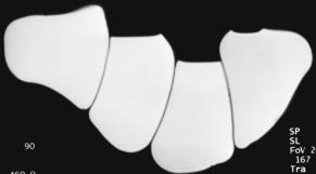
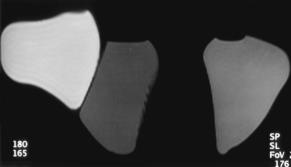
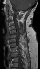
1 Magnetic Resonance Imaging Protons aligned with B0 magnetic filed Longitudinal magnetization - T1 relaxation Transverse magnetization - T2 relaxation Signal measured in the transverse plane Basics of MRI in practice In biological tissues T1>T2 Nandor Pinter M.D. Dent Neurologic Institute, Buffalo NY American Society of Neuroimaging, Los Angeles, Generation of MR signal Generation of MR signal Free induction decay Longitudinal magnetization - T1 relaxation Transverse magnetization - T2 relaxation The two happen simultaneously magnetization signal Generation of MR signal Spin echo imaging What does short T2 mean? y x Short T2 Long T2 The magnetic vector is echoed by the 180 deg pulse 1


2 Spin echo Spin echo Spin echo Long TR versus short TR Signal decrease by T2 From T1 Image weighting effect of TR Image weighting effect of TE From Proton Density To Proton Density To T2 TE=10 TR=400 TE=10 TR=1000 TE=10 TR=1800 TE=10 TR=2600 TE=10 TR=3000 TE=10 TR=3000 TE=40 TR=3000 TE=70 TR=3000 TE=90 TR=3000 TE=150 TR=3000 2


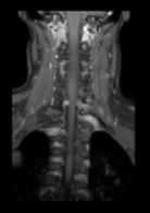
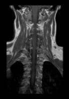
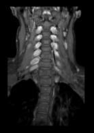
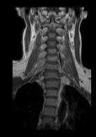
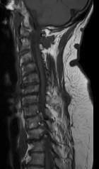
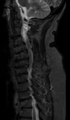
3 Conventional SE T1-weighted scan advantages: resolution Anatomy or Resolution: 0.9-8mm3 Tissues: CSF: dark WM: light grey GM: dark grey Lesions: bright/dark The T1 weighting is optimized for the T1 differences of tissues in the given body part. T x1.25x5mm 0.9x0.9x1mm advantages: Multi Planar Reconstructions (MPR) Conventional T2-weighted scans Sagittal Axial Coronal Axial Coronal Resolution: 1.5-7mm3 Lesion characterization CSF spaces, fluid collection, edema Tissues: CSF: bright WM: dark grey GM: light grey Lesions: mostly bright Inversion recovery sequences Inversion recovery sequences Nulling time of a tissue is 0.69 x T1 time If TI=400ms what is the suppressed tissue? 3


 STIR Lower resolution for higher")


4 Inversion recovery sequences Inversion recovery sequences T1 IR ST IR FLA IR STIR STIR "Fat Suppression in MR Imaging: Techniques and Pitfalls" Delfaut et al. RadioGraphics Vol. 19, No. 2: "Fat Suppression in MR Imaging: Techniques and Pitfalls" Delfaut et al. RadioGraphics Vol. 19, No. 2: STIR Contrast-enhanced (?) STIR Lower resolution for higher signal : smaller matrix size, but keep slice thickness PRE POST 0.8x0.8x3mm 0.9x1.1x3mm 0.7x0.8 x 4 mm 0.9x1.25x4mm 4
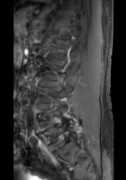
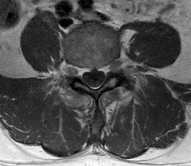
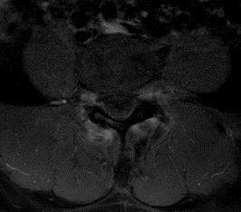
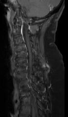
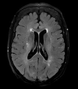
5 Contrast-suppressed STIR Other fat suppression techniques in spine PRE POST Fat saturation: frequency-selective saturation of fat signal Why do CE spine with fat suppression? Krinsky G et al AJR 1996; 166: Other fat suppression techniques in spine Why do CE spine with fat suppression? Other fat suppression techniques in spine Why do CE spine with fat suppression? Standard T1 CE mdixon T1 CE Fluid Attenuated Inversion Recovery (FLAIR) T1 T1 CE T1 CE + Fat Sat STIR CE Lesion characterization or Resolution: mm3 Tissues: CSF: no signal (T2!) WM: dark grey GM: lighter grey Lesions: bright 5


6 3T Philips FLAIR vs. FLAIR : Thin slice Acquisition Axial/Sagittal Axial/Sag/Cor Thickness 4mm axi 3mm sag 2mm Gap 1mm -1mm In plane res. 0.74x0.86 axi 0.84x0.88 sag Pulsation MPR Scan length 4:30 min axi 5:30 min sag 0.9x0.9 4:47 min : Thin slice : Thin slice : No pulsation artifact : False positive from pulsation artifact AXI FLAIR FLAIR SAG FLAIR AXI T2 6

 Inhomogeneity")


7 : Thin slice, no pulsation artifact : Thin slice, no pulsation artifact SWI phase map SWI phase mask Local differences (susceptibility) Inhomogeneity of B0 Geometry E.M. Haacke et al. AJNR Am J Neuroradiol 2009;30: by American Society of Neuroradiology Magnitude Image x Phase mask = SWI image Magnitude Image x Phase mask = SWI image Magnitude Mask X E.M. Haacke et al. AJNR Am J Neuroradiol 2009;30: by American Society of Neuroradiology 7


8 SWI: Effect of filter size SWI: Effect of filter size Filter: 32 Filter: 256 SWI: Effect of filter size Effect of flip angle TE:40ms, TR:60ms FA:10 FA:15 FA:18 FA:20 Effect of flip angle Effect of flip angle TE:40ms, TR:60ms TE:40ms, TR:60ms FA:10 FA:15 FA:20 FA:10 FA:15 FA:18 FA:20 Signal Intensity: ±8.21 SI: ±10.71 SI: ±11.94 SI: ±7.25 Signal Intensity: ±8.21 SI: ±10.71 SI: ±7.25 8


 DRIVE MinIP Sensitive")


9 Effect of bandwidth Effect of bandwidth TE:40ms, TR:60ms, FA:20 BW:72Hz/pixel BW:108Hz/pixel BW:72Hz/pixel BW:108Hz/pixel SI: ±9.66 SI: ±7.25 SI: ±8.39 SI: ±7.30 TE:40ms, TR:60ms, FA:10 Effect of repetition time Effect of repetition time TE:40ms, BW:72Hz/min, FA:20 TR:50ms TR:60ms TE:40ms, BW:72Hz/min, FA:20 TR:50ms TR:60ms SI: ±9.73 SI: ±9.66 Susceptibility Weighted Imaging (SWI) DRIVE MinIP Sensitive for Venous blood (MRV) Blood products Calcifications T2 High contrast between CSF and parenchyma Poor tissue contrast Trauma Stroke Amyloid angiopathy Venous anomalies DVA Cavernoma Postirradiation teleangiectasia Cranial nerves:3, 5, 6, 7&8, 9 Vestibular Schwannoma Meningioma Trigeminal neuralgia Neurovascular compressions IAC and inner ear 9


 MIP S. Haller et al.")


 Intracranial stenosis (less")

10 12/19/2016 Neurovascular compression DRIVE TOF MRA Neurovascular compression DRIVE Merge of DRIVE+TOF TOF MRA Same geometry: FOV=15cm, matrix=300x300, thk=1mm, space=-0.5mm Neurovascular compression MRA of the head Time-of-flight (TOF) MIP S. Haller et al. AJNR Am J Neuroradiol 2016;37: MRV Phase contrast Sensitive for Direction of flow Velocity of flow Imaging of intracranial veins Slow flow Sinus thrombosis Sinus thrombosis vs Hypoplasia Gadolinium contrast is not necessary in most cases! MR angiography COW and vertebrobasilar arteries 5-6 mins No contrast! Sensitive for high velocity flow Rule out aneurysm Other vascular anomalies with high velocity flow (moya moya, AV fistula) Intracranial stenosis (less precise) Not good for DVA! Clinical MRI protocols - Routine brain Indication Non-specific symptoms Rule out: tumor, inflammation, demyelinating disease, vascular lesions, CVJ abnormalities, Chiari, headache. T2 axi and cor, DWI FLAIR 10
 IAC and inner ear T2 axi FLAIR Tumor")


11 12/19/2016 Headache/dizziness T2 axi T2 cor FLAIR DRIVE Vestibular Schwannoma Meningioma Trigeminal neuralgia Neurovascular compressions 4th is too small (mostly) IAC and inner ear T2 axi FLAIR Tumor FLAIR Axi T2 DWI SWI Newly discovered tumor Tumor follow-up Tumor patient with Dizziness Headache Seizure CE T1 Contrast-enhanced study! T2 cor DWI Gadolinium contrast is not necessary in most cases! SWI Seizure Cor T2 Head trauma Dizziness / vertigo as leading symptoms Cranial nerves: 3, 5, 6, 7&8, 9 DWI Trauma Focal cortical dysplasia Other GM abnormality T2, FLAIR Any abnormality CSF spaces Coronal FLAIR and IR Hippocampal sclerosis GM abnormalities Aligned with hippocampus Gadolinium contrast is not necessary in most cases! Dementia w volumetrics Multiple Sclerosis - 1.5T Known MS follow-up Contrast-enhanced study! 11


 Posterior lobe")
12 Multiple Sclerosis - 3T Philips Pituitary T2, DWI Sag FLAIR CE T1 Contrast-enhanced study! Most frequent indications Rule out / follow-up microadenoma Follow-up macroadenoma (1-2cm) Posterior lobe anomalies (diabetes insipidus) Not for craniopharyngeoma! 12
Neuroradiology MR Protocols
 Neuroradiology MR Protocols Brain protocols N 1: Brain MRI without contrast N 2: Pre- and post-contrast brain MRI N 3 is deleted N 4: Brain MRI without or pre-/post-contrast (seizure protocol) N 5: Pre-
MR Advance Techniques. Vascular Imaging. Class II
 MR Advance Techniques Vascular Imaging Class II 1 Vascular Imaging There are several methods that can be used to evaluate the cardiovascular systems with the use of MRI. MRI will aloud to evaluate morphology
Magnetic Resonance Angiography
 Magnetic Resonance Angiography 1 Magnetic Resonance Angiography exploits flow enhancement of GR sequences saturation of venous flow allows arterial visualization saturation of arterial flow allows venous
High Field MR of the Spine
 Department of Radiology University of California San Diego 3T for MR Applications Advantages High Field MR of the Spine Increased signal-to-noise Better fat suppression Increased enhancement with gadolinium
1Pulse sequences for non CE MRA
 MRI: Principles and Applications, Friday, 8.30 9.20 am Pulse sequences for non CE MRA S. I. Gonçalves, PhD Radiology Department University Hospital Coimbra Autumn Semester, 2011 1 Magnetic resonance angiography
Department of Radiology University of California San Diego. MR Angiography. Techniques & Applications. John R. Hesselink, M.D.
 Department of Radiology University of California San Diego MR Angiography Techniques & Applications John R. Hesselink, M.D. Vascular Imaging Arterial flow void Flow enhancement Gadolinium enhancement Vascular
University of Michigan continues fine-tuning neuro ExamCards
 I s s u e 3 1 - M a rc h 2 0 0 7 F i e l d Strength Publication for the Philips MRI Community University of Michigan continues fine-tuning neuro ExamCards Drs. Mukherji, Parmar create Achieva 3.0T ExamCards
Essentials of Clinical MR, 2 nd edition. 99. MRA Principles and Carotid MRA
 99. MRA Principles and Carotid MRA As described in Chapter 12, time of flight (TOF) magnetic resonance angiography (MRA) is commonly utilized in the evaluation of the circle of Willis. TOF MRA allows depiction
NEURO PROTOCOLS MRI NEURO PROTOCOLS (SIEMENS SCANNERS)
") Page 1 NEURO PROTOCOLS Brain Stroke Brain Brain with contrast Brain for seizures Brain for MS Brain for Pineal gland Sella FAST Scan for hydrocephalus MRA/MRV Brain MRA carotids 8 th nerve Cranial nerves
Page 1 NEURO PROTOCOLS Brain Stroke Brain Brain with contrast Brain for seizures Brain for MS Brain for Pineal gland Sella FAST Scan for hydrocephalus MRA/MRV Brain MRA carotids 8 th nerve Cranial nerves
Consortium of MS Centres Guidelines Revised Standardized MRI Protocol. for the Diagnosis and Follow-up of MS. David K.B.
 Consortium of MS Centres Guidelines Revised Standardized MRI Protocol for the Diagnosis and Follow-up of MS David K.B. Li MD FRCPC Indianapolis, Indiana May 27, 2015 Disclosure I have received research
RADIOLOGY TEACHING CONFERENCE
 RADIOLOGY TEACHING CONFERENCE John Athas, MD Monica Tadros, MD Columbia University, College of Physicians & Surgeons Department of Otolaryngology- Head & Neck Surgery September 27, 2007 CT SCAN IMAGING
Methods. Yahya Paksoy, Bülent Oğuz Genç, and Emine Genç. AJNR Am J Neuroradiol 24: , August 2003
 AJNR Am J Neuroradiol 24:1364 1368, August 2003 Retrograde Flow in the Left Inferior Petrosal Sinus and Blood Steal of the Cavernous Sinus Associated with Central Vein Stenosis: MR Angiographic Findings
The Low Sensitivity of Fluid-Attenuated Inversion-Recovery MR in the Detection of Multiple Sclerosis of the Spinal Cord
 The Low Sensitivity of Fluid-Attenuated Inversion-Recovery MR in the Detection of Multiple Sclerosis of the Spinal Cord Mark D. Keiper, Robert I. Grossman, John C. Brunson, and Mitchell D. Schnall PURPOSE:
Index. aneurysm, 92 carotid occlusion, 94 ICA stenosis, 95 intracranial, 92 MCA, 94
 A ADC. See Apparent diffusion coefficient (ADC) Aneurysm cerebral artery aneurysm, 93 CT scan, 93 gadolinium, 93 Angiography, 13 Anoxic brain injury, 25 Apparent diffusion coefficient (ADC), 7 Arachnoid
Keep Imaging Simple: An Introduction To Neuroimaging
 Keep Imaging Simple: An Introduction To Neuroimaging Meghan Elkins, OD, FAAO Please silence all mobile devices and remove items from chairs so others can sit. Unauthorized recording of this session is
MR Imaging with the CCSVI or Haacke protocol
 MR Imaging with the CCSVI or Haacke protocol Reports from the Haacke protocol are often made available to the patients. The report consists of four major components: 1. anatomical images of major neck
High Signal Intensity of the Infundibular Stalk on Fluid-Attenuated Inversion Recovery MR
 High Signal Intensity of the Infundibular Stalk on Fluid-Attenuated Inversion Recovery MR Yutaka Araki, Ryuichirou Ashikaga, Satoru Takahashi, Jun Ueda, and Osamu Ishida PURPOSE: To determine the MR imaging
MRI Imaging of GP Medicare Eligible Conditions
 MRI Imaging of GP Medicare Eligible Conditions By Dr. Andrew Stuart Radiologist Sydney Adventist Hospital 0562/SAH/1112/SAH Learning Objectives Indications for GP referred Medicare eligible MRI scans MRI
Non Contrast MRA. Mayil Krishnam. Director, Cardiovascular and Thoracic Imaging University of California, Irvine
 Non Contrast MRA Mayil Krishnam Director, Cardiovascular and Thoracic Imaging University of California, Irvine No disclosures Non contrast MRA-Why? Limitations of CTA Radiation exposure Iodinated contrast
Why Talk About Technique? MRI of the Knee:
 Why Talk About Technique? MRI of the Knee: Part 1 - Imaging Techniques Mark Anderson, M.D. University of Virginia Health Sciences Center Charlottesville, Virginia Always had an interest teach our fellows
Neurovascular elements of the PCF
 Level I: Neurovascular elements of the PCF Trigeminal and Abducent Superior cerebellar artery and vein Dandy s vein Level II: Facial and Cochleovestibular AICA and internal auditory artery, veins Level
Imaging Acute Stroke and Cerebral Ischemia
 Department of Radiology University of California San Diego Imaging Acute Stroke and Cerebral Ischemia John R. Hesselink, M.D. Causes of Stroke Arterial stenosis Thrombosis Embolism Dissection Hypotension
Speed, Comfort and Quality with NeuroDrive
 Speed, Comfort and Quality with NeuroDrive Echelon Oval provides a broad range of capabilities supporting fast, accurate diagnosis of brain conditions and injuries. From anatomical depiction to vascular
SWI including phase and magnitude images
 On-line Table: MRI imaging recommendation and summary of key features Sequence Pathologies Visible Key Features T1 volumetric high-resolution whole-brain reformatted in axial, coronal, and sagittal planes
Clinical Applications
 C H A P T E R 16 Clinical Applications In selecting pulse sequences and measurement parameters for a specific application, MRI allows the user tremendous flexibility to produce variations in contrast between
MR Angiography in the evaluation of Lower Extremity Arterial Disease
 March 2001 MR Angiography in the evaluation of Lower Extremity Arterial Disease Ted Mau, Harvard Medical School Year III Objectives We will cover: Indications for Magnetic Resonance Angiography (MRA) Basic
Applicable Neuroradiology
 For the Clinical Neurology Clerkship LSU Medical School New Orleans Amy W Voigt, MD Clerkship Director Introduction The field of Radiology first developed following the discovery of X-Rays by Wilhelm Roentgen
MR Flow Imaging in Vascular Malformations Using Gradient Recalled Acquisition
 637 MR Flow Imaging in Vascular Malformations Using Gradient Recalled Acquisition William M. Needell 1 Kenneth R. Maravilla Twenty patients with known or suspected intracranial vascular lesions were evaluated
Basics of MRI Part I
 Basics of MRI Part I Mathew J. Dixon, D.O. Chairman Department of Radiology Memorial Health University Medical Center Savannah, GA Objectives Brief History Concept of MRI Creation of a Magnetic Field Concepts
Objectives 8/17/2011. Challenges in Cardiac Imaging. Challenges in Cardiac Imaging. Basic Cardiac MRI Sequences
 8/17/2011 Traditional Protocol Model for Tomographic Imaging Cardiac MRI Sequences and Protocols Frandics Chan, M.D., Ph.D. Stanford University Medical Center Interpretation Lucile Packard Children s Hospital
Optimized phase contrast MRV technique outperforms timeof-flight in the diagnosis of cerebral venous thrombosis
 Optimized phase contrast MRV technique outperforms timeof-flight in the diagnosis of cerebral venous thrombosis Poster No.: C-3377 Congress: ECR 2010 Type: Topic: Authors: Keywords: DOI: Scientific Exhibit
A New Trend in Vascular Imaging: the Arterial Spin Labeling (ASL) Sequence
 Sequence") A New Trend in Vascular Imaging: the Arterial Spin Labeling (ASL) Sequence Poster No.: C-1347 Congress: ECR 2013 Type: Educational Exhibit Authors: J. Hodel, A. GUILLONNET, M. Rodallec, S. GERBER, R. 1
A New Trend in Vascular Imaging: the Arterial Spin Labeling (ASL) Sequence Poster No.: C-1347 Congress: ECR 2013 Type: Educational Exhibit Authors: J. Hodel, A. GUILLONNET, M. Rodallec, S. GERBER, R. 1
SPINAL MAGNETIC RESONANCE IMAGING INTERPRETATION
 CLINICAL VIGNETTE 2017; 3:2 SPINAL MAGNETIC RESONANCE IMAGING INTERPRETATION Editor-in-Chief: Idowu, Olufemi E. Neurological surgery Division, Department of Surgery, LASUCOM/LASUTH, Ikeja, Lagos, Nigeria.
NEURORADIOLOGY Part I
 NEURORADIOLOGY Part I Vörös Erika University of Szeged Department of Radiology SZEGED BRAIN IMAGING METHODS Plain film radiography Ultrasonography (US) Computer tomography (CT) Magnetic resonance imaging
Role of 3D T2 weighted imaging at 3T in evaluation of cranial nerve pathologies - An overview
 Role of 3D T2 weighted imaging at 3T in evaluation of cranial nerve pathologies - An overview Poster No.: C-1153 Congress: ECR 2013 Type: Educational Exhibit Authors: V. Kasi Arunachalam 1, R. Renganathan
RECENT ADVANCES IN CLINICAL MR OF ARTICULAR CARTILAGE
 In Practice RECENT ADVANCES IN CLINICAL MR OF ARTICULAR CARTILAGE By Atsuya Watanabe, MD, PhD, Director, Advanced Diagnostic Imaging Center and Associate Professor, Department of Orthopedic Surgery, Teikyo
Clinician s Guide To Ordering NeuroImaging Studies
 Clinician s Guide To Ordering NeuroImaging Studies MRI CT South Jersey Radiology Associates The purpose of this general guide is to assist you in choosing the appropriate imaging test to best help your
Anatomic Evaluation of the Circle of Willis: MR Angiography versus Intraarterial Digital Subtraction Angiography
 Anatomic Evaluation of the Circle of Willis: MR Angiography versus Intraarterial Digital Subtraction Angiography K. W. Stock, S. Wetzel, E. Kirsch, G. Bongartz, W. Steinbrich, and E. W. Radue PURPOSE:
How I do it: Non Contrast-Enhanced MR Angiography (syngo NATIVE)
") Clinical How-I-do-it Cardiovascular How I do it: Non Contrast-Enhanced MR Angiography (syngo NATIVE) Manuela Rick, Nina Kaarmann, Peter Weale, Peter Schmitt Siemens Healthcare, Erlangen, Germany Introduction
Clinical How-I-do-it Cardiovascular How I do it: Non Contrast-Enhanced MR Angiography (syngo NATIVE) Manuela Rick, Nina Kaarmann, Peter Weale, Peter Schmitt Siemens Healthcare, Erlangen, Germany Introduction
MR Advance Techniques. Cardiac Imaging. Class III
 MR Advance Techniques Cardiac Imaging Class III Black Blood Imaging & IR Blue= O2 poor blood Red=O2 rich blood Inversion pulses can produce black blood imaging in GRE pulse sequences. Specially on the
Magnetization Preparation Sequences
 Magnetization Preparation Sequences Acquisition method may not give desired contrast Prep block adds contrast (and/or encoding) MP-RAGE = Magnetization prepared rapid acquisition with gradient echo (Mugler,
CARDIAC MRI. Cardiovascular Disease. Cardiovascular Disease. Cardiovascular Disease. Overview
 CARDIAC MRI Dr Yang Faridah A. Aziz Department of Biomedical Imaging University of Malaya Medical Centre Cardiovascular Disease Diseases of the circulatory system, also called cardiovascular disease (CVD),
Laura Tormoehlen, M.D. Neurology and EM-Toxicology Indiana University
 Laura Tormoehlen, M.D. Neurology and EM-Toxicology Indiana University Disclosures! No conflicts of interest to disclose Neuroimaging 101! Plain films! Computed tomography " Angiography " Perfusion! Magnetic
Cerebral MR Venography: Normal Anatomy and Potential Diagnostic Pitfalls
 AJNR Am J Neuroradiol 21:74 78, January 2000 Cerebral MR Venography: Normal Anatomy and Potential Diagnostic Pitfalls R. H. Ayanzen, C. R. Bird, P. J. Keller, F. J. McCully, M. R. Theobald, and J. E. Heiserman
6/23/2009. Inversion Recovery (IR) Techniques and Applications. Variations of IR Technique. STIR, FLAIR, TI and TI Null. Applications of IR
 Techniques and Applications. Variations of IR Technique. STIR, FLAIR, TI and TI Null. Applications of IR") The Anatomy of Basic R Pulse Sequences Inversion Recovery () Techniques and Applications Chen Lin, PhD Indiana University School of edicine & Clarian Health Partners agnetization Preparation Section Chemical
The Anatomy of Basic R Pulse Sequences Inversion Recovery () Techniques and Applications Chen Lin, PhD Indiana University School of edicine & Clarian Health Partners agnetization Preparation Section Chemical
Imaging for Epilepsy Diagnosis December 2, 2011
 Imaging for Epilepsy Diagnosis December 2, 2011 Samuel Wiebe, MD University of Calgary Canada American Epilepsy Society Annual Meeting Disclosure University of Calgary Hopewell Professorship of Clinical
Supplementary Online Content
 Supplementary Online Content Hooshmand B, Magialasche F, Kalpouzos G, et al. Association of vitamin B, folate, and sulfur amino acids with brain magnetic resonance imaging measures in older adults: a longitudinal
Imaging veins, oxygen extraction fraction, arteries and vessel wall using susceptibility weighted imaging (SWI) and susceptibility mapping (SWIM)
 and susceptibility mapping (SWIM)") Imaging veins, oxygen extraction fraction, arteries and vessel wall using susceptibility weighted imaging (SWI) and susceptibility mapping (SWIM) SWI E. Mark Haacke Department of Radiology, Wayne State
Imaging veins, oxygen extraction fraction, arteries and vessel wall using susceptibility weighted imaging (SWI) and susceptibility mapping (SWIM) SWI E. Mark Haacke Department of Radiology, Wayne State
Refractory focal epilepsy: findings by MRI.
 Refractory focal epilepsy: findings by MRI. Doctors Nicolás Sgarbi, Osmar Telis Clinical Radiology Department Hospital de Clínicas Montevideo- Uruguay ABSTRACT Epilepsy is one of the most frequent neurological
Personal use only. MRI Metal Artifact Reduction: Shoulder Implants and Arthroplasty. Reto Sutter, MD
 MRI Metal Artifact Reduction: Shoulder Implants and Arthroplasty Reto Sutter, MD University Hospital Balgrist Zurich University of Zurich Cor PD fat sat 56-year old male patient with positive lift-off
Synthesized Magnetic Resonance Imaging
 Synthesized Magnetic Resonance Imaging and SyMRI Theory and Application A White Paper Marcel Warntjes, PhD. Table of contents SyMRI technical overview Synthesized MR image acquisition and reconstruction
brain MRI for neuropsychiatrists: what do you need to know
 brain MRI for neuropsychiatrists: what do you need to know Christoforos Stoupis, MD, PhD Department of Radiology, Spital Maennedorf, Zurich & Inselspital, University of Bern, Switzerland c.stoupis@spitalmaennedorf.ch
MRI PEDIATRIC PROTOCOLS (Updated 6/19/2018)
") MRI PEDIATRIC PROTOCOLS (Updated 6/19/2018) *Please get or let us know where radiologist can review plain films. *For Texas Orthopedics and other Docs requesting only MSK section read for their pediatric
MRI PEDIATRIC PROTOCOLS (Updated 6/19/2018) *Please get or let us know where radiologist can review plain films. *For Texas Orthopedics and other Docs requesting only MSK section read for their pediatric
Sung Hong Park. M.S. in Electrical Engineering, KAIST, South Korea, Submitted to the Graduate Faculty of
 NONINVASIVE IMAGING OF BRAIN VASCULATURE WITH HIGH RESOLUTION BLOOD OXYGENATION LEVEL DEPENDENT VENOGRAPHY IN MAGNETIC RESONANCE IMAGING: APPLICATIONS TO FUNCTIONAL AND CLINICAL STUDIES by Sung Hong Park
Supplementary Online Content
 Supplementary Online Content Gregg NM, Kim AE, Gurol ME, et al. Incidental cerebral microbleeds and cerebral blood flow in elderly individuals. JAMA Neurol. Published online July 13, 2015. doi:10.1001/jamaneurol.2015.1359.
3D high-resolution MR imaging can provide reliable information
 Published April 11, 2013 as 10.3174/ajnr.A3472 ORIGINAL RESEARCH HEAD & NECK High-Resolution MRI of the Intraparotid Facial Nerve Based on a Microsurface Coil and a 3D Reversed Fast Imaging with Steady-State
醫用磁振學 MRM 肌肉骨骼磁振造影簡介 肌肉骨骼磁振造影. 本週課程內容 General Technical Considerations 肌肉骨骼磁振造影簡介 盧家鋒助理教授國立陽明大學生物醫學影像暨放射科學系
 本週課程內容 http://www.ym.edu.tw/~cflu 肌肉骨骼磁振造影簡介 醫用磁振學 MRM 肌肉骨骼磁振造影 盧家鋒助理教授國立陽明大學生物醫學影像暨放射科學系 alvin4016@ym.edu.tw MRI of the musculoskeletal system (5th/6th edition) Editor: Thomas H. Berquist MD 2 General
Pearls and Pitfalls in Neuroradiology of Cerebrovascular Disease The Essentials with MR and CT
 Pearls and Pitfalls in Neuroradiology of Cerebrovascular Disease The Essentials with MR and CT Val M. Runge, MD Wendy R. K. Smoker, MD Anton Valavanis, MD Control # 823 Purpose The focus of this educational
JMSCR Vol 05 Issue 06 Page June 2017
 www.jmscr.igmpublication.org Impact Factor 5.84 Index Copernicus Value: 83.27 ISSN (e)-2347-176x ISSN (p) 2455-0450 DOI: https://dx.doi.org/10.18535/jmscr/v5i6.64 Comparison of Brain Magnetic Reasoning
Orthopedic Hardware Imaging Part II: MRI v. Metal
 Orthopedic Hardware Imaging Trent Roth, MD And Lauren Ladd, MD Indiana University School of Medicine IU Health Physicians-Radiology Recap: Imaging Techniques Radiography Standard for initial and surveillance
Table 1. Summary of PET and fmri Methods. What is imaged PET fmri BOLD (T2*) Regional brain activation. Blood flow ( 15 O) Arterial spin tagging (AST)
 Regional brain activation. Blood flow ( 15 O) Arterial spin tagging (AST)") Table 1 Summary of PET and fmri Methods What is imaged PET fmri Brain structure Regional brain activation Anatomical connectivity Receptor binding and regional chemical distribution Blood flow ( 15 O)
Table 1 Summary of PET and fmri Methods What is imaged PET fmri Brain structure Regional brain activation Anatomical connectivity Receptor binding and regional chemical distribution Blood flow ( 15 O)
KNEE ALIGNMENT SYSTEM (KAS) MRI Protocol
 MRI Protocol") KNEE ALIGNMENT SYSTEM (KAS) MRI Protocol Sample referral sticker Referral Sticker Insert here Corin 17 Bridge Street Pymble NSW Australia 2073 P: +61 (0)2 9497 7400 F: +61 (0)2 9497 7498 E: KAS.customerservice@coringroup.com
KNEE ALIGNMENT SYSTEM (KAS) MRI Protocol Sample referral sticker Referral Sticker Insert here Corin 17 Bridge Street Pymble NSW Australia 2073 P: +61 (0)2 9497 7400 F: +61 (0)2 9497 7498 E: KAS.customerservice@coringroup.com
mr brain volume analysis using brain assist
 mr brain volume analysis using brain assist This Paper describes the tool named BrainAssist, which can be used for the study and analysis of brain abnormalities like Focal Cortical Dysplasia (FCD), Heterotopia
Non-Contrast MRA. How and When 1996! Why Non-Contrast MRA? Angiography: What are our goals? Inflow Techniques Differences in excitation hx
 A major teaching hospital of Harvard Medical School Angiography: What are our goals? Non-Contrast MRA: How and When Neil M. Rofsky, M.D. Professor of Radiology, Harvard Medical School Director of MRI &
Radiological Appearance of Extra-axial CNS Hemangioma
 Chin J Radiol 2002; 27: 183-190 183 Radiological Appearance of Extra-axial CNS Hemangioma MING-SHIANG YANG CLAYTON CHI-CHANG CHEN WEN-HSIEN CHEN HAO-CHUN HUNG SAN-KAN LEE Department of Radiology, Taichung
ORIGINAL CONTRIBUTION
 ORIGINAL CONTRIBUTION Magnetic Resonance Imaging in Basilar Artery Occlusion Richard du Mesnil de Rochemont, MD; Tobias Neumann-Haefelin, MD; Joachim Berkefeld, MD; Matthias Sitzer, MD; Heinrich Lanfermann,
1 Normal Anatomy and Variants
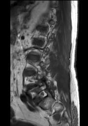
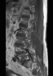
 1 Normal Anatomy and Variants 1.1 Normal Anatomy MR Technique. e standard MR protocol for a routine evaluation of the spine always comprises imaging in sagittal and axial planes, while coronal images are
Surface Appearance of the Vertebrobasilar Artery Revealed on Basiparallel Anatomic Scanning (BPAS) MR Imaging: Its Role for Brain MR Examination
 MR Imaging: Its Role for Brain MR Examination") AJNR Am J Neuroradiol 26:2508 2513, November/December 2005 Surface Appearance of the Vertebrobasilar Artery Revealed on Basiparallel Anatomic Scanning (BPAS) MR Imaging: Its Role for Brain MR Examination
AJNR Am J Neuroradiol 26:2508 2513, November/December 2005 Surface Appearance of the Vertebrobasilar Artery Revealed on Basiparallel Anatomic Scanning (BPAS) MR Imaging: Its Role for Brain MR Examination
Spinal dural AV fistula: One stop shop imaging with MR?
 Spinal dural AV fistula: One stop shop imaging with MR? Poster No.: C-3378 Congress: ECR 2010 Type: Educational Exhibit Topic: Neuro Authors: J. C. Röper-Kelmayr 1, C. Ginthör 1, D. Flöry 1, R. Chapot
Advanced Vascular Imaging: Pulsatile Tinnitus. Disclosures. Pulsatile Tinnitus: Differential Diagnosis. Pulsatile Tinnitus
 Advanced Vascular Imaging: Pulsatile Tinnitus Patrick Turski MD, Zach Clark MD, Tabby Kennedy MD The Objectives of this presentation are to: Review the differential diagnosis of pulsatile tinnitus Discuss
Tips and Tricks of State of the art MRA
 Tips and Tricks of State of the art MRA Mayil Krishnam, MD,MBA, MRCP,FRCR(UK) Professor of Radiology Director, Cardiovascular and Thoracic Imaging University of California, Irvine Objectives Technical
NEUROVASCULAR ANATOMY
 E. Michael Harned, M.D. Assistant Professor of Clinical Radiology ndiana University School of Medicine have nothing to disclose Neurovascular Magnetic Resonance Angiography And Magnetic Resonance Venography
MRI of the Brain: A Primer on What, How, Why, and When. September Amit Malhotra, Harvard Medical School, Year- IV. Gillian Lieberman, MD
 September 2000 MRI of the Brain: A Primer on What, How, Why, and When Hornak, J.P. The Basics of MRI. 1996-2000 Amit Malhotra, Harvard Medical School, Year- IV Magnetic Resonance Imaging A Brief History
Cervical Spondylosis: Three-dimensional Gradient-Echo MR with Magnetization Transfer
 Cervical Spondylosis: Three-dimensional Gradient-Echo MR with Magnetization Transfer Elias R. Melhem, Mark L. Benson, Norman J. Beauchamp, and Roland R. Lee PURPOSE: To compare a three-dimensional Fourier
Revised Dec Spine MR Protocols
 Spine MR Protocols Sp 1: Cervical spine MRI without contrast Sp 2: Pre- and post-contrast cervical spine MRI Sp 3: Pre- and post-contrast cervical spine MRI (multiple sclerosis protocol) Sp 4: Thoracic
Stroke imaging. Why image stroke patients? Stroke. Treatment of infarct. Methods for infarct diagnosis. Treatment of infarct.
 Stroke imaging Stroke Infarct: -Arterial thrombosis/embolus -Hypoxic/ischemic -Venous thrombosis Non-traumatic hemorrhage: -Intracerebral -Subarachnoid Johan Wikström MD PhD Associate Professor of Radiology
The central nervous system
 Sectc.qxd 29/06/99 09:42 Page 81 Section C The central nervous system CNS haemorrhage Subarachnoid haemorrhage Cerebral infarction Brain atrophy Ring enhancing lesions MRI of the pituitary Multiple sclerosis
Comparison of MERGE and Axial T2-Weighted Fast Spin-Echo Sequences for Detection of Multiple Sclerosis Lesions in the Cervical Spinal Cord
 Neuroradiology/Head and Neck Imaging Original Research Martin et al. MRI Detection of Multiple Sclerosis Neuroradiology/Head and Neck Imaging Original Research Nancy Martin 1 David Malfair 2 Yinshan Zhao
SMRT Student Scope Submission
 SMRT Student Scope Submission Title & Author Title: MRI in the Detection and Diagnosis of Acoustic Neuroma Author: Manya Lovesky Andersen, student Massachusetts College of Pharmacy and Health Sciences,
41 year old female with headache. Elena G. Violari MD and Leo Wolansky MD
 41 year old female with headache Elena G. Violari MD and Leo Wolansky MD ? Dural Venous Sinus Thrombosis with Hemorrhagic Venous Infarct Acute intraparenchymal hematoma measuring ~3 cm in diameter centered
Attenuation value in HU From -500 To HU From -10 To HU From 60 To 90 HU. From 200 HU and above
 Brain Imaging Common CT attenuation values Structure Air Fat Water Brain tissue Recent hematoma Calcifications Bone Brain edema and infarction Normal liver parenchyma Attenuation value in HU From -500
Laurie A. Loevner, MD
 Laurie A. Loevner, MD Chief, Division of Neuroradiology UPHS Professor of Radiology, Otorhinolaryngology: Head & Neck Surgery, Neurosurgery, and Ophthalmology University of Pennsylvania Health System Disclosures
Fat Suppression in the Abdomen
 Clinical How I do it? Fat Suppression in the Abdomen Wilhelm Horger Siemens Medical Solutions, Erlangen, Germany Introduction Due to the different chemical environment, hydrogen nuclei in - and in -tissue
Detection of Leptomeningeal CNS Metastases in Children
 Detection of Leptomeningeal CNS Metastases in Children Noah D. Sabin, M.D. Julie H. Harreld M.D. Kathleen J. Helton M.D. Zoltan Patay M.D., Ph.D. St. Jude Children s Research Hospital Memphis, TN Leptomeningeal
Half-Fourier Acquisition Single-Shot Turbo Spin-Echo (HASTE) MR: Comparison with Fast Spin-Echo MR in Diseases of the Brain
 MR: Comparison with Fast Spin-Echo MR in Diseases of the Brain") Half-Fourier Acquisition Single-Shot Turbo Spin-Echo (HASTE) MR: Comparison with Fast Spin-Echo MR in Diseases of the Brain Mahesh R. Patel, Roman A. Klufas, Ronald A. Alberico, and Robert R. Edelman PURPOSE:
Half-Fourier Acquisition Single-Shot Turbo Spin-Echo (HASTE) MR: Comparison with Fast Spin-Echo MR in Diseases of the Brain Mahesh R. Patel, Roman A. Klufas, Ronald A. Alberico, and Robert R. Edelman PURPOSE:
Brain tissue and white matter lesion volume analysis in diabetes mellitus type 2
 Brain tissue and white matter lesion volume analysis in diabetes mellitus type 2 C. Jongen J. van der Grond L.J. Kappelle G.J. Biessels M.A. Viergever J.P.W. Pluim On behalf of the Utrecht Diabetic Encephalopathy
Carlos Torres MD, FRCPC, Associate Professor of Radiology Department of Radiology, University of Ottawa
 Carlos Torres MD, FRCPC, Associate Professor of Radiology Department of Radiology, University of Ottawa catorres@toh.on.ca None 1. Simplify the complex imaging anatomy of the BP using clear anatomical
MR Imaging of Atherosclerotic Plaques
 MR Imaging of Atherosclerotic Plaques Yeon Hyeon Choe, MD Department of Radiology, Samsung Medical Center, Sungkyunkwan University, Seoul MRI for Carotid Atheroma Excellent tissue contrast (fat, fibrous
This presentation is the intellectual property of the author. Contact them for permission to reprint and/or distribute.
 MRI of the Knee Jennifer Swart, M.D. Musculoskeletal Radiology South Texas Radiology Group Outline Coils, Patient Positioning Acquisition Parameters, Planes and Pulse Sequences Knee Arthrography Normal
Susceptibility-weighted MRI ups contrast, offers minute detail 9/15/04 By: Shalmali Pal
 Susceptibility-weighted MRI ups contrast, offers minute detail 9/15/04 By: Shalmali Pal With its flashy sequences and high-speed protocols, there's no shortage of razzle-dazzle in MRI. But learning to
MR imaging at 3.0 tesla of glossopharyngeal neuralgia by neurovascular compression
 MR imaging at 3.0 tesla of glossopharyngeal neuralgia by neurovascular compression Poster No.: C-1281 Congress: ECR 2011 Type: Scientific Exhibit Authors: M. Nishihara 1, T. Noguchi 1, H. Irie 1, K. Sasaguri
Imaging of Hearing Loss
 Contemporary Imaging of Sensorineural Hearing Loss Imaging of Hearing Loss Discussion Outline (SNHL) Imaging Approaches Anatomic Relationships Lesions: SNHL KL Salzman, MD University of Utah School of
HEAD AND NECK IMAGING. James Chen (MS IV)
") HEAD AND NECK IMAGING James Chen (MS IV) Anatomy Course Johns Hopkins School of Medicine Sept. 27, 2011 OBJECTIVES Introduce cross sectional imaging of head and neck Computed tomography (CT) Review head
HEAD AND NECK IMAGING James Chen (MS IV) Anatomy Course Johns Hopkins School of Medicine Sept. 27, 2011 OBJECTIVES Introduce cross sectional imaging of head and neck Computed tomography (CT) Review head
This presentation is the intellectual property of the author. Contact them at for permission to reprint and/or distribute.
 MRI of the Knee Jennifer Swart, M.D. Musculoskeletal Radiology South Texas Radiology Group Financial Disclosure Dr. Jennifer Swart has no relevant financial relationships with commercial interests to disclose.
FOR CMS (MEDICARE) MEMBERS ONLY NATIONAL COVERAGE DETERMINATION (NCD) FOR MAGNETIC RESONANCE IMAGING:
 MEMBERS ONLY NATIONAL COVERAGE DETERMINATION (NCD) FOR MAGNETIC RESONANCE IMAGING:") National Imaging Associates, Inc. Clinical guidelines BONE MARROW MRI Original Date: July 2008 Page 1 of 5 CPT Codes: 77084 Last Review Date: September 2014 NCD 220.2 MRI Last Effective Date: July 2011
National Imaging Associates, Inc. Clinical guidelines BONE MARROW MRI Original Date: July 2008 Page 1 of 5 CPT Codes: 77084 Last Review Date: September 2014 NCD 220.2 MRI Last Effective Date: July 2011
Case Studies in the Skull Base
 Case Studies in the Skull Base Amy C Tsai, MD Neuroradiology Fellow Department of Radiology and Imaging Sciences University of Utah Health Sciences Center Salt Lake City, Utah, USA No disclosures related
Stroke Imaging Basics. Jeremy Hopkin M.D.
 Stroke Imaging Basics Jeremy Hopkin M.D. Goals Introduce the basic physical properties of imaging used in stroke. Understand why each modality is used in the setting of stroke. Understand some strengths
Introduction to Neuroimaging spine. John J. McCormick MD
 Introduction to Neuroimaging spine John J. McCormick MD Neuroanatomy Netter drawings Radiographic Anatomy Cervical Spine Cervical Spine Oblique View Cervical Spine Dens View Thoracic Spine Lumbar Spine
Dynamic 3D MR Angiography of Intra- and Extracranial Vascular Malformations at 3T: A Technical Note
 AJNR Am J Neuroradiol 26:630 634, March 2005 Technical Note Dynamic 3D MR Angiography of Intra- and Extracranial Vascular Malformations at 3T: A Technical Note S. Ziyeh, R. Strecker, A. Berlis, J. Weber,
Non-Traumatic Neuro Emergencies
 Department of Radiology University of California San Diego Non-Traumatic Neuro Emergencies John R. Hesselink, M.D. Nontraumatic Neuroemergencies 1. Acute focal neurological deficit 2. Worst headache of