David Rand, MD April 5, 2012
|
|
|
- Noah McDowell
- 6 years ago
- Views:
Transcription

1 David Rand, MD April 5, 2012
2 History 46-yr-old woman presented to SIUH ophthalmology clinic 1/20/12 for initial visit, referred by endocrinology for routine visual field testing for pituitary adenoma No visual complaints or vision changes PMHx: As above, hypothyroidism POHx: Refractive error FamHx: No Hxglc/blindness Meds: No drops Allergies: NKDA
3 Examination 1/20/12 dvacc 20/25 OU EOM full OU CVF full OU Pupils 5-3 OU, no APD Tap: 14/14 at 10:15 AM SLE WNL OU DFE V clear OU M flat OU CD 0.35/0.35, s/p OU V WNL OU P no evidence of detachment, hole, or tear OU
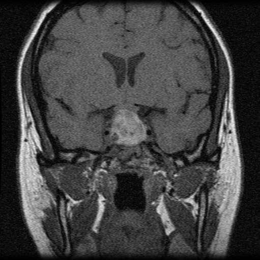
4 1/20/12 MRI sella/pituitary with/without contrast 12/19/11: Pituitary macroadenoma 1.8 x 1.7 cm Plan: --HVF 30-2 within 3 weeks --F/up with endocrinology as previously scheduled
5 3/4/12 Consulted at SIUH ED for vision loss OD Pt had presented on 2/28/12 to the ED c/o severe headache, nausea, and vomiting that started the previous night after coming home from a visual field test that was normal per pt Head CT: sinusitis Pt was discharged home Pt s symptoms persisted, and pt awoke a couple days later with painless total vision loss OD Pt waited 2 days to present to the ED since onset of vision loss on 3/2/12 because she felt delirious as a result of her severe headache and vomiting No eye pain Ophthalmology service only initial consultant
6 Examination in ED 3/4/12 nvasc OD NLP all directions, OS 20/25 EOM + esotropia OD, -4 abduction OD; full OS (right 6 th nerve palsy) CVF full OS Pupils 5mm, non-reactive OD, 5-3 OS, + APD OD Tpalp WNL OU CN III, IV, V1, V2, V3, VII, VIII, IX, X, XI, XII grossly intact bilaterally PLE WNL OU (beside non-reactive pupil OD), no proptosis, no resistance to retropulsion OU, no facial rash DFE V clear OU M flat OU CD 0.35/0.35, s/p OU, mild fullness to disc OD, but no edema V WNL OU P no evidence of detachment, hole, or tear OU
7
8 Differential Diagnosis Pituitary Apoplexy Vasculopathic 6 th Nerve Palsy Lyme Disease Tertiary Syphillus Giant Cell Arteritis Aneurysm/Subarachnoid Hemorrhage Intracranial Neoplasm/Metastases Multiple Sclerosis Early Herpes Zoster Angle-closure glaucoma
9 STAT MRI brain with/without contrast ordered STAT neurology/neurosurgical evaluations suggested Discussed findings with chief resident and attending on call

10 12/19/11 3/4/12
11 Differential Diagnosis Pituitary Apoplexy Vasculopathic 6 th Nerve Palsy Giant Cell Arteritis Lyme Disease Tertiary Syphillus Aneurysm/Subarachnoid Hemorrhage Intracranial Neoplasm/Metastases Multiple Sclerosis Early Herpes Zoster

 and is contiguous with antero-inferior floor of 3 rd")
12 Pituitary/Optic Chiasm Anatomy Chiasm derived from Greek letter chi (x) Optic chiasm is a flattened structure situated approx 10mm above pituitary gland, angled about 45 0 Pituitary gland rests in sellaturcica of sphenoid bone Chiasm and pituitary are separated by suprasellar or inferior chiasmatic cistern Chiasm may be in normal, prefixed (5-12%), or postfixed (4%) orientation relative to sellar structures Chiasm is flanked by supraclinoid segments of the carotid arteries (ICA) and is contiguous with antero-inferior floor of 3 rd ventricle

13 Wilbrand s Knee Originally noted by Michael as early as 1887, described by Wilbrand starting in 1904 Group of crossing, inferior nasal quadrant, extramacular ganglion cell axons that loop anteriorly to posterior contralateral optic nerve before looping posteriorly and laterally to the optic tract However, more recently Horton suggested that it may only be an artifact of previously studied preparations
 Anterior lobe (adenohypophysis) forms embryologically from Rathke s")
14 Anatomy Pituitary infundibulum arises from hypothalamus behind the chiasm, extends to posterior lobe of pituitary (neurohypophysis) Anterior lobe (adenohypophysis) forms embryologically from Rathke s pouch
15

16 Pituitary Adenoma 12-15% of symptomatic intracranial neoplasms Most common lesion causing chiasmal dysfunction Uncommon before age 20, incidence increases after 4 th decade Autopsies revealed that prevalence of asymptomatic adenomas may be as high as 20-27%, and adenomatous hyperplasia may be found in almost every pituitary gland
17 Pituitary Adenoma Classified according to size: Microadenoma: < 10mm in largest diameter Macroadenoma: > 10mm in largest diameter Functional vs. non-functional May also be distinguished anatomically/radiologically: Intrapituitary, intrasellar, diffuse, or invasive Invasive adenomas: Approx 35% of all pituitary neoplasms, may invade dura mater, cranial bone, cavernous sinus, or sphenoid bone
18 Pituitary Adenoma Separation of chiasm from pituitary by suprasellar (or inferior chiasmatic) cistern enables mild-moderate suprasellar extension of tumors to occur without chiasmal visual field loss If visual field loss present, advanced enlargement with expansion beyond diaphragm sella is expected May be endocrine inactive or active; active tumors likely cause systemic signs and symptoms before affecting visual pathways Symptoms include infertility, unexplained breast milk production, impotence, oligomenorrhea, acromegaly, weight loss, increased appetite, tachycardia, tremors, frequent bowel movements, backache, thin skin, flushed face, weak and fragile muscles and bones, wrinkles and stretch marks, excessive hair growth, mood swings, osteoporosis of the rib and vertebrae, hyperglycemia/diabetes, and/or fat build up in the face, back, and chest Important to consider work up for all patients with non-specific headaches and/or endocrine abnormalities
19 Progression of Visual Field Defects Both superotemporal fields are usually affected first as tumor grows upwards and splays anterior chiasmal notch, compressing crossing inferonasal fibers Progress to lower temporal fields, usually asymmetric given irregularity of tumor shape/growth Progression to macular fibers with central vision loss (usually first in eye with greater field deficits) Remember: differential for bitemporal deficits include dermatochalasis of upper lids, tilted discs, optic nerve colobomas, nasal retinoschisis, and nasal retinitis pigmentosa
20 Color Desaturation May occur across vertical midline of monocular visual field Each eye tested separately, patient is asked to compare color of red top as it is brought from nasal to temporal visual field Alternatively, place red tops at symmetric points in nasal and temporal visual fields, ask patient to compare intensity Patient may also miss temporal number on Ishihara testing
21 Pregnancy Pituitary adenomas (especially prolactinomas) are sensitive to increased levels of estrogen and progesterone May enlarge during pregnancy causing visual symptoms may abate after delivery
22 Other Causes of Chiasmal Dysfunction Meningioma (also enlarge during pregnancy) Craniopharyngioma Optic glioma Lymphocytic adenohypophysitis rare, immune-mediated diffuse lymphocytic infiltration of pituitary gland that may cause chiasmal compression from suprasellar extension; reported in women only, over 50% of cases in perinatal period Other tumors/metastases/cysts (chordoma, dysgerminoma, nasopharyngeal tumors, Rathke s pouch cyst, sphenoid mucocele) Cavernous hemangioma/av malformation Hydrocephalus/enlarged 3 rd ventricle Infectious/inflammatory/demyelinating etiologies Trauma Toxins Rarely congenital chiasmal dysplasia

23 Pituitary Apoplexy Apoplexy originates from Latin word meaning stroke or struck Acute, unpredictable, lifethreatening Sudden enlargement of pituitary gland, usually in setting of adenoma, although no increased risk depending on histological subtype of tumor May be caused by hemorrhage or infarction Acute headache, visual loss, ophthalmoplegia, and/or facial pain or numbness Tumor of any size can undergo hemorrhagic necrosis
24 Pituitary Apoplexy Thought to occur in % of all surgically-managed cases of pituitary adenoma Age range is broad, from 1 st to 9 th decade, estimated to peak in 5 th decade No gender predilection Ocular manifestations: usually painful ophthalmoplegia and vision loss Vision loss is variable, may not always produce an APD Visual field defects common Degree of ophthalmoplegia variable, depending on involvement of each cavernous sinus
25 Pituitary Apoplexy Predisposing factors include pregnancy, estrogen therapy, obstetrical hemorrhage (Sheehan syndrome), diabetes, bleeding disorders, long-term anticoagulation, blood dyscrasias, radiation therapy, hypotension trauma, angiography, atheromatous emboli, cardiac surgery, coughing, positive pressure ventilation, and vasoconstrictive agents
26 Pituitary Apoplexy Rapid expansion into structures such as --optic chiasm --hypothalamus --cavernous sinus may occur not uncommonly. This condition is a neurosurgical emergency Most patients experience meningeal irritation Hypofunction of gland common afterward
27 Diagnosis MRI is gold standard for neuroimaging Will delineate tumor and hemorrhage Much more sensitive than CT scan (estimated CT only diagnoses apoplexy 46% of the time) If pituitary apoplexy not found on MRI, MRA or CTA should be performed to rule out expanding or ruptured aneurysm

28 , Practice-based Learning and Improvement

29 Treatment Trans-sphenoidal decompression of sella in patients with neuroophthalmic abnormality Bromocriptine (dopamine agonist) has been advocated in cases with little or no deficit Supplementation of pituitary hormones often necessary for prolonged period after apoplectic event
30 Course/Outcome Good prognosis with surgical decompression Improvement of visual acuity, field deficits, and ophthalmoplegia reported to be as high as 76-91% of cases Endocrine abnormalities, however, remain high with 43-58% requiring some form of hormonal supplementation Mortality with surgical management low
31 Back to Our Patient Pt was placed on IV steroids (dosed per neurosurgery) and was prepared for emergent trans-sphenoidal resection of tumor which was performed urgently Postoperatively in ICU pt reported some vision present in right eye (HM), and 6 th nerve palsy had resolved, Systems-based Practice
32 D Agrawal, A Mahapatra. Visual outcome of blind eyes in pituitary apoplexy after transsphenoidal surgery: a series of 14 eyes. Surgical Neurology Volume 63 Issue 1, Pages 42-46, January blind eyes secondary to pituitary apoplexy, now s/p trans-sphenoidal surgery Average delay to neurosurgical consultation 10 days, total range 4 to 30 days 4 eyes improved to greater than 20/60 2 eyes improved to 20/20 All patients with improvement of vision had undergone operation within 1 week of apoplectic episode, Practice-Based Learning and Improvement
33
34 3/27/12 dvacc OD 20/40 +2, OS 20/20 EOM full OU Complaint of residual superotemporal field defect OD + APD OD
35
36

37 Etiology of 6 th nerve palsy?, Practice-based Learning and Improvement
38 Reflective Practice Pt was treated in a timely manner in life-threatening situation with improvement of symptoms NLP is not always NLP Patients do not always read the textbooks; this presentation reflects importance of a differential diagnosis, in this case ranging from pituitary apoplexy to angle-closure glaucoma, for a consult with chief complaint of vision loss and pain Professionalism
39 Core Competencies Patient Care compassionate, appropriate, and effective in the evaluation and treatment of this patient. Medical Knowledge comprehensive literature search was performed to better understand the evaluation and treatment of this condition. A better understanding of both basic and clinical science of this condition was attained, and relevant topics were discussed among residents and attending physicians Practice-based learning and improvement Care of patient was discussed among residents and faculty. Scientific studies were reviewed to ensure the highest level of evidence-based practice. Suggested protocols in the literature were analyzed and discussed to refine treatment plan for our patient and future patients Interpersonal and communication skills all questions of the patient and family were answered in a complete and caring manner to allay the fears of the patient and family. A professional relationship based on compassion and trust was established for the patient s wellbeing. Pt was followed adequately for consistently excellent medical care and to maintain this relationship. Professionalism Responsibility of potential complications of medical and surgical treatment regimen was accepted by the entire team and communicated to the patient and family. Professional relationships were established and maintained with physicians from other fields. Systems-based Practice communication with physicians from other fields was emphasized in this case to provide multi-specialty care in the best fashion possible. Acute care was provided, follow up appointments were scheduled, contact information was exchanged, and reports to primary care physician given to ensure a successful long-term outcome.
40 Thank you Dr. Nejat Dr. Ferri Dr. Mostafavi Dr. Oren Herman Dr. Adam Bernheim
No Financial Interest
 Pituitary Apoplexy Michael Vaphiades, D.O. Professor Department of Ophthalmology, Neurology, Neurosurgery University of Alabama at Birmingham, Birmingham, AL No Financial Interest N E U R O L O G I C
Laurie A. Loevner, MD
 Laurie A. Loevner, MD Chief, Division of Neuroradiology UPHS Professor of Radiology, Otorhinolaryngology: Head & Neck Surgery, Neurosurgery, and Ophthalmology University of Pennsylvania Health System Disclosures
Visual pathways in the chiasm
 Visual pathways in the chiasm Intracranial relationships of the optic nerve Fixation of the chiasm Chiasmatic pathologies The function of the optic chiasm may be altered by the presence of : 4) Artero
Imaging The Turkish Saddle. Russell Goodman, HMS III Dr. Gillian Lieberman
 Imaging The Turkish Saddle Russell Goodman, HMS III Dr. Gillian Lieberman Learning Objectives Review the anatomy of the sellar region Discuss the differential diagnosis of sellar masses Discuss typical
PITUITARY PARASELLAR LESIONS. Kim Learned, MD
 PITUITARY PARASELLAR LESIONS Kim Learned, MD DIFFERENTIALS Pituitary Sella Clivus, Sphenoid Sinus Suprasellar Optic chiasm, Hypothalamus, Circle of Willis Parasellar Cavernous Sinus Case 1 17 YEAR-OLD
Metastasis. 57 year old with progressive Headache and Right Sided Visual Loss
 Metastasis 1% of sellar/parasellar masses Usually occurs with known primary Can involve third ventricle, hypothalamus, infundibular stalk May be both supra-, intrasellar 57 year old with progressive Headache
A Case of Carotid-Cavernous Fistula
 A Case of Carotid-Cavernous Fistula By : Mohamed Elkhawaga 2 nd Year Resident of Ophthalmology Alexandria University A 19 year old male patient came to our outpatient clinic, complaining of : -Severe conjunctival
Imaging pituitary gland tumors
 November 2005 Imaging pituitary gland tumors Neel Varshney,, Harvard Medical School Year IV Two categories of presenting signs of a pituitary mass Functional tumors present with symptoms due to excess
panhypopituitarism Pattawan Wongwijitsook Maharat Nakhon Ratchasima hospital 17 Nov 2013
 panhypopituitarism Pattawan Wongwijitsook Maharat Nakhon Ratchasima hospital 17 Nov 2013 PITUITARY GLAND (HYPOPHYSIS CEREBRI) The master of endocrine glands master of endocrine glands It is a small oval
NANOS Patient Brochure
 NANOS Patient Brochure Pituitary Tumor Copyright 2015. North American Neuro-Ophthalmology Society. All rights reserved. These brochures are produced and made available as is without warranty and for informational
Case Studies in Sella/Parasellar Region. Child thirsty, increased urination. Imaging. Suprasellar Germ Cell Tumor (Germinoma) No Disclosures
 No Disclosures") Case Studies in Sella/Parasellar Region No Disclosures 2018 Head and Neck Imaging Conference Child thirsty, increased urination Suprasellar Germ Cell Tumor (Germinoma) Midline Pineal >> Suprasellar > Other
Case Studies in Sella/Parasellar Region No Disclosures 2018 Head and Neck Imaging Conference Child thirsty, increased urination Suprasellar Germ Cell Tumor (Germinoma) Midline Pineal >> Suprasellar > Other
Topical Diagnosis of Chiasmal and Retrochiasmal Disorders
 Topical Diagnosis of Chiasmal and Retrochiasmal Disorders Leonard A. Levin CHAPTER 12 TOPICAL DIAGNOSIS OF OPTIC CHIASMAL LESIONS Visual Field Defects Etiologies of the Optic Chiasmal Syndrome Masqueraders
Where Has My Vision Gone? Evaluation of Sellar Lesions. Caleb Stowell,, HMS III Gillian Lieberman, MD November 2008
 Where Has My Vision Gone? Evaluation of Sellar Lesions Caleb Stowell,, HMS III Gillian Lieberman, MD November 2008 Objectives Present a case highlighting the clinical presentation and evaluation of a sellar
Diseases of pituitary gland
 Diseases of pituitary gland A brief introduction Anterior lobe = adenohypophysis Posterior lobe = neurohypophysis The production of most pituitary hormones is controlled in large part by positively and
DISCLOSURES LEARNING OBJECTIVES WE WILL NOT DISCUSS. CSB: Birdseye View MESSAGE NAVIGATING THE SELLA AND CENTRAL SKULL BASE
 NAVIGATING THE SELLA AND CENTRAL SKULL BASE Christopher P. Hess, M.D., Ph.D. DISCLOSURES Research Support, General Electric SLIDES: http://www.radiology.ucsf.edu/research/meetings/rsna LEARNING OBJECTIVES
Sharon maslovitz Lis Maternity Hospital
 Sharon maslovitz Lis Maternity Hospital Case report Chief complaint 27 yo, with PMC @ 31+3w, BCBA twins Complaints of severe rt parietal and retrobulbar headaches Medical background Healthy until 24yo
Mechanism of hyperprolactinemia
 Hyperprolactinemia Mechanism of hyperprolactinemia Causes of hyperprolactinemia Hormone-producing pituitary tumors Prolactinoma Acromegaly Hypothalamic/pituitary stalk lesion Tumors, cysts (craniopharyngeoma,
Urgent and Emergent Pituitary Conditions
 Urgent and Emergent Pituitary Conditions PANKAJ A. GORE, MD DIRECTOR, BRAIN AND SKULL BASE T UMOR SURGERY PROVIDENCE B R AIN AND S PINE I NSTITUTE Urgent and Emergent Pituitary Conditions Neurosurgical
THE OPTIC CHIASM MAY BE DAMAGED BY A VARIETY
 Clinical Features Associated With Lesions Other Than Pituitary Adenoma in Patients With an Optic Chiasmal Syndrome LUIS J. MEJICO, MD, NEIL R. MILLER, MD, AND LI MING DONG, PHD PURPOSE: Pituitary adenomas
Pituitary Apoplexy. Updated: April 22, 2018 CLINICAL RECOGNITION
 Pituitary Apoplexy Zeina C Hannoush, MD. Assistant Professor of Clinical Medicine. Division of Endocrinology, Diabetes and Metabolism. University of Miami, Miller School of Medicine. Roy E Weiss, MD, PhD,
Intrasphenoidal Rathke's Cleft Cyst: Case presentation and review of the literature
 Romanian Neurosurgery Volume XXX Number 4 2016 October - December Article Intrasphenoidal Rathke's Cleft Cyst: Case presentation and review of the literature Umit Kocaman, Muhammet Bahadir Yilmaz, Hakan
TABLES. Imaging Modalities Evidence Tables Table 1 Computed Tomography (CT) Imaging. Conclusions. Author (Year) Classification Process/Evid ence Class
 Imaging. Conclusions. Author (Year) Classification Process/Evid ence Class") TABLES Imaging Modalities Evidence Tables Table 1 Computed Tomography (CT) Imaging Author Clark (1986) 9 Reformatted sagittal images in the differential diagnosis meningiomas and adenomas with suprasellar
TABLES Imaging Modalities Evidence Tables Table 1 Computed Tomography (CT) Imaging Author Clark (1986) 9 Reformatted sagittal images in the differential diagnosis meningiomas and adenomas with suprasellar
Pituitary Macroadenoma with Superior Orbital Fissure Syndrome
 1 CASE REPORT OPEN ACCESS Pituitary Macroadenoma with Superior Orbital Fissure Syndrome Tapan Nagpal, Ankit Singhania ABSTRACT Introduction: Pituitary adenomas are benign tumours which arise within the
Disclosures. Visual Pathways. Visual Pathways. Visual Loss Understanding the Patterns. I have no financial disclosures. Tabby A.
 Visual oss Understanding the Patterns Tabby A. Kennedy, MD University of Wisconsin Department of adiology I have no financial disclosures Acknowledgements: indell Gentry Greg Avey JP Yu Judy Chen Disclosures
In some patients with pituitary macroadenoma, visual acuity
 ORIGINAL RESEARCH A.M. Tokumaru I. Sakata H. Terada S. Kosuda H. Nawashiro M. Yoshii Optic Nerve Hyperintensity on T2-Weighted Images among Patients with Pituitary Macroadenoma: Correlation with Visual
PITUITARY: JUST THE BASICS PART 2 THE PATIENT
 PITUITARY: JUST THE BASICS PART 2 THE PATIENT DISCLOSURE Relevant relationships with commercial entities none Potential for conflicts of interest within this presentation none Steps taken to review and
CHAPTER 13 CLINICAL CASES INTRODUCTION
 2 CHAPTER 3 CLINICAL CASES INTRODUCTION The previous chapters of this book have systematically presented various aspects of visual field testing and is now put into a clinical context. In this chapter,
Case Report Rapid Pituitary Apoplexy Regression: What Is the Time Course of Clot Resolution?
 Case Reports in Radiology Volume 2015, Article ID 268974, 5 pages http://dx.doi.org/10.1155/2015/268974 Case Report Rapid Pituitary Apoplexy Regression: What Is the Time Course of Clot Resolution? Devon
Case Report Successful Pregnancy in a Female with a Large Prolactinoma after Pituitary Tumor Apoplexy
 Case Reports in Obstetrics and Gynecology Volume 2013, Article ID 817603, 4 pages http://dx.doi.org/10.1155/2013/817603 Case Report Successful Pregnancy in a Female with a Large Prolactinoma after Pituitary
Jacqueline Theis, O.D., F.A.A.O.
 Neuro-Ophthalmological Emergencies Presenting in Primary Care Optometry Describes the symptoms, signs, and management of neuro-ophthalmological emergencies. Signs/Symptoms to be Concerned about (especially
Sharon maslovitz Lis Maternity Hospital
 Sharon maslovitz Lis Maternity Hospital Case report Chief complaint 27 yo, with PMC @ 31+3w, BCBA twins Complaints of severe rt parietal and retrobulbar headaches Conditions that may cause episodic headaches:
Picture of patient with apparent lid retraction and poor elevation. Shows you Orbital CT-Scan with muscle involvement including IR and asks is this
 NEUROLOGY Q: MENINGIOMAS AND SKULL (*2) Real skull is given, and you are asked to point to tuberculum sella What kind of meningioma occurs at this location? Where is the anterior clinoid process? Where
A Case of Pituitary Apoplexy in Third Trimester of Pregnancy
 IOSR Journal of Dental and Medical Sciences (IOSR-JDMS) e-issn: 2279-0853, p-issn: 2279-0861.Volume 17, Issue 3 Ver.16 March. (2018), PP 01-06 www.iosrjournals.org A Case of Pituitary Apoplexy in Third
The View through the Nose: ENT considerations for Pituitary/Skull Base Surgery
 The View through the Nose: ENT considerations for Pituitary/Skull Base Surgery Edsel Kim, M.D. Otolaryngology-Head and Neck Surgery The Oregon Clinic Providence Brain and Spine Institute Pituitary, Thyroid
EXPERT DIFFERENTIAL DIAGNOSIS:
 EXPERT DIFFERENTIAL DIAGNOSIS: Sellar Region Anne G. Osborn, M.D. DISCLOSURE: Published RSNA 2008 SELLA, PITUITARY: Normal Gross, 3T Anatomy SELLA, PITUITARY: Anatomically-Based Differential Diagnoses
Optic Pathway Gliomas, Germinomas, Spinal Cord Tumours. Colin Kennedy March 2015
 Optic Pathway Gliomas, Germinomas, Spinal Cord Tumours Colin Kennedy March 2015 Glioma of the optic chiasm. T1-weighted MRI with gadolinium enhancement, showing intense irregular uptake of contrast. The
Optic Nerve Disorders: Structure and Function and Causes
 Optic Nerve Disorders: Structure and Function and Causes Using Visual Fields, OCT and B-scan Ultrasound to Diagnose and Follow Optic Nerve Visual Losses Ohio Ophthalmological Society and Ophthalmic Tech
Factors Influencing Visual Field Recovery after Transsphenoidal Resection of a Pituitary Adenoma
 pissn: 1011-8942 eissn: 2092-9382 Korean J Ophthalmol 2018;32(6):488-496 https://doi.org/10.3341/kjo.2017.0094 Original Article Factors Influencing Visual Field Recovery after Transsphenoidal Resection
Pituitary Tumors: adenoma, craniopharyngioma, rathke cyst
 Pituitary Tumors: adenoma, craniopharyngioma, rathke cyst Overview Tumors that grow from the pituitary gland can affect the whole body by interfering with normal hormone levels. They can also cause headaches
Carotid Cavernous Fistula
 Chief Complaint: Double vision. Carotid Cavernous Fistula Alex W. Cohen, MD, PhD; Richard Allen, MD, PhD May 14, 2010 History of Present Illness: A 46 year old female patient presented to the Oculoplastics
RADIOANATOMY OF SELLA TURCICA
 RADIOANATOMY OF SELLA TURCICA O.BAKKACHA, H.MALAJATI, M.RHISSASSI, H. BENCHAABOUNE, N.CHAKIR, My R. EL HASSANI,M.JIDDANE Department of Neuroradiology specialties Hospital. Rabat Objective: New imaging
Non-arteritic anterior ischemic optic neuropathy (NAION) with segmental optic disc edema. Jonathan A. Micieli, MD Valérie Biousse, MD
 with segmental optic disc edema. Jonathan A. Micieli, MD Valérie Biousse, MD") Non-arteritic anterior ischemic optic neuropathy (NAION) with segmental optic disc edema Jonathan A. Micieli, MD Valérie Biousse, MD A 75 year old white woman lost vision in the inferior part of her visual
Non-arteritic anterior ischemic optic neuropathy (NAION) with segmental optic disc edema Jonathan A. Micieli, MD Valérie Biousse, MD A 75 year old white woman lost vision in the inferior part of her visual
Non-Traumatic Neuro Emergencies
 Department of Radiology University of California San Diego Non-Traumatic Neuro Emergencies John R. Hesselink, M.D. Nontraumatic Neuroemergencies 1. Acute focal neurological deficit 2. Worst headache of
Headache Assessment In Primary Eye Care
 Headache Assessment In Primary Eye Care Spencer Johnson, O.D., F.A.A.O. Northeastern State University Oklahoma College of Optometry johns137@nsuok.edu Course Objectives Review headache classification Understand
Pathology of pituitary gland. By: Shifaa Qa qa
 Pathology of pituitary gland By: Shifaa Qa qa Sella turcica Adenohypophysis (80%): - epithelial cells - acidophil, basophil, chromophobe - Somatotrophs, Mammosomatotrophs, Corticotrophs, Thyrotrophs, Gonadotrophs
10/23/2010. Excludes Single Surgeon Pituitary (N=~140) Skull Base Volume 12 Month UC SF. Patients. Anterior/Midline. Pituitary CSF Leak.
 Skull Base Volume 12 Month UC SF. Patients. Anterior/Midline. Pituitary CSF Leak.") Advances in Pituitary Surgery Ivan El-Sayed MD, FACS Director- Otolaryngology Minimally Invasive Skull Base Surgery Program Otolaryngology-Head and Neck Surgery University of California-San Francisco Minimally
Advances in Pituitary Surgery Ivan El-Sayed MD, FACS Director- Otolaryngology Minimally Invasive Skull Base Surgery Program Otolaryngology-Head and Neck Surgery University of California-San Francisco Minimally
Anatomy of Pituitary Gland
 Anatomy of Pituitary Gland Please view our Editing File before studying this lecture to check for any changes. Color Code Important Doctors Notes Notes/Extra explanation Objectives At the end of the lecture,
The central nervous system
 Sectc.qxd 29/06/99 09:42 Page 81 Section C The central nervous system CNS haemorrhage Subarachnoid haemorrhage Cerebral infarction Brain atrophy Ring enhancing lesions MRI of the pituitary Multiple sclerosis
Evaluation of ONH Pallor in Glaucoma Patients and Suspects. Leticia Rousso, O.D. Joseph Sowka, O.D
 Evaluation of ONH Pallor in Glaucoma Patients and Suspects Leticia Rousso, O.D Joseph Sowka, O.D I. Abstract This case report will evaluate a young glaucoma suspect with unilateral sectoral optic nerve
Learn Connect Succeed. JCAHPO Regional Meetings 2017
 Learn Connect Succeed JCAHPO Regional Meetings 2017 NO FINANCIAL DISCLOSURES Technician s Role in Neuro-Ophthalmology Workup Beth Koch COT, ROUB Cleveland 9/16/2017 What Tests Are You Expected To Perform?
Prolactin-Secreting Pituitary Adenomas (Prolactinomas) The Diagnostic Pathway (11-2K-234)
 The Diagnostic Pathway (11-2K-234)") Prolactin-Secreting Pituitary Adenomas (Prolactinomas) The Diagnostic Pathway (11-2K-234) Common presenting symptoms/clinical assessment: Pituitary adenomas are benign neoplasms of the pituitary gland.
Prolactin-Secreting Pituitary Adenomas (Prolactinomas) The Diagnostic Pathway (11-2K-234) Common presenting symptoms/clinical assessment: Pituitary adenomas are benign neoplasms of the pituitary gland.
OPTIC CHIASM. project ipsilaterally, and thus Zic2 may endow these cells with response properties that influence their trajectory at the
 EMBRYOLOGY, ANATOMY, AND PHYSIOLOGY OF THE AFFERENT VISUAL PATHWAY A 35 B Figure 1.37. Wilbrand s knee in human tissue. A, Woelcke myelin stain of a horizontal section through the optic chiasm from a patient
Visual Fields at Presentation and after Trans-sphenoidal Resection of Pituitary Adenomas
 Original Article Visual Fields at Presentation and after Trans-sphenoidal Resection of Pituitary Adenomas Renu Dhasmana, MD; Ramesh C Nagpal, MD; Rahul Sharma, MD Krishan K Bansal, MD; Harsh Bahadur, MD
33-Year-Old Female With Amenorrhea DISHA KUMAR NARANG, MD PITUITARY ENDORAMA DECEMBER 11, 2014
 33-Year-Old Female With Amenorrhea DISHA KUMAR NARANG, MD PITUITARY ENDORAMA DECEMBER 11, 2014 Our Patient 33-year-old female presents to endocrinology clinic after amenorrhea for 4 years History of Present
Case Follow Up. Sepi Jooniani PGY-1
 Case Follow Up Sepi Jooniani PGY-1 Triage 54 year old M Pt presents to prelim states noticed today he had reddness to eyes, states worse in R eye. Pt denies any pain or itching. No further complaints.
Coincidental aneurysms with tumours of pituitary origin
 Journal ofneurology, Neurosurgery, and Psychiatry, 1978, 41, 972-979 Coincidental aneurysms with tumours of pituitary origin JAN JAKUBOWSKI AND BRIAN KENDALL From the Gough Cooper Department of Neurological
TABLES. Table 1: Imaging. Congress of Neurological Surgeons Author (Year) Description of Study Classification Process / Evidence Class
 Description of Study Classification Process / Evidence Class") TABLES Table 1: Imaging Kremer et al (2002) 2 Study Design: Prospective followed case series. Patient Population: Fifty adult patients with NFPA Study Description: Patients underwent MRI before surgery,
TABLES Table 1: Imaging Kremer et al (2002) 2 Study Design: Prospective followed case series. Patient Population: Fifty adult patients with NFPA Study Description: Patients underwent MRI before surgery,
Pituitary Disease Resident Tutorial 2017
 Pituitary Disease Resident Tutorial 2017 Sarat Sunthornyothin MD Division of Endocrinology and Metabolism King Chulalongkorn Memorial Hospital Pituitary Anatomy hypophyseal portal system direct arterial
Somatotroph Pituitary Adenomas (Acromegaly) The Diagnostic Pathway (11-2K-234)
 The Diagnostic Pathway (11-2K-234)") Somatotroph Pituitary Adenomas (Acromegaly) The Diagnostic Pathway (11-2K-234) Common presenting symptoms/clinical assessment: Pituitary adenomas are benign neoplasms of the pituitary gland. In patients
Somatotroph Pituitary Adenomas (Acromegaly) The Diagnostic Pathway (11-2K-234) Common presenting symptoms/clinical assessment: Pituitary adenomas are benign neoplasms of the pituitary gland. In patients
Transplanum Approach for Suprasellar pathology
 Transplanum Approach for Suprasellar pathology Omar A. El-Banhawy Prof. of otorhinolaryngology El Menoufyia University, Egypt Why Endoscopic Approach For Suprasellar Pathology Constant improvements in
Craniopharyngioma. Michael Gottschalk, MD,PhD University of California San Diego Rady Children s Hospital
 Craniopharyngioma Michael Gottschalk, MD,PhD University of California San Diego Rady Children s Hospital Objectives Incidence Clinical Presentation Treatment Options Perioperative concerns Long-term endocrine
Neurology Case Presentation. Rawan Albadareen, MD 12/20/13
 Neurology Case Presentation Rawan Albadareen, MD 12/20/13 Case presentation A 49 y.o. female presented to the ED after an episode of zigzagging w a jagged bright light crossing through her Rt visual field
JACK L. SNITZER, DO INTERNAL MEDICINE BOARD REVIEW COURSE 2018 PITUITARY
 JACK L. SNITZER, DO INTERNAL MEDICINE BOARD REVIEW COURSE 2018 PITUITARY JACK L. SNITZER, D.O. Peninsula Regional Endocrinology 1415 S. Division Street Salisbury, MD 21804 Phone:410-572-8848 Fax:410-572-6890
Anterior Ischemic Optic Neuropathy (AION)
") Anterior Ischemic Optic Neuropathy (AION) Your doctor thinks you have suffered an episode of anterior ischemic optic neuropathy (AION). This is the most common cause of sudden decreased vision in patients
Anterior Ischemic Optic Neuropathy (AION) Your doctor thinks you have suffered an episode of anterior ischemic optic neuropathy (AION). This is the most common cause of sudden decreased vision in patients
Neuro-Ocular Grand Rounds
 Neuro-Ocular Grand Rounds Anthony B. Litwak,OD, FAAO VA Medical Center Baltimore, Maryland Dr. Litwak is on the speaker and advisory boards for Alcon and Zeiss Meditek COMMON OPTIC NEUROPATHIES THAT CAN
HYPOTHALAMO PITUITARY GONADAL AXIS
 HYPOTHALAMO PITUITARY GONADAL AXIS Physiology of the HPG axis Endogenous opioids and the HPG axis (exerciseinduced menstrual disturbances) Effects of the immune system on the HPG axis (cytokines: interleukins
Typical idiopathic intracranial hypertension Optic nerve appearance and brain MRI findings. Jonathan A. Micieli, MD Valérie Biousse, MD
 Typical idiopathic intracranial hypertension Optic nerve appearance and brain MRI findings Jonathan A. Micieli, MD Valérie Biousse, MD A 24 year old African American woman is referred for bilateral optic
53 year old woman attends your practice for routine exam. She has no past medical history or family history of note.
 Case 1 Normal Tension Glaucoma 53 year old woman attends your practice for routine exam. She has no past medical history or family history of note. Table 1. Right Eye Left Eye Visual acuity 6/6 6/6 Ishihara
Neuro Op Grand Rounds: Fields and Diplopia Utah Optometric Association June 2018
 Disclosures Neuro Op Grand Rounds: Fields and Diplopia Utah Optometric Association June 2018 I have received honorarium from the following: Glaukos CE in Italy Heidelberg Engineering Review of Optometry
Neuro-Ocular Grand Rounds Anthony B. Litwak,OD, FAAO VA Medical Center Baltimore, Maryland
 Neuro-Ocular Grand Rounds Anthony B. Litwak,OD, FAAO VA Medical Center Baltimore, Maryland Dr. Litwak is on the speaker and advisory boards for Alcon and Zeiss Meditek COMMON OPTIC NEUROPATHIES THAT CAN
Pituitary Adenomas: Patterns Of Visual Presentation And Outcome After Transsphenoidal Surgery - An Institutional Experience
 ISPUB.COM The Internet Journal of Ophthalmology and Visual Science Volume 4 Number 2 Pituitary Adenomas: Patterns Of Visual Presentation And Outcome After Transsphenoidal Surgery - An Institutional Experience
Pathologies of postchiasmatic visual pathways and visual cortex
 Pathologies of postchiasmatic visual pathways and visual cortex Optic radiation: anatomy Pathologies of the postchiamsatic visual pathways and visual cortex Characterized by homonymous hemianopsia. This
Pituitary Macroadenoma Joseph Junewick, MD FACR
 Pituitary Macroadenoma Joseph Junewick, MD FACR 08/13/2010 History 12 year old female with headache and visual disturbance. Diagnosis Pituitary Macroadenoma Additional Clinical Markedly elevated growth
Optic Disc: Anatomy, Variants, Unusual discs. Kathleen B. Digre, MD Professor Neurology, Ophthalmology
 Optic Disc: Anatomy, Variants, Unusual discs Kathleen B. Digre, MD Professor Neurology, Ophthalmology THE OPHTHALMOSCOPE DIRECT OPHTHALMOSCOPY Jan Purkinje 1823 Hermann von Helmholtz 1851 Hand held ophthalmoscope
GLMS CME- Cell Group 5 10 April Greenlane Medical Specialists Pui-Ling Chan Endocrinologist
 GLMS CME- Cell Group 5 10 April 2018 Greenlane Medical Specialists Pui-Ling Chan Endocrinologist Pituitary case one Mrs Z; 64F Seen ORL for tinnitus wax impaction MRI Head Pituitary microadenoma (3mm)
OCT : retinal layers. Extraocular muscles. History. Central vs Peripheral vision. History: Temporal course. Optical Coherence Tomography (OCT)
") Optical Coherence Tomography (OCT) OCT : retinal layers 7 Central vs Peripheral vision Extraocular muscles RPE E Peripheral Vision: Rods (95 million) 30% Ganglion cells Central Vision: Cones (5 million)
Optical Coherence Tomography (OCT) OCT : retinal layers 7 Central vs Peripheral vision Extraocular muscles RPE E Peripheral Vision: Rods (95 million) 30% Ganglion cells Central Vision: Cones (5 million)
Pituitary Tumors and Incidentalomas. Bijan Ahrari, MD, FACE, ECNU Palm Medical Group
 Pituitary Tumors and Incidentalomas Bijan Ahrari, MD, FACE, ECNU Palm Medical Group Background Pituitary incidentaloma: a previously unsuspected pituitary lesion that is discovered on an imaging study
Blue-domed cyst with optic nerve compression
 Journal ofneurology, Neurosurgery, and Psychiatry, 1978, 41, 987-991 Blue-domed cyst with optic nerve compression MITCHELL D. BURNBAUM, JOHN W. HARBISON, JOHN B. SELHORST, AND HAROLD F. YOUNG From the
LECTURE # 7 EYECARE REVIEW: PART III
 LECTURE # 7 EYECARE REVIEW: PART III HOW TO TRIAGE EYE EMERGENCIES STEVE BUTZON, O.D. EYECARE REVIEW: HOW TO TRIAGE EYE EMERGENCIES FOR PRIMARY CARE PHYSICIANS Steve Butzon, O.D. Member Director IDOC President
Orbital Tumors - A Clinico Pathological Study
 Orbital Tumors - A Clinico Pathological Study Radha. J. DO, Ani Sreedhar. MS. Little Flower Hospital, Angamaly, Kerala ORIGINAL ARTICLES Abstract: Aim. To study the clinical and histopathological profiles
Objectives. Unexplained Vision Loss: Where Do I Go From Here. History. History. Drug Induced Vision Loss
 Objectives Unexplained Vision Loss: Where Do I Go From Here Denise Goodwin, OD, FAAO Coordinator, Neuro-ophthalmic Disease Clinic Pacific University College of Optometry goodwin@pacificu.edu Know the importance
The dura is sensitive to stretching, which produces the sensation of headache.
 Dural Nerve Supply Branches of the trigeminal, vagus, and first three cervical nerves and branches from the sympathetic system pass to the dura. Numerous sensory endings are in the dura. The dura is sensitive
UC SF. g h. Eye Trauma. Martha Neighbor, MD Emergency Services San Francisco General Hospital University of California
 UC SF Eye Trauma sf g h Martha Neighbor, MD Emergency Services San Francisco General Hospital University of California Goals Recognize vision threatening eye emergencies Treat them when we can Know when
Pituitary apoplexy 台北榮總內分泌新陳代謝科主治醫師林怡君
 Pituitary apoplexy 台北榮總內分泌新陳代謝科主治醫師林怡君 Williams text book of endocrinology 11 th e Anterior pituitary hormone 10-20% of pituitary cells, increase to 40% during AP PRL releasing factors: TRH, oxytocin,
T HE visual field changes that accompany
 J. Neurosurg. / Volume 31 / September, 1969 The Arterial Supply of the Human Optic Chiasm RICHARD BERGLAND, M.D.,* AND BRONSON S. RAY, M.D. Department of Surgery (Neurosurgery), New York Hospital-Cornell
25/06/2010. Scaricato da 1
 Approcci chirurgici al Clivus DIPARTIMENTO DI NEUROCHIRURGIA SECONDA UNIVERSITÀ DI NAPOLI Prof. Aldo Moraci Surgical Anatomy of the Clivus Scaricato da www.sunhope.it 1 Midsagittal Section of the Skull
Grand Rounds. Eddie Apenbrinck M.D. University of Louisville School of Medicine Department of Ophthalmology & Visual Sciences 6/20/2014
 Grand Rounds Eddie Apenbrinck M.D. University of Louisville School of Medicine Department of Ophthalmology & Visual Sciences 6/20/2014 Subjective CC: sudden painless loss of vision OD HPI: 75 year old
Professor Helen Danesh-Meyer. Eye Institute Auckland
 Professor Helen Danesh-Meyer Eye Institute Auckland Bitten by Ophthalmology Emergencies Helen Danesh-Meyer, MBChB, MD, FRANZCO Sir William and Lady Stevenson Professor of Ophthalmology Head of Glaucoma
TRAUMATIC CAROTID &VERTEBRAL ARTERY INJURIES
 TRAUMATIC CAROTID &VERTEBRAL ARTERY INJURIES ALBERTO MAUD, MD ASSOCIATE PROFESSOR TEXAS TECH UNIVERSITY HEALTH SCIENCES CENTER EL PASO PAUL L. FOSTER SCHOOL OF MEDICINE 18TH ANNUAL RIO GRANDE TRAUMA 2017
Pituitary tumour apoplexy within prolactinomas in children: a more aggressive condition?
 https://doi.org/10.1007/s11102-018-0900-8 Pituitary tumour apoplexy within prolactinomas in children: a more aggressive condition? Elizabeth Culpin 1 Matthew Crank 1 Mark Igra 2 Daniel J. A. Connolly 2
Neuroanatomy, Text and Atlas (J. H. Martin), 3 rd Edition Chapter 7, The Visual System, pp ,
, 3 rd Edition Chapter 7, The Visual System, pp ,") Normal CNS, Special Senses, Head and Neck TOPIC: FACULTY: LECTURE: READING: RETINA and CENTRAL VISUAL PATHWAYS P. Hitchcock, Ph.D. Department Cell and Developmental Biology Kellogg Eye Center Friday, 20
Normal CNS, Special Senses, Head and Neck TOPIC: FACULTY: LECTURE: READING: RETINA and CENTRAL VISUAL PATHWAYS P. Hitchcock, Ph.D. Department Cell and Developmental Biology Kellogg Eye Center Friday, 20
Anatomy: There are 6 muscles that move your eye.
 Thyroid Eye Disease Your doctor thinks you have thyroid orbitopathy. This is an autoimmune condition where your body's immune system is producing factors that stimulate enlargement of the muscles that
THE VISUAL PATHWAY FOR DENTAL STUDENTS
 Neuroanatomy Suzanne S. Stensaas, Ph.D. February 16, 2012 Objectives: THE VISUAL PATHWAY FOR DENTAL STUDENTS A. Draw the expected visual fields seen in classic lesions of the nerve, chiasm, thalamus, optic
15 month-old female with a cystic brain lesion. Magdalena Dumin, MD Pediatric Endocrinology Fellow University of Chicago December 4, 2014
 + 15 month-old female with a cystic brain lesion Magdalena Dumin, MD Pediatric Endocrinology Fellow University of Chicago December 4, 2014 + Chief Complaint 15 month-old female admitted to PICU for concern
OCULAR MANIFESTATIONS OF SYSTEMIC DISEASES THUCANH MULTERER, MD
 OCULAR MANIFESTATIONS OF SYSTEMIC DISEASES THUCANH MULTERER, MD UNDERGRADUATE: Philadelphia College of Pharmacy and Science 1996 MEDICAL SCHOOL: MCP Hahnemann School of Medicine, Philadelphia PA 2000 RESIDENCY:
Hypothalamus & Pituitary Gland
 Hypothalamus & Pituitary Gland Hypothalamus and Pituitary Gland The hypothalamus and pituitary gland form a unit that exerts control over the function of several endocrine glands (thyroid, adrenals, and
Five diagnoses you cannot afford to miss. I will not be discussing any off label uses of drugs
 Five diagnoses you cannot afford to miss Andrew G. Lee, MD Chair Ophthalmology, Houston Methodist Hospital, Professor of Ophthalmology, Neurology, & Neurosurgery, Weill Cornell Medical College; Adjunct
Alan G. Kabat, OD, FAAO (901)
") THE SWOLLEN OPTIC DISC: EMERGENCY OR ANOMALY? Alan G. Kabat, OD, FAAO (901) 252-3691 Memphis, Tennessee alan.kabat@alankabat.com Course description: The swollen disc presents a diagnostic dilemma. While
THE SWOLLEN OPTIC DISC: EMERGENCY OR ANOMALY? Alan G. Kabat, OD, FAAO (901) 252-3691 Memphis, Tennessee alan.kabat@alankabat.com Course description: The swollen disc presents a diagnostic dilemma. While
Sudden Vision Loss. Brendan Girschek, MD, FRCSC, FACS Vitreoretinal Surgery Cedar Valley Medical Specialists
 Sudden Vision Loss Brendan Girschek, MD, FRCSC, FACS Vitreoretinal Surgery Cedar Valley Medical Specialists My Credentials -Residency in Ophthalmology at the LSU Eye Center in New Orleans, LA -Fellowship
Unilateral Optic Nerve Hypoplasia in a patient desiring surgical treatment for exotropia
 Unilateral Optic Nerve Hypoplasia in a patient desiring surgical treatment for exotropia Michael S. Floyd, MD, Christy Benson, and Susannah Q. Longmuir, MD June 13, 2011 Chief Complaint: 17- year- old