SCINTIGRAPHY OF THE CENTRAL NERVOUS SYSTEM Part 1: Introduction and BBB studies
|
|
|
- Ursula Armstrong
- 6 years ago
- Views:
Transcription

1 SCINTIGRAPHY OF THE CENTRAL NERVOUS SYSTEM Part 1: Introduction and BBB studies George N. Sfakianakis MD Professor of Radiology and Pediatrics Director, Division of Nuclear Medicine October 2009
2 FIRST PART INTRODUCTION AND METHODOLOGY
3 RADIOISOTOPES IN NUCLEAR MEDICINE CLINICAL USE PATIENT IMAGING Planar and SPECT: Positron Emittion Tomography: PATIENT THERAPY: IN VITRO STUDIES:
4 RADIOISOTOPES IN NUCLEAR MEDICINE CLINICAL USE RADIOISOTOPES PATIENT IMAGING Planar and SPECT: Single Photons emitters (γ, x ) ( 99m Tc, 201 TI ) Positron Emittion Tomography: PATIENT THERAPY: IN VITRO STUDIES: Positron emitters (e + ) ( 18 F, 82 Rb ) Particular radiation emitters (e,α) ( 131 I, 90 Υ ) Low energy emitters (γ,e) ( 125 I, 3 H, 14 C)
5 SCINTIGRAPHY OF THE CENTRAL NERVOUS SYSTEM DYNAMIC FIRST PASS FLOW STUDY CISTERNOGRAPHY SHUNTOGRAM TUMOR IMAGING FUNCTIONAL SPECT/PET OF THE BRAIN
6 SCINTIGRAPHY OF THE CENTRAL NERVOUS SYSTEM DYNAMIC FIRST PASS FLOW STUDY Diagnosis of Cerebral Death CISTERNOGRAPHY Normal Pressure Hydrocephalus, CSF leaks SHUNTOGRAM Patency/Function of shunts TUMOR IMAGING FUNCTIONAL SPECT/PET OF THE BRAIN
7 SCINTIGRAPHY OF THE CENTRAL NERVOUS SYSTEM DYNAMIC FIRST PASS FLOW STUDY Diagnosis of Cerebral Death CISTERNOGRAPHY Normal Pressure Hydrocephalus, CSF leaks SHUNTOGRAM Patency/Function of shunts TUMOR IMAGING Grading, Differentiation, Recurrence FUNCTIONAL SPECT/PET OF THE BRAIN Regional Cerebral Blood Flow Regional Cerebral Metabolism (Glucose/AA/F-DOPA) Regional Cerebral Receptor Studies
8 BASIC BIOLOGIC CONCEPTS FOR BRAIN IMAGING THE BLOOD-BRAIN BARRIER (BBB) a) Imaging Agents, which do not cross the normal BBB (normal brain appears non-active) but they cross the broken BBB and show lesions as hot spots b) Imaging Agents, which cross the normal BBB (normal brain appears active) and show some lesions as cold spots and some as hot spots
9 A. RADIOPHARMACEUTICALS LABELED WITH SINGLE PHOTON EMISSION RADIONUCLIDES a) With agents which DO NOT CROSS THE NORMAL BBB and DO NOT ACCUMULATE IN THE NORMAL BRAIN ( 99m Tc-PTC, DTPA, GH and MAG 3 Brain Studies, 201 TI ) b) With agents which CROSS THE NORMAL BBB and ACCUMULATE WITHIN THE NORMAL BRAIN (99m Tc-HMPAO, 99m Tc-ECD, 123 I-IBZM, 123 I-IQNB) c) With agents for INTRATHECAL INJECTION (111 In -DTPA)
10 B. RADIOPHARMACEUTICALS LABELED WITH POSITRON EMISSION RADIONUCLIDES THEY ALL CROSS THE NORMAL BBB 11 C(20min)-Carbon Monoxide Blood Pool-Volume 11 C-Carbon Dioxide Tissue ph 11 C- Dopamine Neuroreceptors 11 C- N-methyl-spiperone Neuroreceptors 13 N(10min)- Ammonia Perfusion 13 N(10)- Amino Acids Tumors (2min) - Oxygen Metabolism/Tumors 15 0H 2 - Water Blood Flow 18 F(110min)-2 Deoxy-D-glucose Metabolism/Tumors 18 F-Haloperidol Neuroreceptors 18 F-DOPA Basal Ganglia / Tumors
11 POSITRON EMISSION TOMOGRAPHY (PET) (COINCIDENCE DETECTION PRINCIPLE) (TIME OF FLIGHT PRINCIPLE) Future plans: Currently used: Near Future plans:
12 PROPERTIES OF POSITRON EMITTERS CHEMICAL Can Label Metabolic Molecules (Glucose, AA, etc) PHYSICAL Small: Do not alter the Biochemical Properties of the Metabolic Molecules (Biochemical Recognition Maintained) Therefore allow Molecular Imaging Emit Positrons: Need special Cameras (PET cameras) Allow Tomography and Quantitation Have short half lives: Low Radiation Exposure Need Cyclotrons to produce them (or special generators)
13 EXAMPLES OF BRAIN SCINTIGRAPHY
 anterior cerebral arteries middle")

14 NORMAL BRAIN FLOW STUDY with agents which do not cross the Normal BBB (with 99m Tc-PTc, DTPA, GH, MAG 3 ) anterior cerebral arteries middle cerebral artery carotid artery arterial phase Superior sagittal sinus capillary phase Superior sagittal sinus venous phase First Pass Flow Study Static Images
.")
15 Normal Study with agents which do not cross the Normal BBB (Thallium-201). The Normal brain is not visualized. The skull it is Normal Study with agents which cross the Normal BBB (HMPAO,ECD,FDG). The Normal brain (cortex) is visualized.
 arterial phase capillary phase venous phase First Pass Flow Study Static")
16 NORMAL BRAIN FLOW STUDY with agents which cross the Normal BBB (with 99m Tc-HMPAO, 99m Tc-ECD) arterial phase capillary phase venous phase First Pass Flow Study Static Images

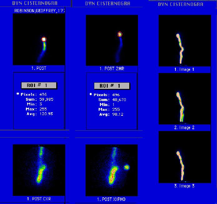
17 NORMAL CISTERNOGRAPHY PLANAR 1. Timely filling and emptying of the cisternae and the subarachnoid space 2. No filling of the ventricles (= no reversal of the CSF flow.

18 NORMAL CISTERNOGRAPHY SPECT Three Plane Tomograms Volume Reprojection Frames
19 SHUNTOGRAM NORMAL


20 FDG / PET NORMAL AND IN ALZHEIMER S DISEASE NORMAL ALZHEIMER S

 amidst necrotic")
21 PET FDG IN BRAIN TUMORS MRI cannot differentiate between recurrence and necrosis FDG/PET shows recurrence (arrows) amidst necrotic (non-radioactive) area


22 SEROTONIN RECEPTORS Normal Brain Study After 3mo on Ecstasy
23 SECOND PART CLINICAL APPLICATIONS
24 SCINTIGRAPHY OF THE BRAIN A. WITH RADIOPHARMACEUTICALS WHICH DO NOT CROSS THE NORMAL BLOOD-BRAIN-BARRIER
25 DIAGNOSIS OF CEREBRAL DEATH DYNAMIC FIRST PASS FLOW STUDY 99m Tc-PTc, DTPA, GH, MAG 3 First Pass Flow ( 99m Tc- HMPAO or ECD )
26 BRAIN DEATH: DEFINITION Brain Death results from total cessation of cerebral blood flow and global infarction of the brain at a time when respiration is preserved with artificial support and the heart continues to function. It is the only irrevocable loss of brain function currently recognized by law as death. Brain Death is a clinical diagnosis! Laboratory tests serve as confirmatory signs Brain Death is a Critical diagnosis whenever organ donation for transplantation is considered and the donor has to be transferred to the operating room while cardiac and respiratory function persists
27 BRAIN DEATH: CONFIRMATORY TESTS Isoelectric Electro Encephalo Gram (Must exclude profound hypothermic, hypotensive, or drug- induced CNS depression) Doppler measurements of brain flow Four vessel Cerebral Angiogram CT imaging after inhalation of stable xenon MRA First Pass Radionuclide Cerebral Blood Flow Study
28 BRAIN (CEREBRAL) DEATH Cessation of Cerebral Blood Flow is a Confirmatory Sign of Brain Death First-Pass Radionuclide Flow study: Portable γ-camera, 99m Tc-MAG 3, (or other BBB) 5min study Non-visualization of arterial, capillary and SSS activity during the first pass is a confirmatory sign of Cerebral Death This is the last sign to develop in the process of Brain Death
29 BRAIN DEATH ASSESSMENT BY SCINTIGRAPHY First Pass Radionuclide Cerebral Blood Flow Study Reliable, safe, and cost-effective bedside Adults and children The last objective finding to develop It is not due to an increase in intracranial pressure and it is not associated necessarily with thrombosis of the vessels
30 FIRST PASS RADIONUCLIDE CEREBRAL BLOOD FLOW STUDY: Technical Issues Adequate Dose (5-30mCi MAG 3 )or HMPAO or ECD Good bolus Must see flow to both common carotids Some use an elastic band around the head just above the orbits. It is our experience that MAG 3 is the agent of choice, not ECD etc.
31 A study is presented to you for evaluation for cerebral death
32 FIRST PASS RADIONUCLIDE CEREBRAL BLOOD FLOW STUDY MCA CCA Capillary Circulation SSS SSS Tc-GLUCOHEPTONATE DOES NOT CROSS BBB There is evidence of Cerebral Blood Flow SSS
33 A study is presented to you for evaluation for cerebral death

34 FIRST PASS RADIONUCLIDE CEREBRAL BLOOD FLOW STUDY MAG 3, DOES NOT CROSS THE BBB CA MCA ACA Capillary There is evidence of Cerebral Blood Flow SSS SSS
35 A study is presented to you for evaluation for cerebral death

36 FIRST PASS RADIONUCLIDE CEREBRAL BLOOD FLOW STUDY Tc-HMPAO, DOES CROSS THE BBB MCA ACA CA There is evidence of Cerebral Blood Flow Capillary Activity fixed in the cortex
37 FIRST PASS RADIONUCLIDE CEREBRAL BLOOD FLOW STUDY In the presence of activity in the carotid arteries, a Lack of Cerebral Perfusion is a Positive Result and this is diagnostic of Cerebral Death It is the Last test to become Positive It is Irreversible and equivalent to Cerebral Death However, in the presence of Cerebral Death, Brain Stem and Cerebellum may still be alive The test must be of excellent quality
38 A study is presented to you for evaluation for cerebral death

39 FIRST PASS RADIONUCLIDE CEREBRAL BLOOD FLOW STUDY GH, DOES NOT CROSS THE BBB Patient with no flow but late visualization of the SSS Lack of Cerebral Perfusion CA No arterial flow No visualization of the SSS SSS
40 A study is presented to you for evaluation for cerebral death


41 FIRST PASS RADIONUCLIDE CEREBRAL BLOOD FLOW STUDY MAG3, DOES NOT CROSS THE BBB Lack of Cerebral Perfusion CA No arterial flow No visualization of the SSS Is this the SSS? No, Superficial Vein
42 A study is presented to you for evaluation for cerebral death
43 FIRST PASS RADIONUCLIDE CEREBRAL BLOOD FLOW STUDY GH, DOES NOT CROSS THE BBB There is evidence of Cerebral Blood Flow Patient sp stroke: Right Middle Cerebral Artery Infarct CA SSS L. MCA No Flow R. MCA Flow
44 A study is presented to you for evaluation for cerebral death


 Flow Study Flow Ischemia")
45 FIRST PASS RADIONUCLIDE AND SPECT CEREBRAL BLOOD FLOW STUDY HMPAO DOES CROSS THE BBB CA Flow Ischemia ACA MCA Capillary SSS First Pass Flow Study There is evidence of Cerebral Blood Flow in the right hemisphere Tomographic (SPECT) Flow Study Flow Ischemia Static Planar Study
46 NORMAL PRESSURE HYDROCEPHALUS LEAKS OF CEREBROSPINAL FLUID CISTERNOGRAPHY
47 CISTERNOGRAPHY METHOD Intrathecal injection (L 4 -L 5 ) mCi 111 In-DTPA, which normally does not cross the ependyma and does not enter the ventricular system Imaging at hrs to study CSF kinetics and search for ventricular filling or other abnormalities INDICATIONS Diagnosis of Normal (Low) Pressure Hydrocephalus and Cerebrospinal Fluid Leaks
48 A patient with mental and walking problems and urine leaks CT: Hydrocephalus CSF pressure was found normal Evaluate for NPH
49 CISTERNOGRAPHY: Principle and Methodology 111In-DTPA does not cross the ependyma It is removed from the Subarachnoid Space by the arachnoid granulation (pacchionian bodies) which normally drain the Cerebrospinal Fluid into the Superior Sagittal Sinus (SSS) It does not enter the ventricular system Imaging Technique: Combination of Planar and SPECT imaging Planar images at hrs, and a SPECT at 24 hrs to confirm or exclude ventricular filling and localize leaks Normal CSF Kinetics: After an Intrathecal Injection of mci 111 In-DTPA at 4 hr, the activity is in the basal cisternae (cerebellopontine) at 24hr activity is in the silvian cisternae and around the brain at 48hr it is mostly around the convexities and the SSS and at 72hr it remains only around the convexities and the SSS

50 NORMAL (?) CISTERNOGRAPHY PLANAR 1. Timely filling and emptying of the cisternae and the subarachnoid space 2. No filling of the ventricles(?) (= no reversal of the CSF flow?)
51 NORMAL (!) CISTERNOGRAPHY PLANAR and SPECT Timely filling and emptying of the cisternae and the subarachnoid space (from the planar study) Three Plane Tomograms No filling of the ventricles! (= no reversal of the CSF flow! )
52 A patient with mental and walking problems and urine leaks CSF pressure was found low CT: Hydrocephalus Evaluate for NPH
53 NORMAL PRESSURE HYDROCEPHALUS Pathophysiology Adhesions in the Subarachnoid Space around the convexities lead to temporary impairment of drainage of CSF and increased pressure, which causes Reversal of Flow into the Ventricles and Hydrocephalus with eventual compensation of the system (pressure becomes normal) Characteristics Preservation of a Normal CSF Pressure Slow Kinetics of the CSF and Ventricular Filling after intrathecal injection of 111 In-DTPA

54 CISTERNOGRAPHY-PLANAR NORMAL PRESSURE HYDROCEPHALUS Ant 24 hr Ant 48 hr Lat 24 hr Lat 48 hr Ventricular filling, Slow clearance: NPH

55 CISTERNOGRAPHY-SPECT NORMAL PRESSURE HYDROCEPHALUS Marked Hydrocephalus, Confirmation of Ventricular Filling
56 A 7 yo boy had a history of repeated meningitis Cerebrospinal Fluid Leak was suspected

57 CEREBROSPINAL FLUID RHINORHEA 24 hr Lateral View of Cisternogram CSF Rhinorhea
58 A 44 yo male following a benign tumor removal from the thoracic spine developed headaches and walking problems. CSF pressure was found low

59 CSF Leak from the upper thoracic spine CISTERNOGRAPHY FOR CSF LEAK Ant Lat CSF Leak
60 PATENCY OF SHUNTS SHUNTOGRAM
61 A patient with a ventriculo-peritoneal shunt was sent to be evaluated for patency
62 SHUNT STUDIES Dynamic studies performed after injection of 99m Tc-DTPA into the reservoir of a ventriculo-peritoneal shunt Follow the rate of emptying Laboratories have to develop standards and data base for normalcy

63 NORMAL SHUNTOGRAM Reservoir of a ventriculoperitoneal shunt shuntogram draining T 1 / 2 =12min Shunt Shunt Shunt Spinal canal

64 SHUNTOGRAM: OBSTRUCTION shuntogram showed no draining
65 BRAIN TUMORS THALLIUM, GALLIUM, OCTREOSCAN
66 A patient with a treated malignant meningioma had a questionable MRI study and was sent for evaluation with Thallium-201


67 SPECT Th-201 TUMOR IMAGING: Recurrence DD Necrosis v/s Tumor recurrence. Can Th-201 SPECT help? Th-201 SPECT c/w Recurrence
68 CLINICAL APPLICATIONS OF BRAIN TUMOR SCINTIGRAPHY PREOPERATIVE TISSUE CHARACTERIZATION DIFFERENTIATION OF TUMORS FROM INFECTIONS THERAPY PLANNING EVALUATION OF EFFECTIVENESS OF TREATMENT DIAGNOSIS OF RECURRENCE PROGNOSIS BEFORE AND AFTER THERAPY
 Patient A survived 6 months Patient B survived 38 months")
69 SPECT I-Methyl Tyrosine: Prognostic Information 123 Ι-methyl-tyrosine Tumor Scans Scans (Post Operatively) Patient A survived 6 months Patient B survived 38 months
70 SCINTIGRAPHY OF THE CENTRAL NERVOUS SYSTEM George N. Sfakianakis MD Professor of Radiology and Pediatrics Director, Division of Nuclear Medicine
Nuclear neurology. Zámbó Katalin Department of Nuclear Medicine
 Nuclear neurology Zámbó Katalin Department of Nuclear Medicine To refresh your memory Brain has a high rate of oxidative metabolism. It has no reserves either of oxygen or of glucose and has a very limited
OTHER NON-CARDIAC USES OF Tc-99m CARDIAC AGENTS Tc-99m Sestamibi for parathyroid imaging, breast tumor imaging, and imaging of other malignant tumors.
 DEFINITION OF CARDIAC RADIOPHARMACEUTICAL: A radioactive drug which, when administered for purpose of diagnosis of heart disease, typically elicits no physiological response from the patient. Even though
Applicable Neuroradiology
 For the Clinical Neurology Clerkship LSU Medical School New Orleans Amy W Voigt, MD Clerkship Director Introduction The field of Radiology first developed following the discovery of X-Rays by Wilhelm Roentgen
Radionuclides in Medical Imaging. Danielle Wilson
 Radionuclides in Medical Imaging Danielle Wilson Outline Definitions History and development Radionuclide applications & techniques in imaging Conclusion Definition #1 : Radionuclide An unstable nucleus
2010 Radiology Prior Authorization List for UnitedHealthcare s HealthChoice Members
 70336 MR TEMPOROMANDIBULAR JOINT 70450 CT, HEAD OR BRAIN; WITHOUT MATERIAL 70460 CT HEAD/BRAIN W/ 70470 CT HEAD/BRAIN W/O & W/ 70480 CT, ORBIT, SELLA, OR POSTERIOR FOSSA OR OUTER, MID 70481 CT ORBIT W/
Laura Tormoehlen, M.D. Neurology and EM-Toxicology Indiana University
 Laura Tormoehlen, M.D. Neurology and EM-Toxicology Indiana University Disclosures! No conflicts of interest to disclose Neuroimaging 101! Plain films! Computed tomography " Angiography " Perfusion! Magnetic
THE PARATHYROID GLAND THEORY AND NUCLEAR MEDICINE PRACTICE
 THE PARATHYROID GLAND THEORY AND NUCLEAR MEDICINE PRACTICE George N. Sfakianakis MD Professor of Radiology and Pediatrics Director, Division of Nuclear Medicine UM/JMMC Miami FL October 2009 ENDONCRINE
HEALTHFIRST 2011 RADIOLOGY PROGRAM CODE LIST
 HEALTHFIRST 2011 RADIOLOGY PROGRAM CODE LIST Outpatient Radiology utilization call Carecore at 1-877-773-6964 Modality CPT CODE Description CT SCANS 70450 CT HEAD/BRAIN W/O CONTRAST CT SCANS 70460 CT HEAD/BRAIN
CISTERNOGRAPHY (CEREBRO SPINAL FLUID IMAGING): A VERSATILE DIAGNOSTIC PROCE DURE
: A VERSATILE DIAGNOSTIC PROCE DURE") VOL. 115, No. i E D I T 0 R I A L CISTERNOGRAPHY (CEREBRO SPINAL FLUID IMAGING): A VERSATILE DIAGNOSTIC PROCE DURE C ISTERNOGRAPHY (CSF imaging) is a diagnostic study based on the premise that certain
VOL. 115, No. i E D I T 0 R I A L CISTERNOGRAPHY (CEREBRO SPINAL FLUID IMAGING): A VERSATILE DIAGNOSTIC PROCE DURE C ISTERNOGRAPHY (CSF imaging) is a diagnostic study based on the premise that certain
Brain Meninges, Ventricles and CSF
 Brain Meninges, Ventricles and CSF Lecture Objectives Describe the arrangement of the meninges and their relationship to brain and spinal cord. Explain the occurrence of epidural, subdural and subarachnoid
Description MRI, TMJ C T Head Without Contrast C T Head With Contrast C T Head Without & With Contrast
 s Requiring Prior Authorization for the Advanced Imaging 70336 MRI, TMJ 70450 C T Head Without Contrast 70460 C T Head With Contrast 70470 C T Head Without & With Contrast 70480 C T Orbit Without Contrast
HIP RADIOLOGY PROGRAM CODE LISTS
 EFFECTIVE OCTOBER 1, 2012 70336 MAGNETIC RESONANCE IMAGING TMJ 70450 COMPUTED TOMOGRAPHY HEAD/BRAIN WITHOUT 70460 COMPUTED TOMOGRAPHY HEAD/BRAIN WITH 70470 COMPUTED TOMOGRAPHY HEAD/BRAIN WITHOUT AND WITH
Cardiac Imaging Tests
 Cardiac Imaging Tests http://www.medpagetoday.com/upload/2010/11/15/23347.jpg Standard imaging tests include echocardiography, chest x-ray, CT, MRI, and various radionuclide techniques. Standard CT and
M555 Medical Neuroscience Blood Flow in CNS Meninges Blood Brain Barrier CSF
 M555 Medical Neuroscience Blood Flow in CNS Meninges Blood Brain Barrier CSF Arterial Blood Flow to CNS approximately % of what goes wrong within the skull that produces neurological deficits is vascular
MOLINA HEALTHCARE OF MICHIGAN PRIOR AUTHORIZATION / PRE-SERVICE REVIEW GUIDE IMAGING CODES REQUIRING PRIOR AUTHORIZATION EFFECTIVE 1/1/2014
 70336 MRI MRI, temporomandibular joint(s) 70450 CT/CTA CT, head or brain; without contrast material 70460 CT/CTA CT, head or brain; with contrast material(s) 70470 CT/CTA CT, head or brain; without contrast
Medical imaging X-ray, CT, MRI, scintigraphy, SPECT, PET Györgyi Műzes
 Medical imaging X-ray, CT, MRI, scintigraphy, SPECT, PET Györgyi Műzes Semmelweis University, 2nd Dept. of Medicine Medical imaging: definition technical process of creating visual representations about
Meninges and Ventricles
 Meninges and Ventricles Irene Yu, class of 2019 LEARNING OBJECTIVES Describe the meningeal layers, the dural infolds, and the spaces they create. Name the contents of the subarachnoid space. Describe the
CSF. Cerebrospinal Fluid(CSF) System
 System") Cerebrospinal Fluid(CSF) System By the end of the lecture, students must be able to describe Physiological Anatomy of CSF Compartments Composition Formation Circulation Reabsorption CSF Pressure Functions
Cerebrospinal Fluid(CSF) System By the end of the lecture, students must be able to describe Physiological Anatomy of CSF Compartments Composition Formation Circulation Reabsorption CSF Pressure Functions
Cerebral hemisphere. Parietal Frontal Occipital Temporal
 Cerebral hemisphere Sulcus / Fissure Central Precental gyrus Postcentral gyrus Lateral (cerebral) Parieto-occipital Cerebral cortex Frontal lobe Parietal lobe Temporal lobe Insula Amygdala Hippocampus
Last Updated: 2/10/2017 Implementation date: 4/3/2017 Radiology & Cardiology Prior Authorization / Utilization Management Procedure List
 Last Updated: 2/10/2017 Implementation date: 4/3/2017 Radiology & Cardiology Prior Authorization / Utilization Management Procedure List Deal Sheet Group Product Category CPT CPT Description 3D Imaging
Blood Supply of the CNS
 Blood Supply of the CNS Lecture Objectives Describe the four arteries supplying the CNS. Follow up each artery to its destination. Describe the circle of Willis and its branches. Discuss the principle
Anthem Blue Cross and Blue Shield Virginia Advanced Imaging Procedures Requiring Precertification Revised 02/13/2013
 Anthem Blue Cross and Blue Shield Virginia Advanced Imaging Procedures Requiring Precertification Revised 02/13/2013 Modality and CT Head CTA Head: Cerebrovascular MRI Head MRA Head: Cerebrovascular Functional
AMERICAN IMAGING MANAGEMENT
 2012 CPT Codes Computerized Tomography (CT) CPT Description Abdomen 74150 CT abdomen; w/o 74160 CT abdomen; with 74170 CT abdomen; w/o followed by Chest 71250 CT thorax; w/o 71260 CT thorax; with 71270
AMERICAN IMAGING MANAGEMENT
 2010 BCBS of Georgia CPT Codes With Grouper Numbers Computerized Tomography (CT) CPT Description Abdomen 74150 CT abdomen; w/o contrast 6 74160 CT abdomen; with contrast 74170 CT abdomen; w/o contrast
MRI and CT of the CNS
 MRI and CT of the CNS Dr.Maha ELBeltagy Assistant Professor of Anatomy Faculty of Medicine The University of Jordan 2018 Computed Tomography CT is used for the detection of intracranial lesions. CT relies
Recent initiatives of the FANC. Michel Biernaux Health Protection Service Health and Environment Department
 Recent initiatives of the FANC Michel Biernaux Michel.biernaux@fanc.fgov.be Health Protection Service Health and Environment Department Reminder objectives of the national survey : 1.Review the average
AIM 2014 CPT Radiology & Cardiac Codes Requiring Review
 AIM 2014 CPT Radiology & Cardiac Codes Requiring Review Modality Body Part CT Head 1 70480 CT orbit, sella or posterior fossa; w/o contrast 1 CT Head 1 70481 CT orbit, sella or posterior fossa; with CT
Index. aneurysm, 92 carotid occlusion, 94 ICA stenosis, 95 intracranial, 92 MCA, 94
 A ADC. See Apparent diffusion coefficient (ADC) Aneurysm cerebral artery aneurysm, 93 CT scan, 93 gadolinium, 93 Angiography, 13 Anoxic brain injury, 25 Apparent diffusion coefficient (ADC), 7 Arachnoid
Enhancement of Cranial US: Utility of Supplementary Acoustic Windows and Doppler Harriet J. Paltiel, MD
 Enhancement of Cranial US: Utility of Supplementary Acoustic Windows and Doppler Harriet J. Paltiel, MD Boston Children s Hospital Harvard Medical School None Disclosures Conventional US Anterior fontanelle
Basics of nuclear medicine
 Basics of nuclear medicine Prof. dr. Davor Eterović Prof. dr. Vinko Marković Radioisotopes are used both in diagnostics and in therapy Diagnostics gamma emitters are used since gamma rays can penetrate
Ventricles, CSF & Meninges. Steven McLoon Department of Neuroscience University of Minnesota
 Ventricles, CSF & Meninges Steven McLoon Department of Neuroscience University of Minnesota 1 Coffee Hour Thursday (Sept 14) 8:30-9:30am Surdyk s Café in Northrop Auditorium Stop by for a minute or an
Gastrointestinal tract
 Gastrointestinal tract Colloidal liver-spleen imaging Presented by: Jehad Felemban Introduction: To obtain better anatomic display of liver and spleen architecture, we use (CT Ultrasound). (Radionuclide
2012 CPT Radiology Codes Requiring Review Blue Cross and Blue Shield of Louisiana
 2012 CPT Radiology Codes Requiring Review Blue Cross and Blue Shield of Louisiana CT Head 70480 CT orbit, sella or posterior fossa; w/o CT Head 70481 CT orbit, sella or posterior fossa; with CT Head 70482
Radiology Codes Requiring Authorization*
 70336 Magnetic resonance (eg, proton) imaging, temporomandibular joint(s) 70450 Computed tomography, head or brain; without contrast material 70460 Computed tomography, head or brain; with contrast material(s)
RENAL SCINTIGRAPHY IN THE 21 st CENTURY
 RENAL SCINTIGRAPHY IN THE 21 st CENTURY 99m Tc- MAG 3 with zero time injection of Furosemide (MAG 3 -F 0 ) : A Fast and Easy Protocol, One for All Indications Introduction George N. Sfakianakis MD Professor
Water Any Inulin IV Normal renal function Inulin IV Abnormal renal function Glucose IV Normal Carfentanil IV 17.
 Table 1: Effective Dose Equivalent (HE) values for common procedures involving the use of radiopharmaceuticals. Listings (last column) are averaged over both sexes, and are computed for adult subjects.
05/02/ CPT Preauthorization Groupings Effective May 2, Computerized Tomography (CT) Abdomen 6. CPT Description SEGR CT01
 Abdomen 6. CPT Description SEGR CT01") Computerized Tomography (CT) 6 & 101 5 Upper Extremity 11 Lower Extremity 12 Head 3 Orbit 1 Sinus 2 Neck 4 7 Cervical Spine 8 Thoracic Spine 9 Lumbar Spine 10 Colon 13 CPT Preauthorization Groupings CPT
Computerized Tomography (CT) 6 & 101 5 Upper Extremity 11 Lower Extremity 12 Head 3 Orbit 1 Sinus 2 Neck 4 7 Cervical Spine 8 Thoracic Spine 9 Lumbar Spine 10 Colon 13 CPT Preauthorization Groupings CPT
Sample page. Radiology. Cross Coder. Essential links from CPT codes to ICD-10-CM and HCPCS
 Cross Coder 2018 Radiology Essential links from CPT codes to ICD-10-CM and HCPCS POWER UP YOUR CODING with Optum360, your trusted coding partner for 32 years. Visit optum360coding.com. Contents Introduction...
STROKE - IMAGING. Dr RAJASEKHAR REDDY 2nd Yr P.G. RADIODIAGNOSIS KIMS,Narkatpalli.
 STROKE - IMAGING Dr RAJASEKHAR REDDY 2nd Yr P.G. RADIODIAGNOSIS KIMS,Narkatpalli. STROKE Describes a clinical event that consists of sudden onset of neurological symptoms Types Infarction - occlusion of
PTA 106 Unit 1 Lecture 3
 PTA 106 Unit 1 Lecture 3 The Basics Arteries: Carry blood away from the heart toward tissues. They typically have thicker vessels walls to handle increased pressure. Contain internal and external elastic
Pediatric emergencies (SHOCK & COMA) Dr Mubarak Abdelrahman Assistant Professor Jazan University
 Dr Mubarak Abdelrahman Assistant Professor Jazan University") Pediatric emergencies (SHOCK & COMA) Dr Mubarak Abdelrahman Assistant Professor Jazan University SHOCK Definition: Shock is a syndrome = inability to provide sufficient oxygenated blood to tissues. Oxygen
Pediatric emergencies (SHOCK & COMA) Dr Mubarak Abdelrahman Assistant Professor Jazan University SHOCK Definition: Shock is a syndrome = inability to provide sufficient oxygenated blood to tissues. Oxygen
Blood Supply. Allen Chung, class of 2013
 Blood Supply Allen Chung, class of 2013 Objectives Understand the importance of the cerebral circulation. Understand stroke and the types of vascular problems that cause it. Understand ischemic penumbra
NEURO IMAGING 2. Dr. Said Huwaijah Chairman of radiology Dep, Damascus Univercity
 NEURO IMAGING 2 Dr. Said Huwaijah Chairman of radiology Dep, Damascus Univercity I. EPIDURAL HEMATOMA (EDH) LOCATION Seventy to seventy-five percent occur in temporoparietal region. CAUSE Most likely caused
Diagnostic Imaging Utilization Management and Consultation Management Programs Imaging Code Listing for Connecticut, Maine and New Hampshire
 Diagnostic Imaging Utilization Management and Consultation Management Programs Imaging Code Listing for Connecticut, Maine and New Hampshire The grid below contains the CPT * codes that are subject to
PHYSICS 2: HSC COURSE 2 nd edition (Andriessen et al) CHAPTER 20 Radioactivity as a diagnostic tool (pages 394-5)
 CHAPTER 20 Radioactivity as a diagnostic tool (pages 394-5)") PHYSICS 2: HSC COURSE 2 nd edition (Andriessen et al) CHAPTER 20 Radioactivity as a diagnostic tool (pages 394-5) 1. (a) A radioisotope is an isotope that is unstable and will emit particles from the nucleus
PHYSICS 2: HSC COURSE 2 nd edition (Andriessen et al) CHAPTER 20 Radioactivity as a diagnostic tool (pages 394-5) 1. (a) A radioisotope is an isotope that is unstable and will emit particles from the nucleus
Multiple Gated Acquisition (MUGA) Scanning
 Scanning") Multiple Gated Acquisition (MUGA) Scanning Dmitry Beyder MPA, CNMT Nuclear Medicine, Radiology Barnes-Jewish Hospital / Washington University St. Louis, MO Disclaimers/Relationships Standard of care research
Multiple Gated Acquisition (MUGA) Scanning Dmitry Beyder MPA, CNMT Nuclear Medicine, Radiology Barnes-Jewish Hospital / Washington University St. Louis, MO Disclaimers/Relationships Standard of care research
Austin Radiological Association Nuclear Medicine Procedure BONE MINERAL STUDY (Tc-99m-MDP, Tc-99m-HMDP)
") Austin Radiological Association Nuclear Medicine Procedure BONE MINERAL STUDY (Tc-99m-MDP, Tc-99m-HMDP) Overview The Bone Mineral Study, with either Tc-99m-MDP or Tc-99m-HMDP, depicts the distribution
Austin Radiological Association Nuclear Medicine Procedure BONE MINERAL STUDY (Tc-99m-MDP, Tc-99m-HMDP) Overview The Bone Mineral Study, with either Tc-99m-MDP or Tc-99m-HMDP, depicts the distribution
Nuclear pulmonology. Katalin Zámbó Department of Nuclear Medicine
 Nuclear pulmonology Katalin Zámbó Department of Nuclear Medicine Imaging techniques Morphology Physiology Metabolism Molecules X-ray / CT MRI NM - SPECT/ PET MR spectroscopy fmri Ultrasound Hybrid imaging:
The central nervous system
 Sectc.qxd 29/06/99 09:42 Page 81 Section C The central nervous system CNS haemorrhage Subarachnoid haemorrhage Cerebral infarction Brain atrophy Ring enhancing lesions MRI of the pituitary Multiple sclerosis
Non-Invasive Techniques
 Non-Invasive Techniques Key: Does not hurt the organism Psychology 372 Physiological Psychology Steven E. Meier, Ph.D. Listen to the audio lecture while viewing these slides or view the video presentation
Non-Invasive Techniques
 Many Procedures Non-Invasive Techniques Key: Does not hurt the organism Psychology 372 Physiological Psychology Steven E. Meier, Ph.D. Listen to the audio lecture while viewing these slides or view the
CEREBRAL BLOOD FLOW AND METABOLISM
 Supported by: HURO/0901/069/2.3.1 HU-RO-DOCS CEREBRAL BLOOD FLOW AND METABOLISM Part 3 Modern imaging methods SPECT, PET, nmri History of Nuclear Medicine Starts with the invention of the X-ray 1946: radioactive
Nuclear Medicine in the Diabetic Foot
 26.11.2015, Uniklinik Balgrist Nuclear Medicine in the Diabetic Foot Martin Hüllner Nuklearmedizin und Neuroradiologie, USZ / UZH Outline A. Imaging modalities brief technical overview B. Nuclear medicine
FOR CMS (MEDICARE) MEMBERS ONLY NATIONAL COVERAGE DETERMINATION (NCD) FOR MAGNETIC RESONANCE IMAGING:
 MEMBERS ONLY NATIONAL COVERAGE DETERMINATION (NCD) FOR MAGNETIC RESONANCE IMAGING:") National Imaging Associates, Inc. Clinical guidelines SINUS MRI Original Date: November 2007 Page 1 of 5 CPT Codes: 70540, 70542, 70543 Last Review Date: July 2014 NCD 220.2 MRI Last Effective Date: July
National Imaging Associates, Inc. Clinical guidelines SINUS MRI Original Date: November 2007 Page 1 of 5 CPT Codes: 70540, 70542, 70543 Last Review Date: July 2014 NCD 220.2 MRI Last Effective Date: July
BlueAdvantage SM. & BlueChoice SM Radiology Prior Authorization Program Code List CPT /HCPS
 BlueAdvantage SM & BlueChoice SM Radiology Prior Authorization Program Code List CPT /HCPS 70336 MRI TMJ 70450 CT Head Without Contrast 70460 CT Head With Contrast 70470 CT Head Without & With Contrast
Nuclear Medicine: Manuals. Nuclear Medicine. Nuclear imaging. Emission imaging: study types. Bone scintigraphy - technique
 Nuclear Medicine - Unsealed radioactive preparations the tracer mixes with the patients body fluids on a molecular level (e.g. after intravenous injection) - 3 main fields: - In vitro : measuring concentrations
Dr Alfred O Ankrah FCNP
 Dr Alfred O Ankrah FCNP Outline Introduction Brief history of Nuclear Medicine in Ghana Current situation of Nuclear Medicine in Ghana Use of Nuclear medicine in various disciplines Future of Nuclear Medicine
Introduction, use of imaging and current guidelines. John O Brien Professor of Old Age Psychiatry University of Cambridge
 Introduction, use of imaging and current guidelines John O Brien Professor of Old Age Psychiatry University of Cambridge Why do we undertake brain imaging in AD and other dementias? Exclude other causes
Click here for Link to References: CMS Website HOPPS CY 2018 Final Rule. CMS Website HOPPS CY2018 Final Rule Updated November 2017.
 Final Compared to 3Q 2017 Rates Medicare Hospital Outpatient Prospective Payment System HOPPS () Nuclear Cardiology Procedures, Radiopharmaceuticals, and Drugs Click here for Link to References: CMS Website
Myocardial viability testing. What we knew and what is new
 Myocardial viability testing. What we knew and what is new Dr B K S Sastry, MD, DM. CARE Hospitals, Hyderabad What is Viability Viability Dysfunctional myocardium subtended by diseased coronary arteries
Positron Emission Tomography Computed Tomography (PET/CT)
") Positron Emission Tomography Computed Tomography (PET/CT) What is Positron Emission Tomography Computed Tomography (PET/CT) Scanning? What are some common uses of the procedure? How should I prepare for
Positron Emission Tomography Computed Tomography (PET/CT) What is Positron Emission Tomography Computed Tomography (PET/CT) Scanning? What are some common uses of the procedure? How should I prepare for
Department of Nuclear Medicine with Positron Emission Tomography
 (PET) Unit [1] Contact information: Registration: +48 41 367 4850 Main office: +48 41 367 4860 Fax: +48 41 367 4887 e-mail: zmnsco@onkol.kielce.pl [2] Head of the Department: Professor Janusz Braziewicz
Cardiac Nuclear Medicine
 Cardiac Nuclear Medicine What is Cardiac Nuclear Medicine? What are some common uses of the procedure? How should I prepare? What does the equipment look like? How does the procedure work? How is the procedure
Contrast Agents and Radiopharmaceuticals 2017
 Contrast Agents and Radiopharmaceuticals 207 Covered: Code Code Description Allow with Code(s) Code Description Max Units A464 Radiopharmaceutical, diagnostic, not otherwise classified n/a Invoice Req'd
Molecular Imaging and the Brain
 Molecular imaging technologies are playing an important role in neuroimaging, a branch of medical imaging, by providing a window into the living brain. Where CT and conventional MR imaging provide important
Brain Atrophy. Brain Atrophy
 Aging Central Nervous System Processes Age related brain atrophy Non-age related brain atrophy Cerebrovascular disease Cerebral infarction Hypertensive hemorrhage Carotid artery stenosis and occlusion
2017 St. Luke's-Roosevelt Nuclear Medicine Department Procedure Catalog. meds
 2017 St. Luke's-Roosevelt Nuclear Medicine Department Procedure Catalog Procedure Name Patient Preparation Cardiac Studies Stress Myocardial Perfusion Study Gated pool study (MUGA) Myocardial Cell Damage
What Is an Arteriovenous malformation (AVM)?
?") American Society of Neuroradiology What Is an Arteriovenous malformation (AVM)? From the Cerebrovascular Imaging and Intervention Committee of the American Heart Association Cardiovascular Council Randall
American Society of Neuroradiology What Is an Arteriovenous malformation (AVM)? From the Cerebrovascular Imaging and Intervention Committee of the American Heart Association Cardiovascular Council Randall
Organization of the nervous system. [See Fig. 48.1]
![Organization of the nervous system. [See Fig. 48.1]](/thumbs/90/103926552.jpg "Organization of the nervous system. [See Fig. 48.1]") Nervous System [Note: This is the text version of this lecture file. To make the lecture notes downloadable over a slow connection (e.g. modem) the figures have been replaced with figure numbers as found
Nervous System [Note: This is the text version of this lecture file. To make the lecture notes downloadable over a slow connection (e.g. modem) the figures have been replaced with figure numbers as found
What Are We Going to Do? Fourth Year Meds Clinical Neuroanatomy. Hydrocephalus and Effects of Interruption of CSF Flow. Tube Blockage Doctrine
 Fourth Year Meds Clinical Neuroanatomy Ventricles, CSF, Brain Swelling etc. David A. Ramsay, Neuropathologist, LHSC What Are We Going to Do? Hydrocephalus and some effects of the interruption of CSF flow
Central nervous system (CNS): brain and spinal cord Collections of cell body and dendrites (grey matter) are called nuclei/nucleus Nucleus can also
: brain and spinal cord Collections of cell body and dendrites (grey matter) are called nuclei/nucleus Nucleus can also") Chapter 3 Part 1 Orientation Directions in the nervous system are described relatively to the neuraxis An imaginary line drawn through the center of the length of the central nervous system, from the bottom
Chapter 3 Part 1 Orientation Directions in the nervous system are described relatively to the neuraxis An imaginary line drawn through the center of the length of the central nervous system, from the bottom
RADIOTRACERS FOR MYOCARDIAL PERFUSION IMAGING
 RADIOTRACERS FOR MYOCARDIAL PERFUSION IMAGING RAYMOND TAILLEFER, M.D. FRCP(c), ABNM DIRECTOR, DEPARTMENT OF NUCLEAR MEDICINE HOPITAL ST-JEAN-SUR-RICHELIEU Disclosures to Report: Grant Research Support:
NANOS Patient Brochure
 NANOS Patient Brochure Pseudotumor Cerebri Copyright 2016. North American Neuro-Ophthalmology Society. All rights reserved. These brochures are produced and made available as is without warranty and for
Chapter 1. Introduction
 Chapter 1 Introduction 1.1 PRINCIPLES OF NUCLEAR MEDICINE Nuclear medicine techniques use radioactive tracers and imaging devices, mainly to provide diagnostic information, but in some cases also for therapeutic
Cerebrovascular diseases-2
 Cerebrovascular diseases-2 Primary angiitis of CNS - Other causes of infarction i. Hypercoagulable states ii. Drug-abuse such as amphetamine, heroin and cocain Note - The venous side of the circulation
Cardiovascular nuclear imaging employs non-invasive techniques to assess alterations in coronary artery flow, and ventricular function.
 National Imaging Associates, Inc. Clinical guidelines CARDIOVASCULAR NUCLEAR MEDICINE -MYOCARDIAL PERFUSION IMAGING -MUGA Original Date: October 2015 Page 1 of 9 FOR CMS (MEDICARE) MEMBERS ONLY CPT4 Codes:
Renal. Prof John Buscombe
 Renal Prof John Buscombe Renal nuclear Medicine Only consistent test of kidney func7on Many good tests for renal anatomy Ultrasound good looking at cysts and renal pelvis CT can look at perfusion, size
High Tech Imaging Quick Reference Guide
 High Tech Imaging Quick Reference Guide 1 High Tech Imaging Authorizations may now be requested through our secure provider portal, BlueAccess. Getting Started Step 1: Log into BlueAccess from www.bcbst.com
Diagnostic Imaging Utilization Management and Consultation Management Programs Imaging Code Listing for Connecticut, Maine and New Hampshire
 Diagnostic Imaging Utilization Management and Consultation Management Programs Imaging Code Listing for Connecticut, Maine and New Hampshire The grid below contains the CPT * codes that are subject to
Overview Blood supply of the brain What is moyamoya disease? > 1
 Moyamoya Disease Overview Moyamoya disease is caused by blocked arteries at the base of the brain. The name "moyamoya" means "puff of smoke" in Japanese and describes the appearance of tiny vessels that
Fellows on this rotation are expected to attend nuclear conferences and multimodality imaging conference.
 Rotation: Imaging 1 Imaging 1 provides COCATS Level 1 experience for nuclear cardiology (including SPECT and PET) and cardiac CT. Fellows will administer, process, and read cardiac nuclear studies with
An Introduction to Imaging the Brain. Dr Amy Davis
 An Introduction to Imaging the Brain Dr Amy Davis Common reasons for imaging: Clinical scenarios: - Trauma (NICE guidelines) - Stroke - Tumours - Seizure - Neurological degeneration memory, motor dysfunction,
CNS Imaging. Dr Amir Monir, MD. Lecturer of radiodiagnosis.
 CNS Imaging Dr Amir Monir, MD Lecturer of radiodiagnosis www.dramir.net Types of radiological examinations you know Plain X ray X ray with contrast GIT : barium (swallow, meal, follow through, enema) ERCP
THE SNM PRACTICE GUIDELINE FOR BRAIN DEATH SCINTIGRAPHY V2.0
 THE SNM PRACTICE GUIDELINE FOR BRAIN DEATH SCINTIGRAPHY V2.0 Revised 2011 PREAMBLE These guidelines are an educational tool designed to assist practitioners in providing appropriate care for patients.
POSITRON EMISSION TOMOGRAPHY (PET)
") Status Active Medical and Behavioral Health Policy Section: Radiology Policy Number: V-27 Effective Date: 08/27/2014 Blue Cross and Blue Shield of Minnesota medical policies do not imply that members should
Status Active Medical and Behavioral Health Policy Section: Radiology Policy Number: V-27 Effective Date: 08/27/2014 Blue Cross and Blue Shield of Minnesota medical policies do not imply that members should
Fidelis Care: Cardiology, Radiology, and Ultrasound CPT Code List
 Fidelis Care: Cardiology, Radiology, and Ultrasound CPT Code List CPT Code CPT Code Description Requires PA 75557 Cardiac magnetic resonance imaging for morphology and function without contrast material
Cardiac Imaging. Kimberly Delcour, DO, FACC. Mahi Ashwath, MD, FACC, FASE. Director, Cardiac CT. Director, Cardiac MRI
 Cardiac Imaging Kimberly Delcour, DO, FACC Director, Cardiac CT Mahi Ashwath, MD, FACC, FASE Director, Cardiac MRI Cardiac Imaging Discuss the clinical applications of and indications for: Cardiac CT Nuclear
Option D: Medicinal Chemistry
 Option D: Medicinal Chemistry Basics - unstable radioactive nuclei emit radiation in the form of smaller particles alpha, beta, positron, proton, neutron, & gamma are all used in nuclear medicine unstable
Diagnostic Imaging Prior Review Code List 2 nd Quarter 2018
 Computerized Tomography (CT) Abdomen 6 Abdomen/Pelvis Combination 101 Service 74150 CT abdomen; w/o 74160 CT abdomen; with 74170 CT abdomen; w/o followed by 74176 Computed tomography, abdomen and pelvis;
General Nuclear Medicine
 General Nuclear Medicine What is General Nuclear Medicine? What are some common uses of the procedure? How should I prepare? What does the equipment look like? How does the procedure work? How is the procedure
Brain Tumors. What is a brain tumor?
 Scan for mobile link. Brain Tumors A brain tumor is a collection of abnormal cells that grows in or around the brain. It poses a risk to the healthy brain by either invading or destroying normal brain
Department of Cognitive Science UCSD
 Department of Cognitive Science UCSD Verse 1: Neocortex, frontal lobe, Brain stem, brain stem, Hippocampus, neural node, Right hemisphere, Pons and cortex visual, Brain stem, brain stem, Sylvian fissure,
Hydrocephalus 1/16/2015. Hydrocephalus. Functions of Cerebrospinal fluid (CSF) Flow of CSF
 Flow of CSF") Hydrocephalus Hydrocephalus Ruth Arms, MSN, CNS-BC, SCRN Hydrocephalus is the buildup of fluid in the cavities (ventricles) deep within the brain. The excess fluid increases the size of the ventricles
Hydrocephalus Hydrocephalus Ruth Arms, MSN, CNS-BC, SCRN Hydrocephalus is the buildup of fluid in the cavities (ventricles) deep within the brain. The excess fluid increases the size of the ventricles
MANAGEMENT OF CSF. Steven D. Schaefer, MD, FACS. Department of Otolaryngology New York Eye and Ear Infirmary
 MANAGEMENT OF CSF RHINORRHEA, MENIGIOCELES, Steven D. Schaefer, MD, FACS Professor and Chair Department of Otolaryngology New York Eye and Ear Infirmary New York Medical College Anatomy and Physiology
2014 CPT Radiology Codes Requiring Review
 CT Head 1 70480 CT orbit, sella or posterior fossa; w/o contrast 1 CT Head 1 70481 CT orbit, sella or posterior fossa; with CT orbit, sella or posterior fossa; w/o contrast CT Head 1 70482 followed by
Functional aspects of anatomical imaging techniques
 Functional aspects of anatomical imaging techniques Nilendu Purandare Associate Professor & Consultant Radiologist Tata Memorial Centre Functional/metabolic/molecular imaging (radioisotope scanning) PET
CEREBRO SPINAL FLUID ANALYSIS IN BRAIN TUMOUR
 CEREBRO SPINAL FLUID ANALYSIS IN BRAIN TUMOUR Sankar K 1, Shankar N 2, Anushya 3, ShymalaDevi 4, Purvaja 5 3,4,5 III Biomedical Student, Alpha college of Engineering, Chennai. kssankar10@yahoo.co.in 1,
Cardiovascular nuclear imaging employs non-invasive techniques to assess alterations in coronary artery flow, and ventricular function.
 National Imaging Associates, Inc. Clinical guidelines CARDIOVASCULAR NUCLEAR MEDICINE -MYOCARDIAL PERFUSION IMAGING -MUGA CPT4 Codes: Refer to pages 6-9 LCD ID Number: L33960 J 15 = KY, OH Responsible
Brain Perfusion SPECT
 APPROVED BY: Director of Radiology Page 1 of 5 Brain Perfusion SPECT Primary Indications: Brain perfusion SPECT is most commonly performed (1) to aid in identification of the epileptogenic focus in patients
[(PHY-3a) Initials of MD reviewing films] [(PHY-3b) Initials of 2 nd opinion MD]
![[(PHY-3a) Initials of MD reviewing films] [(PHY-3b) Initials of 2 nd opinion MD]](/thumbs/89/98619893.jpg "[(PHY-3a) Initials of MD reviewing films] [(PHY-3b) Initials of 2 nd opinion MD]") 2015 PHYSICIAN SIGN-OFF (1) STUDY NO (PHY-1) CASE, PER PHYSICIAN REVIEW 1=yes 2=no [strictly meets case definition] (PHY-1a) CASE, IN PHYSICIAN S OPINION 1=yes 2=no (PHY-2) (PHY-3) [based on all available
2015 PHYSICIAN SIGN-OFF (1) STUDY NO (PHY-1) CASE, PER PHYSICIAN REVIEW 1=yes 2=no [strictly meets case definition] (PHY-1a) CASE, IN PHYSICIAN S OPINION 1=yes 2=no (PHY-2) (PHY-3) [based on all available