LESSON ASSIGNMENT. Positioning for Exams of the Cranium, Sinuses, and Mandible. After completing this lesson, you should be able to:
|
|
|
- Jeffry Murphy
- 6 years ago
- Views:
Transcription
1 LESSON ASSIGNMENT LESSON 5 Positioning for Exams of the Cranium, Sinuses, and Mandible. LESSON ASSIGNMENT Paragraphs 5-1 through 5-9. LESSON OBJECTIVES After completing this lesson, you should be able to: 5-1. Identify specifications for proper placement of the anatomical structures of the cranium (skull), sinuses, mastoids, and mandible listed below: Posteroanterior Skull (Caldwell method). Bilateral skull. Anterior-posterior axial skull (Chamberlain Towne or CT skull). PA projection (Caldwell method). Parientoacanthial projection (Waters view). Lateral sinuses. PA mandible. Axiolateral (oblique, bilateral) mandible. SUGGESTION After reading and studying the assignment, complete the exercises. These exercises will help you to achieve the lesson objectives. MD
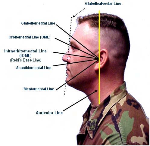
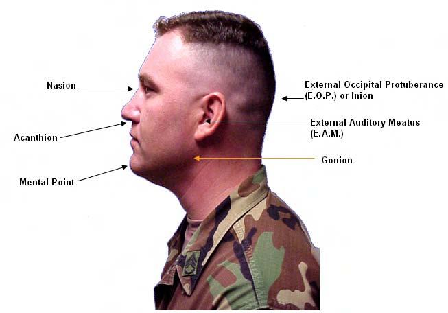
2 LESSON 5 POSITIONING FOR EXAMS OF THE CRANIUM, SINUSES, AND MANDIBLE Section I. PROJECTIONS OF THE CRANIUM (SKULL) 5-1. SURFACE ANATOMY AND REFERENCE POINTS There are 21 terms pertaining to surface anatomy and reference points given in table 5-1. You will need to know these terms in order to comprehend the specifications for the cranium projections that follow. Please take a moment to familiarize yourself with the terms that appear in the table below and on the following page. Be sure to refer to the graphics on the pages following the table. These graphics depict cranial reference points. Radiographic Surface Anatomy and Reference Points 1. Vertex The most superior portion of the skull 2. Superciliary (arch) The ridge or arch of the bone extending across the forehead directly above each eye 3. Glabella Smooth prominence between the eyebrow 4. Nasion The depression at the bridge of the nose 5. Acanthion 6. Gonion 7. External auditory meatus (EAM) (see also #20, tragus.) 8. External occipital protuberance (EOP) 9. Mental point The junction of the upper lip and the nasal septum. The angle of the mandible. The most inferior posterior and lateral points on the external angle of the mandible. The opening of the external ear canal The bump along the midline of the lower back of the head; also referred to as the inion. The midpoint of the triangular area of the chin; also referred to as the mental protuberance. 10. External (outer canthus) The lateral junction of the eyelids Table 5-1. Terminology (continued). MD
3 Radiographic Surface Anatomy and Reference Points 11. Infraorbital margin 12. Midsagittal plane 13. Acanthiomeatal line (AML) 14. Orbitalmeatal line (OML) 15. Infraorbitmealtal line (IOML) 16. Interpupillary line (IPPL) 17. Glabellomeatal line (GML) The inferior rim of the orbit (the bony cavity of the skull that contains the eye). Plane that divides the body into right and left halves. Cranial reference points are external occipital protuberance (EOP) and the glabella. Line that connects the acanthion and the external auditory meatus (EAM), the opening of the ear. Also know as the occlusal plane. Line that connects the external (outer) canthus of the eye and auditory meatus (EAM). Line that connects the infraorbital margin and the external auditory meatus (EAM). It is also referred to as the Reid s base line of the cranium. Line that connects the pupils of the eye. In a true lateral position, the IPPL must be perpendicular to the table. Line that connects the glabella and the external auditory meatus (EAM). 18. Gabelloalveolar line (facial plane) 19. Two-way heal alignment (TWHA) 20. Tragus 21. Mental meatal line (MML) Line that connects the glabella and the mental point. Two or more planes of the skull are used to ensure proper alignment, e.g., the median plane, the facial plane, the OML, the IOML, the AML and IPPL. The cartilaginous projection anterior to the external opening of the ear. (The tragus and EAM are the same.) Line that connects the mental point and the EAM. Table 5-1. Terminology (concluded). MD

4 MD

5 MD
, lateral skull (bilateral), and AP axial (Chamberlain Town or CT).")
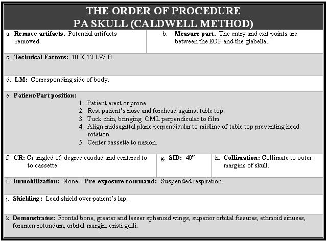
6 5-2. POSTEROANTERIOR SKULL (CALDWELL) The routine views of the cranium are PA skull (Caldwell), lateral skull (bilateral), and AP axial (Chamberlain Town or CT). MD

7 MD
 MD0962")
8 5-3. LATERAL SKULL (BI-LATERAL) MD

9 MD
 MD0962")
10 5-4. ANTERIOR-POSTERIOR AXIAL (CHAMBERLAIN TOWNE, CT) MD


11 Continue with Exercises MD
12 EXERCISES, LESSON 5, SECTION I MATCHING: For exercises 1 through 3, match the position with the anatomical structure(s) that the position demonstrates. Enter the letter that corresponds to your choice in the space provided. (There is an extra alternative that will not be selected.) 1. PA skull. a. Foramen magnum with dorsum sallae, petrous pyramids, occipital bone, posterior clinoid process. 2. Lateral skull. b. Frontal bone, greater and lesser sphenoid wings, superior orbital fissures, ethmoid sinuses, foramen rotundeum, orbital margin, cristi galli. 3. AP axial skull c. Lateral cranium closest to the film, sella (Towne method). turcica, anterior and posterior clinoids, dorsum sellae, greater and lesser wings of sphenoid. d. Petrous ridge in profile, bony labyrinth, tympanic cavity, internal auditory canal, and mastoid air cell. MULTIPLE- CHOICE. For exercises 4 through 8, select the ONE word or phrase that BEST completes the statement or BEST answers the question. 4. For a lateral skull the is perpendicular to the film. a. IOML. b. AML. c. OML. d. Midsagittal plane. MD
13 5. The proper patient and part position for a PA skull is: a. Patient in semi-prone position; midsagittal plane parallel to film; IOML perpendicular to front edge of cassette; midpoint between the EOP and the glabella is over the midline of the table. b. Patient in prone position (nose-forehead position; meidsagittal plane perpendicular and over the midline of the film; orbitomeatal line perpendicular to the film. c. Patient in supine position (posterior skull against table top); midsagittal plane perpendicular and over the midline of the film; OML perpendicular to the film. d. Patient prone (nose-forehead position); midsagittal plane perpendicular and over midline of film; infraorbitomeatal line perpendicular to film. 6. What is centered for a lateral skull? a. Midsagittal plane, 2.5 inches above the superciliary arch. b. Nasion. c. A point 2 inches superior to the EAM. d. EAM. 7. What is perpendicular to the film for a PA (Caldwell) skull? a. IOML. b. AML. c. OML. d. IPPL. MD
14 8. Which of the following names go together? a. PA and Towne. b. AP axial and Towne. c. AP axial and Caldwell. d. Lateral and Caldwell. Check Your Answers on Next Page MD
15 SOLUTION, LESSON 5, SECTION I 1. b (para 5-2) 2. c (para 5-3) 3. a (para 5-4) 4. a (para 5-3) 5. b (para 5-2) 6. c (para 5-3) 7. c (para 5-2) 8. b (para 5-4) MD
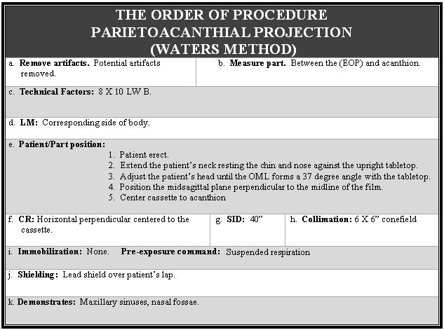
, parietoacanthial projection (Waters), and lateral sinuses. b. The Sinuses.")
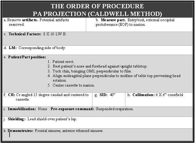
16 Section II. PROJECTIONS OF THE SINUSES 5-5. POSTEROANTERIOR PROJECTION (CALDWELL METHOD) a. Sinus Routine. The routine views are the PA projection (Caldwell method), parietoacanthial projection (Waters), and lateral sinuses. b. The Sinuses. The four sinuses are the frontal sinus, the ethmoid sinus, the sphenoid sinus, and the maxillary sinus. c. Best Demonstrated. The PA projection (Caldwell method) best demonstrates the frontal and ethmoid sinuses. The Waters best demonstrates the maxillary sinuses. The lateral projection best demonstrates all four sinuses. MD

17 MD
")
18 5-6. PARIETOACNTHIAL PROJECTION (WATERS METHOD) MD

19 MD

20 5-7. LATERAL SINUSES MD


21 Continue with Exercises MD
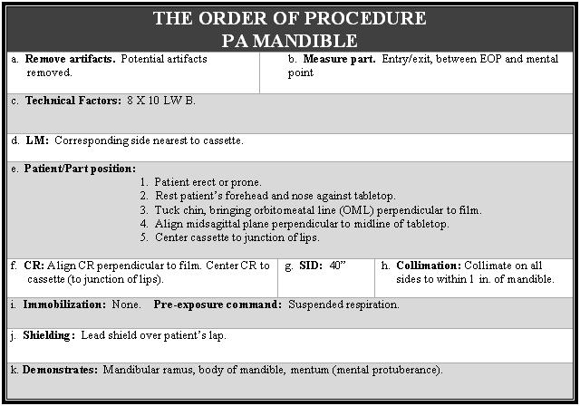
22 EXERCISES, LESSON 5, SECTION II MATCHING: For exercises 1 through 3, match the position with the anatomical structure(s) that the position demonstrates. Enter the letter that corresponds to your choice in the space provided. (There is an extra alternative that will not be selected.) 1. PA sinuses. a. Maxillary sinuses and nasal fossae. (Caldwell method) 2. Parietoacanthia b. Frontal sinuses and anterior ethmoid sinuses. sinuses (Waters method). 3. Lateral sinuses. c. Sphenoid sinuses, superimposed frontal sinuses, ethmoid sinuses, maxillary sinuses, sella turcica, orbital roofs. d. Petrous ridge in profile, bony labyrinth, tympanic cavity, internal auditory canal, and mastooid air cell. MULTIPLE- CHOICE. For exercises 4 through 7, select the ONE word or phrase that BEST completes the statement or BEST answers the question. 4. The technical factors for the three sinus projections taught in this lesson are: a. 8 x 10 LW NB. b. 8 x 10 LW B. c. 10 x 12 CW B. d. 10 x 12 LW B 5. The part centered for the parietoacanthial sinus (Waters method) is: a. Nasion to the center of the film b. Acanthion to the center of the cassette. c. Midway between the outer canthus and the EAM to the center of the cassette. d. Glabella to the center of the cassette. MD
23 6. The proper patient and part position for a PA sinus (Caldwell method) is: a. Patient erect; midsagittal plane perpendicular to the midline of film; OML perpendicular to the film. b. Patient erect; midsagittal plane perpendicular to the midline of the film; IOML perpendicular to the film. c. Patient erect, head in true lateral position; midsagittal plane parallel to film; IPPL perpendicular to film; IOML perpendicular to front edge of cassette. d. Patient erect; chin against table or upright Bucky, MML perpendicular to film; OML at a 37 degree angle; midsagittal plane perpendicular to film. 7. For a parietoacanthial projection (Waters), the angle of the OML is: a. 15 degrees. b. 25 degrees. c. 30 degrees. d. 37 degrees. Check Your Answers on Next Page MD
24 SOLUTIONS, LESSON 5, SECTION II 1. b (para 5-5) 2. a (para 5-6) 3. c (para 5-7) 4. b (paras 5-5, 5-6, 5-7) 5. b (para 5-6) 6. a (para 5-5) 7. d (para 5-6) MD

 are the routine views for the")
25 Section III. PROJECTIONS OF THE MANDIBLE 5-8. POSTEROANTERIOR MANDIBLE The posterior-anterior (PA) and the axiolateral (oblique) are the routine views for the mandible. MD

26 MD


27 5-9. AXIOLATERAL (OBLIQUE) MANDIBLE MD

28 Continue with Exercises Return to Table of Contents MD
29 EXERCISES, LESSON 5, SECTION III MATCHING: For exercises 1 through 2, match the position with the anatomical structure(s) that the position demonstrates. Enter the letter that corresponds to your choice in the space provided. (There is an extra alternative that will not be selected.) 1. PA mandible. a. An end-on view of the petrous portion, EAM canal, mastoid air cells, and mastoid antrum. 2. Axiolateral oblique b. Mandibular ramus, body of mandible, mentum. mandible. c. Mandibular rami, body of mentum of mandible positioned closest to film. MULTIPLE-CHOICE. For exercises 3 through 6, select the ONE word or phrase that BEST completes the statement or BEST answers the question. 3. When measuring the part for a PA mandible, the entry/exit point is: a. Between the external occipital protuberance (EOP) and the mental point. b. Laterally between the midpoint of the mandible. c. Between the EOP and tragus. d. Between the vertex of the skull and the tragus. 4. The proper patient and part position for an axiolateral oblique mandible is: a. Patient in true lateral recumbent position, knees flexed, arms at right angle to body; radiolucent material under lower dorsal region, coronal plane perpendicular to the table. b. Patient supine, midsagittal plane perpendicular to the table. c. Patient seated or standing (nose-forehead position); midsagittal plane perpendicular and over the center line of the film; OML perpendicular to the film. d. Patient erect or prone; head in a lateral position, with side of interest closest to cassette; IPPL perpendicular to table; midsagittal plane (chin) rotated 30 degrees face down towards the mandible body being demonstrated. MD
30 5. The technical factors for the PA mandible is: a. 10 x 12 LW NB b. 10 x 12 LW B c. 8x 10 CW NB d. 8 x 10 LW B 6. The proper patient and part position for a PA mandible is: a. Patient in true lateral recumbent position, knees flexed, arms at right angle to body; radiolucent material under lower dorsal region; coronal plane perpendicular to the table. b. Patient prone, midsagittal plane perpendicular to table top, OML perpendicular to film. c. Patient seated or standing (not forehead position); midsagittal plane perpendicular and over the centerline of the film OML perpendicular to the film. d. Patient semi-prone, one arm down by the side and the other supporting the side up. Shoulder in down and out of CF chin extended until the inferior margin of side up is parallel with lower edge of cassette. Midsagittal plane of skull tilted 15 degrees vertex down. Check Your Answers on Next Page MD
31 SOLUTIONS, LESSON 5, SECTION III 1. b (para 5-8) 2. c (para 5-9) 3. a (para 5-8) 4. d (para 5-9) 5. d (paras 5-8 and 5-9) 6. b (para 5-9) Return to Table of Contents MD
LESSON ASSIGNMENT. Positioning for Exams of the Spine. After completing this lesson, you should be able to identify:
 LESSON ASSIGNMENT LESSON 4 Positioning for Exams of the Spine. LESSON ASSIGNMENT Paragraphs 4-1 through 4-15. LESSON OBJECTIVES After completing this lesson, you should be able to identify: 4-1. Identify
Bones of the skull & face
 Bones of the skull & face Cranium= brain case or helmet Copyright The McGraw-Hill Companies, Inc. Permission required for reproduction or display. The cranium is composed of eight bones : frontal Occipital
Chapter 7 Part A The Skeleton
 Chapter 7 Part A The Skeleton Why This Matters Understanding the anatomy of the skeleton enables you to anticipate problems such as pelvic dimensions that may affect labor and delivery The Skeleton The
COURSE SYLLABUS RT 1244 RADIOGRAPHIC POSITIONING FILM CRITIQUE AND MEDICAL TERMINOLOGY II
 COURSE SYLLABUS RT 1244 RADIOGRAPHIC POSITIONING FILM CRITIQUE AND MEDICAL TERMINOLOGY II CLASS HOURS: 4 CREDIT HOURS: 4 LABORATORY HOURS: 4 CATALOG COURSE DESCRIPTION: This course is the second of a three
Extraoral radiography Introduction: Extraoral radiographs (outside the mouth) are taken when large areas of the skull or jaw must be examined or when
 are taken when large areas of the skull or jaw must be examined or when") Extraoral radiography Introduction: Extraoral radiographs (outside the mouth) are taken when large areas of the skull or jaw must be examined or when patients are unable to open their mouths for film placement.
Extraoral radiography Introduction: Extraoral radiographs (outside the mouth) are taken when large areas of the skull or jaw must be examined or when patients are unable to open their mouths for film placement.
Biology 218 Human Anatomy. Adapted from Martini Human Anatomy 7th ed. Chapter 6 The Skeletal System: Axial Division
 Adapted from Martini Human Anatomy 7th ed. Chapter 6 The Skeletal System: Axial Division Introduction The axial skeleton: Composed of bones along the central axis of the body Divided into three regions:
Anatomy and Physiology. Bones, Sutures, Teeth, Processes and Foramina of the Human Skull
 Anatomy and Physiology Chapter 6 DRO Bones, Sutures, Teeth, Processes and Foramina of the Human Skull Name: Period: Bones of the Human Skull Bones of the Cranium: Frontal bone: forms the forehead and the
AXIAL SKELETON SKULL
 AXIAL SKELETON SKULL CRANIAL BONES (8 total flat bones w/ 2 paired) 1. Frontal forms forehead & upper portion of eyesocket (orbital) 2. Parietal paired bones; form superior & lateral walls of cranium 3.
Skeletal System -Axial System. Chapter 7 Part A
 Skeletal System -Axial System Chapter 7 Part A Skeleton Learn: Names of the s. Identify specific landmarks that allow: Bones to fit into each other, Organs to fit into the cavities, Muscles to attach,
Technique. Simpo PDF Merge and Split Unregistered Version -
 extraoral radiographic examinations both ~.~ x-ray source and image receptor (film or electronic sensor) are placed outside the patient's mouth. This chapter describes the most common extraoral radiographic
Cranium Facial bones. Sternum Rib
 Figure 7.1 The human skeleton. Skull Thoracic cage (ribs and sternum) Cranium Facial bones Sternum Rib Bones of pectoral girdle Vertebral column Sacrum Vertebra Bones of pelvic girdle (a) Anterior view
SKULL / CRANIUM BONES OF THE NEUROCRANIUM (7) Occipital bone (1) Sphenoid bone (1) Temporal bone (2) Frontal bone (1) Parietal bone (2)
 Occipital bone (1) Sphenoid bone (1) Temporal bone (2) Frontal bone (1) Parietal bone (2)") Important! 1. Memorizing these pages only does not guarantee the succesfull passing of the midterm test or the semifinal exam. 2. The handout has not been supervised, and I can not guarantee, that these
Important! 1. Memorizing these pages only does not guarantee the succesfull passing of the midterm test or the semifinal exam. 2. The handout has not been supervised, and I can not guarantee, that these
Skull-2. Norma Basalis Interna Norma Basalis Externa. Dr. Heba Kalbouneh Associate Professor of Anatomy and Histology
 Skull-2 Norma Basalis Interna Norma Basalis Externa Dr. Heba Kalbouneh Associate Professor of Anatomy and Histology Norma basalis interna Base of the skull- superior view The interior of the base of the
Skull-2. Norma Basalis Interna. Dr. Heba Kalbouneh Assistant Professor of Anatomy and Histology
 Skull-2 Norma Basalis Interna Dr. Heba Kalbouneh Assistant Professor of Anatomy and Histology Norma basalis interna Base of the skull- superior view The interior of the base of the skull is divided into
Skull basic structures. Neurocranium
 Assoc. Prof. Květuše Lovásová, M.V.D., PhD. Skull basic structures Skull consists of two groups of bones: neurocranium (bones forming the brain box) splanchnocranium (bones forming the facial skeleton)
SKULL AS A WHOLE + ANTERIOR CRANIAL FOSSA
 SKULL AS A WHOLE + ANTERIOR CRANIAL FOSSA LEARNING OBJECTIVES At the end of this lecture, the student should be able to know: Parts of skeleton (axial and appendicular) Parts of skull Sutures of skull
Radiography of The Skull
 Radiography of The Skull By N.J.Oldnall Tameside General Hospital. August 8, 1996 NJO / SKULL ISO PENDO TECHNIQUE 1998.doc 1 Basic Osteology, Revision Basic Anatomy: The skull encloses and protects the
Dr. Sami Zaqout, IUG Medical School
 The skull The skull is composed of several separate bones united at immobile joints called sutures. Exceptions? Frontal bone Occipital bone Vault Cranium Sphenoid bone Zygomatic bones Base Ethmoid bone
Skeletal System: Skull.
 Skeletal System: Skull www.fisiokinesiterapia.biz Bones of the Skull SPLANCHNOCRANIUM Nasal (2) Maxilla (2) Lacrimal (2) Zygomatic (2) Palatine (2) Inferior concha (2) Vomer Mandible NEUROCRANIUM Frontal
Chapter 7. Skeletal System
 Chapter 7 Skeletal System 1 Skull A. The skull is made up of 22 bones: 8 cranial bones, 13 facial bones, and the mandible. B. The Cranium encloses and protects the brain, provides attachments for muscles,
OUTLINE ANATOMY, RADIOGRAPHY,
 20 SKULL OUTLINE SUMMARY OF PROJECTIONS, 262 ANATOMY, 263 Skull, 263 Cranial bones, 267 Ear, 277 Facial bones, 278 Articulations of the skull, 281 SUMMARY OF ANATOMY, 282 SUMMARY OF PATHOLOGY, 284 EXPOSURE
Introduction to Local Anesthesia and Review of Anatomy
 5-Sep Introduction and Anatomy Review 12-Sep Neurophysiology and Pain 19-Sep Physiology and Pharmacology part 1 26-Sep Physiology and Pharmacology part 2 Introduction to Local Anesthesia and Review of
Course Description. This course provides the student with instruction in the radiographic anatomy and
 Course Title Course Code RA 331 Prerequisites RA 230 Course Website Instructor Office Location Office Phone # 26879 Office Hours E-mail Teaching Assistant(s) Jordan University of Science and Technology
View of a Skull, 1489 by Leonardo Da Vinci. Kaan Yücel M.D., Ph.D Tuesday
 View of a Skull, 1489 by Leonardo Da Vinci Kaan Yücel M.D., Ph.D. 26.11.2013 Tuesday 1.SKULL skeleton of the head cranium 22 bones excluding ossicles of the ear 1.SKULL Mandible Lower jaw bone Neurocranium
APPENDICULAR SKELETON 126 AXIAL SKELETON SKELETAL SYSTEM. Cranium. Skull. Face. Skull and associated bones. Auditory ossicles. Associated bones.
 SKELETAL SYSTEM 206 AXIAL SKELETON 80 APPENDICULAR SKELETON 26 Skull Skull and associated s 29 Cranium Face Auditory ossicles 8 4 6 Associated s Hyoid Thoracic cage 25 Sternum Ribs 24 Vertebrae 24 column
Dr.Noor Hashem Mohammad Lecture (5)
") Dr.Noor Hashem Mohammad Lecture (5) 2016-2017 If the mandible is discarded, the anterior part of this aspect of the skull is seen to be formed by the hard palate. The palatal processes of the maxillae
Dr.Noor Hashem Mohammad Lecture (5) 2016-2017 If the mandible is discarded, the anterior part of this aspect of the skull is seen to be formed by the hard palate. The palatal processes of the maxillae
Proteus XR/f Patient positioning guide
 Proteus XR/f Patient positioning guide PROTEUS XR/F Now a single digital x-ray room accommodates nearly all your radiographic studies. With extended tube coverage and wireless detectors, Proteus XR/f gives
Chapter 7: Head & Neck
 Chapter 7: Head & Neck Osteology I. Overview A. Skull The cranium is composed of irregularly shaped bones that are fused together at unique joints called sutures The skull provides durable protection from
Anatomy images for MSS practical exam- 2019
 Anatomy images for MSS practical exam- 2019 Ilium Ischium Pubis Acetabulaum Iliac crest Iliac tubercle ASIS (muscle and ligament attached) AIIS (muscle attached) PSIS PIIS Ischial spine Ischial tuberosity
Anatomy Made Easy MSS
 Anatomy Made Easy MSS part #1 هذا الملف يشمل تفريغ المحاضرة الثانية لعون بدءا من الصفحة 11 وحتى األخير Done By :MohamedA. Diabat Edited by Awn Academic team The Axial Skeleton The axial skeleton consist
CLASS HOURS: 4 CREDIT HOURS: 4 LABORATORY HOURS: 2
 Revised 9-2010 COURSE SYLLABUS RT 2442 RADIOLOGY SEMINAR I CLASS HOURS: 4 CREDIT HOURS: 4 LABORATORY HOURS: 2 CATALOG COURSE DESCRIPTION: This is the first of a two-course sequence in advanced radiographic
Adaptive Radiography: Tips and Tricks
 Adaptive Radiography: Tips and Tricks WCEC 20 th Student Educator Radiographer Conference Dennis Bowman, RT(R), CRT (R)(F) Community Hospital of the Monterey Peninsula (CHOMP) - Staff Radiographer Owner/Consultant
Major Anatomic Components of the Orbit
 Major Anatomic Components of the Orbit 1. Osseous Framework 2. Globe 3. Optic nerve and sheath 4. Extraocular muscles Bony Orbit Seven Bones Frontal bone Zygomatic bone Maxillary bone Ethmoid bone Sphenoid
Structure Location Function
 Frontal Bone Cranium forms the forehead and roof of the orbits Occipital Bone Cranium forms posterior and inferior portions of the cranium Temporal Bone Cranium inferior to the parietal bone forms the
POSTERIOR 1. situated behind: situated at or toward the hind part of the body :
 ANATOMICAL LOCATION Anatomy is a difficult subject with a large component of memorization. There is just no way around that, but we have made every effort to make this course diverse and fun. The first
LESSON ASSIGNMENT. After completing this lesson, you should be able to: 3-1. Identify body part terminology.
 LESSON ASSIGNMENT LESSON 3 Positioning Terminology. LESSON ASSIGNMENT Paragraphs 3-1 through 3-23. LESSON OBJECTIVES After completing this lesson, you should be able to: 3-1. Identify body part terminology.
Radiography Log Book
 Radiography Log Book Radiography teaching occurs in constituent hospitals as part of working week. This unit of the log book deals with diagnostic radiography. Tuition in radiography will be carried out
CHAPTER 7, PART II (BONES)
") Anatomy Name: CHAPTER 7, PART II (BONES) Entry #: INSTRUCTIONS: 1) READ Chapter 7, pg. 140-161. 2) Using the outline, make a note card for each underlined bone name or phrase. 3) On each note card, put
Anatomy Name: CHAPTER 7, PART II (BONES) Entry #: INSTRUCTIONS: 1) READ Chapter 7, pg. 140-161. 2) Using the outline, make a note card for each underlined bone name or phrase. 3) On each note card, put
Bones of the Skull Lateral View
 Bones of the Skull Lateral View Frontal Bone Parietal Bone Occipital Bone Temporal Bone Sphenoid Bone Pterion Sutures of the Skull Lateral View Coronal Suture Lambdoid Suture Squamous Suture Sutures of
THE SKELETAL SYSTEM. Focus on the Skull
 THE SKELETAL SYSTEM Focus on the Skull Review Anatomical Terms Anterior/Posterior Dorsal/Ventral Medial/Lateral Superior/Inferior Bone Markings - Review Projections for attachment of muscles, ligaments
Chapter 7: Skeletal System: Gross Anatomy
 Chapter 7: Skeletal System: Gross Anatomy I. General Considerations A. How many bones in an average adult skeleton? B. Anatomic features of bones are based on II. Axial Skeleton A. Skull 1. Functionally
Extraoral Radiology October 10th, 2008
 Extraoral Radiology October 10th, 2008 Steven R. Singer, DDS srs2@columbia.edu 212.305.5674 November 8 th, 1895 Extraoral Projections Images can be produced in the dental office X-ray source can be Intraoral
YOU MUST BRING YOUR OWN GLOVES FOR THIS ACTIVITY.
 ACTIVITY 3: AXIAL SKELETON AND LONG BONE DISSECTION Objectives: 1) How to get ready: Read Chapter 7, McKinley et al., Human Anatomy, 5e. All text references are for this textbook. Learning the meanings
University of Palestine. Midterm Exam 2013/2014 Total Grade:
 Course No: DNTS2208 Course Title: Head and Neck Anatomy Date: 09/11/2013 No. of Questions: (50) Time: 1hour Using Calculator (No) University of Palestine Midterm Exam 2013/2014 Total Grade: Instructor
Bones Ethmoid bone Inferior nasal concha Lacrimal bone Maxilla Nasal bone Palatine bone Vomer Zygomatic bone Mandible
 splanchnocranium - Consists of part of skull that is derived from branchial arches - The facial bones are the bones of the anterior and lower human skull Bones Ethmoid bone Inferior nasal concha Lacrimal
University of Palestine. Midterm Exam 2013/2014 Total Grade:
 [ Course No: DNTS2208 Course Title: Head and Neck Anatomy Date: 17/11/1024 No. of Questions: (52) Time: 2hours Using Calculator (No) University of Palestine Midterm Exam 2013/2014 Total Grade: Instructor
The orbit-1. Dr. Heba Kalbouneh Assistant Professor of Anatomy and Histology
 The orbit-1 Dr. Heba Kalbouneh Assistant Professor of Anatomy and Histology Orbital plate of frontal bone Orbital plate of ethmoid bone Lesser wing of sphenoid Greater wing of sphenoid Lacrimal bone Orbital
ACTIVITY 3: AXIAL SKELETON AND LONG BONE DISSECTION COW BONE DISSECTION
 ACTIVITY 3: AXIAL SKELETON AND LONG BONE DISSECTION Objectives: 1) How to get ready: Read Chapter 7, McKinley et al., Human Anatomy, 4e. All text references are for this textbook. Learning the meanings
o Diaphysis o Area where red marrow is found o Area where yellow marrow is found o Epiphyseal plate AXIAL SKELETON Skull
 64 Anatomy & Physiology Coloring Workbook 7. Figure 5-2A is a midlevel, cross-sectional view of the diaphysis of the femur. Label the membrane that lines the cavity and the membrane that covers the outside
3-Deep fascia: is absent (except over the parotid gland & buccopharngeal fascia covering the buccinator muscle)
") The Face 1-Skin of the Face The skin of the face is: Elastic Vascular (bleed profusely however heal rapidly) Rich in sweat and sebaceous glands (can cause acne in adults) It is connected to the underlying
The Face 1-Skin of the Face The skin of the face is: Elastic Vascular (bleed profusely however heal rapidly) Rich in sweat and sebaceous glands (can cause acne in adults) It is connected to the underlying
Basic Chiropractic Assistant Radiologic Certification Program
 Basic Chiropractic Assistant Radiologic Certification Program Module 3 Instructor: Dr. Roxzanne Breland Special Notice THE PRINTED MATERIALS USED IN THIS CLASS ARE COPYRIGHTED AND MAY NOT BE COPIED, REPRODUCED
The Skull DANIL HAMMOUDI.MD
 The Skull DANIL HAMMOUDI.MD summary of bones/structures in Chapter 15 of the manual need tp be print as soon as possible http://www.mnsu.edu/emuseum/biology/humananatomy/skeletal/skul l/frontal/frontal.html
The Axial Skeleton. C h a p t e r. PowerPoint Lecture Slides prepared by Jason LaPres Lone Star College - North Harris
 C h a p t e r 7 The Axial Skeleton PowerPoint Lecture Slides prepared by Jason LaPres Lone Star College - North Harris Copyright 2009 Pearson Education, Inc., publishing as Pearson Benjamin Cummings An
Unit 18: Cranial Cavity and Contents
 Unit 18: Cranial Cavity and Contents Dissection Instructions: The calvaria is to be removed without damage to the dura mater which is attached to the inner surface of the calvaria. Cut through the outer
Learning Objectives (1&2)
") Learning Objectives (1&2) By the end of the session, students should be able to: 1) Identify anatomical position seated, standing, prone, supine. 2) Pronounce, define and be able to use directional and
Learning Objectives (1&2) By the end of the session, students should be able to: 1) Identify anatomical position seated, standing, prone, supine. 2) Pronounce, define and be able to use directional and
Important Parts of Bones
 Important Parts of Bones For 2015 Know: Humerus (posterior) Clavical Femur (Anterior) Foot Hand Mandible Os Coxa Scapula Skull (Anterior, Inferior, Lateral) Sternum Humerus (posterior) A. olecranon fossa
Bone Flashcards for 10a
 Bone Flashcards for 0a CLAVICLE (collar bone). Sternal extremity (end) flat end. Acromial extremity (end) rounded end. SCAPULA (shoulder blade). Right or left scapula?. Superior border (superior margin).
Core Curriculum Syllabus Emergencies in Otolaryngology-Head and Neck Surgery FACIAL FRACTURES
 Core Curriculum Syllabus Emergencies in Otolaryngology-Head and Neck Surgery A. General Considerations FACIAL FRACTURES Look for other fractures like skull and/or cervical spine fractures Test function
Human Anatomy and Physiology - Problem Drill 07: The Skeletal System Axial Skeleton
 Human Anatomy and Physiology - Problem Drill 07: The Skeletal System Axial Skeleton Question No. 1 of 10 Which of the following statements about the axial skeleton is correct? Question #01 A. The axial
THIEME. Scalp and Superficial Temporal Region
 CHAPTER 2 Scalp and Superficial Temporal Region Scalp Learning Objectives At the end of the dissection of the scalp, you should be able to identify, understand and correlate the clinical aspects: Layers
Superior View of the Skull (Norma Verticalis) Anteriorly the frontal bone articulates with the two parietal bones AT THE CORONAL SUTURE
 Anteriorly the frontal bone articulates with the two parietal bones AT THE CORONAL SUTURE") Superior View of the Skull (Norma Verticalis) Anteriorly the frontal bone articulates with the two parietal bones AT THE CORONAL SUTURE 1 The two parietal bones articulate in the midline AT THE SAGITTAL
Superior View of the Skull (Norma Verticalis) Anteriorly the frontal bone articulates with the two parietal bones AT THE CORONAL SUTURE 1 The two parietal bones articulate in the midline AT THE SAGITTAL
Gross Anatomy of the. TEMPORAL BONE, EXTERNAL EAR, and MIDDLE EAR. Assignment: Head to Toe Temporomandibular Joint (TMJ)
") Gross Anatomy the TEMPORAL BONE, EXTERNAL EAR, and MIDDLE EAR M1 Gross and Developmental Anatomy 9:00 AM, December 11, 2008 Dr. Milton M. Sholley Pressor Anatomy and Neurobiology Assignment: Head to Toe
Gross Anatomy the TEMPORAL BONE, EXTERNAL EAR, and MIDDLE EAR M1 Gross and Developmental Anatomy 9:00 AM, December 11, 2008 Dr. Milton M. Sholley Pressor Anatomy and Neurobiology Assignment: Head to Toe
External Occipital Protuberance
 Osteology Exterior Skull Frontal Bone Glabella Superciliary Arch Supraorbital Notch/Foramen Nasion (junction w/ Nasal bone) Frontal/Metopic Suture (usually absent in adult, b/w ossification centers of
TEST YOURSELF- Chapter 7
 TEST YOURSELF- Chapter 7 Cranial Bones 1. Give the name of the bone for each of the following markings. Some of the markings are found on more than one bone. List all that apply. Cranium a. Frontal squama:
in compact bone, large vertical canals carrying blood vessels and nerves. in compact bone, large horizontal canals carrying blood vessels and nerves.
 Carl Christensen, PhD Skeletal System (Bones`) Bio. 2304 Human Anatomy 1. Identify a term for each of the following: shaft of a long bone ends of a long bone ossified remnant of the "growth plate" connective
External Acoustic Meatus. Mastoid Process. Zygomatic Process. Temporal Bone
 Bone lab review 1. Frontal Bone 2. Supra-Orbital Foramen 3. Orbit (Orbital Cavity) 4. Superior Orbital Fissure 5. Inferior Orbital Fissure 6. Zygomatic Bone 7. Infra-Orbital Foramen 8. Maxilla 9. Mandible
Gross Anatomy of the. TEMPORAL BONE, EXTERNAL EAR, and MIDDLE EAR
 Gross Anatomy of the TEMPORAL BONE, EXTERNAL EAR, and MIDDLE EAR M1 Gross and Developmental Anatomy 9:00 AM, December 11, 2008 Dr. Milton M. Sholley Professor of Anatomy and Neurobiology Assignment: Head
The Human Body: An Orientation
 The Human Body: An Orientation Body standing upright Anatomical Position feet slightly apart palms facing forward thumbs point away from body Directional Terms Superior and inferior toward and away from
Routine Guide EXAMINATION PROJECTION CASSETTE SIZE NOTES PRINT ORIENTATION. 14x17 CW* 14x17LW 14x17LW. 14x17LW 14x17LW 14x17LW
 EXAMINATION PROJECTION CASSETTE SIZE NOTES PRINT ORIENTATION A-C Joints without weights with weights 14x17 CW* One 14x17 divided; both shoulders on one exposure. *If part does not fit, do 10x12s CW. Both
ANATOMY & PHYSIOLOGY I Laboratory Version B Name Section. REVIEW SHEET Exercise 10 Axial Skeleton
 ANATOMY & PHYSIOLOGY I Laboratory Version B Name Section REVIEW SHEET Exercise 10 Axial Skeleton 1 POINT EACH. THE SKULL MULTIPLE CHOICE 1. The major components of the axial skeleton include the 7. The
Radiology Positioning Practical Test #2 Table (By Jung Park):
:") Radiology Positioning Practical Test #2 Table (By Jung Park): (Lower Extremity): patient is fully gowned / no artifacts / properly shielded (exposure for femur and below : hold still, don t move ) (exposure
Radiology Positioning Practical Test #2 Table (By Jung Park): (Lower Extremity): patient is fully gowned / no artifacts / properly shielded (exposure for femur and below : hold still, don t move ) (exposure
For educational and institutional use. This test bank is licensed for noncommercial, educational inhouse or online educational course use only in
 For educational and institutional use. This test bank is licensed for noncommercial, educational inhouse or online educational course use only in educational and corporate institutions. Any broadcast,
BONE CHALLENGE DANIL HAMMOUDI.MD
 BONE CHALLENGE DANIL HAMMOUDI.MD Bone Basic functions? A. support B. protection C. movement assistance in D. RBC formation-hemopoiesis E. mineral homeostasis +importance of calcium F. energy supply -yellow
Blair Radiology Exam Examination Packet
 Blair Radiology Exam Examination Packet This packet is made of up five sections: Examiner s Instructions, Applicant Requirements, Analysis Rubric, Overall Result and Comments and Exam Form. The Exam Form
Nasal region. cartilages: septal cartilage (l); lateral nasal cartilage (2); greater alar cartilages (2); lesser alar cartilages (?
; lateral nasal cartilage (2); greater alar cartilages (2); lesser alar cartilages (?") Nasal region skull bones: nasal and frontal processes of maxilla cartilages: septal cartilage (l); lateral nasal cartilage (2); greater alar cartilages (2); lesser alar cartilages (?) 1 Nasal cavity Roof
Nasal region skull bones: nasal and frontal processes of maxilla cartilages: septal cartilage (l); lateral nasal cartilage (2); greater alar cartilages (2); lesser alar cartilages (?) 1 Nasal cavity Roof
PH-2 Whole Body Phantom PBU-50. PH-2B CT Whole Body Phantom PBU-60. Contents. Point of X-ray Photography Head radiography P.1 ~ P.5 P.6 ~ P.
 PH-2 Whole Body Phantom PBU-50 PH-2B CT Whole Body Phantom PBU-60 Production supervision: Kyoto College of Medical Science Photography cooperation: Kyoto College of Medical Science Toshifumi Kasai Keiko
NEUROCRANIUM VISCEROCRANIUM VISCEROCRANIUM VISCEROCRANIUM
 LECTURE 4 SKULL NEUROCRANIUM VISCEROCRANIUM VISCEROCRANIUM VISCEROCRANIUM CRANIUM NEUROCRANIUM (protective case around brain) VISCEROCRANIUM (skeleton of face) NASOMAXILLARY COMPLEX MANDIBLE (DESMOCRANIUM)
The sebaceous glands (glands of Zeis) open directly into the eyelash follicles, ciliary glands (glands of Moll) are modified sweat glands that open
 open directly into the eyelash follicles, ciliary glands (glands of Moll) are modified sweat glands that open") The Orbital Region The orbits are a pair of bony cavities that contain the eyeballs; their associated muscles, nerves, vessels, and fat; and most of the lacrimal apparatus upper eyelid is larger and more
The Orbital Region The orbits are a pair of bony cavities that contain the eyeballs; their associated muscles, nerves, vessels, and fat; and most of the lacrimal apparatus upper eyelid is larger and more
Anatomy and Physiology 1 Chapter 7 self quiz Pro, Dima Darwish,MD.
 Anatomy and Physiology 1 Chapter 7 self quiz Pro, Dima Darwish,MD. 1) How many bones make up the axial skeleton? A) 50 B) 60 C) 70 D) 80 E) 90 2) Which of the following is a function of the axial skeleton?
HBA THE BODY Head & Neck Written Examination October 23, 2014
 HBA 531 - THE BODY Head & Neck Written Examination October 23, 2014 Name: NOTE 2: When asked to trace nerve, artery, or vein pathways, do so by using arrows, e.g., structure a structure b structure c...
LESSON ASSIGNMENT. Positioning for Exams of the Upper Extremities. After completing this lesson, you should be able to:
 LESSON ASSIGNMENT LESSON 5 Positioning for Exams of the Upper Extremities. LESSON ASSIGNMENT Paragraphs 5-1 through 5-25. LESSON OBJECTIVES After completing this lesson, you should be able to: 5-1. Identify
Dentalelle Tutoring - Faulty Radiographs
 Dentalelle Tutoring - Faulty Radiographs Errors in improperly exposing or processing dental films can produce undesirable dental radiographs of nondiagnostic quality. These are known as faulty radiographs.
Infratemporal fossa: Tikrit University college of Dentistry Dr.Ban I.S. head & neck Anatomy 2 nd y.
 Infratemporal fossa: This is a space lying beneath the base of the skull between the lateral wall of the pharynx and the ramus of the mandible. It is also referred to as the parapharyngeal or lateral pharyngeal
PTERYGOPALATINE FOSSA
 PTERYGOPALATINE FOSSA Outline Anatomical Structure and Boundaries Foramina and Communications with other spaces and cavities Contents Pterygopalatine Ganglion Especial emphasis on certain arteries and
Bisection of Head & Nasal Cavity 頭部對切以及鼻腔. 解剖學科馮琮涵副教授 分機
 Bisection of Head & Nasal Cavity 頭部對切以及鼻腔 解剖學科馮琮涵副教授 分機 3250 E-mail: thfong@tmu.edu.tw Outline: The structure of nose The concha and meatus in nasal cavity The openings of paranasal sinuses Canals, foramens
بسم هللا الرحمن الرحيم السالم عليكم ورحمة هللا وبركاته
 بسم هللا الرحمن الرحيم السالم عليكم ورحمة هللا وبركاته Lecture one Introduction to General Anatomy By Anatomist Dr. Hatem A. Hatem Department of Anatomy, Histology& Embryology College of Dentistry-Ibn
Anatomic Relations Summary. Done by: Sohayyla Yasin Dababseh
 Anatomic Relations Summary Done by: Sohayyla Yasin Dababseh Anatomic Relations Lecture 1 Part-1 - The medial wall of the nose is the septum. - The vestibule lies directly inside the nostrils (Nares). -
Perpendicular Plate Zygomatic Bone. Mental Foramen Mandible
 Glabella Frontal Middle Nasal Concha Nasal Lacrimal Perpendicular Plate Zygomatic Inferior Nasal Concha Maxilla Mental Mandible Skull (anterior view) Squamosal Suture Coronal Suture Frontal Parietal Nasal
TRAUMA TO THE FACE AND MOUTH
 Dr.Yahya A. Ali 3/10/2012 F.I.C.M.S TRAUMA TO THE FACE AND MOUTH Bailey & Love s 25 th edition Injuries to the orofacial region are common, but the majority are relatively minor in nature. A few are major
An Introduction to the Axial Skeleton. Copyright 2009 Pearson Education, Inc., publishing as Pearson Benjamin Cummings
 An Introduction to the Axial Skeleton Copyright 2009 Pearson Education, Inc., publishing as Pearson Benjamin Cummings Terms: Structures of Bones Articulations: Contacts with other bones Landmarks (Bone
Skeletal system. Prof. Abdulameer Al-Nuaimi. E. mail:
 Skeletal system Prof. Abdulameer Al-Nuaimi E-mail: a.al-nuaimi@sheffield.ac.uk E. mail: abdulameerh@yahoo.com Functions of Bone and The Skeletal System Support: The skeleton serves as the structural framework
Skull. Sphenoid and Ethmoid bones
 Skull. Sphenoid and Ethmoid bones PhD., Dr. David Lendvai Department of Anatomy, Histology and Embriology Semmelweis University, Faculty of Medicine 2018. Skeletal system Structure of the skull Border
BLUE SKY SCHOOL OF PROFESSIONAL MASSAGE AND THERAPEUTIC BODYWORK. Musculoskeletal Anatomy & Kinesiology I TERMINOLOGY, STRUCTURES, & SKELETAL OVERVIEW
 BLUE SKY SCHOOL OF PROFESSIONAL MASSAGE AND THERAPEUTIC BODYWORK Musculoskeletal Anatomy & Kinesiology I TERMINOLOGY, STRUCTURES, & SKELETAL OVERVIEW MSAK101-I Session 1 Learning Objectives: 1. Define
revised originals as separate pages on I://DX(all folders)/trauma X Manual and project
/trauma X Manual and project") 06-25-14 revised originals as separate pages on I://DX(all folders)/trauma X Manual and project Procedure for Suspected Child Abuse Imaging Trauma X GUIDELINES: Every effort should be made to request studies
06-25-14 revised originals as separate pages on I://DX(all folders)/trauma X Manual and project Procedure for Suspected Child Abuse Imaging Trauma X GUIDELINES: Every effort should be made to request studies
Crafton Hills College Human Anatomy & Physiology Axial Skeleton
 A. Major Divisions Crafton Hills College Human Anatomy & Physiology Axial keleton 1. Axial: Part of skeleton lies along long axis of body 2. Appendicular: Bones & features of the appendages B. AXIAL KELETON
Jefferson Cephalometric Analysis--Face and Health Focused
 Jefferson Cephalometric Analysis--Face and Health Focused Google: Jefferson Ceph Analysis Video Instruction for video instruction. Note: video instruction teaches how to find Center O. Center O is now
Dr. Sami Zaqout Faculty of Medicine IUG
 The Nose External Nose Nasal Cavity External Nose Blood and Nerve Supplies of the External Nose Blood Supply of the External Nose The skin of the external nose Branches of the ophthalmic and the maxillary
The Language of Anatomy. (Anatomical Terminology)
") The Language of Anatomy (Anatomical Terminology) Terms of Position The anatomical position is a fixed position of the body (cadaver) taken as if the body is standing (erect) looking forward with the upper
The Language of Anatomy (Anatomical Terminology) Terms of Position The anatomical position is a fixed position of the body (cadaver) taken as if the body is standing (erect) looking forward with the upper