Biology 340 Comparative Embryology Lecture 11 Dr. Stuart Sumida. Overview of Embryology of the Vertebrate Skull. Emphasis on Amniota
|
|
|
- Janice Merritt
- 6 years ago
- Views:
Transcription
1 Biology 340 Comparative Embryology Lecture 11 Dr. Stuart Sumida Overview of Embryology of the Vertebrate Skull Emphasis on Amniota

2 Initial introduction to components parts of a vertebrate head.

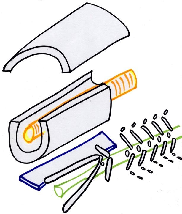
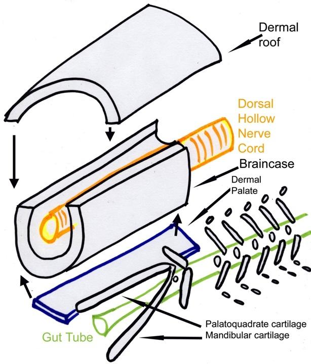
3 This lecture will revolve around the early embryology of the vertebrate skull. One of the landmark achievments in this was the summary of that topic based initially the developing embryonic shark head by Edwin S. Goodrich. Thus it has come to be known as the Goodrich Diagram. We will draw our own version of it before we go on to a slide review.
4 If you re working with the PowerPoint files, save space in your notes to draw here.

5
6
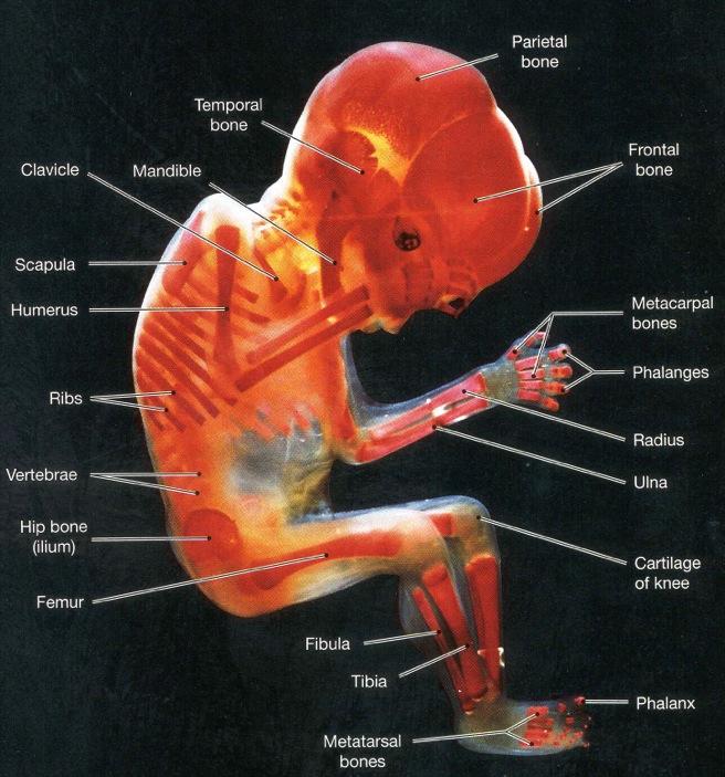
7 The developing skull has three component origins: Condrocranium (base of skull / braincase) Dermatocranium (flat bones of skull) Splanchnocranium (bones derived from gill arch elements)
8
9
10 Mode of Germ Layer Formation Origin Condrocranium Endochondral Mesoderm & Neural Crest Dermatocranium Intramembranous Mesoderm & Neural Crest Splanchnocranium Endochondral Neural Crest
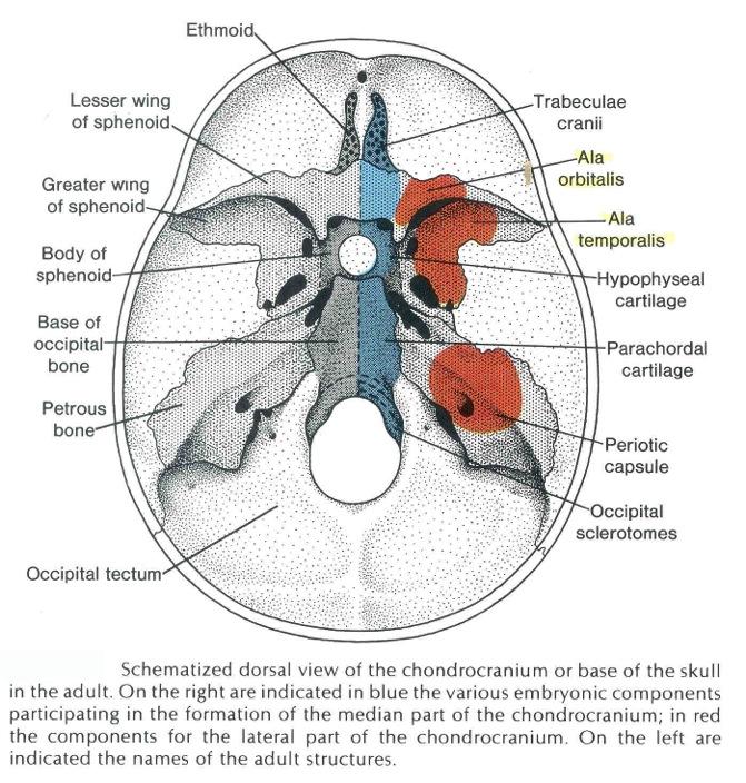
11 CHONDROCRANIUM: Bones of the base of the skull. Most major cranial nerves escape the skull through these. Endochondral Neural Crest rostrally; Mesodermal caudally Include: ethmoid, sphenoid (part), occipital (part) right and left temporal (parts).
12 Chondrocranium precursors Dorsal view


13 Initial component parts of the vertebrate braincase.
14
15 Ethmoid Sphenoid Temporal (otic region) Occipital (base)
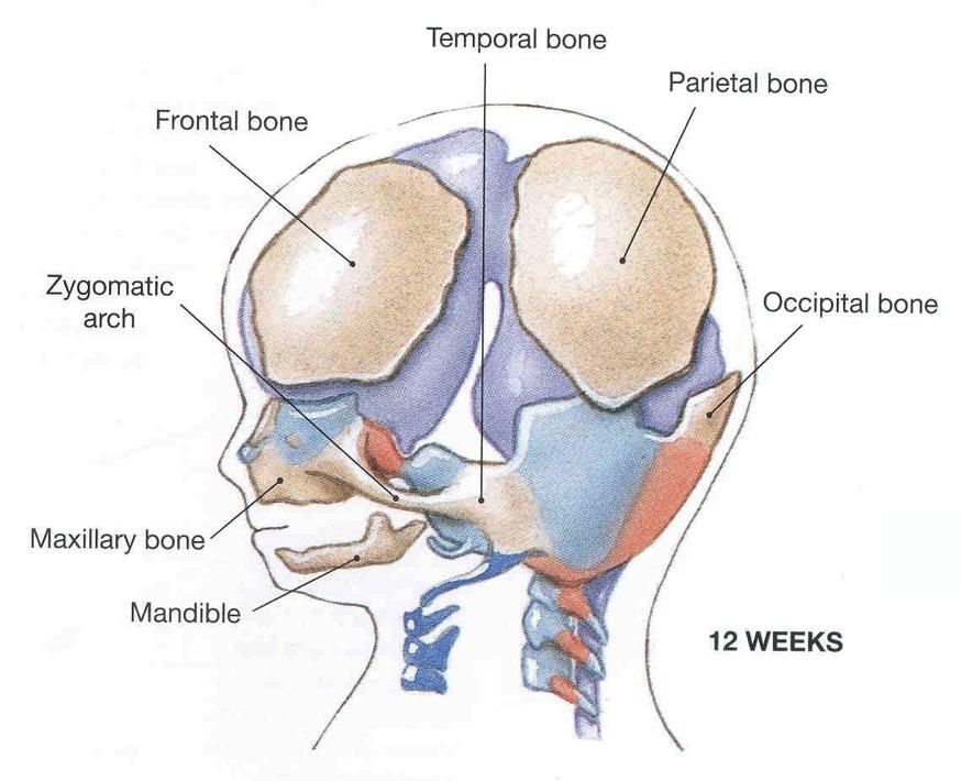
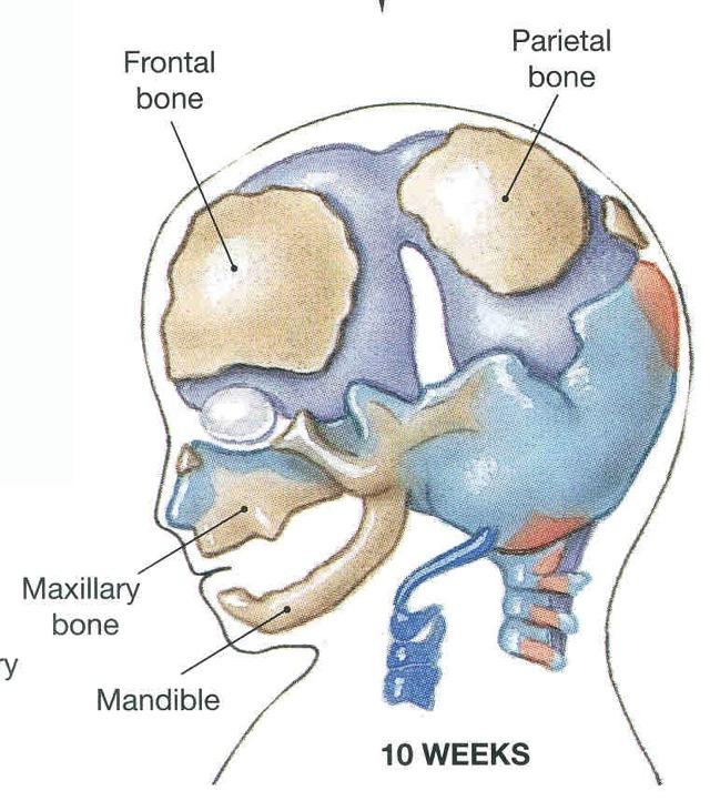
16 How the parts of the braincase grow together.
17
18

19 Flat bones of skull: DERMATOCRANIUM (These and others.)
20
21
22 The junction between neural crest and mesodermal contributions to the dermatocranium has changed through vertebrate evolution. Generally in region of frontal-parietal complex.

23 The junction between neural crest and mesodermal contributions to the dermatocranium has changed through vertebrate evolution. Generally in region of frontal-parietal complex
24

25 Red: Mesoderm Blue: Neural crest

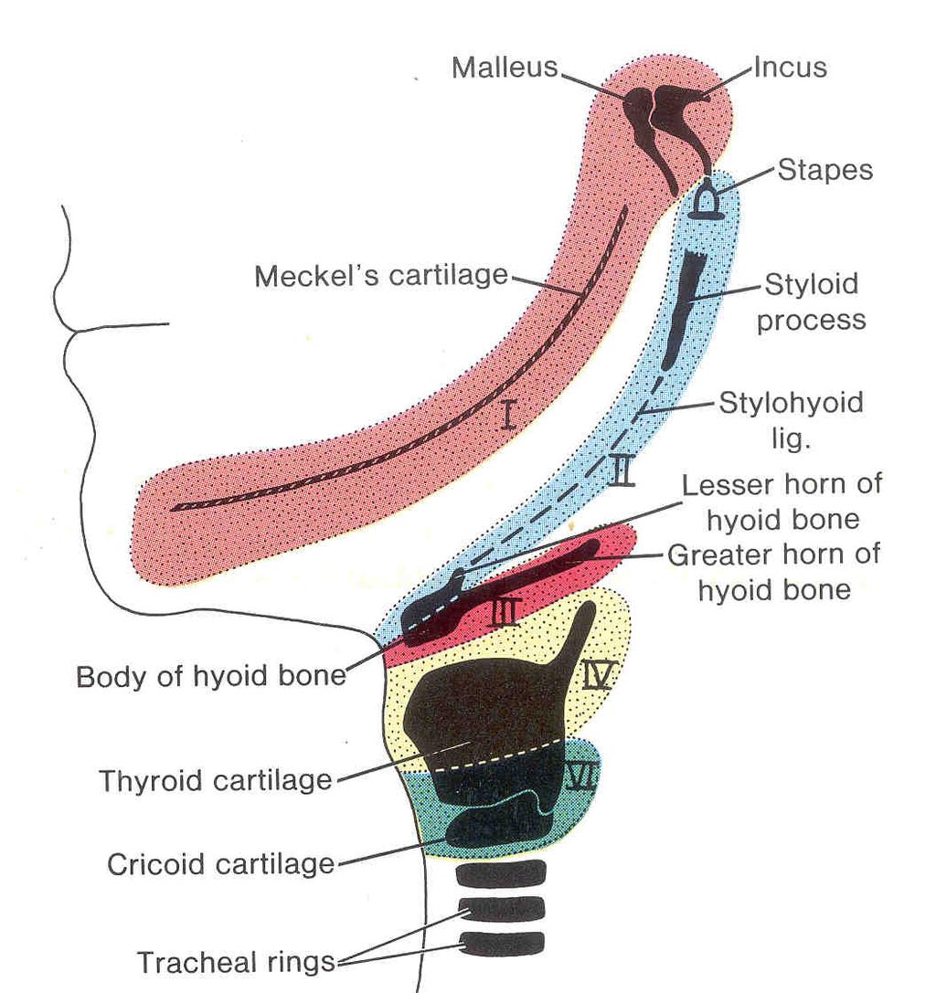
26 Gill slit bones: become SPLANCHNOCRANIUM
27
28
29 Contribution of somite-derived musculature and gillslit associated musculature in the vertebrate head.

30 Contribution of somite-derived musculature and gillslit associated musculature in the vertebrate head.
31 Biology 323 Human Anatomy for Biology Majors Week 10; Lecture 1; Tuesday Dr. Stuart S. Sumida Cranial Nerves and Other Soft Tissues of the Skull
32 Start with BRAIN STUFF

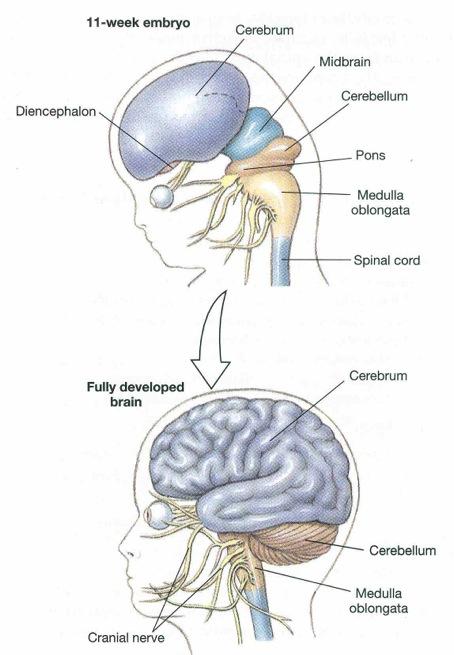
33 The Brain and Cranial Nerves
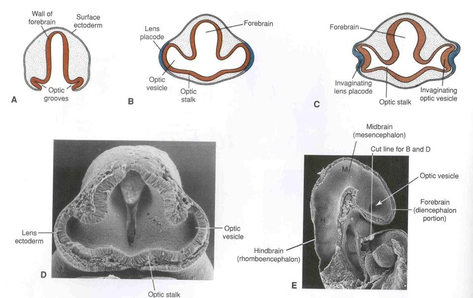
34 FOREBRAIN MIDBRAIN HINDBRAIN
35
36 Forebrain: Cerebrum Perception, movement of somatopleure, sensoro-motor integration, emotion, memory, learning. Diencephalon Homeostasis, behavioral drives in hypothalamus; sensory relay and modification in thalamus; melatonin secretion in pineal gland. Midbrain (Mesencephalon) Control of eye movement. Hindbrain Cerebellum and Pons control of movement, proprioreceptive input; relays visual and auditory reflexes in pons. Medulla Oblongata Involuntry functions: blood pressure, sleep, breathing, vomiting.
37 See in Part 3 of your Laboratory Protocols...
38 Development Special Sense organs = nose, eyes and ears, begin as small outcrops of ectoderm called placodes

39 Development Placode 1 = nose Placode 2 = eye Placode 3 = ear
40 Development In the nose, the ectoderm become nerve cells that send their fibres through the cribriform plate of the ethmoid, back to the brain This is Cranial Nerve I = the Olfactory Nerve
41 Development The second placode becomes the lens of the eye. It sinks below the surface of the skin, and an outgrowth of the brain wraps around it. The outgrowth is the retina, and the stalk connecting it is Cranial Nerve II = The Optic Nerve
42 Eye starts out as photosensitive lobe of brain underlying surface of skin. Lobe eventually becomes two-layered cup = retina. Connected to brain by stalk that is the OPITC NERVE (cranial nerve II). Lens from ectodermal placode. Marginal cells of retina become specialized as MUSCLE CELLS that regulate opening of pupil: Sphinctor pupillae (parasympathetically regulated) Dilator pupillae (sympathetically regulated)
43

44

45 Developing Retina Developing Lens
46 Ventral Root Cranial Nerves Somite Associated
47 Development Head somites can be divided into 2 sets. Pre-otic and post-otic
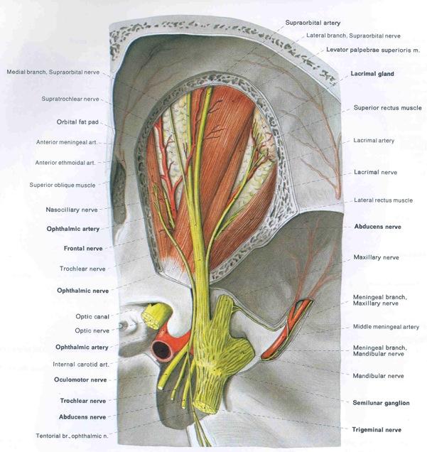
48 Development The sklerotomes of the post otic somites form the floor of the brain case
49 Development.and their myotomes develop into muscles of the tongue
50 Development The myotomes of the pre-otic somites form the muscles that move the eyeballs.
51 Development Each is supplied by a different cranial nerve:
52 Development Cranial Nerve III = Occulomotor Nerve
53 Development Cranial Nerve IV = Trochlear Nerve

54 Development Cranial Nerve VI = Abducens Nerve
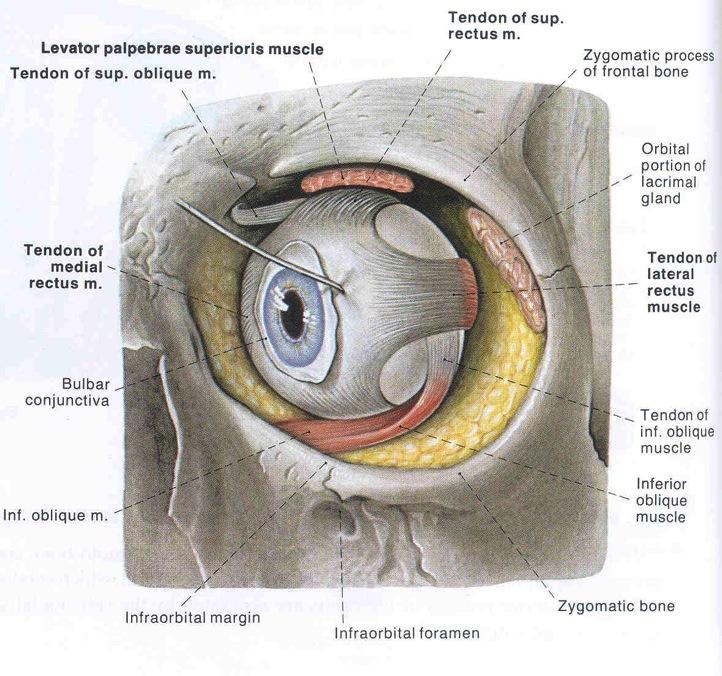
55 EYEBALL MOVING MUSCLES: Rectus Muscles Superior rectus - III Inferior rectus - III Lateral rectus - VI Medial rectus - III Oblique muscles Superior oblique - IV Inferior oblique - III Lavator palpebrae superioris - III
56
57
58 Dorsal Root Cranial Nerves Gill Slit Associated

59 Development Gill Arch Derivatives
60 Mandibular arch Development
61 Cranial Nerve V: The Trigeminal Nerve (3 branches) V1 Opthalmic, V2 Maxillary, V3 Mandibular
62 Hyoid arch Development
63 Cranial Nerve VII: Facial nerve
64 Development The Inner ear starts out as a lens, but turns into a fluid filled sac Receptor organs of hearing and balance. Cranial Nerve VIII = Auditory or Vestibulocochlear Nerve
 Otic")
65 Cranial Nerve VIII Vestibulocochlear Nerve (Evolutionary branch of VII) Otic Vesicle

66 Early Development of the Ear
67 Next arch Development
68 Cranial nerve IX: Glossopharyngeal Nerve
69 Development Remaining arches

70 Cranial nerve X: The Vagus Nerve
71 Is there a #0 nerve? The Nervus Terminalis (Nerve Zero) has been suggested as a primitive vertebrate structure serving the vomeraonasal organ.
72 I II III IV V VI VII VIII IX X XI XII Olfactory Optic Occulomotor Trochlear Trigeminal Abducens Facial Vestibulochochlar Glossopharyngeal Vagus Accessory Hypoglossal
73 Motor, sensory, or both
74 I O Sensory II O Sensory III O Mainly motor IV T Mainly motor V T Both VI A Mainly motor VII F Both VIII A Sensory IX G Both X V Both XI A Mainly motor XII H Mainly motor
75 For YEARS developmental biologists tried to figure out the correspondence between specific head somites and specific pharyngeal gill slits. Eventually, it was thought that somites and gill slits were such fundamentally different types of primary organizing segmentation that one could not be ties to the other. However, in the mid 1990s on, the study of structures of the brain called BRAIN NEUROMERES serial swellings of the dorsla hollow nerve cord - showed that: 1) Certain somites of the head were associated with certain neuromeres, thus certain ventral root cranial neves were associated with certain neurmeres. 2) Certain gill slits were also associated with certain neuromeres. 3) Thus, although somites and gill slits are not causally related to one another, they do follow an even more primal head segmentation, that of the neuromeres of the brain.

76 Recall. FOREBRAIN MIDBRAIN HINDBRAIN
77 NEUROMERE Head Somites & Visceral Arch & Associated Associated Cranial Nerve Cranial Nerve IV V VI VII, VIII VI IX Occipital Somites XII X,XI Is III perhaps associated with V 1?
Biology 323 Human Anatomy for Biology Majors Week 10; Lecture 1; Tuesday Dr. Stuart S. Sumida. Cranial Nerves and Soft Tissues of the Skull
 Biology 323 Human Anatomy for Biology Majors Week 10; Lecture 1; Tuesday Dr. Stuart S. Sumida Cranial Nerves and Soft Tissues of the Skull FOREBRAIN MIDBRAIN HINDBRAIN Forebrain: Cerebrum Perception,
C h a p t e r PowerPoint Lecture Slides prepared by Jason LaPres North Harris College Houston, Texas
 C h a p t e r 15 The Nervous System: The Brain and Cranial Nerves PowerPoint Lecture Slides prepared by Jason LaPres North Harris College Houston, Texas Copyright 2009 Pearson Education, Inc., publishing
Principles of Anatomy and Physiology
 Principles of Anatomy and Physiology 14 th Edition CHAPTER 14 The Brain and Cranial Nerves Introduction The purpose of the chapter is to: 1. Understand how the brain is organized, protected, and supplied
This lab activity is aligned with Visible Body s Human Anatomy Atlas app.
 1 This lab activity is aligned with Visible Body s Human Anatomy Atlas app. Learn more at visiblebody.com/professors We've split our Cranial Nerves lab activity into two parts. Part 1 is pre-lab exercises
Unit VIII Problem 3 Neuroanatomy: Brain Stem, Cranial Nerves and Scalp
 Unit VIII Problem 3 Neuroanatomy: Brain Stem, Cranial Nerves and Scalp - Brain stem: It is connected to the cerebellum and cerebral hemispheres. Rostral end of brain stem: diencephalon is the area which
b. The groove between the two crests is called 2. The neural folds move toward each other & the fuse to create a
 Chapter 13: Brain and Cranial Nerves I. Development of the CNS A. The CNS begins as a flat plate called the B. The process proceeds as: 1. The lateral sides of the become elevated as waves called a. The
Laboratory Exercise 3 Laboratory Examination of the Chicken Embryo Part II
 Biology 340, Comparative Embryology, Laboratory Exercise 3 Page 16 S. S. SUMIDA BIOLOGY 340 Comparative Embryology Laboratory Exercise 3 Laboratory Examination of the Chicken Embryo Part II Introduction
Brain and Cranial Nerves (Ch. 15) Human Anatomy lecture. caudal = toward the spinal cord)
 Human Anatomy lecture. caudal = toward the spinal cord)") Insight: Some cranial nerve disorders Brain and Cranial Nerves (Ch. 15) Human Anatomy lecture I. Overview (Directional terms: rostral = toward the forehead caudal = toward the spinal cord) A. 3 Major parts
Insight: Some cranial nerve disorders Brain and Cranial Nerves (Ch. 15) Human Anatomy lecture I. Overview (Directional terms: rostral = toward the forehead caudal = toward the spinal cord) A. 3 Major parts
Cranial Nerves. Steven McLoon Department of Neuroscience University of Minnesota
 Cranial Nerves Steven McLoon Department of Neuroscience University of Minnesota 1 Course News Change in Lab Sequence Week of Oct 2 Lab 5 Week of Oct 9 Lab 4 2 Sensory and Motor Systems Sensory Systems:
Bellringer: The central nervous system is comprised of: What is the name of the outermost layer of the brain? a. Brain. b.
 Bellringer: The central is comprised of: a. Brain b. Spinal cord c. Sensory receptors d. Both a and b What is the name of the outermost layer of the brain? a. Pia mater b. Dura mater c. Arachnoid d. Pons
Review of Nervous System Anatomy
 For the real amazement, if you wish to be amazed, is this process. You start out as a single cell derived from the coupling of a sperm and an egg; this divides in two, then four, then eight, and so on,
Brain and spinal nerve. By: shirin Kashfi
 Brain and spinal nerve By: shirin Kashfi Nervous system: central nervous system (CNS) peripheral nervous system (PNS) Brain (cranial) nerves Spinal nerves Ganglions (dorsal root ganglions, sympathetic
Nervous System. Student Learning Objectives:
 Nervous System Student Learning Objectives: Identify the primary parts of the neuron Identify the major structures of the central nervous system Identify the major structures of the peripheral nervous
M555 Medical Neuroscience Lab 1: Gross Anatomy of Brain, Crainal Nerves and Cerebral Blood Vessels
 M555 Medical Neuroscience Lab 1: Gross Anatomy of Brain, Crainal Nerves and Cerebral Blood Vessels Anatomical Directions Terms like dorsal, ventral, and posterior provide a means of locating structures
The Nervous System PART B
 7 The Nervous System PART B PowerPoint Lecture Slide Presentation by Jerry L. Cook, Sam Houston University ESSENTIALS OF HUMAN ANATOMY & PHYSIOLOGY EIGHTH EDITION ELAINE N. MARIEB Central Nervous System
Peripheral Nervous System
 Peripheral Nervous System Sensory Receptors Motor Endings Cranial Nerves The Four Plexuses Extremities Review of Reflexes Fast, preprogrammed, inborn, automatic responses Occur in the CNS at the spinal
Blood supply to the brain Blood brain barrier isolates neural tissue from general circulation
 The Brain and Cranial Nerves Objectives Name the major regions of the brain and describe their functions. Discuss the formation, circulation, and functions of the CSF. List the main components of the medulla
A&P 1 Brain & Cranial Nerves Guide #1 - Pre-Lab Exercises
 A&P 1 Brain & Cranial Nerves Guide #1 - Pre-Lab Exercises In this "Pre-lab Guide", we will be looking at the brain & cranial nerves. This should be done before lab, so we don't waste time in lab! This
Course: Physical Assessment II Date: October 17, 2008 Doc: Practice Quiz 1
 Course: Physical Assessment II Date: October 17, 2008 Doc: Practice Quiz 1 This is the practice quiz we did in Class 4. The answers are at the end of the quiz should you wish to test yourself. Complete
Cranial nerves.
 Cranial nerves eaglezhyxzy@163.com Key Points of Learning Name Components Passing through Peripheral distribution Central connection Function Cranial nerves Ⅰ olfactory Ⅱ optic Ⅲ occulomotor Ⅳ trochlear
DEVELOPMENT OF BRAIN
 Ahmed Fathalla OBJECTIVES At the end of the lecture, students should: List the components of brain stem. Describe the site of brain stem. Describe the relations between components of brain stem & their
Chapter 14: Nervous System Guided Notes (A-day)
") Chapter 14: Nervous System Guided Notes (A-day) Nervous System Overview Major Function: Control the body's and. Divided into the Nervous System (CNS=Brain and Spinal Cord) and the Nervous System (PNS=Cranial
Chapter 14: Nervous System Guided Notes (A-day) Nervous System Overview Major Function: Control the body's and. Divided into the Nervous System (CNS=Brain and Spinal Cord) and the Nervous System (PNS=Cranial
The Brain and Cranial Nerves Pg. 129
 The Brain and Cranial Nerves Pg. 129 Three Main Regions of the Brain Forebrain Cerbral hemispheres Diencephalon Midbrain Brain stem Hindbrain Pons Cerebellum Medulla oblongata Forebrain Interprets sensory
Stanley Pruisinger 1980's
 Neuroanatomy Prion disease cerebellum chapter b/c cerebellar ataxia here as a warning for obvious reasons. Creutzfeldt - Jakob Disease (CJD) "Spongiform" (brain turns to sponge) Jews in Lybia who ate
The Brain and Cranial Nerves Pg Three Main Regions of the Brain. Forebrain
 The Brain and Cranial Nerves Pg. 129 Three Main Regions of the Brain Forebrain Cerbral hemispheres Diencephalon Midbrain Brain stem Hindbrain Pons Cerebellum Medulla oblongata Interprets sensory inputs
Cranial Nerves and Spinal Cord Flashcards
 1. Name the cranial nerves and their Roman numeral. 2. What is Cranial Nerve I called, and what does it 3. Scientists who are trying to find a way to make neurons divide to heal nerve injuries often study
Nervous System The Brain and Spinal Cord Unit 7b
 Nervous System The Brain and Spinal Cord Unit 7b Chetek High School Mrs. Michaelsen 9.12 Meninges A. Meninges 1. The organs of the CNS are covered by membranes a. The meninges are divided into 3 layers:
Ms. K. GOWRI. M.Pharm., Lecturer.
 Ms. K. GOWRI. M.Pharm., Lecturer. CENTRAL NERVOUS SYSTEM (CNS) central nervous system consists of brain and spinal cord membrane covering the brain and spinal cord are surrounded by three membrane Meninges
PSY 302: CHAPTER 3 NOTES THE BRAIN (PART II) - 9/5/17. By: Joseline
 - 9/5/17. By: Joseline") PSY 302: CHAPTER 3 NOTES THE BRAIN (PART II) - 9/5/17 By: Joseline Left 3 MAJOR FISSURES : 2HEMISPHERES Right Lateral Ventricle Central Fissure Third Ventricle Sulcus Lateral Fissure Gyros Fissure- Fissures
PSY 302: CHAPTER 3 NOTES THE BRAIN (PART II) - 9/5/17 By: Joseline Left 3 MAJOR FISSURES : 2HEMISPHERES Right Lateral Ventricle Central Fissure Third Ventricle Sulcus Lateral Fissure Gyros Fissure- Fissures
Nervous System: An Introduction. HAP Susan Chabot Lemon Bay High School
 Nervous System: An Introduction HAP Susan Chabot Lemon Bay High School Function of the Nervous System 3 overlapping functions SENSORY INPUT - Monitor changes inside and outside of the body; these changes
Human Nervous System:
 OLLI Brain: Making Sense of Our World: Lecture 3 Human Nervous System: The Motor & Sensory Divisions Copyright 2004 Pearson Education, Inc., publishing as Benjamin Cummings Organization of the Nervous
Examination and Diseases of Cranial Nerves
 Cranial nerve evaluation is an important part of a neurologic exam. There are some differences in the assessment of cranial nerves with different species but the general principles are the same. Going
PERIPHERAL NERVOUS SYSTEM
 CHAPTER 13 PERIPHERAL NERVOUS SYSTEM Functional division of nervous system = afferent info to the CNS ascending spinal cord = efferent info from CNS descending spinal cord somatic skin, muscles visceral
Lab 16: PNS: Nerves and Autonomic NS Hamilton Answers to Pre- Lab Assignments
 Lab 16: PNS: Nerves and Autonomic NS Hamilton Answers to Pre- Lab Assignments Pre-Lab Activity 1: 1. a. olfactory nerve b. optic nerve c. oculomotor nerve d. abducens nerve e. trochlear nerve f. trigeminal
ACTIVITY 7: NERVOUS SYSTEM HISTOLOGY, BRAIN, CRANIAL NERVES
 ACTIVITY 7: NERVOUS SYSTEM HISTOLOGY, BRAIN, CRANIAL NERVES LABORATORY OBJECTIVES: 1. Histology: Identify structures indicated on three different slides or images of nervous system tissue. These images
Nervous Systems. Brain Development
 Nervous Systems Brain Development 2007-2008 Nervous system Central nervous system Peripheral nervous system Brain Spinal cord Sensory pathways Motor pathways Sympathetic arousal & energy production fight
Nervous and Endocrine System Exam Review
 Directions: Read each question and complete the statement using the multiple choice responses I. Nervous System 1. The interpretation of olfactory receptor information would fall under which general function
8.3 The Central Nervous System. SBI4U Ms. Ho-Lau
 8.3 The Central Nervous System SBI4U Ms. Ho-Lau The Central Nervous System the structural and functional centre for the entire nervous system the site of neural integration and processing The Central
Laboratory Manual for Comparative Anatomy and Physiology Figure 15.1 Transparency Master 114
 Neuron Capillary Astrocyte Microglial cell Neuron Fluid-filled cavity Process of oligodendrocyte Ependymal cells Brain or spinal cord tissue Myelin sheath Nerve fibers Figure 15.1 Transparency Master 114
Biological Bases of Behavior. 3: Structure of the Nervous System
 Biological Bases of Behavior 3: Structure of the Nervous System Neuroanatomy Terms The neuraxis is an imaginary line drawn through the spinal cord up to the front of the brain Anatomical directions are
Peripheral Nervous System Dr. Gary Mumaugh
 Peripheral Nervous System Dr. Gary Mumaugh Spinal Nerves Overview Thirty-one pairs of spinal nerves are connected to the spinal cord No special names; numbered by level of vertebral column at which they
The Nervous System PART B
 7 The Nervous System PART B PowerPoint Lecture Slide Presentation by Jerry L. Cook, Sam Houston University ESSENTIALS OF HUMAN ANATOMY & PHYSIOLOGY EIGHTH EDITION ELAINE N. MARIEB The Reflex Arc Reflex
Nervous System. Lecture 4
 Nervous System Lecture 4 Neurons Functional unit of the nervous system Also called the nerve cell Soma or body Axon Dendrites Neuroglial cells support cells Schwann cells produce myelin in PNS Oligodendrocytes
Protection of the Brain. Overview of the Brain. Visual Anatomy & Physiology First Edition. Martini & Ober. Chapter 13. Lecture 20
 Visual Anatomy & Physiology First Edition Martini & Ober Chapter 13 Brain and Cranial Nerves Lecture 20 1 Overview of the Brain Functions Major Parts regulates visceral activities cerebrum (two hemispheres)
Nervous System: Part IV The Central Nervous System The Brain
 Nervous System: Part IV The Central Nervous System The Brain Can you survive when part of your brain is destroyed? 2 Essential Knowledge 3.D.2 2. Cells communicate with each other through direct contact
Chapter 10 The Nervous System: The Brain and Cranial Nerves
 Chapter 10 The Nervous System: The Brain and Cranial Nerves Copyright 2015 Wolters Kluwer Health Lippincott Williams & Wilkins Overview Key Terms aphasia corpus callosum meninges basal nuclei diencephalon
Biology 3201 Nervous System #2- Anatomy. Components of a Nervous System
 Biology 3201 Nervous System #2- Anatomy Components of a Nervous System In any nervous system, there are 4 main components: (1) sensors: gather information from the external environment (sense organs) (2)
1/10/2013. What do neurons look like? Topic 14: Spinal Cord & Peripheral Nerves. How do neurons work? The nervous impulse. Specialized Neurons
 Topic 4: Spinal Cord & Peripheral Nerves What do neurons look like? Neurons What do they look like? How do they work? Neuronal and spinal organization What is the difference between neuron & nerve? How
Tracing the Cranial Nerves Osteologically
 CN I II III IV V 1 Supra-orbital ethmoidal nn. Ext. nasal V 2 Tracing the Cranial Nerves Osteologically Nucleus of Origin Olfactory tracts of frontal lobe of cerebrum Optic tracts from optic chiasma and
Review or skim Ch 12 on the vascular supply of the brain. Just look at pictures and legends for the clinical part at the end.
 Dental Neuroanatomy January 20 and 27, 10-12, 2011 Suzanne S. Stensaas, Ph.D. Dear Students: Please print these notes and bring them with you. My style is to use a Tablet PC and I draw on either a Word
Instructor s Review for Final Exams. The Nervous System
 Instructor s Review for Final Exams The Nervous System Divisions of the Central Nervous System? Brain and spinal cord. Key word, central. Divisions of the nervous system Central and Peripheral Coverings
Doctor Osama Asa ad Khader. Mohammad Alsalem
 6 Doctor 2015 Osama Asa ad Khader Mohammad Alsalem A quick revision for the spinal cord blood supply: Arterial Blood supply of spinal cord The spinal cord got its arterial supply by two ways: Longitudinal
PHYSIOLOHY OF BRAIN STEM
 PHYSIOLOHY OF BRAIN STEM Learning Objectives The brain stem is the lower part of the brain. It is adjoining and structurally continuous with the spinal cord. 1 Mid Brain 2 Pons 3 Medulla Oblongata The
LEARNING OBJECTIVES: PART 3 ASC171 NERVOUS SYSTEM MODULE 5 MAJOR SUBDIVISIONS AND ANATOMICAL LANDMARKS OF THE BRAIN: LO8 PROTECTION OF THE CNS
 LEARNING OBJECTIVES: PART 3 ASC171 NERVOUS SYSTEM MODULE 5 Part 3 Part 4 8. List the major subdivisions and anatomical landmarks of the brain 9. List the meninges & the order in which they are found around
Parts of the Brain. Hindbrain. Controls autonomic functions Breathing, Heartbeat, Blood pressure, Swallowing, Vomiting, etc. Upper part of hindbrain
 Parts of the Brain The human brain is made up of three main parts: 1) Hindbrain (or brainstem) Which is made up of: Myelencephalon Metencephalon 2) Midbrain Which is made up of: Mesencephalon 3) Forebrain
THE CENTRAL NERVOUS SYSTE M
 THE CENTRAL NERVOUS SYSTE M Structure and Functio n THIRD EDITIO N PER BRODAL A Brief Survey, x i Studying the Structures and Function of the Nervous System, xii i Animal Experiments Crucial for Progress,
The Nervous System: Central Nervous System
 The Nervous System: Central Nervous System I. Anatomy of the nervous system A. The CNS & the body by: 1. monitoring of the body 2. & information between parts of the body 3. acting as a to gather, store,
The CNS and PNS: How is our Nervous System Organized?
 Honors Biology Guided Notes Chapter 28 Nervous System Name 28.10 28.19 The CNS and PNS: How is our Nervous System Organized? ANIMAL NERVOUS SYSTEMS Define Cephalization and Centralization. What type of
Unit 18: Cranial Cavity and Contents
 Unit 18: Cranial Cavity and Contents Dissection Instructions: The calvaria is to be removed without damage to the dura mater which is attached to the inner surface of the calvaria. Cut through the outer
Model 3-50B or 3-88 III VIII. Olfactory Nerve. Optic Nerve. Oculomotor Nerve. Trochlear Nerve. Trigeminal Nerve. Abducens Nerve.
 Model 3-50B or 3-88 I Olfactory Nerve II Optic Nerve Oculomotor Nerve III IV Trochlear Nerve Trigeminal Nerve V VI Abducens Nerve Glossopharyngeal Nerve IX VII Facial Nerve VIII Vestibocochlear Nerve or
Laboratory Exercise 4 Laboratory Examination of the Pig Embryo
 S. S. SUMIDA BIOLOGY 340 Comparative Embryology Laboratory Exercise 4 Laboratory Examination of the Pig Embryo Introduction With the use of serial cross-sections of chicken embryos, you recently completed
CRANIAL NERVES. Dr. Amani A. Elfaki Associate Professor Department of Anatomy
 CRANIAL NERVES Dr. Amani A. Elfaki Associate Professor Department of Anatomy LEARNING OBJECTIVES Named the cranial nerves Identify the funcunal component of each cranial nerve Identify the effect of each
NEUROCRANIUM VISCEROCRANIUM VISCEROCRANIUM VISCEROCRANIUM
 LECTURE 4 SKULL NEUROCRANIUM VISCEROCRANIUM VISCEROCRANIUM VISCEROCRANIUM CRANIUM NEUROCRANIUM (protective case around brain) VISCEROCRANIUM (skeleton of face) NASOMAXILLARY COMPLEX MANDIBLE (DESMOCRANIUM)
Autonomic Nervous System (the visceral motor system) Steven McLoon Department of Neuroscience University of Minnesota
 Steven McLoon Department of Neuroscience University of Minnesota") Autonomic Nervous System (the visceral motor system) Steven McLoon Department of Neuroscience University of Minnesota 1 Course News Coffee Hour Monday, Nov 6, 9:00-10:00am Surdyk s Café in Northrop Auditorium
Autonomic Nervous System (the visceral motor system) Steven McLoon Department of Neuroscience University of Minnesota 1 Course News Coffee Hour Monday, Nov 6, 9:00-10:00am Surdyk s Café in Northrop Auditorium
Cranial Cavity REFERENCES: OBJECTIVES OSTEOLOGY. Stephen A. Gudas, PT, PhD
 Stephen A. Gudas, PT, PhD Cranial Cavity REFERENCES: Moore and Agur, Essential Clinical Anatomy (ECA), 3rd ed., pp. 496 498; 500 507; 512 514 Grant s Atlas 12 th ed., Figs 7.6; 7.19 7.30. Grant s Dissector
Page 1. Neurons Transmit Signal via Action Potentials: neuron At rest, neurons maintain an electrical difference across
 Chapter 33: The Nervous System and the Senses Neurons: Specialized excitable cells that allow for communication throughout the body via electrical impulses Neuron Anatomy / Function: 1) Dendrites: Receive
Brainstem. By Dr. Bhushan R. Kavimandan
 Brainstem By Dr. Bhushan R. Kavimandan Development Ventricles in brainstem Mesencephalon cerebral aqueduct Metencephalon 4 th ventricle Mylencephalon 4 th ventricle Corpus callosum Posterior commissure
Chapter 14: The Brain and Cranial Nerves. Copyright 2009, John Wiley & Sons, Inc.
 Chapter 14: The Brain and Cranial Nerves Development of the Brain Three to four-week embryo: prosencephalon, mesencephalon and rhombencephalon. Five-week embryo: telencephalon (cerebrum), diencephalon
The orbit-1. Dr. Heba Kalbouneh Assistant Professor of Anatomy and Histology
 The orbit-1 Dr. Heba Kalbouneh Assistant Professor of Anatomy and Histology Orbital plate of frontal bone Orbital plate of ethmoid bone Lesser wing of sphenoid Greater wing of sphenoid Lacrimal bone Orbital
CHAPTER 48: NERVOUS SYSTEMS
 CHAPTER 48: NERVOUS SYSTEMS Name I. AN OVERVIEW OF NERVOUS SYSTEMS A. Nervous systems perform the three overlapping functions of sensory input, integration, and motor output B. Networks of neurons with
CHAPTER 13&14: The Central Nervous System. Anatomy of the CNS
 CHAPTER 13&14: The Central Nervous System Anatomy of the CNS in human consists of brain and spinal cord as stated earlier neurons have little support from their extracellular matrix and depend on glial
Overview of the Nervous System (some basic concepts) Steven McLoon Department of Neuroscience University of Minnesota
 Steven McLoon Department of Neuroscience University of Minnesota") Overview of the Nervous System (some basic concepts) Steven McLoon Department of Neuroscience University of Minnesota 1 Coffee Hour Tuesday (Sept 11) 10:00-11:00am Friday (Sept 14) 8:30-9:30am Surdyk s
Overview of the Nervous System (some basic concepts) Steven McLoon Department of Neuroscience University of Minnesota 1 Coffee Hour Tuesday (Sept 11) 10:00-11:00am Friday (Sept 14) 8:30-9:30am Surdyk s
The Nervous System 7PART B. PowerPoint Lecture Slide Presentation by Patty Bostwick-Taylor, Florence-Darlington Technical College
 PowerPoint Lecture Slide Presentation by Patty Bostwick-Taylor, Florence-Darlington Technical College The Nervous System 7PART B What is a reflex? What is a reflex? What is meant by the statement that
Note to Self. The Brain and Cranial Nerves. Organization of the Brain
 Note to Self Despite what you think you can get through this chapter in 1 class period, or one with only a few slides left over The Brain and Cranial Nerves Chapter 14 Organization of the Brain The brain
a) Central sulcus- shallow groove that runs across brain sagitally
 Central sulcus- shallow groove that runs across brain sagitally") KEY BRAIN Brain Gross Anatomy Terms 1) Explain each of the following in terms of structure of the brain a) Central sulcus- shallow groove that runs across brain sagitally b) Lateral fissure- deep groove
KEY BRAIN Brain Gross Anatomy Terms 1) Explain each of the following in terms of structure of the brain a) Central sulcus- shallow groove that runs across brain sagitally b) Lateral fissure- deep groove
The dura is sensitive to stretching, which produces the sensation of headache.
 Dural Nerve Supply Branches of the trigeminal, vagus, and first three cervical nerves and branches from the sympathetic system pass to the dura. Numerous sensory endings are in the dura. The dura is sensitive
Nervous System. The Peripheral Nervous System Agenda Review of CNS v. PNS PNS Basics Cranial Nerves Spinal Nerves Reflexes Pathways
 Nervous System Agenda Review of CNS v. PNS PNS Basics Cranial Nerves Spinal Nerves Sensory Motor Review of CNS v. PNS Central nervous system (CNS) Brain Spinal cord Peripheral nervous system (PNS) All
REVIEW OF HEAD AND NECK CRANIAL NERVES AND EVERYTHING ELSE
 REVIEW OF HEAD AND NECK CRANIAL NERVES AND EVERYTHING ELSE OLFACTORY NERVE CN I ANTERIOR CRANIAL FOSSA CRISTA GALLI OF ETHMOID OLFACTORY FORAMINA IN CRIBIFORM PLATE OF ETHMOID BONE CN I OLFACTORY NERVE
THE NERVOUS SYSTEM CONCEPT 2: THE VERTEBRATE BRAIN IS REGIONALLY SPECIALIZED
 THE NERVOUS SYSTEM CONCEPT 2: THE VERTEBRATE BRAIN IS REGIONALLY SPECIALIZED Images of the human brain in popular culture almost always focus on the cerebrum, the part of the brain whose surface lies just
Major Anatomic Components of the Orbit
 Major Anatomic Components of the Orbit 1. Osseous Framework 2. Globe 3. Optic nerve and sheath 4. Extraocular muscles Bony Orbit Seven Bones Frontal bone Zygomatic bone Maxillary bone Ethmoid bone Sphenoid
Synapse Homework. Back page last question not counted. 4 pts total, each question worth 0.18pts. 26/34 students answered correctly!
 Synapse Homework Back page last question not counted 26/34 students answered correctly! 4 pts total, each question worth 0.18pts Business TASS hours extended! MWF 1-2pm, Willamette 204 T and Th 9:30-10:30am,
Chapter 7 Nervous System
 Chapter 7 Nervous System Two message centers: Functions of these systems: 1. * 2. * Overview of the Nervous System Parts: General Functions: Functions Sensory input: Sensation via nerves Integration: interpretation
Arnab Rana. Last updated 27th November The Skull Base. Arnab Rana. Background. Skull base foramina. Skull base bones. Self learning exercise
 Last updated 27th November 2017 Table of contents 1 2 3 4 5 Aberdeen MBChB learning guide links Year 2 Systems II (Part 1) ENT Pg 24 Aberdeen MBChB learning guide links Year 2 Systems II (Part 1) ENT Pg
Nervous System - PNS and CNS. Bio 105
 Nervous System - PNS and CNS Bio 105 Outline I. Central Nervous System vs Peripheral Nervous System II. Peripheral Nervous System A. Autonomic Nervous Systems B. Somatic Nervous Systems III. Autonomic
The Nervous System PART C. PowerPoint Lecture Slide Presentation by Patty Bostwick-Taylor, Florence-Darlington Technical College
 PowerPoint Lecture Slide Presentation by Patty Bostwick-Taylor, Florence-Darlington Technical College The Nervous System 7 PART C Protection of the Central Nervous System Scalp and skin Skull and vertebral
Topic/Objective: Identify the structures and functions of
 Cornell Notes Textbook Diagrams Video notes Topic/Objective: Identify the structures and functions of the human nervous system. Name: Class/Period: Date: Essential Question: How does the nervous system
The neurvous system senses, interprets, and responds to changes in the environment. Two types of cells makes this possible:
 NERVOUS SYSTEM The neurvous system senses, interprets, and responds to changes in the environment. Two types of cells makes this possible: the neuron and the supporting cells ("glial cells"). Neuron Neurons
3/15/17. Outline. Nervous System - PNS and CNS. Two Parts of the Nervous System
 Nervous System - PNS and CNS Bio 105 Outline I. Central Nervous System vs Peripheral Nervous System II. Peripheral Nervous System A. Autonomic Nervous Systems B. Somatic Nervous Systems III. Autonomic
Sheep Brain Dissection
 Sheep Brain Dissection Mammalian brains have many features in common. Human brains may not be available, so sheep brains often are dissected as an aid to understanding the mammalian brain since he general
Biology 3201 Unit 1: Maintaining Dynamic Equilibrium II
 Biology 3201 Unit 1: Maintaining Dynamic Equilibrium II Ch. 12 The Nervous System (Introduction and Anatomy) The Nervous System - Introduction Cells, tissues, organs and organ systems must maintain a biological
Brainstem. Telencephalon Diencephalon Cerebellum Brain stem
 Brainstem Brainstem 脑 脊髓 Brainstem Telencephalon Diencephalon Cerebellum Brain stem Ventral view Lateral view 10 pairs of the cranial nerves are attached to the brain stem The brainstem Midbrain Pons Medulla
Skull-2. Norma Basalis Interna. Dr. Heba Kalbouneh Assistant Professor of Anatomy and Histology
 Skull-2 Norma Basalis Interna Dr. Heba Kalbouneh Assistant Professor of Anatomy and Histology Norma basalis interna Base of the skull- superior view The interior of the base of the skull is divided into
Pharyngeal Apparatus. Pouches Endoderm Grooves Ectoderm Arch Neural Crest Somitomeres Aortic Arch - Vessel
 Pharyngeal Apparatus Pouches Endoderm Grooves Ectoderm Arch Neural Crest Somitomeres Aortic Arch - Vessel Segmental Organization Humans: Arch 1-4 prominent Arch 5 absent Arch 6 - transient First Arch Face
Medical Neuroscience Tutorial Notes
 Medical Neuroscience Tutorial Notes Cranial Nerve Nuclei MAP TO NEUROSCIENCE CORE CONCEPTS 1 NCC1. The brain is the body's most complex organ. LEARNING OBJECTIVES After study of the assigned learning materials,
Embryonic Brain Development
 Chapter 14 The Brain and Cranial Nerves Largest organ in the body? Brain functions in sensations, memory, emotions, decision making, behavior 19-1 19-2 Embryonic Brain Development Principal Parts of the
Brain ميهاربا لض اف دمح ا د The Meninges 1- Dura Mater of the Brain endosteal layer does not extend meningeal layer falx cerebri tentorium cerebelli
 .احمد د فاضل ابراهيم Lecture 15 Brain The Meninges Three protective membranes or meninges surround the brain in the skull: the dura mater, the arachnoid mater, and the pia mater 1- Dura Mater of the Brain
213: HUMAN FUNCTIONAL ANATOMY: PRACTICAL CLASS 12 Cranial cavity, eye and orbit
 213: HUMAN FUNCTIONAL ANATOMY: PRACTICAL CLASS 12 Cranial cavity, eye and orbit OSTEOLOGY Identify the bones which comprise the walls of the orbit: maxilla, zygomatic, ethmoid, lachrymal, frontal, and
Remember from the first year embryology Trilaminar disc has 3 layers: ectoderm, mesoderm, and endoderm
 Development of face Remember from the first year embryology Trilaminar disc has 3 layers: ectoderm, mesoderm, and endoderm The ectoderm forms the neural groove, then tube The neural tube lies in the mesoderm
Brainstem and Cranial Nerves II. Nerves covered in other lectures. A reminder about embryology. Prof. Stuart Bunt
 Brainstem and Cranial Nerves II Prof. Stuart Bunt Nerves covered in other lectures 1 Olfactory 2 Optic 3,4,6 Extraocular eye muscles 8 Vestibulo-cochlear 5 Motor and Sensory to the face and muscles of
I. Anatomy of the Brain A. Cranial Meninges and Ventricles of the Brain 1. Meninges a. Dura mater 1) Endosteal/Periosteal Layer - Outer 2) Meningeal
 Endosteal/Periosteal Layer - Outer 2) Meningeal") I. Anatomy of the Brain A. Cranial Meninges and Ventricles of the Brain 1. Meninges a. Dura mater 1) Endosteal/Periosteal Layer - Outer 2) Meningeal Layer - Inner 3) Falx cerebri a) Superior sagittal sinus
I. Anatomy of the Brain A. Cranial Meninges and Ventricles of the Brain 1. Meninges a. Dura mater 1) Endosteal/Periosteal Layer - Outer 2) Meningeal Layer - Inner 3) Falx cerebri a) Superior sagittal sinus