The Skull DANIL HAMMOUDI.MD
|
|
|
- Johnathan Ball
- 6 years ago
- Views:
Transcription


1 The Skull DANIL HAMMOUDI.MD
2 summary of bones/structures in Chapter 15 of the manual need tp be print as soon as possible l/frontal/frontal.html p/mainframe.htm
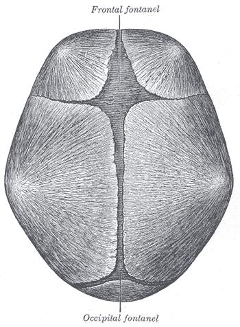
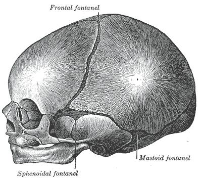
3 SUGGESTION HOW TO STUDY THE SKULL 1/RECOGNIZE THE TWO PARTS OF THE SKULL, CRANIAL AND FACIAL, 2/ FIND THE FONTANELS 3/FIND THE SUTURES 4/RECOGNIZED EACH CRANIAL BONE 5/RECOGNIZED EACH FACIAL BONES 6/RECOGNIZED THE fossa s AND FORAMINA
4
5 he skeleton has two parts: the axial skeleton and the appendicular skeleton. The axial skeleton includes the skull, the hyoid bone, the vertebral column (spine, sacrum, and coccyx), the sternum, and the ribs. Its components are aligned along the long axis of the body. The appendicular skeleton includes the bones of the upper extremities (arms, forearms, and hands), the pectoral (shoulder) girdle, the pelvic (hip) girdle, and the bones of the lower extremities (thigh, knee, leg, and foot). Its components are outside the body main axis.
6 Major bones of the Axial Skeleton: 1. Frontal 2. Parietal 5. Zygomatic 7. Mandible 9. Nasal 10. Sternum 14. Vertebrae 28. Ribs
7 11. Humerus 12. Ulna 13. Radius 18. Femur 19. Patella 20. Tibia 21. Fibula 25. Clavicle Appendicular:
8 THE SKULL = 22 BONES ALL FUSED EXCEPT ONE.:HYOID BONE JOINTS = SUTURES CRANIAL CAVITY 8 BONES=CRANIAL BONES 14 LEFT = FACIAL BONES 3 MORE IN THE EARS
9

10
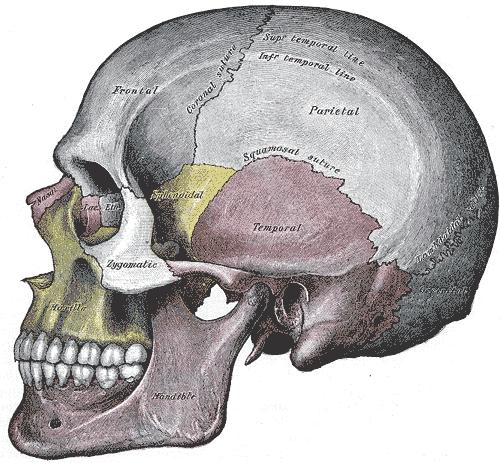
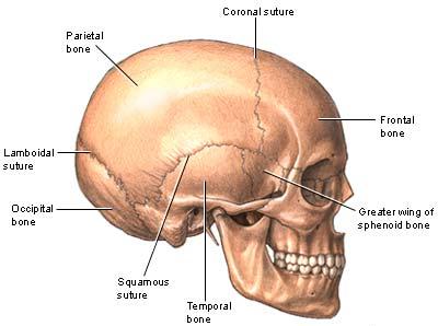
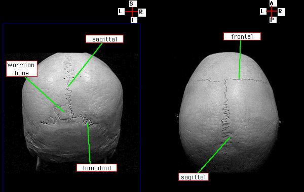
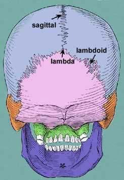
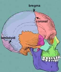
11 SUTURES Primarily visible from the side (norma lateralis) 1. Coronal suture - between the frontal and parietal bones 2. Lambdoid suture - between the parietal bones and the occipital bone 3. Occipitomastoid suture 4. Parietomastoid suture 5. Sphenofrontal suture 6. Sphenoparietal suture 7. Sphenosquamosal suture 8. Sphenozygomatic suture 9. Squamosal suture - between the parietal and the temporal bone 10. Zygomaticotemporal suture 11. Zygomaticofrontal suture
12 Primarily visible from front (norma frontalis) or above (norma verticalis) 1. Frontal suture / Metopic suture - between the two frontal bones, prior to the fusion of the two into a single bone 2. Sagittal suture - along the midline, between parietal bones Primarily visible from below (norma basalis) or inside 1. Frontoethmoidal suture 2. Petrosquamous suture 3. Sphenoethmoidal suture 4. Sphenopetrosal suture
13
14
15


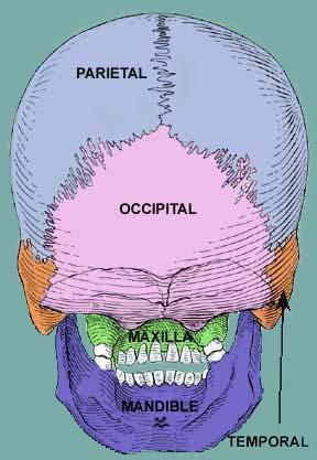
16 Coronal Suture Articulation between the parietal bones and the frontal bone. Squamous Suture: Articulation between the temporal bones with the parietal bones. Lambdoid Suture: Articulation of the parietal bones and the occipital bone. Occipitomastoid Suture: Articulation between the occipital bone and the mastoid process of the temporal bone. Sagittal Suture: You can't really see this one, but it is on the very top of the cranium. The articulation between the parietal bones.
17
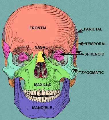
18 In the skull (22): Cranial bones: frontal bone parietal bone (2) temporal bone (2) occipital bone sphenoid bone ethmoid bone In the middle ears (6): malleus (2) incus (2) stapes (2) Facial bones: zygomatic bone (2) superior and inferior maxilla nasal bone (2) mandible palatine bone (2) lacrimal bone (2) vomer bone inferior nasal conchae (2) In the throat (1): hyoid bone
19 Paired Cranial Bones: Parietals Temporals Unpaired Cranial Bones: Frontal Occipital Sphenoid Ethmoid Paired Facial Bones: Lacrimals Nasals Zygomatics Maxillae Palatines Inferior Nasal Conchae Unpaired Facial Bones: Vomer Mandible Hyoid
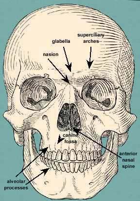
20
21
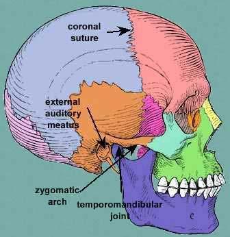
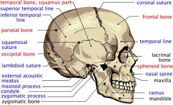
22
23 Anatomical Landmarks Cranial landmarks

24 HYOID BONE

25 is suspended from the tips of the styloid processes of the temporal bones by the stylohyoid ligaments. It is supported by the muscles of the neck and in turn supports the root of the tongue. Due to its position, the hyoid bone is not usually easy to fracture in most situations. In cases of suspicious death, however, a fractured hyoid is a strong sign of strangulation.
26

27 Frontal View. Legend: 1- Mental tubercle. 2- Body of mandible. 3- Ramus of mandible. 4- Anterior nasal spine. 5- Canine fossa. 6- Infra-orbital foramen. 7- Zygomatic-facial foramen. 8- Orbital surface of maxilla. 9- Temporal fossa. 10- Lateral surface of ethmoid. 11- Superior orbital fissure. 12- Lacrimal bone and groove. 13- Optic foramen. 14- Ethmoidal foramina. 15- Temporal line. 16- Supraorbital notch. 17- Glabella. 18- Frontal tuberosity. 19- Superciliary arch. 20- Parietal bone. 21- Naso-frontal suture. 22- Pterion. 23- Great wing of sphenoid. 24- Orbital surface of great wing of sphenoid. 25- Squamous part of temporal. 26- Left nasal bone. 27- Zygomatic bone. 28- Inferior orbital fissure. 27- Zygomatic bone. 28- Inferior orbital fissure. 29- Zygomatic arch. 30- Apertura piriformis, displaying nasal septum and infeior and middle concha. 31- Mastoid process. 32- Incisive fossa. 33- Angle of mandible. 34- Mental foramen. 35- Symphysis menti.
28

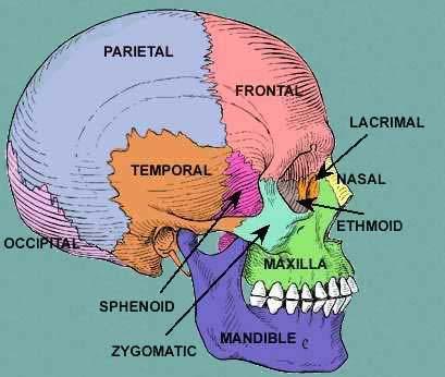
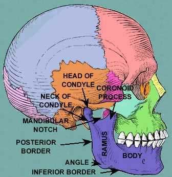
29 Lateral View. Legend: 1- Mental foramen. 2- Body of the mandible. 3- Maxilla. 4- Ramus of the mandible. 5- Zygomatic arch. 6- Styloid process. 7- External acoustic meatus. 8- Mastoid process. 9- Asterion. 10- Superior nuchal line. 11- External occipital protuberance. 12- Lambdoid suture. 13- Occipital bone. 14- Lambda. 15- Obelion. 16- Parietal bone. 17- Lower temporal line. 18- Upper temporal line. 19- Squamous temporal. 20- Bregma. 21- Coronal suture. 22- Stephanion. 23- Frontal bone. 24- Pterion. 25- Temporal fossa. 26- Great wing of sphenoid. 27- Zygomatic bone. 28- Zygomatic-facial foramen. 29- Fossa sacci lacrimalis. 30- Nasal bone. 31- Infraorbital foramen. 32- Anterior nasal spine.

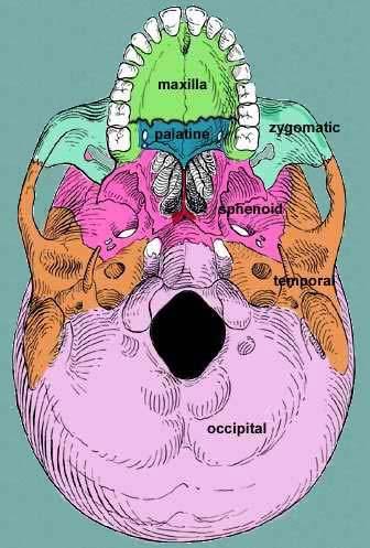
30 Basal View of the lower surface of the cranium (mandible is removed). Legend: 1- External Occipital Crest. 2- Superior nuchal line of the occipital bone. 3- Foramen magnum. 4- Occipital condyle. 5- Mastoid notch. 6- Mastoid process. 7- External acoustic meatus. 8- Styloid process. 9- Mandibular fossa. 10- Foramen spinosum. 11- Angular spine of the sphenoid. 12- Foramen ovale. 13- Lateral pterygoid lamina. 14- Hamulus of medial pterygoid lamina. 15- Vomer. 16- Posterior nasal spine. 17- Horzontal part of palate bone. 18- Palatine process of maxilla. 19- Incisive foramen. 20- Intermaxillary suture. 21- Greater palatine foramen. 22- Zygomatic process of maxilla. 23- Inferior orbital fissure. 24- Infra-temporal fossa. 25- Zygomatic arch. 26- Left choana. 27- Pterygoid fossa. 28- Scaphoid fossa. 29- Foramen lacerum. 30- Opening of osseous part of auditory tube. 31- Carotid canal. 32- Jugular fossa. 33- Stylo-mastoid foramen. 34- Jugular process of occipital bone. 35- Groove for occipital artery. 36- Mastoid foramen. 37- Canalis condyloideus. 38- Inferior nuchal line of occipital bone. 39- External occipital protuberance.

31 Middle Cranial Fossa

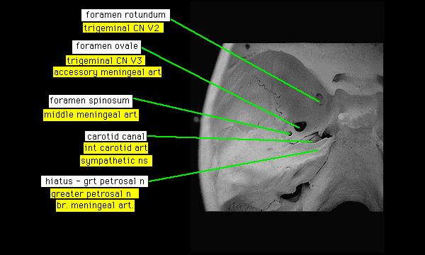
32 Skull Foramina
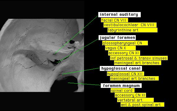
33 Basal Foramen Location Transmits Olfactory foramen Anterior crainal fossa CN I - Olfactory Optic foramen Middle Crainial fossa, inferior aspect of lesser wings of sphenoid CN II - Optic Superior Orital fussure Foramen Rotundum Foramen Ovale Foramen Spinosum Foramen Lacerum Carotid Foramen B/w greater and lesser wing of sphenoid Middle Crainial Fossa, greater wing of sphenoid Middle Crainial fossa, posterolateral to foramen rotundum Middle Crainial fossa, posterolateral to foramen ovale Middle crainial fossa, lateral to dorsum sallae Middle crainial fossa, petrous bone CN III, IV, V1, VI - Occularmotor, Trochlear, Trigeninal Opthalmic, Abdeuces CN V2 - Trigeminal Maxillary CN V3 - Trigeminal Mandibular Middle meninggeal artery Carotid artery Leads to foramen lacerum, carotid artery Foramen Magnum Posterior crainial fossa spinal cord and vertebral arteries Jugular Foramen Internal Auditory Foramen Hypoglossal Foramen Posterior crainial fossa, b/w petrous ridges and occipital bone, antero-lateral to foramen magnum Posterior crainial fossa, posterolateral aspect of petrous ridegs Posterior crainial fossa, lateral aspect of foramen magnum Jugular vein, CN IX, X, XI - Glossopharengeal, vagus, accessory CN VII, VIII - Facial, Vestibulochochlear CN XII - hypoglossal

34 Posterior Cranial Fossa

35 Frontal Bone, Frontal view, showing top of eye sockets and nasal cavity

36 Occipital Bone from the inside. The Foramen Magnum is where the spinal cord passes through; the fossa labeled are where lobes of the brain fit.

37 Temporal bone, outer right side showing features
, its greater wings (2), the external pterygoid plate (5), and the internal pterygoid place (6).")
38 Sphenoid bone from the rear, with Parietal (3) and Temporal bones (4, mastoid process) attached. The sphenoid consists of the body of the sphenoid (1), its greater wings (2), the external pterygoid plate (5), and the internal pterygoid place (6). The frontal bone fits on the upper front part of this assembly (distant in the image) and the occipital bone fits on the rear (near in the image). The Ethmoid bone attaches to the front (distant) part of the Sphenoid.

39 Ethmoid bone showing features
APPENDICULAR SKELETON 126 AXIAL SKELETON SKELETAL SYSTEM. Cranium. Skull. Face. Skull and associated bones. Auditory ossicles. Associated bones.
 SKELETAL SYSTEM 206 AXIAL SKELETON 80 APPENDICULAR SKELETON 26 Skull Skull and associated s 29 Cranium Face Auditory ossicles 8 4 6 Associated s Hyoid Thoracic cage 25 Sternum Ribs 24 Vertebrae 24 column
Biology 218 Human Anatomy. Adapted from Martini Human Anatomy 7th ed. Chapter 6 The Skeletal System: Axial Division
 Adapted from Martini Human Anatomy 7th ed. Chapter 6 The Skeletal System: Axial Division Introduction The axial skeleton: Composed of bones along the central axis of the body Divided into three regions:
BONE CHALLENGE DANIL HAMMOUDI.MD
 BONE CHALLENGE DANIL HAMMOUDI.MD Bone Basic functions? A. support B. protection C. movement assistance in D. RBC formation-hemopoiesis E. mineral homeostasis +importance of calcium F. energy supply -yellow
Structure Location Function
 Frontal Bone Cranium forms the forehead and roof of the orbits Occipital Bone Cranium forms posterior and inferior portions of the cranium Temporal Bone Cranium inferior to the parietal bone forms the
Chapter 7 Part A The Skeleton
 Chapter 7 Part A The Skeleton Why This Matters Understanding the anatomy of the skeleton enables you to anticipate problems such as pelvic dimensions that may affect labor and delivery The Skeleton The
Anatomy and Physiology. Bones, Sutures, Teeth, Processes and Foramina of the Human Skull
 Anatomy and Physiology Chapter 6 DRO Bones, Sutures, Teeth, Processes and Foramina of the Human Skull Name: Period: Bones of the Human Skull Bones of the Cranium: Frontal bone: forms the forehead and the
Cranium Facial bones. Sternum Rib
 Figure 7.1 The human skeleton. Skull Thoracic cage (ribs and sternum) Cranium Facial bones Sternum Rib Bones of pectoral girdle Vertebral column Sacrum Vertebra Bones of pelvic girdle (a) Anterior view
Bone Flashcards for 10a
 Bone Flashcards for 0a CLAVICLE (collar bone). Sternal extremity (end) flat end. Acromial extremity (end) rounded end. SCAPULA (shoulder blade). Right or left scapula?. Superior border (superior margin).
Bones of the skull & face
 Bones of the skull & face Cranium= brain case or helmet Copyright The McGraw-Hill Companies, Inc. Permission required for reproduction or display. The cranium is composed of eight bones : frontal Occipital
Skeletal System: Skull.
 Skeletal System: Skull www.fisiokinesiterapia.biz Bones of the Skull SPLANCHNOCRANIUM Nasal (2) Maxilla (2) Lacrimal (2) Zygomatic (2) Palatine (2) Inferior concha (2) Vomer Mandible NEUROCRANIUM Frontal
Skull-2. Norma Basalis Interna Norma Basalis Externa. Dr. Heba Kalbouneh Associate Professor of Anatomy and Histology
 Skull-2 Norma Basalis Interna Norma Basalis Externa Dr. Heba Kalbouneh Associate Professor of Anatomy and Histology Norma basalis interna Base of the skull- superior view The interior of the base of the
External Acoustic Meatus. Mastoid Process. Zygomatic Process. Temporal Bone
 Bone lab review 1. Frontal Bone 2. Supra-Orbital Foramen 3. Orbit (Orbital Cavity) 4. Superior Orbital Fissure 5. Inferior Orbital Fissure 6. Zygomatic Bone 7. Infra-Orbital Foramen 8. Maxilla 9. Mandible
Chapter 7: Skeletal System: Gross Anatomy
 Chapter 7: Skeletal System: Gross Anatomy I. General Considerations A. How many bones in an average adult skeleton? B. Anatomic features of bones are based on II. Axial Skeleton A. Skull 1. Functionally
THE SKELETAL SYSTEM. Focus on the Skull
 THE SKELETAL SYSTEM Focus on the Skull Review Anatomical Terms Anterior/Posterior Dorsal/Ventral Medial/Lateral Superior/Inferior Bone Markings - Review Projections for attachment of muscles, ligaments
Anatomy images for MSS practical exam- 2019
 Anatomy images for MSS practical exam- 2019 Ilium Ischium Pubis Acetabulaum Iliac crest Iliac tubercle ASIS (muscle and ligament attached) AIIS (muscle attached) PSIS PIIS Ischial spine Ischial tuberosity
Skeletal System -Axial System. Chapter 7 Part A
 Skeletal System -Axial System Chapter 7 Part A Skeleton Learn: Names of the s. Identify specific landmarks that allow: Bones to fit into each other, Organs to fit into the cavities, Muscles to attach,
Biology 2401 The Skeletal System
 Biology 2401 The Skeletal System Purpose: The lab will describe the microscopic and gross anatomy of bone, identify bones of the body, and identify important bone markings. I. Overview of the Skeleton
SKULL / CRANIUM BONES OF THE NEUROCRANIUM (7) Occipital bone (1) Sphenoid bone (1) Temporal bone (2) Frontal bone (1) Parietal bone (2)
 Occipital bone (1) Sphenoid bone (1) Temporal bone (2) Frontal bone (1) Parietal bone (2)") Important! 1. Memorizing these pages only does not guarantee the succesfull passing of the midterm test or the semifinal exam. 2. The handout has not been supervised, and I can not guarantee, that these
Important! 1. Memorizing these pages only does not guarantee the succesfull passing of the midterm test or the semifinal exam. 2. The handout has not been supervised, and I can not guarantee, that these
Axial skeleton bones and markings
 Axial skeleton bones and markings Skull Cranial bones Frontal x 1 Supraorbital foramen Occipital x 1 Foramen magnum Occipital condyles Superior nuchal line Inferior nuchal line Anterior cranial fossa External
University of Palestine. Midterm Exam 2013/2014 Total Grade:
 Course No: DNTS2208 Course Title: Head and Neck Anatomy Date: 09/11/2013 No. of Questions: (50) Time: 1hour Using Calculator (No) University of Palestine Midterm Exam 2013/2014 Total Grade: Instructor
the Skeletal System provided by Academic Web Services Grand Canyon University
 Anatomy Resource Center Study Guides the Skeletal System HEAD & NECK REGIONAL VIEW SKULL BONES CRANIUM FACE SKULL LANDMARKS ANTERIOR SIDE SUPERIOR/INFERIOR VERTEBRAL COLUMN VERTEBRAL REGIONS CERVICAL C1
AXIAL SKELETON SKULL
 AXIAL SKELETON SKULL CRANIAL BONES (8 total flat bones w/ 2 paired) 1. Frontal forms forehead & upper portion of eyesocket (orbital) 2. Parietal paired bones; form superior & lateral walls of cranium 3.
SKULL AS A WHOLE + ANTERIOR CRANIAL FOSSA
 SKULL AS A WHOLE + ANTERIOR CRANIAL FOSSA LEARNING OBJECTIVES At the end of this lecture, the student should be able to know: Parts of skeleton (axial and appendicular) Parts of skull Sutures of skull
Skull basic structures. Neurocranium
 Assoc. Prof. Květuše Lovásová, M.V.D., PhD. Skull basic structures Skull consists of two groups of bones: neurocranium (bones forming the brain box) splanchnocranium (bones forming the facial skeleton)
YOU MUST BRING YOUR OWN GLOVES FOR THIS ACTIVITY.
 ACTIVITY 3: AXIAL SKELETON AND LONG BONE DISSECTION Objectives: 1) How to get ready: Read Chapter 7, McKinley et al., Human Anatomy, 5e. All text references are for this textbook. Learning the meanings
Chapter 7. Skeletal System
 Chapter 7 Skeletal System 1 Skull A. The skull is made up of 22 bones: 8 cranial bones, 13 facial bones, and the mandible. B. The Cranium encloses and protects the brain, provides attachments for muscles,
Spring Written By: J. E. Sutton. Contents: I. Overview of the Skeleton: II. Appendicular Skeleton III. Axial Skeleton IV.
 Spring 2012 Written By: J. E. Sutton Contents: I. Overview of the Skeleton: II. Appendicular Skeleton III. Axial Skeleton IV. Articulations Overview of the Skeleton: I. Orientation to Human Skeleton: a.
ACTIVITY 3: AXIAL SKELETON AND LONG BONE DISSECTION COW BONE DISSECTION
 ACTIVITY 3: AXIAL SKELETON AND LONG BONE DISSECTION Objectives: 1) How to get ready: Read Chapter 7, McKinley et al., Human Anatomy, 4e. All text references are for this textbook. Learning the meanings
Dr.Noor Hashem Mohammad Lecture (5)
") Dr.Noor Hashem Mohammad Lecture (5) 2016-2017 If the mandible is discarded, the anterior part of this aspect of the skull is seen to be formed by the hard palate. The palatal processes of the maxillae
Dr.Noor Hashem Mohammad Lecture (5) 2016-2017 If the mandible is discarded, the anterior part of this aspect of the skull is seen to be formed by the hard palate. The palatal processes of the maxillae
Perpendicular Plate Zygomatic Bone. Mental Foramen Mandible
 Glabella Frontal Middle Nasal Concha Nasal Lacrimal Perpendicular Plate Zygomatic Inferior Nasal Concha Maxilla Mental Mandible Skull (anterior view) Squamosal Suture Coronal Suture Frontal Parietal Nasal
Exercise 10. The Axial Skeleton
 Exercise 10 The Axial Skeleton The Axial Skeleton Consists of the skeletal structures found along the midline of the body. Includes the skull, hyoid, vertebrae, ribs, sternum, and sacrum. The cartilages
Introduction to Local Anesthesia and Review of Anatomy
 5-Sep Introduction and Anatomy Review 12-Sep Neurophysiology and Pain 19-Sep Physiology and Pharmacology part 1 26-Sep Physiology and Pharmacology part 2 Introduction to Local Anesthesia and Review of
11/25/2012. Chapter 7 Part 2: Bones! Skeletal Organization. The Skull. Skull Bones to Know Cranium
 Chapter 7 Part 2: Bones! 5) Distinguish between the axial and appendicular skeletons and name the major parts of each 6) Locate and identify the bones and the major features of the bones that compose the
The Axial Skeleton. C h a p t e r. PowerPoint Lecture Slides prepared by Jason LaPres Lone Star College - North Harris
 C h a p t e r 7 The Axial Skeleton PowerPoint Lecture Slides prepared by Jason LaPres Lone Star College - North Harris Copyright 2009 Pearson Education, Inc., publishing as Pearson Benjamin Cummings An
Skeletal system. Prof. Abdulameer Al-Nuaimi. E. mail:
 Skeletal system Prof. Abdulameer Al-Nuaimi E-mail: a.al-nuaimi@sheffield.ac.uk E. mail: abdulameerh@yahoo.com Functions of Bone and The Skeletal System Support: The skeleton serves as the structural framework
Lab Unit One Flashcards
 CLAVICLE (collar bone). Sternal extremity (end) flat end. Acromial extremity (end) rounded end.. Conoid tubercle near round end SCAPULA (shoulder blade). Right or left scapula?. Superior border (superior
BIO 137 AXIAL SKELETON BONE STUDY THE HUMAN SKELETON
 BIO 137 THE AXIAL SKELETON MARY CATHERINE FLATH, Ph.D. THE HUMAN SKELETON AXIAL SKULL HYOID THORACIC CAGE VERTEBRAL COLUMN APPENDICULAR PECTORAL GIRDLE UPPER LIMBS PELVIC GIRDLE LOWER LIMBS AXIAL SKELETON
Chapter 8A. The Skeletal System: The Axial Skeleton. The Skeletal System: The Axial Skeleton. Types of Bones. Types of Bones
 Chapter 8A The Skeletal System: The Axial Skeleton The Skeletal System: The Axial Skeleton 206 named bones Axial Skeleton 80 bones lie along longitudinal axis skull, hyoid, vertebrae, ribs, sternum, ear
Biology 210 Chapter 8: Skeletal Tissues Supplement 1
 Biology 210 Chapter 8: Skeletal Tissues Supplement 1 By John McGill Material contributed by Beth Wyatt & Jack Bagwell DIVISIONS OF THE SKELETAL SYSTEM AXIAL SKELETON (80 BONES) Bones of the Head, Neck,
University of Palestine. Midterm Exam 2013/2014 Total Grade:
 [ Course No: DNTS2208 Course Title: Head and Neck Anatomy Date: 17/11/1024 No. of Questions: (52) Time: 2hours Using Calculator (No) University of Palestine Midterm Exam 2013/2014 Total Grade: Instructor
Bone List Anatomy
 1 Frontal Bone Skull 2 Parietal Bone Skull 3 Occipital Bone Skull 4 Temporal Bone Skull 5 Coronal Suture Skull 6 Sagittal Suture Skull 7 Squamous suture Skull 8 Lambdoid Suture Skull 9 Surpaorbital Ridge
TEST YOURSELF- Chapter 7
 TEST YOURSELF- Chapter 7 Cranial Bones 1. Give the name of the bone for each of the following markings. Some of the markings are found on more than one bone. List all that apply. Cranium a. Frontal squama:
Dr. Sami Zaqout, IUG Medical School
 The skull The skull is composed of several separate bones united at immobile joints called sutures. Exceptions? Frontal bone Occipital bone Vault Cranium Sphenoid bone Zygomatic bones Base Ethmoid bone
View of a Skull, 1489 by Leonardo Da Vinci. Kaan Yücel M.D., Ph.D Tuesday
 View of a Skull, 1489 by Leonardo Da Vinci Kaan Yücel M.D., Ph.D. 26.11.2013 Tuesday 1.SKULL skeleton of the head cranium 22 bones excluding ossicles of the ear 1.SKULL Mandible Lower jaw bone Neurocranium
Labs 9 and 10. Classification of Bones. Bone Shapes 1/05/13. Skeletal system overview. Bone are identified by:
 Labs 9 and 10 Skeletal system overview Classification of Bones Bone are identified by: shape internal tissues bone markings 1. Flat bones 2. Long bones 3. Short bones 4. Irregular bones 5. Sutural bones
Skeletal System - Prelab 1
 Skeletal System - Prelab 1 1. Which bones contain the paranasal sinuses? What function do the sinuses serve? 2. What two areas are separated from each other by the hard palate? Name the two bones that
Important Parts of Bones
 Important Parts of Bones For 2015 Know: Humerus (posterior) Clavical Femur (Anterior) Foot Hand Mandible Os Coxa Scapula Skull (Anterior, Inferior, Lateral) Sternum Humerus (posterior) A. olecranon fossa
Skeletal system overview. Classification of Bones
 Skeletal system overview BIOL241 Lab #9 Classification of Bones Bone are identified by: shape internal tissues bone markings 1 1. Flat bones 2. Long bones 3. Short bones 4. Irregular bones 5. Sutural bones
CHAPTER 7, PART II (BONES)
") Anatomy Name: CHAPTER 7, PART II (BONES) Entry #: INSTRUCTIONS: 1) READ Chapter 7, pg. 140-161. 2) Using the outline, make a note card for each underlined bone name or phrase. 3) On each note card, put
Anatomy Name: CHAPTER 7, PART II (BONES) Entry #: INSTRUCTIONS: 1) READ Chapter 7, pg. 140-161. 2) Using the outline, make a note card for each underlined bone name or phrase. 3) On each note card, put
Skull-2. Norma Basalis Interna. Dr. Heba Kalbouneh Assistant Professor of Anatomy and Histology
 Skull-2 Norma Basalis Interna Dr. Heba Kalbouneh Assistant Professor of Anatomy and Histology Norma basalis interna Base of the skull- superior view The interior of the base of the skull is divided into
Bones of the Skull Lateral View
 Bones of the Skull Lateral View Frontal Bone Parietal Bone Occipital Bone Temporal Bone Sphenoid Bone Pterion Sutures of the Skull Lateral View Coronal Suture Lambdoid Suture Squamous Suture Sutures of
Anatomy and Physiology 1 Chapter 7 self quiz Pro, Dima Darwish,MD.
 Anatomy and Physiology 1 Chapter 7 self quiz Pro, Dima Darwish,MD. 1) How many bones make up the axial skeleton? A) 50 B) 60 C) 70 D) 80 E) 90 2) Which of the following is a function of the axial skeleton?
Nervous & Skeletal Systems. Virtual Science University
 Nervous & Skeletal Systems Virtual Science University 1 Nervous & Skeletal Systems Texas TEK B.10(A) The student will interpret the function of systems in organisms (humans) including the nervous and skeletal
ANATOMY & PHYSIOLOGY I Laboratory Version B Name Section. REVIEW SHEET Exercise 10 Axial Skeleton
 ANATOMY & PHYSIOLOGY I Laboratory Version B Name Section REVIEW SHEET Exercise 10 Axial Skeleton 1 POINT EACH. THE SKULL MULTIPLE CHOICE 1. The major components of the axial skeleton include the 7. The
The Skeletal System: Axial Skeleton
 The Skeletal System: Axial Skeleton The Big Idea The Axial Skeleton & Homeostasis The bones of the axial skeleton contribute to homeostasis by protecting many of the body s organs such as the brain, spinal
Chapter 7: Head & Neck
 Chapter 7: Head & Neck Osteology I. Overview A. Skull The cranium is composed of irregularly shaped bones that are fused together at unique joints called sutures The skull provides durable protection from
Crafton Hills College Human Anatomy & Physiology Axial Skeleton
 A. Major Divisions Crafton Hills College Human Anatomy & Physiology Axial keleton 1. Axial: Part of skeleton lies along long axis of body 2. Appendicular: Bones & features of the appendages B. AXIAL KELETON
Lab Exercise #04 The Skeletal System Student Performance Objectives
 Lab Exercise #04 The Skeletal System Student Performance Objectives The material that you are required to learn in this exercise can be found in either the lecture text or the supplemental materials provided
Human Anatomy and Physiology - Problem Drill 07: The Skeletal System Axial Skeleton
 Human Anatomy and Physiology - Problem Drill 07: The Skeletal System Axial Skeleton Question No. 1 of 10 Which of the following statements about the axial skeleton is correct? Question #01 A. The axial
Riverside Community College Anatomy & Physiology 2B SPRING 2012 EXAM #1-ABC (Nervous System)
") Riverside Community College Anatomy & Physiology 2B SPRING 2012 EXAM #1-ABC (Nervous System) Name: 1) This vertebra is an example of a(n). 1) A) thoracic B) axis C) atlas D) lumbar E) sacral 1 2) W hich
Riverside Community College Anatomy & Physiology 2B SPRING 2012 EXAM #1-ABC (Nervous System) Name: 1) This vertebra is an example of a(n). 1) A) thoracic B) axis C) atlas D) lumbar E) sacral 1 2) W hich
BLUE SKY SCHOOL OF PROFESSIONAL MASSAGE AND THERAPEUTIC BODYWORK. Musculoskeletal Anatomy & Kinesiology I TERMINOLOGY, STRUCTURES, & SKELETAL OVERVIEW
 BLUE SKY SCHOOL OF PROFESSIONAL MASSAGE AND THERAPEUTIC BODYWORK Musculoskeletal Anatomy & Kinesiology I TERMINOLOGY, STRUCTURES, & SKELETAL OVERVIEW MSAK101-I Session 1 Learning Objectives: 1. Define
Lab-1. Miss. Lina Al-Onazy & samar Al-Wgeet =)
") Lab-1 Introduction The human skeleton is composed of 300 bones at birth and by the time adulthood is reached, some bones have fused together to give a total of 206 bones in the body. The human skeleton
Lab-1 Introduction The human skeleton is composed of 300 bones at birth and by the time adulthood is reached, some bones have fused together to give a total of 206 bones in the body. The human skeleton
Lab 6, 7, 8: Skeletal System
 107 Lab 6, 7, 8: Skeletal System Adult Skull Bony orbit (FLEZMS) Frontal bone supraorbital foramen frontal sinus Lacrimal bone Ethmoid bone perpendicular plate of ethmoid middle nasal conchae cribriform
10/11/2009. What is wrong with this picture? BONE PICTURE EXAM MARS / IN WHAT PART OF THE BONE DO YOU FIND THIS TISSUE SLIDE?
 What is wrong with this picture? BONE PICTURE EXAM MARS 2008 1/ IN WHAT PART OF THE BONE DO YOU FIND THIS TISSUE SLIDE? 1 2/ IDENTIFY THIS TISSUE? 3/what is wrong in this picture? 2 4/ Identify these cells
locomotice system Plastinated specimensⅠ: Silicone specimens Regional specimens and organs
 locomotice system Plastinated specimensⅠ: Silicone specimens Regional specimens and organs Art-No. Name Description The locomotor system SL001 Two hundred pieces of plastinated bones (without six The bones
Anatomy & Physiology Skeletal System Worksheet
 1. Name the five functions of the skeleton. c) d) e) Anatomy & Physiology Skeletal System Worksheet 2. The term for the shaft of a bone is:. 3. The bony struts found in spongy bone are called. 4. In ossification,
Bio 5/6 5 The Skeletal System Study Guide
 Name: THE SKELETAL SYSTEM: 5 The Skeletal System Study Guide Period: The skeleton is constructed of two of the most supportive tissues found in the human body - cartilage and bone. Besides supporting and
Anatomy Lab: The skeletal system. Part I: Vertebrae and Thoracic cage
 ANA Lab: Bone 1 Anatomy Lab: The skeletal system Part I: Vertebrae and Thoracic cage Spine (Vertebrae) Body Vertebral arch Vertebral canal Pedicle Lamina Spinous process Transverse process Sup. articular
Human Anatomy & Physiology I Dr. Sullivan Unit VIIIa The Axial Skeleton Chapter 8 (Sections )
") Human Anatomy & Physiology I Dr. Sullivan Unit VIIIa The Axial Skeleton Chapter 8 (Sections 8.1-8.3) I. Divisions of the skeletal system a) An adult human skeleton has 206 named bones b) Most are paired
Human Anatomy & Physiology I Dr. Sullivan Unit VIIIa The Axial Skeleton Chapter 8 (Sections 8.1-8.3) I. Divisions of the skeletal system a) An adult human skeleton has 206 named bones b) Most are paired
Anatomy and Physiology II. Review Spine and Neck
 Anatomy and Physiology II Review Spine and Neck Spine regions How many cervical vertibrae are there? 7 The curvature is the cervical region posterior? Concave posterior How many thoracic? And curvature?
SKELETON FUNCTIONS OF BONE:
 SKELETON FUNCTIONS OF BONE: SKELETON: 1. Performs a mechanical function in forming the skeletal support of the body and in forming a leverage system whereby work and movement are possible. 2. Serves as
Labs 6, 7, 8: Skeletal System
 153 Labs 6, 7, 8: Skeletal System Unit 6: Skeletal System: Bone tissue, Bones and Joints (p. 105-152) Ex. 6-1: Histology of Osseous Tissue, p. 113 Model: Osteon Tiss Lamella Osteocyte Lacunae Canaliculi
bio4165 lab quiz 1 Posterior View Anterior View Lateral View Anterior View bio fall.quarter lab.quiz.1...page.1 of 6
 B A Posterior View D C E Lateral View bio.4165...fall.quarter.2005...lab.quiz.1...page.1 of 6 F I G 35 Posterior View H bio.4165...fall.quarter.2005...lab.quiz.1...page.2 of 6 J Posterior View L K Inferior
Major Anatomic Components of the Orbit
 Major Anatomic Components of the Orbit 1. Osseous Framework 2. Globe 3. Optic nerve and sheath 4. Extraocular muscles Bony Orbit Seven Bones Frontal bone Zygomatic bone Maxillary bone Ethmoid bone Sphenoid
Axial Skeleton BONE TERMINOLOGY FEATURES
 Axial Skeleton BONE TERMINOLOGY FEATURES Tuberosity Rounded area on bone often roughened for muscle attachment. Tubercle Rounded projection on bone. This is called a tuberosity on the femur. Crest Ridgeline
The SKELETAL System. The framework of bones and cartilage which protect organs, and provides a lever system that allows locomotion.
 The SKELETAL System The framework of bones and cartilage which protect organs, and provides a lever system that allows locomotion. Functions of the Skeletal System Support Protection Movement Facilitation
BIOLOGY 113 LABORATORY Skeletal System
 BIOLOGY 113 LABORATORY Skeletal System Objectives Distinguish between the axial and appendicular skeleton. Distinguish between the cranium and facial skeleton. Locate and name the bones of the skull and
Average # of bones = major subdivisions: Axial skeleton Appendicular Skeleton (appendages) 1. Number and major subdivisions of bones
 1. Number and major subdivisions of bones") Osteology of the Human Body Without the skeletal system, you would be unable to engage in activities such as walking or grasping objects in your hand. Since the skeleton forms the internal framework of
Osteology of the Human Body Without the skeletal system, you would be unable to engage in activities such as walking or grasping objects in your hand. Since the skeleton forms the internal framework of
Skeletal System. It s all about the bones!!!
 Skeletal System It s all about the bones!!! The Skeletal System in Action!! The Skeletal System in Action! https://www.youtube.com/watch?v=icwllrqkv cg&list=plzile25upgebvru0jneppcabh0fhktgt Q 1. FYI 5
Human Anatomy - Problem Drill 06: The Skeletal System Axial Skeleton & Articualtions
 Human Anatomy - Problem Drill 06: The Skeletal System Axial Skeleton & Articualtions Question No. 1 of 10 Instructions: (1) Read the problem and answer choices carefully, (2) Work the problems on paper
in compact bone, large vertical canals carrying blood vessels and nerves. in compact bone, large horizontal canals carrying blood vessels and nerves.
 Carl Christensen, PhD Skeletal System (Bones`) Bio. 2304 Human Anatomy 1. Identify a term for each of the following: shaft of a long bone ends of a long bone ossified remnant of the "growth plate" connective
Anatomy Skull and Spinal Cord
 1 Anatomy Skull and Spinal Cord 1. Skull The skull is anterior to the spinal column and is the bony structure that encases the brain. Its purpose is to protect the brain and allow attachments for the facial
o Diaphysis o Area where red marrow is found o Area where yellow marrow is found o Epiphyseal plate AXIAL SKELETON Skull
 64 Anatomy & Physiology Coloring Workbook 7. Figure 5-2A is a midlevel, cross-sectional view of the diaphysis of the femur. Label the membrane that lines the cavity and the membrane that covers the outside
Objectives continued- Answer each of the objectives on a separate sheet of paper to demonstrate content mastery. Attach answers to back of packet.
 Anatomy and Physiology Chapter 5: The Skeletal System Name: Objectives- By the end of this chapter I will be able to: 1. Identify the subdivisions of the skeleton as axial or appendicular. 2. List at least
The Axial Skeleton Hyoid Bone. Lecture Overview. Marieb s Human Anatomy and Physiology. Chapter 7 The Axial and Appendicular Skeleton Lecture 14
 Marieb s Human Anatomy and Physiology Marieb Hoehn Chapter 7 The Axial and Appendicular Skeleton Lecture 14 1 Axial Skeleton Hyoid bone Bones of the orbit Paranasal sinuses Infantile skull Vertebral column
Chapter 7: Skeletal System
 Chapter 7: Skeletal System The Skeletal System Introduction P. 182 Bone is an organ made up of tissues: It is made up of the following components. Cartilage Blood Nerves Bone Connective Bone Classification
Bones Ethmoid bone Inferior nasal concha Lacrimal bone Maxilla Nasal bone Palatine bone Vomer Zygomatic bone Mandible
 splanchnocranium - Consists of part of skull that is derived from branchial arches - The facial bones are the bones of the anterior and lower human skull Bones Ethmoid bone Inferior nasal concha Lacrimal
Cornell Notes Name: Date: Topic: CH 5. Subject: The Skeletal System
 Cornell Notes Name: Date: Topic: CH 5 Questions/Main Ideas: Record Notes: We are revisiting Ch 3B on Connective Tissue prior to our study of Ch 5 Skeletal start on p.91-95 I. Types of Connective A. Bone
Lab 1 Introduction to the Vertebrate Skeleton
 G404 Geobiology Fall 2013 Name Lab 1 Introduction to the Vertebrate Skeleton In this lab you will familiarize yourself with the morphology and terminology of the vertebrate skeleton. Using the skeletons
Musculoskeletal System (Part A-1) Module 7 -Chapter 10 Overview. Functions
 Module 7 -Chapter 10 Overview. Functions") Musculoskeletal System (Part A-1) Module 7 -Chapter 10 Overview Susie Turner, M.D. 1/8/13 Muscles Attachments Bones Bone types Surface features of bones Divisions of the skeletal system Joints or Articulations
Musculoskeletal System (Part A-1) Module 7 -Chapter 10 Overview Susie Turner, M.D. 1/8/13 Muscles Attachments Bones Bone types Surface features of bones Divisions of the skeletal system Joints or Articulations
Ch. 5 - Skeletal System
 Ch. 5 - Skeletal System Bones are living, ever-changing structures. This allows them grow and adapt to new situations that the body encounters. The functions of the skeletal system: 1) support bones are
Chapter 7 The Skeletal System:The Axial Skeleton
 Chapter 7 The Skeletal System:The Axial Skeleton Axial Skeleton 80 bones lie along longitudinal axis skull, hyoid, vertebrae, ribs, sternum, ear ossicles Appendicular Skeleton 126 bones upper & lower limbs
Longitudinal fissure separates right and left hemispheres.
 L 10 A B O R A T O R Y Brain/Skull CEREBRAL CORTEX (telencephalon) Longitudinal fissure separates right and left hemispheres. Identify the following structures of the frontal lobe: lateral sulcus central
Chapter 07. Lecture Outline. See separate PowerPoint slides for all figures and tables pre-inserted into PowerPoint without notes and animations.
 Chapter 07 Lecture Outline See separate PowerPoint slides for all figures and tables pre-inserted into PowerPoint without notes and animations. 1-1 Chapter 7 Skeletal System Gross Anatomy 7.1 Skeletal
Hole s Human Anatomy and Physiology
 Hole s Human Anatomy and Physiology 1 Chapter 7 Skeletal System Bone Classification Long Bones Short Bones Flat Bones Irregular Bones Sesamoid (Round) Bones 2 Parts of a Long Bone epiphysis distal proximal
Anatomy images for MSS practical exam- 2019
 Anatomy images for MSS practical exam- 2019 Ilium Ischium Pubis Acetabulaum Iliac crest Iliac tubercle ASIS (muscle and ligament attached) AIIS (muscle attached) PSIS PIIS Ischial spine Ischial tuberosity
Temporal fossa Infratemporal fossa Pterygopalatine fossa Terminal branches of external carotid artery Pterygoid venous plexus
 Outline of content Temporal fossa Infratemporal fossa Pterygopalatine fossa Terminal branches of external carotid artery Pterygoid venous plexus Boundary Content Communication Mandibular division of trigeminal
OUTLINE ANATOMY, RADIOGRAPHY,
 20 SKULL OUTLINE SUMMARY OF PROJECTIONS, 262 ANATOMY, 263 Skull, 263 Cranial bones, 267 Ear, 277 Facial bones, 278 Articulations of the skull, 281 SUMMARY OF ANATOMY, 282 SUMMARY OF PATHOLOGY, 284 EXPOSURE
Frontal Anterior cranium Supraorbital margins. Glabella Frontal sinus Coronal suture Parietal Superior/lateral cranium Sagittal suture
 The Skeleton Outline PART 1: THE AXIAL SKELETON I. The skull consists of 8 cranial bones and 14 facial bones (pp. 200 218; Figs. 7.1 7.18; Table 7.1). A. The cranial and facial bones form the framework
The skeletal system is the framework for the muscular system to attach to so we can move.
 Skeletal System The skeletal system is the framework for the muscular system to attach to so we can move. BONE: A rigid connective tissue Helps to move & support the body Protect the organs (skull, ribs)