Optic Disc Evaluation: Is the Optic Disc Glaucomatous and Has it Progressed?
|
|
|
- Lydia Robertson
- 6 years ago
- Views:
Transcription

1 Optic Disc Evaluation: Is the Optic Disc Glaucomatous and Has it Progressed? Jody Piltz-Seymour, M.D. Clinical Professor Perelman School of Medicine University of Pennsylvania Wills Glaucoma Service Valley Eye Professionals
2 Lecture Objective To learn to identify the glaucomatous optic nerve and detect progressive glaucomatous optic disc damage.
3 Question 1 All discs with a cup-to-disc ratio of.8 are glaucomatous True False
4 Question 2 Disc hemorrhages have no prognostic significance for glaucoma True False
5 Question 3 The most important characteristic to evaluate in the glaucomatous optic nerve is the color of the rim. True False
6 Question 4 All of the following are signs of optic disc progression EXCEPT: Notching Collateral development Vessel shifts Nerve fiber layer swelling
7 Goals of Optic Disc Evaluation Is there glaucomatous optic disc damage? Has there been ongoing glaucomatous optic disc progression

8 What is normal? Rim tissue composed of ganglion cell axons, glial tissue and blood vessels. Axons pass through the pores of the lamina cribrosa

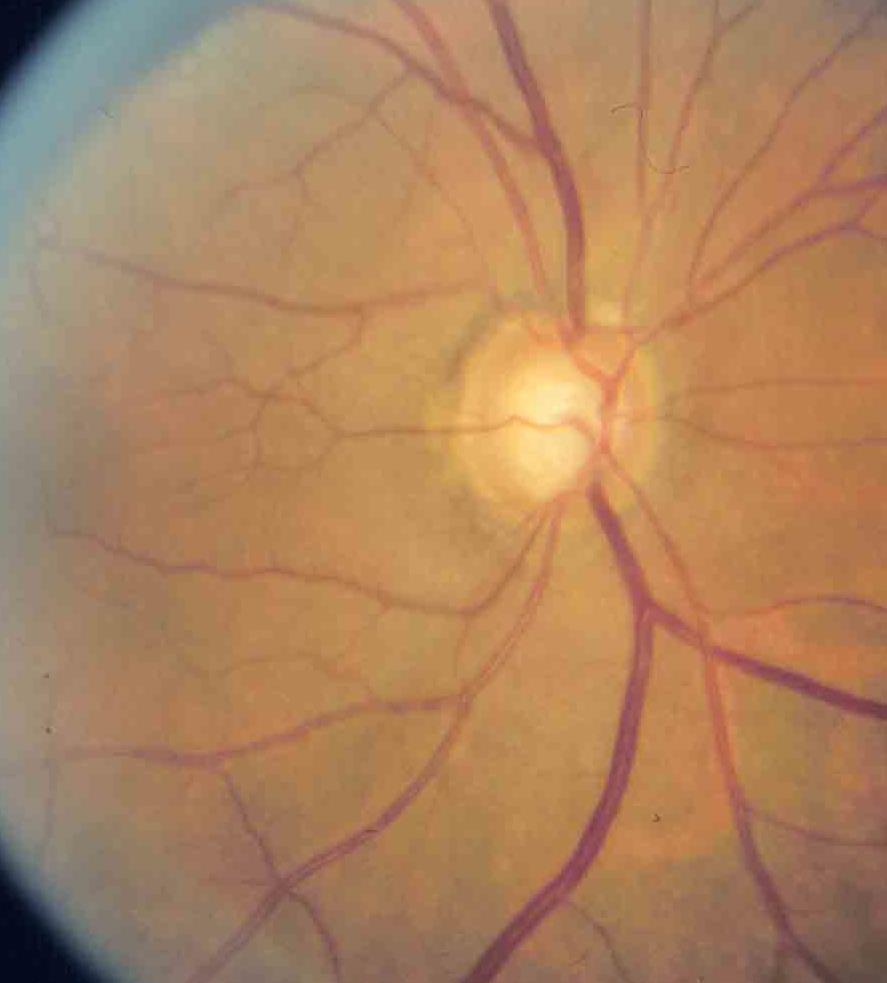
9 Normal Optic Nerve Vertically oval ISN T Rule C/D < 0.5 Armaly: Archives Ophthalmol 78, 1967
10 Assessment of Optic Disc Is the disc glaucomatous? SHIP Size Hemorrhages ISN T Parapapillary atrophy Bonus: RNFL

11 Size Is Important Great variation in the size of the normal optic nerve.


12 Disc Size Is Important Size of cup varies with size of disc Large discs have large cups in healthy eyes
13 Jonas, Graefe s 226: 332, 1998
14 Why is Disc Size Important? Size of the cup is linearly related to the size of the disc Britton AJO 103: 499, 1987 Jonas, Graefe s 226: 522, 198

15 Disc Size Risk of not recognizing disc size: Missing the diagnosis of glaucoma in eyes with small optic discs. Mis-diagnosing glaucoma in eyes with large or asymmetric discs.


16 Small Optic Discs Easy to miss early cupping Even a small cup may be significant β-ppa is often the first finding

17 Small Optic Disc

18 Large Optic Nerve


19 Asymmetric Disc Size Beware if asymmetric cupping

20 Techniques to Measure Disc Size Direct ophthalmoscope 5 o degree spot 1.5 mm diameter 1.8 mm 2 Fundus Lens 90 diopter lens Average 1.4 mm Large > 1.6 mm Small < 1.2 mm
21 Assessment of Optic Disc Is the disc glaucomatous? SHIP Size Hemorrhages ISN T Focal Rim thinning Generalized Rim thinning Saucerization Parapapillary atrophy Bonus: RNFL


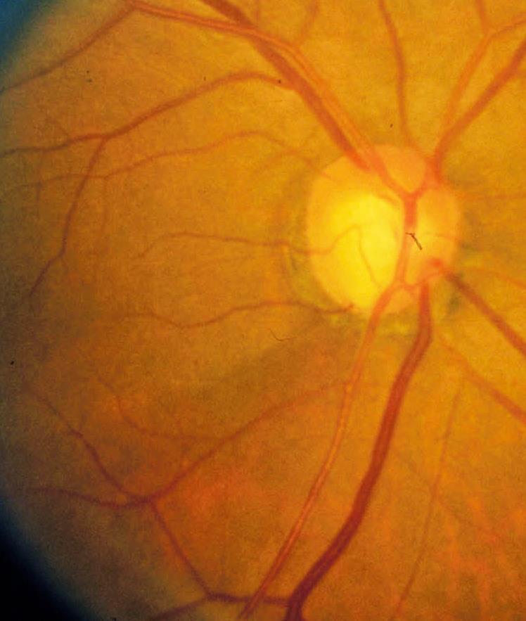
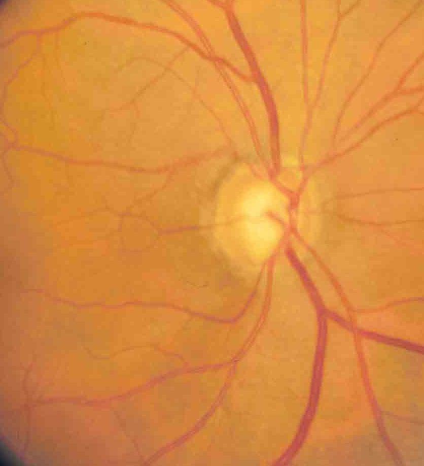
22 Generalized Concentric Loss Common form of cupping High IOP Normal visual field

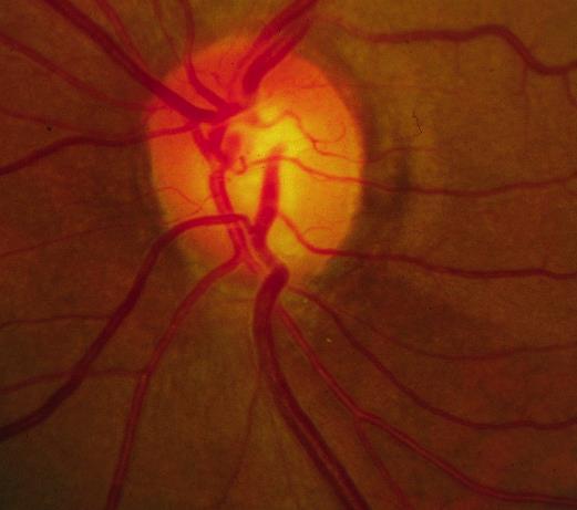
23 Focal Loss Localized thinning of rim Inferior > superior Vertical elongation of cup
24 baseline 6 years later

25 Focal Loss May form acquired pit Earlier VF loss Associated with disc hemorrhage

26 Mixed Concentric and Focal Most common form of cupping
27

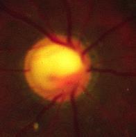
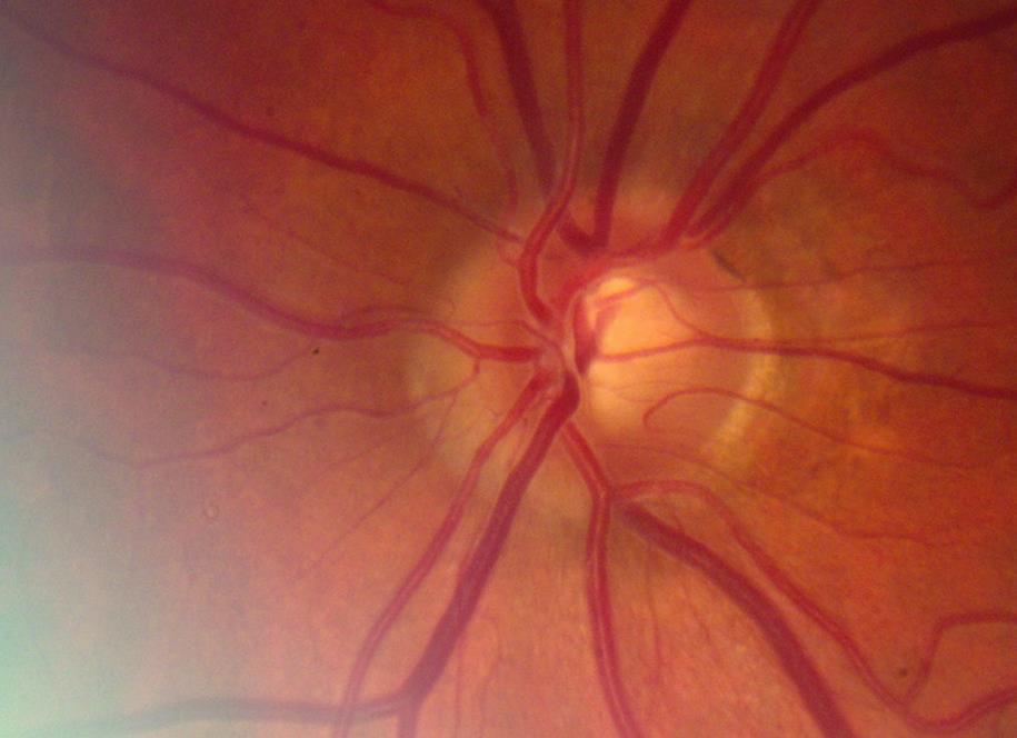
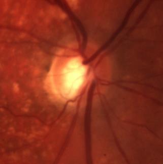
28 Pallor Sometimes seen in association with cupping. May be a marker of nonglaucomatous pathology May be a marker of prior acute, transient IOP spikes


29 Pallor Often follows episodes of high IOP
30

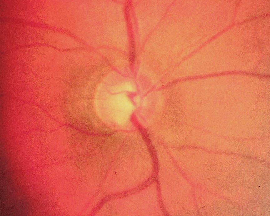
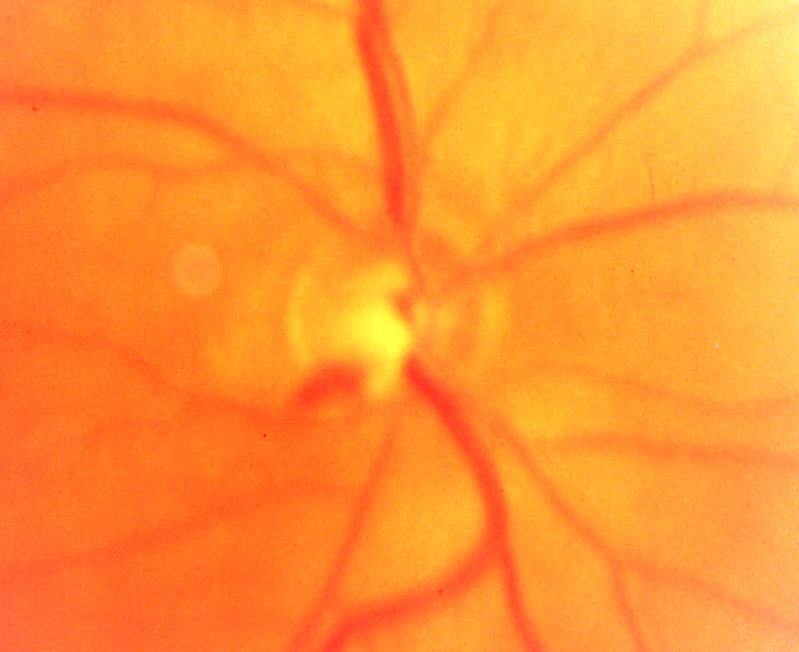
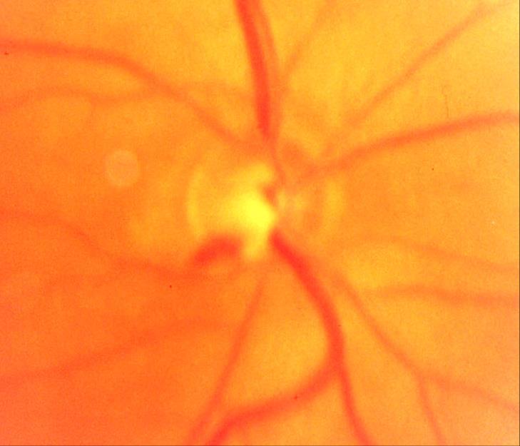
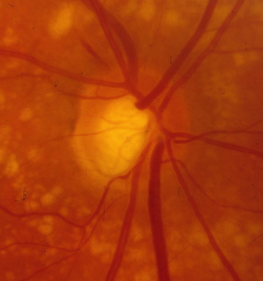
31 Saucerized: Senile Sclerotic Pale saucerization Moth eaten rim Older patients Atherosclerotic disease b - parapapillary atrophy Difficult to see changes

32 Myopic Tilted Discs May be difficult to interpret May have congenital VF abnormalities. AquiredVF defects may be adjacent to fixation.

33 Advanced Glaucomatous Disc Damage Excavation Exposure of the scleral insertion the scleral ring of Elshnig.

34 Scleral Ring of Elschnig
35 Assessment of Optic Disc Is the disc glaucomatous? SHIP Size Hemorrhages ISN T Parapapillary atrophy Bonus: RNFL


36 Optic Disc Hemorrhages Giant red flag for progression Very specific for glaucoma damage. Most common in NTG IT > ST Transient, recurrent
37 Hemorrhages: DDx Posterior vitreous detachment Systemic disease Vascular occlusive disease Anticoagulation
38 Hemorrhages: Prognosis Increased rim loss --> notching Increased VF progression Increased RNFL defects Increased peripapillary atrophy

39 Risk of developing POAG 6x greater in OHT eyes with DH Independent risk factor for glaucoma 3.7x increase in risk in multivariate analysis independent of other significant baseline variables
40 Hemorrhages: Prognosis OHTS 8 year cumulative risk of POAG 5.2% in eyes without disc hemorrhage 13.6% in eyes with disc hemorrhage Patients with DH older thinner corneas larger baseline CDR Only 16% of photographically documented DH were detected with dilated fundus exam!!!

41 baseline 6 years later
42
43 Assessment of Optic Disc Is the disc glaucomatous? SHIP Size Hemorrhages ISN T Focal Rim thinning Generalized Rim thinning Saucerization Parapapillary atrophy Bonus: RNFL
44 Assessment of Optic Disc Is the disc glaucomatous? SHIP Size Hemorrhages ISN T Focal Rim thinning Generalized Rim thinning Saucerization Parapapillary atrophy Bonus: RNFL
45

46 baseline 25 years later
47 Progression: What to look for? Thinning rim Widening notch Hemorrhages Vessel Shifts Increased β-peripapillary atrophy Pallor Widening NFLD


48 Vessel Shifts and Rim Thinning baseline 8 years later 14 years later Berger, J.W., Patel, T.R., Shin, D.S., Piltz, J.R., Stone, R.A.: Computerized stereochronoscopy and alternation flicker to detect optic nerve head contour change. Am A Ophthalmol. 107: , 2000


49 Rim thinning, vessel shifts, widening NFLD baseline 14 years later

50 Increased B PPA Baseline 8 years later
51 Vessel Shifts, Rim thinning


52 Collateral Development baseline 23 years later


53 Collaterals Piltz, JR. Venous collateral remodeling in a patient with post-traumatic glaucoma. Am J Ophthalmol. 128: , 1999.


54 Compare Stereo Pairs
55 SHIP Size Heme Isn t Peripapillary atrophy (RNFL)

56 SHIP Size Heme ISN T Peripapillary atrophy (RNFL)

57 SHIP Size Heme ISN T PPA (RNFL)

")
58 SHIP Size Heme ISN T PPA (RNFL)

")
59 SHIP Size Heme ISN T PPA (RNFL)

")
60 SHIP Size Heme ISN T PPA (RNFL)

61 SHIP Size Heme Isn t PPA

62 SHIP Size Heme Isn t PPA

63
64
65 S normal H yes I thinning rim P none S yes baseline 1 year later 6 years later
66 Question 1 All discs with a cup-to-disc ratio of.8 are glaucomatous True False
67 Question 2 Disc hemorrhages have no prognostic significance for glaucoma True False
68 Question 3 The most important characteristic to evaluate in the glaucomatous optic nerve is the color of the rim. True False
69 Question 4 All of the following are signs of optic disc progression EXCEPT: Notching Collateral development Vessel shifts Nerve fiber layer swelling
The Optic Nerve Head In Glaucoma. Clinical Pearl #1. Characteristics of Normal Disk 9/26/2017. Initial detectable damage Structure vs function
 The Optic Nerve Head In Glaucoma Clinical Pearl #1 Eric E. Schmidt, O.D., F.A.A.O. Omni Eye Specialists Wilmington,NC schmidtyvision@msn.com Glaucoma is an optic neuropathy Initial detectable damage Structure
Intro to Glaucoma/2006
 Intro to Glaucoma/2006 Managing Patients with Glaucoma is Exciting Interesting Challenging But can often be frustrating! Clinical Challenges To identify patients with risk factors for possible glaucoma.
Beyond the C/D Ratio: Evaluating a Glaucomatous Optic Nerve. Marcus Gonzales, OD, FAAO Cedar Springs Eye Clinic COPE ID#: GL
 Beyond the C/D Ratio: Evaluating a Glaucomatous Optic Nerve Marcus Gonzales, OD, FAAO Cedar Springs Eye Clinic COPE ID#: 27809-GL Points to Remember Glaucoma affects the ONH in characteristic patterns
Early Detection Of Glaucoma Clinical Clues. Points To Live By. Glaucoma Risk Factors 10/3/2014
 Early Detection Of Glaucoma Clinical Clues Eric E. Schmidt, O.D. Omni Eye Specialists Wilmington, NC schmidtyvision@msn.com Points To Live By 25% of G pxs NEVER have IOP >21mm 50% of G pxs have trough
A Review Of Risk Factors. Early Detection Of Glaucoma Clinical Clues. A risk factor analysis is critical. Points To Live By
 A Review Of Risk Factors Early Detection Of Glaucoma Clinical Clues Eric E. Schmidt, O.D. Omni Eye Specialists Wilmington, NC schmidtyvision@msn.com FINDACAR Family history IOP Nearsightedness Diabetes/Vascular
10/27/2013. Optic Red Herrings
 Optic Red Herrings 1 Optic neuropathy Compressive Inflammatory Toxic Glaucomatous Ischemic Post traumatic GLAUCOMATOUS OPTIC NEUROPATHY Glaucoma: Traditionally defined as a progressive optic neuropathy
Optic Nerve: Clinical Examination
 Optic Nerve: Clinical Examination Marcelo T. Nicolela 2 Core Messages Optic disc evaluation is of fundamental importance in the management of glaucoma. Clinical examination of the optic disc is best performed
Ophthalmoscopic Evaluation of the Optic Nerve Head JOST B. JONAS, MD, WIDO M. BUDDE, MD, AND SONGHOMITRA PANDA-JONAS, MD
 SURVEY OF OPHTHALMOLOGY VOLUME 43 NUMBER 4 JANUARY FEBRUARY 1999 MAJOR REVIEW Ophthalmoscopic Evaluation of the Optic Nerve Head JOST B. JONAS, MD, WIDO M. BUDDE, MD, AND SONGHOMITRA PANDA-JONAS, MD Department
The Role of the RNFL in the Diagnosis of Glaucoma
 Chapter 1. The Role of the RNFL in the Diagnosis of Glaucoma Introduction Glaucoma is an optic neuropathy characterized by a loss of of retinal ganglion cells and their axons, the Retinal Nerve Fiber Layer
Optic Nerve Evaluation in Glaucoma (TPG)
") Optic Nerve Evaluation in Glaucoma (TPG) Introduction Glaucoma is a progressive optic nerve disease characterized by retinal ganglion cell death and resultant axon loss, ultimately manifesting as excavation
Optic Nerve Evaluation in Glaucoma (TPG) Introduction Glaucoma is a progressive optic nerve disease characterized by retinal ganglion cell death and resultant axon loss, ultimately manifesting as excavation
Dr/ Marwa Abdellah EOS /16/2018. Dr/ Marwa Abdellah EOS When do you ask Fluorescein angiography for optic disc diseases???
 When do you ask Fluorescein angiography for optic disc diseases??? 1 NORMAL OPTIC DISC The normal optic disc on fluorescein angiography is fluorescent due to filling of vessels arising from the posterior
Is NTG different from POAG?
 Is NTG different from POAG? Sunita Radhakrishnan, M.D Glaucoma Center of San Francisco Glaucoma Research and Education Group Subset of POAG 1 Connective tissue structure within ONH Ganglion cell susceptibility
53 year old woman attends your practice for routine exam. She has no past medical history or family history of note.
 Case 1 Normal Tension Glaucoma 53 year old woman attends your practice for routine exam. She has no past medical history or family history of note. Table 1. Right Eye Left Eye Visual acuity 6/6 6/6 Ishihara
The evaluation of the optic nerve and retinal nerve
 Five rules to evaluate the optic disc and retinal nerve fiber layer for glaucoma Murray Fingeret, O.D., a,b Felipe A. Medeiros, M.D., c Remo Susanna, Jr, M.D., d and Robert N. Weinreb, M.D. c a Department
Evaluating Optic Nerve Damage: Pearls and Pitfalls
 54 The Open Ophthalmology Journal, 9, 3, 54-58 Evaluating Optic Nerve Damage: Pearls and Pitfalls Open Access Paul J. Mackenzie * and Frederick S. Mikelberg Division of Glaucoma, Department of Ophthalmology
Two Pits in a Pod: Using EDI-OCT to evaluate the lamina cribrosa in a patient with openangle glaucoma and multiple optic pits
 Two Pits in a Pod: Using EDI-OCT to evaluate the lamina cribrosa in a patient with openangle glaucoma and multiple optic pits Abstract: EDI-OCT imaging is used to evaluate glaucoma by examining the thinning
OCT in the Diagnosis and Follow-up of Glaucoma
 OCT in the Diagnosis and Follow-up of Glaucoma Karim A Raafat MD. Professor Of Ophthalmology Cairo University Hmmmm! Do I have Glaucoma or not?! 1 Visual Function 100% - N Gl Structure : - 5000 axon /
GLAUCOMA SUMMARY BENCHMARKS FOR PREFERRED PRACTICE PATTERN GUIDELINES
 SUMMARY BENCHMARKS FOR PREFERRED PRACTICE PATTERN GUIDELINES Introduction These are summary benchmarks for the Academy s Preferred Practice Pattern (PPP) guidelines. The Preferred Practice Pattern series
The Missing Piece in Glaucoma?
 Open Journal of Ophthalmology, 2016, 6, 56-62 Published Online February 2016 in SciRes. http://www.scirp.org/journal/ojoph http://dx.doi.org/10.4236/ojoph.2016.61008 The Missing Piece in Glaucoma? Syed
Evaluation of ONH Pallor in Glaucoma Patients and Suspects. Leticia Rousso, O.D. Joseph Sowka, O.D
 Evaluation of ONH Pallor in Glaucoma Patients and Suspects Leticia Rousso, O.D Joseph Sowka, O.D I. Abstract This case report will evaluate a young glaucoma suspect with unilateral sectoral optic nerve
Optic Disc Findings in Normal Tension Glaucoma
 ELSEVIER Optic Disc Findings in Normal Tension Glaucoma Yoshio Yamazaki,* Fukuko Hayamizu,* Satoshi Miyamoto,* Takako Nakagami,* Chizuru Tanaka* and Shigeri Inui t *Department of Ophthalmology, Nihon University
Structural abnormality of the optic disc neural rim is a. Presence of an Optic Disc Notch and Glaucoma ORIGINAL STUDY
 ORIGINAL STUDY Paul R. Healey, MMed (Clin Epi), PhD, FRANZCO and Paul Mitchell, MD, PhD, FRANZCO Purpose: To assess the prevalence and associations of a notch in the optic disc neural rim. Methods: Stereo-photographs
Macular Ganglion Cell Complex Measurement Using Spectral Domain Optical Coherence Tomography in Glaucoma
 Med. J. Cairo Univ., Vol. 83, No. 2, September: 67-72, 2015 www.medicaljournalofcairouniversity.net Macular Ganglion Cell Complex Measurement Using Spectral Domain Optical Coherence Tomography in Glaucoma
Clinical Study Visual Field Loss Morphology in High- and Normal-Tension Glaucoma
 Journal of Ophthalmology Volume 2012, Article ID 327326, 8 pages doi:10.1155/2012/327326 Clinical Study Visual Field Loss Morphology in High- and Normal-Tension Glaucoma Michele Iester, 1, 2 Fabio De Feo,
The Effect of Pupil Dilation on Scanning Laser Polarimetry With Variable Corneal Compensation
 C L I N I C A L S C I E N C E The Effect of Pupil Dilation on Scanning Laser Polarimetry With Variable Corneal Compensation Amjad Horani, MD; Shahar Frenkel, MD, PhD; Eytan Z. Blumenthal, MD BACKGROUND
Is this glaucoma? Leo Semes, OD Michael Chaglasian, OD Danica Marrelli, OD. Optometry s Meeting 2015 Seattle, WA
 Is this glaucoma? Leo Semes, OD Michael Chaglasian, OD Danica Marrelli, OD Optometry s Meeting 2015 Seattle, WA Case 1. 54 WM Engineer is referred to UAB Eye Care as a glaucoma suspect. Mild myopic refractive
Il contributo dell'angio-oct: valutazione integrata della componente nervosa e vascolare della malattia glaucomatosa
 SIMPOSIO G.O.A.L. - LE NUOVE FRONTIERE DIAGNOSTICHE E LE LINEE DI INDIRIZZO AMBULATORIALI DEL GLAUCOMA Coordinatore e moderatore: D. Mazzacane Presidente: L. Rossetti Il contributo dell'angio-oct: valutazione
The Glaucoma Suspect. Evaluating the Suspect Disk. Dr Michael Forrest. ! the usual suspects: ! is it glaucoma? ! is it swollen?
 Evaluating the Suspect Disk Dr Michael Forrest Senior Lecturer, The University of Queensland Northside Eye Specialists, Nundah Visiting Ophthalmologist, Mater Hospital, Brisbane Australian Vision Convention
Elevated intraocular pressure (IOP) is a major risk factor for
 is a major risk factor for") Glaucoma The Association between Retinal Vessel Diameter and Retinal Nerve Fiber Layer Thickness in Asymmetric Normal Tension Glaucoma Patients Joon Mo Kim, 1 Mo Sae Kim, 2 Hyo Ju Jang, 1 Ki Ho Park, 3
Glaucoma The Association between Retinal Vessel Diameter and Retinal Nerve Fiber Layer Thickness in Asymmetric Normal Tension Glaucoma Patients Joon Mo Kim, 1 Mo Sae Kim, 2 Hyo Ju Jang, 1 Ki Ho Park, 3
CHAPTER 13 CLINICAL CASES INTRODUCTION
 2 CHAPTER 3 CLINICAL CASES INTRODUCTION The previous chapters of this book have systematically presented various aspects of visual field testing and is now put into a clinical context. In this chapter,
3/16/2018. Optic Nerve Examination. Hassan Eisa Swify FRCS Ed (Ophthalmology) Air Force Hospital
 Air Force Hospital") Optic Nerve Examination Hassan Eisa Swify FRCS Ed (Ophthalmology) Air Force Hospital 1 Examination Structure ( optic disc) Function Examination of the optic disc The only cranial nerve (brain tract) which
Optic Nerve Examination Hassan Eisa Swify FRCS Ed (Ophthalmology) Air Force Hospital 1 Examination Structure ( optic disc) Function Examination of the optic disc The only cranial nerve (brain tract) which
Glaucoma Diagnosis. Definition of Glaucoma. Diagnosing Glaucoma. Vision Institute Annual Fall Conference
 Glaucoma Diagnosis Vision Institute Annual Fall Conference Mitchell W. Dul, OD, MS, FAAO mdul@sunyopt.edu Richard J. Madonna, MA, OD, FAAO rmadonna@sunyopt.edu Definition of Glaucoma Glaucoma can be regarded
Science & Technologies
 STANDARD COMPUTERIZED PERIMETRY IN FUNCTION OF DIAGNOSTIC GLAUCOMA Iljaz Ismaili, 1 Gazepov Strahil, 2, Goshevska Dashtevska Emilija 1 1 University Eye Clinic,Skopje 2 Clinical Hospital, Shtip Abstract
Study of Retinal Nerve Fiber Layer Thickness Within Normal Hemivisual Field in Primary Open-Angle Glaucoma and Normal-Tension Glaucoma
 Study of Retinal Nerve Fiber Layer Thickness Within Normal Hemivisual Field in Primary Open-Angle Glaucoma and Normal-Tension Glaucoma Chiharu Matsumoto, Shiroaki Shirato, Mai Haneda, Hiroko Yamashiro
Detection of Glaucoma Based on CDR, CAR and Blood Vessel Extraction
 402 P a g e INTERNATIONAL RESEARCH JOURNAL IN ADVANCED ENGINEERING AND TECHNOLOGY (IRJAET) www.irjaet.com ISSN (PRINT) : 2454-4744 ISSN (ONLINE): 2454-4752 Vol. 1, Issue 4, pp.402-411, December, 2015 Detection
Targeting Intraocular Pressure in Glaucoma: a Teaching Case Report 1
 Targeting Intraocular Pressure in Glaucoma: a Teaching Case Report 1 By: Andrew Kemp, OD, Marcus Gonzales, OD, FAAO, Joe DeLoach, OD, FAAO, and Zanna Kruoch, OD FAAO Background Glaucoma is a range of conditions
STRUCTURE & FUNCTION An Integrated Approach for the Detection and Follow-up of Glaucoma. Module 3a GDx
 STRUCTURE & FUNCTION An Integrated Approach for the Detection and Follow-up of Glaucoma Module 3a GDx Educational Slide Deck Carl Zeiss Meditec, Inc. November 2005 1 Structure & Function Modules Module
Neuro-Ocular Grand Rounds
 Neuro-Ocular Grand Rounds Anthony B. Litwak,OD, FAAO VA Medical Center Baltimore, Maryland Dr. Litwak is on the speaker and advisory boards for Alcon and Zeiss Meditek COMMON OPTIC NEUROPATHIES THAT CAN
Localised retinal nerve fibre layer defects in. chronic experimental high pressure glaucoma. rhesus monkeys. Jost B Jonas, Sohan Singh Hayreh
 Br J Ophthalmol 1999;83:1291 1295 1291 Department of Ophthalmology and Eye Hospital, Friedrich-Alexander- University Erlangen-Nürnberg in Erlangen, Schwabachanlage 6, 91054 Erlangen, Germany J B Jonas
Influence of Myopic Disc Shape on the Diagnostic Precision of the Heidelberg Retina Tomograph
 Influence of Myopic Disc Shape on the Diagnostic Precision of the Heidelberg Retina Tomograph Yoshio Yamazaki,* Keiji Yoshikawa, Shiho Kunimatsu, Nobuyuki Koseki, Yasuyuki Suzuki, Shun Matsumoto and Makoto
Collaboration in the care of glaucoma patients and glaucoma suspects. Barry Emara MD FRCS(C) Nico Ristorante November 29, 2012
 Nico Ristorante November 29, 2012") Collaboration in the care of glaucoma patients and glaucoma suspects Barry Emara MD FRCS(C) Nico Ristorante November 29, 2012 Goals of Collaboration Patient-centred and evidence based approach Timely access
Collaboration in the care of glaucoma patients and glaucoma suspects Barry Emara MD FRCS(C) Nico Ristorante November 29, 2012 Goals of Collaboration Patient-centred and evidence based approach Timely access
A vailability of the scanning laser polarimeter now permits
 70 CLINICAL SCIENCE Specificity and sensitivity of glaucoma detection in the Japanese population using scanning laser polarimetry Shigeo Funaki, Motohiro Shirakashi, Kiyoshi Yaoeda, Haruki Abe, Shiho Kunimatsu,
Moving forward with a different perspective
 Moving forward with a different perspective The Leader In Vision Diagnostics Offers A New Perspective Marco has served the eyecare community by offering exceptional lane products and automated high tech
Optic Disc: Anatomy, Variants, Unusual discs. Kathleen B. Digre, MD Professor Neurology, Ophthalmology
 Optic Disc: Anatomy, Variants, Unusual discs Kathleen B. Digre, MD Professor Neurology, Ophthalmology THE OPHTHALMOSCOPE DIRECT OPHTHALMOSCOPY Jan Purkinje 1823 Hermann von Helmholtz 1851 Hand held ophthalmoscope
Structural examina.on: Imaging
 ManaMa: Glaucoma Structural examina.on: Imaging Luís Abegão Pinto, MD, PhD Department of Ophthalmology CHLC Lisbon Faculty of Medicine, Lisbon University 1 11-10- 2013 Structural changes Qualitative changes
Parapapillary Atrophy and Retinal Vessel Diameter in Nonglaucomatous Optic Nerve Damage
 Investigative Ophthalmology & Visual Science, Vol. 32, No. 11, October 1991 Copyright Association for Research in Vision and Ophthalmology Parapapillary Atrophy and Retinal Vessel Diameter in Nonglaucomatous
Current Therapy in Ocular Disease -The Vision Institute of Canada-
 Current Therapy in Ocular Disease -The Vision Institute of Canada- Separating the Good, the Bad and the Ugly: Is It Glaucoma or Not Reducing the Pressure on Glaucoma Decision-Making Update and Clinical
Sequential non-arteritic anterior ischemic optic neuropathy (NAION) Jonathan A. Micieli, MD Valérie Biousse, MD
 Jonathan A. Micieli, MD Valérie Biousse, MD") Sequential non-arteritic anterior ischemic optic neuropathy (NAION) Jonathan A. Micieli, MD Valérie Biousse, MD A 68 year old white woman had a new onset of floaters in her right eye and was found to have
Sequential non-arteritic anterior ischemic optic neuropathy (NAION) Jonathan A. Micieli, MD Valérie Biousse, MD A 68 year old white woman had a new onset of floaters in her right eye and was found to have
C a t a r a c t G l a u c o m a R e t i n a R e f r a c t i v e. The GDxVCC Early answers and ongoing assessment for glaucoma
 C a t a r a c t G l a u c o m a R e t i n a R e f r a c t i v e The GDxVCC Early answers and ongoing assessment for glaucoma The quantifiable approach to quality care Only Humphrey GPA software Early insight
7/25/2018. You talk about glaucoma in cup-to-disc ratios ASSESSING THE OPTIC DISC: IS PHOTOGRAPHY STILL NECESSARY IN THE OCT ERA?
 MAXIMIZING YOUR DIAGNOSTIC TECHNOLOGIES: SOMETHING OLD, NEW, BORROWED AND BLUE Greg Caldwell, OD Joseph Sowka, OD Jessica Steen, OD ASSESSING THE OPTIC DISC: IS PHOTOGRAPHY STILL NECESSARY IN THE OCT ERA?
Noel de Jesus Atienza, MD, MSc and Joseph Anthony Tumbocon, MD
 Original Article Philippine Journal of OPHTHALMOLOGY Diagnostic Accuracy of the Optical Coherence Tomography in Assessing Glaucoma Among Filipinos. Part 1: Categorical Outcomes Based on a Normative Database
(the 'laminar dots' sign (Reed and Spaeth, 1974));
);") Brit. Y. Ophthal. (1976) 6o, 778 The optic disc i: Classification in glaucoma R. A. HITCHINGS Moorfields Eye Hospital, City Road, London AND G. L. SPAETH Wills Eye Hospital, Spring Garden Street, Philadelphia
Brit. Y. Ophthal. (1976) 6o, 778 The optic disc i: Classification in glaucoma R. A. HITCHINGS Moorfields Eye Hospital, City Road, London AND G. L. SPAETH Wills Eye Hospital, Spring Garden Street, Philadelphia
Snellen VA 2 IOP 3 Ocular Examination * Only dilate if repeat photos required
 Page 1 of 7 Visit Date: Check Completed Modules Page Number Snellen VA 2 IOP 3 Ocular Examination * Only dilate if repeat photos required Humphrey VFs (if required) 5 Disc Photography (if required) 6 OCT
The Pathology and Pathogenesis of Acute Glaucoma in Dogs. Richard R Dubielzig
 The Pathology and Pathogenesis of Acute Glaucoma in Dogs Richard R Dubielzig Overview of Glaucoma Intraocular Pressure too High to Support a Healthy Optic Nerve Terminology used in the classification of
Evaluation of retinal nerve fiber layer thickness parameters in myopic population using scanning laser polarimetry (GDxVCC)
") Dada T et al Original article Evaluation of retinal nerve fiber layer thickness parameters in myopic population using scanning laser polarimetry (GDxVCC) Dada T1, Aggarwal A1, Bali SJ2, Sharma A1, Shah
Dada T et al Original article Evaluation of retinal nerve fiber layer thickness parameters in myopic population using scanning laser polarimetry (GDxVCC) Dada T1, Aggarwal A1, Bali SJ2, Sharma A1, Shah
Behandlungsstrategien beim Offenwinkelglaukom. F. Bochmann, Augenklinik LUKS
 Behandlungsstrategien beim Offenwinkelglaukom F. Bochmann, Augenklinik LUKS What is strategy? what is our goal? where are we? how can we achieve our goal? Mission Statement The goal of glaucoma management
CLINICAL SCIENCES. Steven L. Mansberger, MD; Pamela A. Sample, PhD; Linda Zangwill, PhD; Robert N. Weinreb, MD
 CLINICAL SCIENCES Achromatic and Short-Wavelength Automated Perimetry in Patients With Glaucomatous Large Cups Steven L. Mansberger, MD; Pamela A. Sample, PhD; Linda Zangwill, PhD; Robert N. Weinreb, MD
Advances in OCT Murray Fingeret, OD
 Disclosures Advances in OCT Murray Fingeret, OD Consultant Alcon, Allergan, Bausch & Lomb, Carl Zeiss Meditec, Diopsys, Heidelberg Engineering, Reichert, Topcon Currently Approved OCT Devices OCT Devices
Singapore Eye Research Institute, Singapore
 AGLAIA System Architecture for Glaucoma Diagnosis J. Liu *, D.W.K. Wong *, N.M. Tan *, Z. Zhang *, F. Yin *, J. Cheng *, B.H. Lee *, Z.Y. Liang *, J.H. Lim *, H. Li *, T.Y. Wong * Institute For Infocomm
Glaucoma Clinical Update. Barry Emara MD FRCS(C) Giovanni Caboto Club October 3, 2012
 Giovanni Caboto Club October 3, 2012") Glaucoma Clinical Update Barry Emara MD FRCS(C) Giovanni Caboto Club October 3, 2012 Objectives Understand the different categories of glaucoma Recognize the symptoms and signs of open angle and angle-closure
Glaucoma Clinical Update Barry Emara MD FRCS(C) Giovanni Caboto Club October 3, 2012 Objectives Understand the different categories of glaucoma Recognize the symptoms and signs of open angle and angle-closure
Glaucoma Evaluation. OCT Pearls for Glaucoma. OCT: Retinal Nerve Fiber Layer. Financial Disclosures. OCT: Macula. Case Example
 OCT Pearls for Glaucoma using OCT of the macula for glaucoma Glaucoma Evaluation Right eye Visual Acuity 20/25 20/25 IOP 13 13 Central corneal 530 530 thickness Anterior exam Normal with PCIOL Normal with
Goals. Glaucoma PARA PEARL TO DO. Vision Loss with Glaucoma
 Glaucoma Janet R. Fett, OD Drs. Kincaid, Fett and Tharp So Sioux City, NE eyewear21@hotmail.com Goals Understand Glaucoma Disease process Understand how your data (objective and subjective) assists in
International Journal Of Basic And Applied Physiology
 A STUDY TO CORRELATE OPTIC CUP/DISC RATIO WITH VISUAL FIELD DEFECTS IN PRIMARY OPEN ANGLE GLAUCOMA Nilay B. Patel, Jayendrasinh M. Jadeja 2, Purvi Bhagat, Jagdeepkaur S. Dani 4, Arjunkumar Jakasania Harsiddh
Follow up of focal narrowing of retinal arterioles in glaucoma
 Br J Ophthalmol 1999;83:285 289 285 Department of Ophthalmology and Eye Hospital, Friedrich-Alexander- University Erlangen-Nürnberg, Erlangen, Germany K I Papastathopoulos J B Jonas Correspondence to:
Retinal nerve fiber layer measured by Heidelberg retina tomograph and nerve fiber analyzer
 European Journal of Ophthalmology / Vol. 15 no. 2, 2005 / pp. 246-254 Retinal nerve fiber layer measured by Heidelberg retina tomograph and nerve fiber analyzer M. IESTER 1,2, A. MERMOUD 1 1 Hopital Ophtalmique
THE EYE: RETINA AND GLOBE
 Neuroanatomy Suzanne Stensaas February 24, 2011, 10:00-12:00 p.m. Reading: Waxman Ch. 15. Your histology and gross anatomy books should be useful. Reading: Histology of the Eye from any histology book
Heidelberg Retina Tomography Analysis in Optic Disks with Anatomic Particularities
 Journal of Medicine and Life Vol. 3, No.4, October December 2010, pp.359 364 Heidelberg Retina Tomography Analysis in Optic Disks with Anatomic Particularities A. M. Dascalu*, C. Alexandrescu*, R. Pascu*,
Structure and Function in Early Glaucoma
 Structure and Function in Early Glaucoma by Yuan-Hao Ho A thesis presented to the University of Waterloo in fulfillment of the thesis requirement for the degree of Doctor of Philosophy in Vision Science
1/25/2019 OCT & OCTA RETINAL IMAGING: HOW TO PREVENT RAGING GLAUCOMA! THE ORIGINAL RAGING GLAUCOMA OCT RETINAL IMAGING OPTIC NERVE HEAD EXAMINATION
 OCT & OCTA RETINAL IMAGING: HOW TO PREVENT RAGING GLAUCOMA! Craig Thomas, O.D. 3900 West Wheatland Road Dallas, Texas 75237 972-780-7199 thpckc@yahoo.com THE ORIGINAL RAGING GLAUCOMA 47-year-old Black
Recent studies have suggested that the pattern of loss of
 Influence of Cilioretinal Arteries on Neuroretinal Rim and Parapapillary Atrophy in Glaucoma Wido M. Budde 1,2 and Jost B. Jonas 1,2 PURPOSE. The pattern of neuroretinal rim loss and increase in the area
Step 4: Ask permission to turn off lights or draw the curtains
 STEPS OF EYE EXAMINATION - FUNDUS Step 1: Approach the patient Read the instructions carefully for clues Shake hands, introduce yourself Ask permission to examine him I would like to examine your eyes,
Retinal Nerve Fiber Analysis: the Role It Plays in Assessing Glaucoma Grace Martin, CPOT
 Retinal Nerve Fiber Analysis: the Role It Plays in Assessing Glaucoma Grace Martin, CPOT Every day, patients experiencing visual difficulties are seen in the optometric practice. This can range from a
The optic disc in glaucoma, III: diffuse optic disc pallor with raised intraocular pressure
 British Journal of Ophthalmology, 1978, 62, 670-675 The optic disc in glaucoma, III: diffuse optic disc pallor with raised intraocular pressure R. A. HITCHINGS From the Institute of Ophthalmology, Moorfields
Case Report Optic Disk Pit with Sudden Central Visual Field Scotoma
 Case Reports in Ophthalmological Medicine Volume 2016, Article ID 1423481, 4 pages http://dx.doi.org/10.1155/2016/1423481 Case Report Optic Disk Pit with Sudden Central Visual Field Scotoma Nikol Panou
Clinical Discussions in Glaucoma
 Clinical Discussions in Glaucoma Joseph W. Sowka, OD, FAAO, Diplomate Professor of Optometry Nova Southeastern University, College of Optometry 3200 South University Drive Fort Lauderdale, Florida 33328
T he optic disc as the bottleneck of the whole visual
 189 CLINICAL SCIENCE Optic disc morphology in south India: the Vellore Eye Study J B Jonas, R Thomas, R George, E Berenshtein, J Muliyil... See end of article for authors affiliations... Correspondence
Correlation of Blue Chromatic Macular Sensitivity with Optic Disc Change in Early Glaucoma Patients
 Correlation of Blue Chromatic Macular Sensitivity with Optic Disc Change in Early Glaucoma Patients Yoshio Yamazaki, Kenji Mizuki, Fukuko Hayamizu and Chizuru Tanaka Department of Ophthalmology, Nihon
Pearls, Pitfalls and Advances in Neuro-Ophthalmology
 Pearls, Pitfalls and Advances in Neuro-Ophthalmology Nancy J. Newman, MD Emory University Atlanta, GA Consultant for Gensight Biologics, Santhera Data Safety Monitoring Board for Quark AION Study Medical-legal
Scanning Laser Tomography to Evaluate Optic Discs of Normal Eyes
 Scanning Laser Tomography to Evaluate Optic Discs of Normal Eyes Hiroshi Nakamura,* Toshine Maeda,* Yasuyuki Suzuki and Yoichi Inoue* *Eye Division of Olympia Medical Clinic, Tokyo, Japan; Department of
Optic Disk Pit with Sudden Central Visual Field Scotoma
 Optic Disk Pit with Sudden Central Visual Field Scotoma The Harvard community has made this article openly available. Please share how this access benefits you. Your story matters. Citation Published Version
OCCLUSIVE VASCULAR DISORDERS OF THE RETINA
 OCCLUSIVE VASCULAR DISORDERS OF THE RETINA Learning outcomes By the end of this lecture the students would be able to Classify occlusive vascular disorders (OVD) of the retina. Correlate the clinical features
O ptic disc cupping detected in glaucoma patients by
 251 EXTENDED REPORT Alterations in the morphology of lamina cribrosa pores in glaucomatous eyes G Tezel, K Trinkaus, M B Wax... See end of article for authors affiliations... Correspondence to: Gülgün
Corporate Medical Policy
 Corporate Medical Policy Glaucoma, Evaluation by Ophthalmologic Techniques File Name: Origination: Last CAP Review: Next CAP Review: Last Review: glaucoma_evaluation_by_ophthalmologic_techniques 3/2001
Jong Chul Han, Da Ye Choi, and Changwon Kee. 1. Introduction
 Ophthalmology Volume 2015, Article ID 641204, 7 pages http://dx.doi.org/10.1155/2015/641204 Clinical Study The Different Characteristics of Cirrus Optical Coherence Tomography between Superior Segmental
T he lamina cribrosa is a sieve-like membrane in the
 1299 EXTENDED REPORT Three dimensional analysis of the lamina cribrosa in glaucoma J Morgan-Davies, N Taylor, A R Hill, P Aspinall, CJ O Brien, A Azuara-Blanco... See end of article for authors affiliations...
Preperimetric glaucoma diagnosis by confocal scanning laser tomography of the optic disc
 Br J Ophthalmol 1999;83:299 304 299 Department of Ophthalmology and Eye Hospital, Friedrich-Alexander- University Erlangen-Nürnberg, Erlangen, Germany C Y Mardin F K Horn J B Jonas W M Budde Correspondence
S uperior segmental optic hypoplasia (SSOH), also termed
, also termed") 910 CLINICAL SCIENCE Optical coherence tomography of superior segmental optic hypoplasia K Unoki, N Ohba, W F Hoyt... See end of article for authors affiliations... Correspondence to: Kazuhiko Unoki, MD,
910 CLINICAL SCIENCE Optical coherence tomography of superior segmental optic hypoplasia K Unoki, N Ohba, W F Hoyt... See end of article for authors affiliations... Correspondence to: Kazuhiko Unoki, MD,
Morphometric Features of Laminar Pores in Lamina Cribrosa Observed by Scanning Laser Ophthalmoscopy
 Morphometric Features of Laminar Pores in Lamina Cribrosa Observed by Scanning Laser Ophthalmoscopy Hidetaka Maeda, Makoto Nakamura and Misao Yamamoto Department of Ophthalmology, Kobe University School
SOUTH-EAST EUROPEAN JOURNAL of OPHTHALMOLOGY 2015; 1 (1) 34 40
 34 40") Review article SOUTH-EAST EUROPEAN JOURNAL of OPHTHALMOLOGY 2015; 1 (1) 34 40 Retinal nerve fiber layer versus peripapillary capillary density assessment A powerful tool for detecting optic nerve head
Review article SOUTH-EAST EUROPEAN JOURNAL of OPHTHALMOLOGY 2015; 1 (1) 34 40 Retinal nerve fiber layer versus peripapillary capillary density assessment A powerful tool for detecting optic nerve head
The optic nerve head ONH) is an important anatomic
 is an important anatomic") Glaucoma Deep Optic Nerve Head Morphology Is Associated With Pattern of Glaucomatous Visual Field Defect in Open-Angle Glaucoma Jong Chul Han, Jae Hwan Choi, Do Young Park, Eun Jung Lee, and Changwon Kee
Glaucoma Deep Optic Nerve Head Morphology Is Associated With Pattern of Glaucomatous Visual Field Defect in Open-Angle Glaucoma Jong Chul Han, Jae Hwan Choi, Do Young Park, Eun Jung Lee, and Changwon Kee
Relationship between central corneal thickness and parameters of optic nerve head topography in healthy subjects
 European Journal of Ophthalmology / Vol. 18 no. 1, 2008 / pp. 32-38 Relationship between central corneal thickness and parameters of optic nerve head topography in healthy subjects A.B. CANKAYA, U. ELGIN,
Seiji T. Takagi, Yoshiyuki Kita, Asuka Takeyama, and Goji Tomita. 1. Introduction. 2. Subjects and Methods
 Ophthalmology Volume 2011, Article ID 914250, 5 pages doi:10.1155/2011/914250 Clinical Study Macular Retinal Ganglion Cell Complex Thickness and Its Relationship to the Optic Nerve Head Topography in Glaucomatous
Maximizing Your Diagnostic Technologies: Something Old, New, Borrowed and Blue Greg Caldwell, OD Joseph Sowka, OD Jessica Steen, OD
 Maximizing Your Diagnostic Technologies: Something Old, New, Borrowed and Blue Greg Caldwell, OD Joseph Sowka, OD Jessica Steen, OD Please silence all mobile devices and remove items from chairs so others
Financial Disclosure. Maximizing OCT in Diagnosis and Management of Glaucoma. Case 1: Dennis, 65yo WM DIAGNOSIS: CASES 1 2 7/29/2016
 Financial Disclosure Maximizing OCT in Diagnosis and Management of Glaucoma Aerie Pharmaceuticals Alcon Laboratories Allergan Carl Zeiss Meditec Danica J. Marrelli, OD, FAAO AAO Diplomate, Glaucoma University
Glaucoma Pearls and Grand Rounds Vision Expo East 2016
 Glaucoma Pearls and Grand Rounds Vision Expo East 2016 Murray Fingeret, Ben Gaddie, Richard Madonna Disclosures Murray Fingeret - Consultant Alcon, Allergan, Bausch & Lomb, Carl Zeiss Meditec, Dyopsys,
Neuro-Ocular Grand Rounds Anthony B. Litwak,OD, FAAO VA Medical Center Baltimore, Maryland
 Neuro-Ocular Grand Rounds Anthony B. Litwak,OD, FAAO VA Medical Center Baltimore, Maryland Dr. Litwak is on the speaker and advisory boards for Alcon and Zeiss Meditek COMMON OPTIC NEUROPATHIES THAT CAN
PREVALENCE OF GLAUCOMA AMONG FISHERMEN COMMUNITY OF MUNDRA TALUKA OF KUTCH DISTRICT- A CROSS- SECTIONAL STUDY
 ORIGINAL RESEARCH PREVALENCE OF GLAUCOMA AMONG FISHERMEN COMMUNITY OF MUNDRA TALUKA OF KUTCH DISTRICT- A CROSS- SECTIONAL STUDY Sanjay Upadhyay 1, Jayantilal Shah 2 1 Assistant Professor, 2 Associate Professor,
Heidelberg Retina Tomograph und Papille Medline Abstracts November 1996 bis August 2001
 Heidelberg Retina Tomograph und Papille Medline Abstracts November 1996 bis August 2001 Invest Ophthalmol Vis Sci 2001 Aug;42(9):1993-2003 Detecting early glaucoma by assessment of retinal nerve fiber