CT INTERPRETATION COURSE
|
|
|
- Giles Lloyd
- 6 years ago
- Views:
Transcription
1 CT INTERPRETATION COURSE Refresher Course ASTRACAT October 2012
2 Stroke is a Clinical Diagnosis A clinical syndrome characterised by rapidly developing clinical symptoms and/or signs of focal loss of cerebral function lasting more than 24 hours.
3 DEFINITIONS Stroke is classified into two major types Brain Ischaemia - due to thrombosis, embolism, or systemic hypoperfusion Brain Haemorrhage - due to intracerebral haemorrhage or subarachnoid haemorrhage A stroke is the acute neurologic injury that occurs as a result of one of these pathologic processes Approximately 80 percent of strokes are due to ischaemic cerebral infarction and 20 percent to brain haemorrhage iwt
4 What s the point of imaging? Exclude haemorrhage Determine the mechanism/cause Differentiate infarcted tissue from salvageable tissue Identify intravascular thrombi Patient selection for therapy Assess risk of complications Haemorrhagic transformation Hydrocephalus in posterior circulation infarction Assist with prognosis
5 CT is the best test in the acute/subacute phase It confidently detects or excludes haemorrhage Confirms the diagnosis in most cases Quick & patient friendly Easy to interpret Readily available
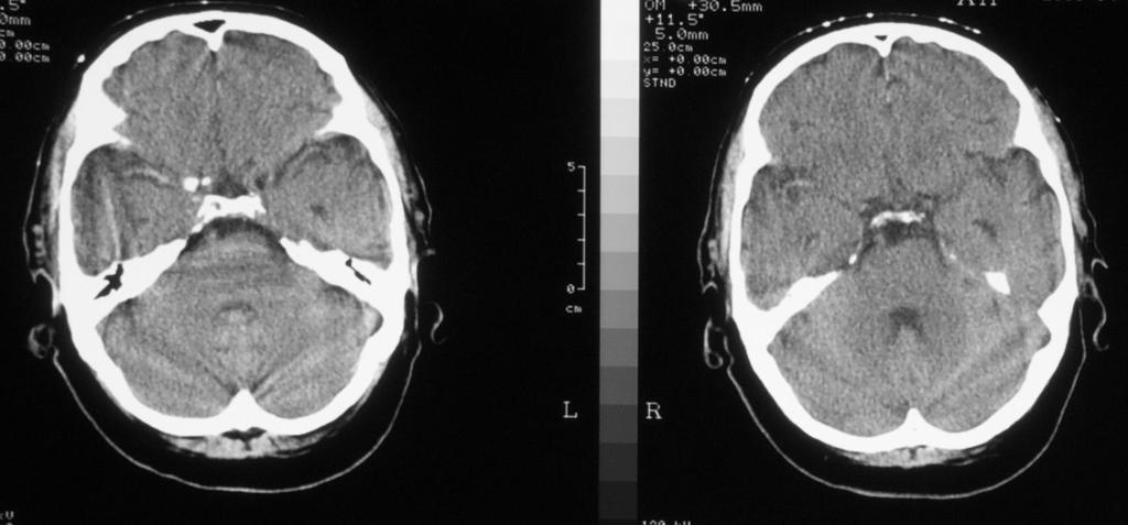
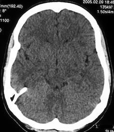
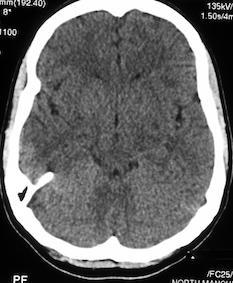
6 How to Recognise Haemorrhage on CT
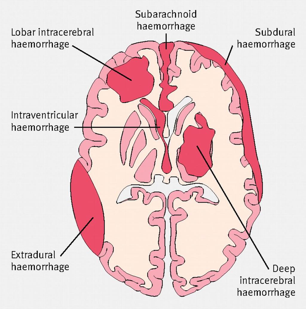
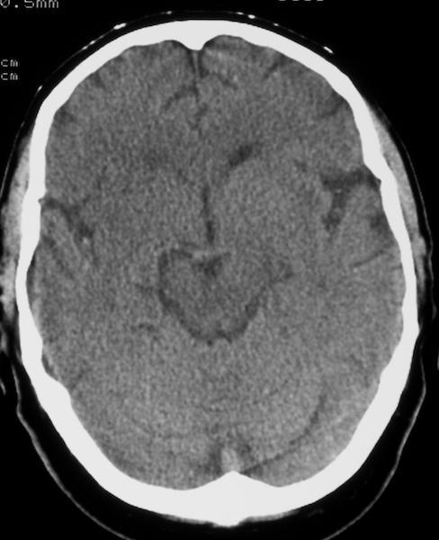
7

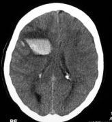
8 Dense white blob Only seen after blood clots Minimal oedema
9

10 Complex haematoma White blob not homogeneous Thalamic haematoma

11 Complex haematoma Blob not homogeneous More white matter oedema

12 Unclotted blood same density as brain clotted blood white NB Normal white Matter in a young person NOT oedema
13 Beware the Resolving Haematoma
14 Acute

15 Fading, subacute ICH


16 CE CT on FU Without previous CT, could be taken for SOL


17 Final FU atrophy at haematoma site


18 Post contrast medium CT looks like SOL
19 Non contrast CT 2 weeks earlier

20 (Infarct) ischaemic oedema Haematoma
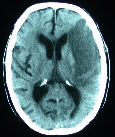
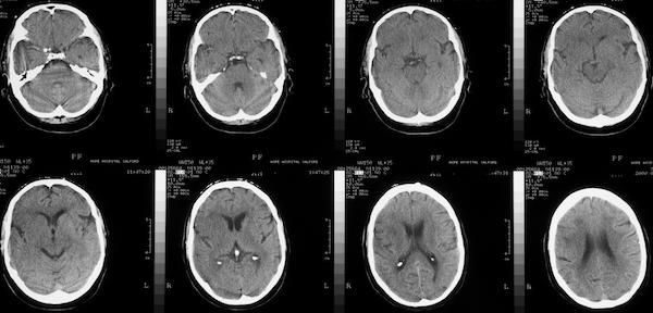
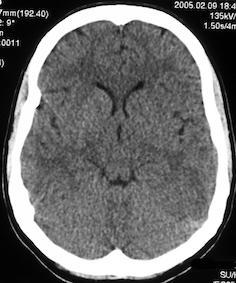
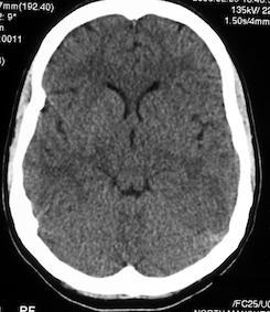
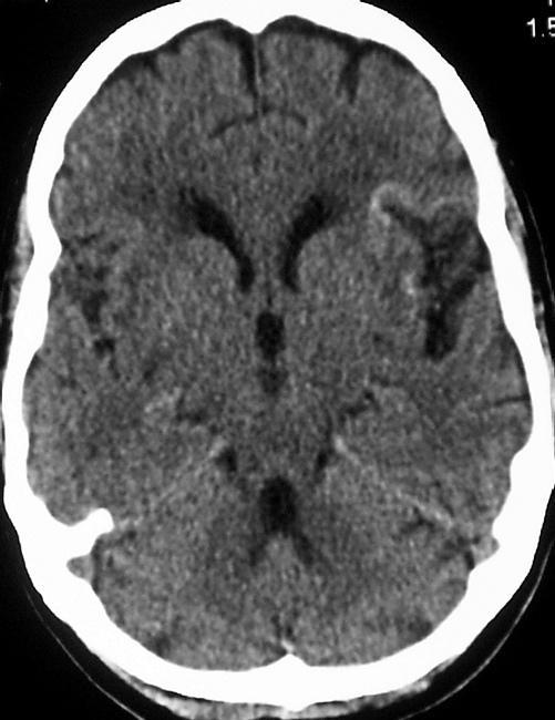
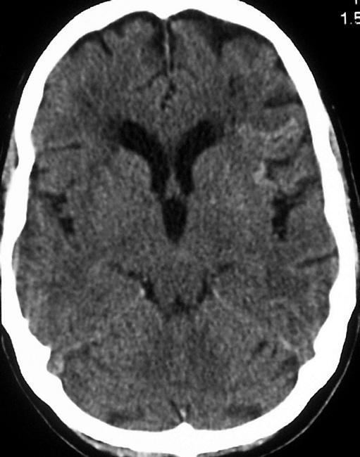
21 Normal Anatomy
22 CN Insular Ribbon Th LN Th = Thalamus
23 Insular Ribbon Int. Caps.
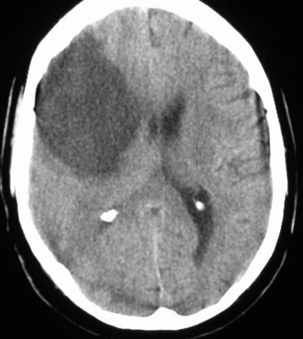
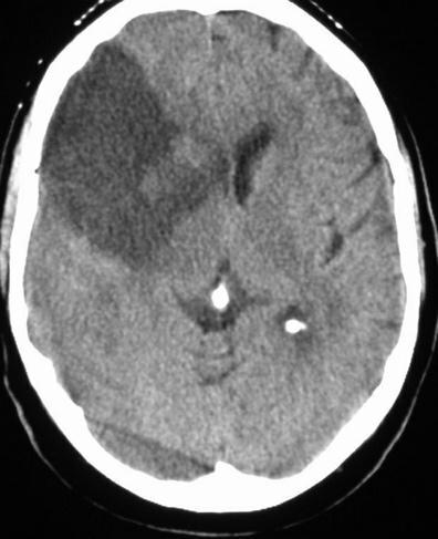
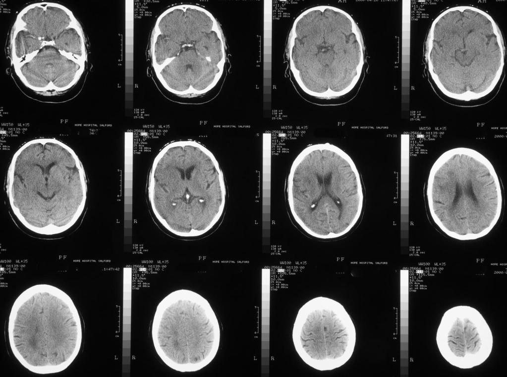
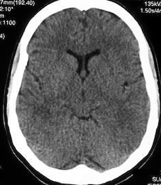
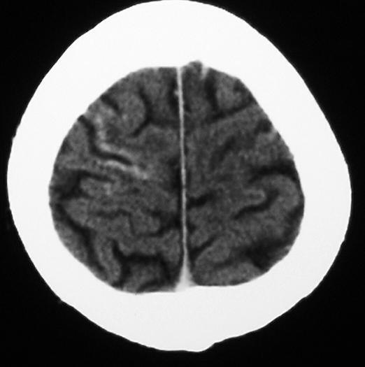
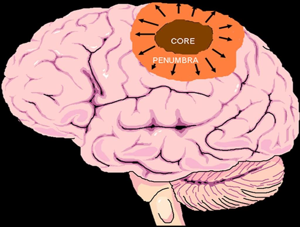
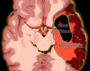
24 How to Recognise an Infarct Actually Ischaemic Oedema Dead infarct (core) indistinguishable from salvageable ischaemic oedema surrounding it (penumbra)

25 Low density Wedge shaped Grey & white matter Within known arterial vascular territory Proportionally little mass effect
26
27

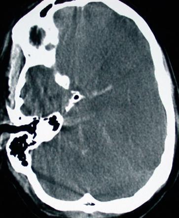
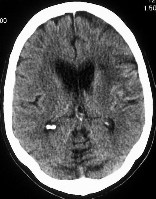
28 Subcortical infarct NB Grey matter not confined to the cortex CN Ant. LN Int. caps obliterated
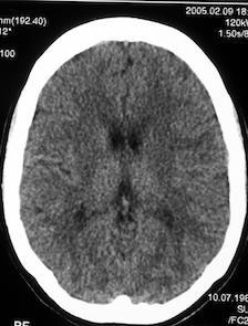
29 Stroke oedema?

30 Stroke oedema?
31
32 Stroke oedema?

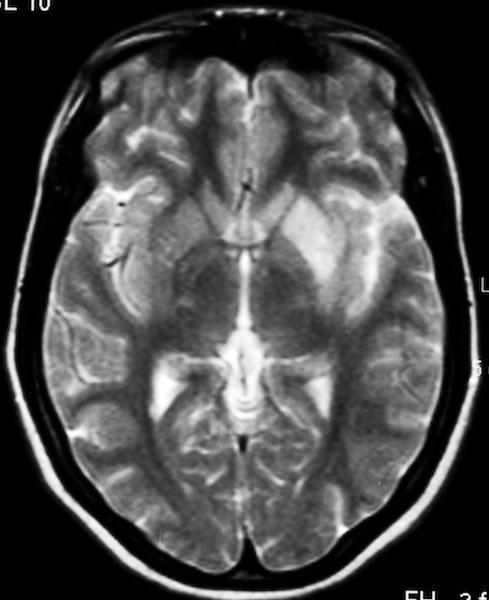
33 This small haematoma caused the stroke Stroke oedema?

34 Stroke oedema? Complex SOL
35 Hyperdense MCA
36
37
38

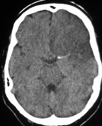

39 Dense ICA Plus fleck of Calcification in Vessel wall Dense MCA
40 More Subtle Examples of Early Infarction Basic neuroanatomy to support early diagnosis
41
42
43
44


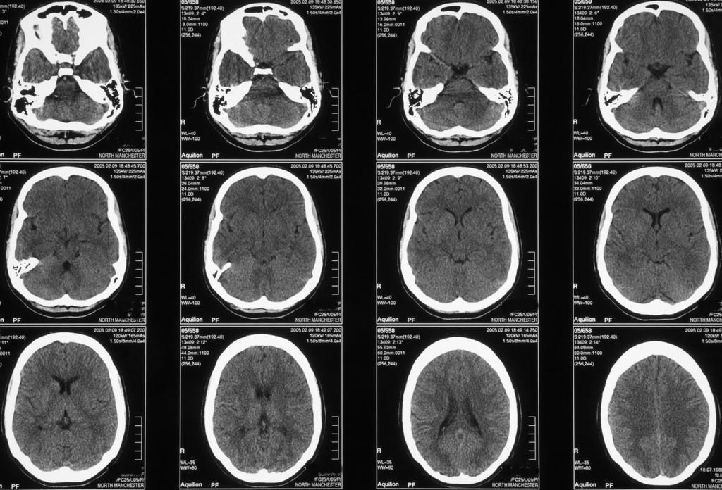
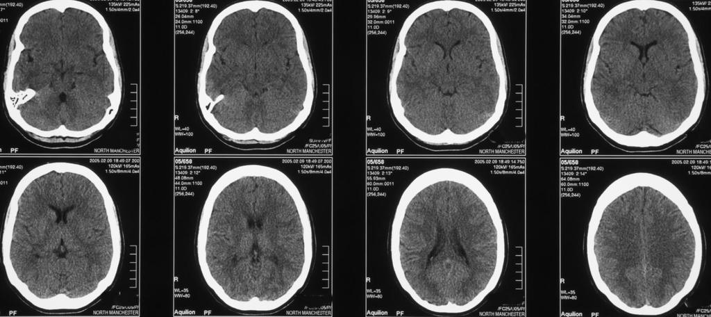
45 Insular Ribbon, BG & CN obliterated on Right. Normal on Left. Blue stars = Insula. Yellow star = BG CN


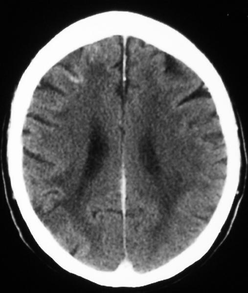
46 Reduced attenuation (low density) obliterates grey/white differentiation
47 Next day
48
49
50
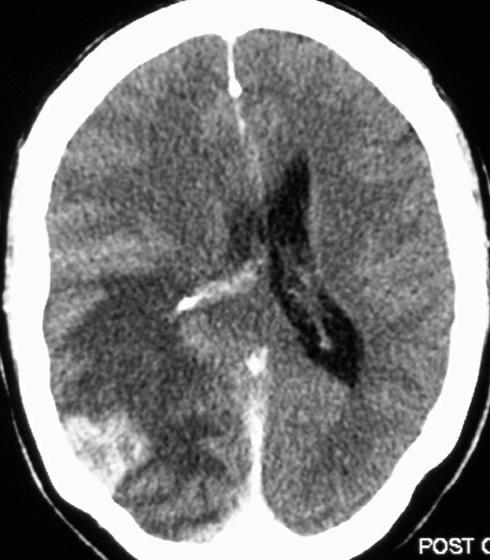
51
52
53
54
55

56 Ext caps CN LN
57
58 Haemorrhagic Infarction and how to tell it from a primary intracerebral haemorrhage


59 Pt with SBE throwing off multiple emboli New H gic infarct Old Infarct Why?
60
61 Plain CT haemorrhagic infarction
62
63
64 ICH
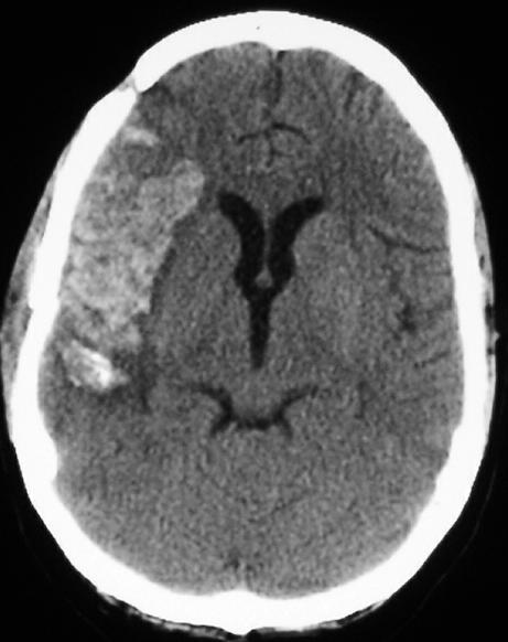
65 Watershed Infarcts ie Border Zone
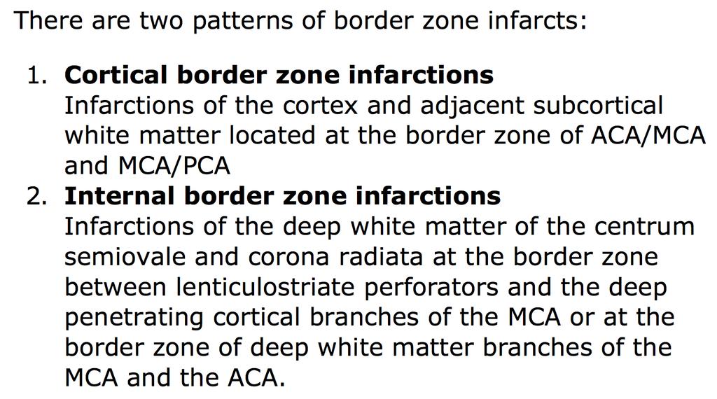
66

67 Radiology Assistant
68 How to Recognise the Different Vascular Territories

69 Green = ACA Red = MCA Purple = PCA lat midline from top from below
70 Circle of Willis rarely a true circle

71 No circle at all No Post.Comm. Arteries Ant & Post circulation isolated iwt

72 Dominant Post Comm

73 ICA occlusion ACA & MCA infarcts
74 iwt
75
76 MCA & PCA infarcts because of dominant Post Comm Art on Right
77 iwt
78 iwt
79 Carotid Artery Dissection No infarct Circle of Willis protects brain


80 Polo Mint thrombus in arterial wall, end on Thrombus en face


81 C of W protects via Ant Comm No infarct
82
83 How to Select Patients for thrombolysis? Clinical NIHSS Infarct size ASPECT Score Distinguishing dead tissue from living, but stunned, brain CT Perfusion iwt
84 How to Select Patients for thrombolysis? Clinical NIHSS Infarct size ASPECT Score Distinguishing dead tissue from living, but stunned, brain CT Perfusion iwt
85
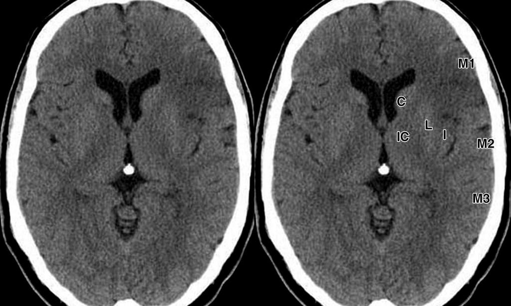
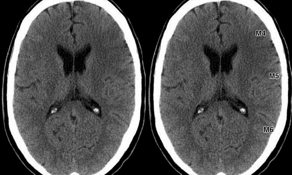
86 ASPECT Scoring System A = ACA; P = PCA; M = MCA MCA territory (10 points is Normal) Subtract one point for each: M1, M2, M3 M4, M5, M6 Caudate, Int Caps, LN, Insula

87 Level with foramina of Monro c ic LN In

88 M1 Sylvian fissure Level with 3 rd V M2 M3

89 M4 Level with top of Lat Vs M5 M6 (VRS)

90 ASPECTS iwt
91 iwt
92 iwt

93 Examples from paper
94 How to Select Patients for thrombolysis? Clinical NIHSS Infarct size ASPECT Score Distinguishing dead tissue from living, but stunned, brain CT Perfusion iwt
95 Penumbra Unlike muscle, brain tissue exquisitely sensitive to ischaemia Absence of neuronal energy stores In complete absence of blood flow, available energy can sustain neuronal viability for 2-3 minutes In acute stroke, ischaemia incomplete Collateral blood supply from uninjured arterial & leptomeningeal territories Results in central infarcted tissue surrounded by peripheral stunned cells (penumbra)
96
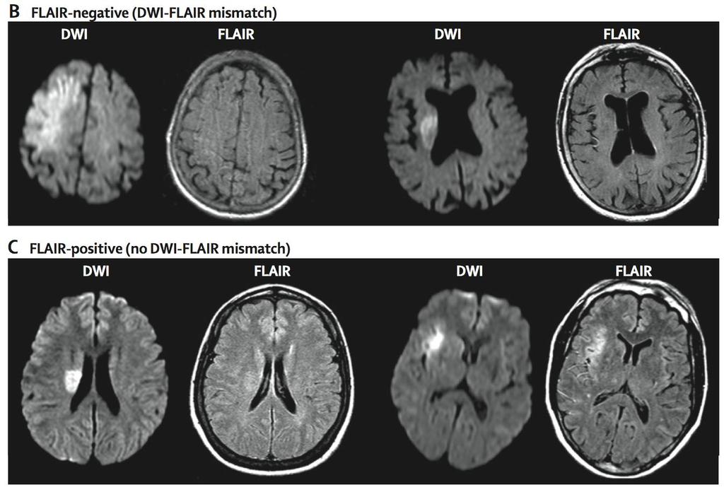
97 Imaging of the Penumbra CT MRI Discrepancy in perfusion parameters Mismatch between diffusion & perfusion parameters (DWI/PWI)
98
99
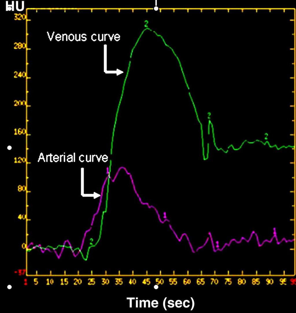
100 Pericallosal artery Sigmoid (venous) sinus
101

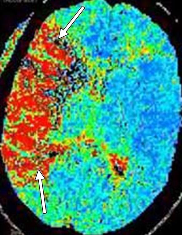
102 CTP CBV CBF MTT

103 Normal CT CTP perfusion defect CTA ICA dissection radiology assistant
104 Goals of Acute Stroke Imaging The Four Ps Parenchyma Assess early signs of acute stroke Rule out haemorrhage Pipes look for intravascular thrombus Extracranial circulation (neck) Intracranial circulation Perfusion Cerebral blood volume Cerebral blood flow Mean transit time Penumbra Tissue at risk
105 Conclusions Stroke is a clinical diagnosis CT is best and will suffice in majority MR for CT neg. stroke or for definitive diagnosis when suggested clinically eg dissection or where CT suggests alternative diagnosis eg SOL Advanced MR techniques best left to specialist units
106
107

108 LancetNeurol November 2011
109
110
CT INTERPRETATION COURSE
 CT INTERPRETATION COURSE Introductory Lecture on Basic Principles ASTRACAT 2012 Part One Stroke is a Clinical Diagnosis A clinical syndrome characterised by rapidly developing clinical symptoms and/or
NEURORADIOLOGY Part I
 NEURORADIOLOGY Part I Vörös Erika University of Szeged Department of Radiology SZEGED BRAIN IMAGING METHODS Plain film radiography Ultrasonography (US) Computer tomography (CT) Magnetic resonance imaging
Acute stroke. Ischaemic stroke. Characteristics. Temporal classification. Clinical features. Interpretation of Emergency Head CT
 Ischaemic stroke Characteristics Stroke is the third most common cause of death in the UK, and the leading cause of disability. 80% of strokes are ischaemic Large vessel occlusive atheromatous disease
An Introduction to Imaging the Brain. Dr Amy Davis
 An Introduction to Imaging the Brain Dr Amy Davis Common reasons for imaging: Clinical scenarios: - Trauma (NICE guidelines) - Stroke - Tumours - Seizure - Neurological degeneration memory, motor dysfunction,
On Call Guide to CT Perfusion. Updated: March 2011
 On Call Guide to CT Perfusion Updated: March 2011 CT Stroke Protocol 1. Non contrast CT brain 2. CT perfusion: contrast 40cc bolus dynamic imaging at 8 slice levels ~ 60 sec creates perfusion color maps
STROKE - IMAGING. Dr RAJASEKHAR REDDY 2nd Yr P.G. RADIODIAGNOSIS KIMS,Narkatpalli.
 STROKE - IMAGING Dr RAJASEKHAR REDDY 2nd Yr P.G. RADIODIAGNOSIS KIMS,Narkatpalli. STROKE Describes a clinical event that consists of sudden onset of neurological symptoms Types Infarction - occlusion of
Advanced Neuroimaging for Acute Stroke
 Advanced Neuroimaging for Acute Stroke E. Bradshaw Bunney, MD, FACEP Professor Department Of Emergency Medicine University of Illinois at Chicago Swedish American Belvidere Hospital Disclosures FERNE Board
How to interpret an unenhanced CT brain scan. Part 2: Clinical cases
 How to interpret an unenhanced CT brain scan. Part 2: Clinical cases Thomas Osborne a, Christine Tang a, Kivraj Sabarwal b and Vineet Prakash c a Radiology Registrar; b Radiology Foundation Year 1 Doctor;
Place for Interventional Radiology in Acute Stroke
 Place for Interventional Radiology in Acute Stroke Dr Lakmalie Paranahewa MBBS, MD(Radiology), FRCR Consultant Interventional Radiologist Asiri Group of Hospitals Objectives Imaging in Stroke Neurovascular
ACUTE STROKE IMAGING
 ACUTE STROKE IMAGING Mahesh V. Jayaraman M.D. Director, Inter ventional Neuroradiology Associate Professor Depar tments of Diagnostic Imaging and Neurosurger y Alper t Medical School at Brown University
ISCHEMIC STROKE IMAGING
 ISCHEMIC STROKE IMAGING ผศ.พญ พญ.จ ร ร ตน ธรรมโรจน ภาคว ชาร งส ว ทยา คณะแพทยศาสตร มหาว ทยาล ยขอนแก น A case of acute hemiplegia Which side is the abnormality, right or left? Early Right MCA infarction
AMSER Case of the Month: March 2019
 AMSER Case of the Month: March 2019 62 year-old male with left-sided weakness Ashley Graziano OMS IV, Lake Erie College of Osteopathic Medicine Erik Yannone MD, Charles Q. Li MD, Warren Chang MD, Matthew
Acute Ischemic Stroke Imaging. Ronald L. Wolf, MD, PhD Associate Professor of Radiology
 Acute Ischemic Stroke Imaging Ronald L. Wolf, MD, PhD Associate Professor of Radiology Title of First Slide of Substance An Illustrative Case 2 Disclosures No financial disclosures Off-label uses of some
11/1/2018. Disclosure. Imaging in Acute Ischemic Stroke 2018 Neuro Symposium. Is NCCT good enough? Keystone Heart Consultant, Stock Options
 Disclosure Imaging in Acute Ischemic Stroke 2018 Neuro Symposium Keystone Heart Consultant, Stock Options Kevin Abrams, M.D. Chief of Radiology Medical Director of Neuroradiology Baptist Hospital, Miami,
Cerebro-vascular stroke
 Cerebro-vascular stroke CT Terminology Hypodense lesion = lesion of lower density than the normal brain tissue Hyperdense lesion = lesion of higher density than normal brain tissue Isodense lesion = lesion
NEURO IMAGING 2. Dr. Said Huwaijah Chairman of radiology Dep, Damascus Univercity
 NEURO IMAGING 2 Dr. Said Huwaijah Chairman of radiology Dep, Damascus Univercity I. EPIDURAL HEMATOMA (EDH) LOCATION Seventy to seventy-five percent occur in temporoparietal region. CAUSE Most likely caused
Disclosure. + Outline. What is a stroke? Role of imaging in stroke Ischemic stroke Venous infarct Current topics
 + Kathleen R. Fink, MD University of Washington 5 th Nordic Emergency Radiology Course May 21, 2015 + Disclosure My spouse receives research salary support from: Bracco BayerHealthcare Guerbet Thank you
Review Article Differentiating between Hemorrhagic Infarct and Parenchymal Intracerebral Hemorrhage
 Hindawi Publishing Corporation Radiology Research and Practice Volume 2012, Article ID 475497, 11 pages doi:10.1155/2012/475497 Review Article Differentiating between Hemorrhagic Infarct and Parenchymal
What to expect on post mechanical thrombectomy CT - a guide to correct diagnosis.
 What to expect on post mechanical thrombectomy CT - a guide to correct diagnosis. Poster No.: C-2257 Congress: ECR 2017 Type: Educational Exhibit Authors: T. Buende Tchokouako, H. Nejadhamzeeigilani, A.
NEURORADIOLOGY DIL part 4
 NEURORADIOLOGY DIL part 4 Strokes and infarcts K. Agyem MD, G. Hall MD, D. Palathinkal MD, Alexandre Menard March/April 2015 OVERVIEW Introduction to Neuroimaging - DIL part 1 Basic Brain Anatomy - DIL
Stroke/TIA. Tom Bedwell
 Stroke/TIA Tom Bedwell tab1g11@soton.ac.uk The Plan Definitions Anatomy Recap Aetiology Pathology Syndromes Brocas / Wernickes Investigations Management Prevention & Prognosis TIAs Key Definitions Transient
IMAGING IN ACUTE ISCHEMIC STROKE
 IMAGING IN ACUTE ISCHEMIC STROKE Timo Krings MD, PhD, FRCP (C) Professor of Radiology & Surgery Braley Chair of Neuroradiology, Chief and Program Director of Diagnostic and Interventional Neuroradiology;
Background. Recommendations for Imaging of Acute Ischemic Stroke: A Scientific Statement From the American Heart Association
 for Imaging of Acute Ischemic Stroke: A Scientific Statement From the American Heart Association An Scientific Statement from the Stroke Council, American Heart Association and American Stroke Association
For Emergency Doctors. Dr Suzanne Smallbane November 2011
 For Emergency Doctors Dr Suzanne Smallbane November 2011 A: Orbit B: Sphenoid Sinus C: Temporal Lobe D: EAC E: Mastoid air cells F: Cerebellar hemisphere A: Frontal lobe B: Frontal bone C: Dorsum sellae
Neuroradiology: Imaging and Stroke
 Neuroradiology: Imaging and Stroke Stroke 2017 William Gallmann January 28, 2017 Stroke Arterial ischemia/infarct accounts for ~85% Cerebral venous occlusions - 0.5-1% Spontaneous intracranial hemorrhage
Acute Ischaemic Stroke
 Acute Ischaemic Stroke CT or MR SCA READIG FORM SCA ID: DATE OF READIG: SCA QUALIT: Good Moderate Poor Comment: READER ID: TPE OF SCA: CT: Without contrast: With contrast: MR: Diffusion: Perfusion ote
Case Conference: Neuroradiology. Case 1: Tumor Case 1: 22yo F w/ HA and prior Seizures
 Case Conference: Neuroradiology Case 1: 22yo F w/ HA and prior Seizures David E. Rex, MD, PhD Stanford University Hospital Department of Radiology Case 1: Tumor Most likely gangiloglioma, oligodendroglioma,
HYPERACUTE STROKE CASE STUDIES. By Mady Roman Hyper Acute Stroke Nurse Practitioner RHH
 HYPERACUTE STROKE CASE STUDIES By Mady Roman Hyper Acute Stroke Nurse Practitioner RHH MC Case study 82 years old, lady 9:30 well, in touch with her son 11:30 hairdresser came to her house and found her
IMAGING IN ACUTE ISCHEMIC STROKE
 IMAGING IN ACUTE ISCHEMIC STROKE Timo Krings MD, PhD, FRCP (C) Professor of Radiology & Surgery Braley Chair of Neuroradiology, Chief and Program Director of Diagnostic and Interventional Neuroradiology;
[(PHY-3a) Initials of MD reviewing films] [(PHY-3b) Initials of 2 nd opinion MD]
![[(PHY-3a) Initials of MD reviewing films] [(PHY-3b) Initials of 2 nd opinion MD]](/thumbs/89/98619893.jpg "[(PHY-3a) Initials of MD reviewing films] [(PHY-3b) Initials of 2 nd opinion MD]") 2015 PHYSICIAN SIGN-OFF (1) STUDY NO (PHY-1) CASE, PER PHYSICIAN REVIEW 1=yes 2=no [strictly meets case definition] (PHY-1a) CASE, IN PHYSICIAN S OPINION 1=yes 2=no (PHY-2) (PHY-3) [based on all available
2015 PHYSICIAN SIGN-OFF (1) STUDY NO (PHY-1) CASE, PER PHYSICIAN REVIEW 1=yes 2=no [strictly meets case definition] (PHY-1a) CASE, IN PHYSICIAN S OPINION 1=yes 2=no (PHY-2) (PHY-3) [based on all available
Stroke imaging. Why image stroke patients? Stroke. Treatment of infarct. Methods for infarct diagnosis. Treatment of infarct.
 Stroke imaging Stroke Infarct: -Arterial thrombosis/embolus -Hypoxic/ischemic -Venous thrombosis Non-traumatic hemorrhage: -Intracerebral -Subarachnoid Johan Wikström MD PhD Associate Professor of Radiology
Imaging in Stroke. D Nagaraja, N Karthik
 Imaging in Stroke D Nagaraja, N Karthik Cerebro-vascular disease (stroke) is the second leading cause of death. Prior to CT era, diagnosis was essentially clinical supported by angio and lumbar puncture.
Acute Ischemic Stroke Imaging Innovations
 Acute Ischemic Stroke Imaging Innovations Guilherme Dabus, MD, FAHA Director, Fellowship NeuroInterventional Surgery Miami Cardiac & Vascular Institute Baptist Neuroscience Center Baptist Neuroscience
occlusions. Cerebral perfusion is driven fundamentally by regional cerebral
 Appendix Figures Figure A1. Hemodynamic changes that may occur in major anterior circulation occlusions. Cerebral perfusion is driven fundamentally by regional cerebral perfusion pressure (CPP). In response
Blood Supply. Allen Chung, class of 2013
 Blood Supply Allen Chung, class of 2013 Objectives Understand the importance of the cerebral circulation. Understand stroke and the types of vascular problems that cause it. Understand ischemic penumbra
Title: Stability of Large Diffusion/Perfusion Mismatch in Anterior Circulation Strokes for 4 or More Hours
 Author's response to reviews Title: Stability of Large Diffusion/Perfusion Mismatch in Anterior Circulation Strokes for 4 or More Hours Authors: Ramon G. Gonzalez (rggonzalez@partners.org) Reza Hakimelahi
Assessment Of Collateral Pathways In Acute Ischemic Cerebrovascular Stroke Using A Mansour Grading Scale; A New Scale, A Pilot Study
 ISPUB.COM The Internet Journal of Interventional Medicine Volume 3 Number 1 Assessment Of Collateral Pathways In Acute Ischemic Cerebrovascular Stroke Using A Mansour Grading Scale; A New Scale, A Pilot
How to Interpret CT/CTA for Acute Stroke in the Age of Endovascular Clot Retrieval
 How to Interpret CT/CTA for Acute Stroke in the Age of Endovascular Clot Retrieval Peter Howard MD FRCPC Disclosures No conflicts to disclose How to Interpret CT/CTA for Acute Stroke in the Age of Endovascular
Imaging Acute Stroke and Cerebral Ischemia
 Department of Radiology University of California San Diego Imaging Acute Stroke and Cerebral Ischemia John R. Hesselink, M.D. Causes of Stroke Arterial stenosis Thrombosis Embolism Dissection Hypotension
Interventions in the Management of Acute Stroke. Dr Md Shafiqul Islam Associate Professor Neurosurgery Dhaka Medical College Hospital
 Interventions in the Management of Acute Stroke Dr Md Shafiqul Islam Associate Professor Neurosurgery Dhaka Medical College Hospital Acute stroke intervention Number of stroke patients increasing day by
Debbie Summers, MSN, RN, ACNS-BC, CNRN, SCRN. Debbie Summers, MSN, ACNS-BC Nothing
 Debbie Summers, MSN, RN, ACNS-BC, CNRN, SCRN Debbie Summers, MSN, ACNS-BC Nothing Identify anatomical location of stroke on CT, MRI, and perfusion scans and angiography. Relate underlying pathology to
Comparison of Five Major Recent Endovascular Treatment Trials
 Comparison of Five Major Recent Endovascular Treatment Trials Sample size 500 # sites 70 (100 planned) 316 (500 planned) 196 (833 estimated) 206 (690 planned) 16 10 22 39 4 Treatment contrasts Baseline
STATE OF THE ART IMAGING OF ACUTE STROKE
 STATE OF THE ART IMAGING OF ACUTE STROKE Marin Penkov UH St Ivan Rilski Sofia RadiologyTogether 2-3 June 2017 GOALS The concept and significance of penumbra CT and MRI Basic Principles Clinical application
LATEST IMAGING FOR ACUTE ISCHEMIC STROKE AND INTRACEREBRAL HEMORRHAGE
 LATEST IMAGING FOR ACUTE ISCHEMIC STROKE AND INTRACEREBRAL HEMORRHAGE Brian A. Stettler, MD Department of Emergency Medicine, University of Cincinnati Cincinnati, OH OBJECTIVES: 1) To describe the role
BGS Spring Conference 2015
 Neuroradiology in hyperacute stroke: what is the UK position? Dr Shelley Renowden Bristol NICE HIS July, 2013 The current evidence on mechanical clot retrieval for treating acute ischaemic stroke shows
ACUTE ISCHEMIC STROKE. Current Treatment Approaches for Acute Ischemic Stroke
 ACUTE ISCHEMIC STROKE Current Treatment Approaches for Acute Ischemic Stroke EARLY MANAGEMENT OF ACUTE ISCHEMIC STROKE Rapid identification of a stroke Immediate EMS transport to nearest stroke center
NEURORADIOLOGY Part I
 NEURORADIOLOGY Part I Vörös Erika University of Szeged Department of Radiology SZEGED DISEASES OF CNS BRAIN Developmental anomalies Cerebrovascular disorders Tumours Inflammatory diseases Trauma DISEASES
Imaging Stroke: Is There a Stroke Equivalent of the ECG? Albert J. Yoo, MD Director of Acute Stroke Intervention Massachusetts General Hospital
 Imaging Stroke: Is There a Stroke Equivalent of the ECG? Albert J. Yoo, MD Director of Acute Stroke Intervention Massachusetts General Hospital Disclosures Penumbra, Inc. research grant (significant) for
The DAWN of a New Era for Wake-up Stroke
 The DAWN of a New Era for Wake-up Stroke Alan H. Yee, D.O. Stroke and Critical Care Neurology Department of Neurology University of California Davis Medical Center Objectives Review Epidemiology and Natural
UPSTATE Comprehensive Stroke Center. Neurosurgical Interventions Satish Krishnamurthy MD, MCh
 UPSTATE Comprehensive Stroke Center Neurosurgical Interventions Satish Krishnamurthy MD, MCh Regional cerebral blood flow is important Some essential facts Neurons are obligatory glucose users Under anerobic
Neuropathology lecture series. III. Neuropathology of Cerebrovascular Disease. Physiology of cerebral blood flow
 Neuropathology lecture series III. Neuropathology of Cerebrovascular Disease Physiology of cerebral blood flow Brain makes up only 2% of body weight Percentage of cardiac output: 15-20% Percentage of O
Cerebrovascular Disease
 Neuropathology lecture series Cerebrovascular Disease Physiology of cerebral blood flow Brain makes up only 2% of body weight Percentage of cardiac output: 15-20% Percentage of O 2 consumption (resting):
EFFECTIVENESS OF CTA AND CTP IN GUIDING MANAGEMENT DECISIONS FOR ACUTE STROKE
 EFFECTIVENESS OF CTA AND CTP IN GUIDING MANAGEMENT DECISIONS FOR ACUTE STROKE Jon Benton MS-4, Chirag Dani MD, Vincent Persaud MD, Bilal A. Manzer, Macksood Aftab DO, MHA OBJECTIVES 1. Utility of advanced
Stroke & the Emergency Department. Dr. Barry Moynihan, March 2 nd, 2012
 Stroke & the Emergency Department Dr. Barry Moynihan, March 2 nd, 2012 Outline Primer Stroke anatomy & clinical syndromes Diagnosing stroke Anterior / Posterior Thrombolysis Haemorrhage The London model
Carotid Embolectomy and Endarterectomy for Symptomatic Complete Occlusion of the Carotid Artery as a Rescue Therapy in Acute Ischemic Stroke
 This is an Open Access article licensed under the terms of the Creative Commons Attribution-NonCommercial-NoDerivs 3.0 License (www.karger.com/oa-license), applicable to the online version of the article
NEURO IMAGING OF ACUTE STROKE
 1 1 NEURO IMAGING OF ACUTE STROKE ALICIA RICHARDSON, MSN, RN, ACCNS-AG, ANVP-BC WENDY SMITH, MA, RN, MBA, SCRN, FAHA LYNN HUNDLEY, APRN, CNRN, CCNS, ANVP-BC 2 2 1 DISCLOSURES Alicia Richardson: Stryker
Practical Considerations in the Early Treatment of Acute Stroke
 Practical Considerations in the Early Treatment of Acute Stroke Matthew E. Fink, MD Neurologist-in-Chief Weill Cornell Medical College New York-Presbyterian Hospital mfink@med.cornell.edu Disclosures Consultant
Carotid artery stenting for long CTO and pseudo occlusion of carotid artery -2 case reports-
 Carotid artery stenting for long CTO and pseudo occlusion of carotid artery -2 case reports- Katsutoshi Takayama, MD, Ph.D Department of Radiology and Interventional Neuroradiology Ishinkai Yao General
The central nervous system
 Sectc.qxd 29/06/99 09:42 Page 81 Section C The central nervous system CNS haemorrhage Subarachnoid haemorrhage Cerebral infarction Brain atrophy Ring enhancing lesions MRI of the pituitary Multiple sclerosis
Acute stroke imaging
 Acute stroke imaging Aims Imaging modalities and differences Why image acute stroke Clinical correlation to imaging appearance What is stroke Classic definition: acute focal injury to the central nervous
The Language of Stroke
 The Language of Stroke Examination / Imaging / Diagnosis / Treatment Dr Suzanne Busch A lot of letters! CBF CVA ICH CVD CBV DWI US MRI/MRA CAA CTA CTP ICA MCA SAH WMD TIA MCA Agnosia A lot of big words!
Diagnostic improvement from average image in acute ischemic stroke
 Diagnostic improvement from average image in acute ischemic stroke N. Magne (1), E.Tollard (1), O. Ozkul- Wermester (2), V. Macaigne (1), J.-N. Dacher (1), E. Gerardin (1) (1) Department of Radiology,
secondary effects and sequelae of head trauma.
 Neuroimaging of vascular/secondary secondary effects and sequelae of head trauma. Andrès Server Alonso Department of Neuroradiology Division of Radiology Ullevål University Hospital Oslo, Norway. Guidelines
Mechanical thrombectomy in Plymouth. Will Adams. Will Adams
 Mechanical thrombectomy in Plymouth Will Adams Will Adams History Intra-arterial intervention 1995 (NINDS) iv tpa improved clinical outcome in patients treated within 3 hours of ictus but limited recanalisation
Tutorials. By Dr Sharon Truter
 Tutorials By Dr Sharon Truter To the Tutorials By Dr Sharon Truter What to expect from the Tutorials What to expect from these tutorials Outlines, structure, guided reading, explanations, mnemonics Begin
Stroke: The First Critical Hour. Alina Candal, RN, PCC, MICN Kevin Andruss, MD, FACEP
 Stroke: The First Critical Hour Alina Candal, RN, PCC, MICN Kevin Andruss, MD, FACEP Disclosures We have no actual or potential conflicts of interest in relation to this presentation. Objectives Discuss
IV. Cerebrovascular diseases
 IV. Cerebrovascular diseases - Cerebrovascular disease denotes brain disorders caused by pathologic processes involving the blood vessels. - The three main pathogenic mechanisms are: 1. Thrombotic occlusion
L M Thornton, MD; L Lanier, MD; C L Sistrom, MD; D Rajderkar, MD; A Mancuso, MD; IM Schmalfuss, MD University of Florida, Gainesville Department of
 L M Thornton, MD; L Lanier, MD; C L Sistrom, MD; D Rajderkar, MD; A Mancuso, MD; IM Schmalfuss, MD University of Florida, Gainesville Department of Radiology RSNA Annual Meeting 2016 Trainee call readiness
Cerebrovascular Disease
 Neuropathology lecture series Cerebrovascular Disease Kurenai Tanji, M.D., Ph.D. December 11, 2007 Physiology of cerebral blood flow Brain makes up only 2% of body weight Percentage of cardiac output:
What Are We Going to Do? Fourth Year Meds Clinical Neuroanatomy. Hydrocephalus and Effects of Interruption of CSF Flow. Tube Blockage Doctrine
 Fourth Year Meds Clinical Neuroanatomy Ventricles, CSF, Brain Swelling etc. David A. Ramsay, Neuropathologist, LHSC What Are We Going to Do? Hydrocephalus and some effects of the interruption of CSF flow
Endovascular Treatment of Ischemic Stroke
 Endovascular Treatment of Ischemic Stroke William Thorell, MD Associate Professor Neurosurgery UNMC Co-Director Stroke and Neurovascular Center Nebraska Medicine Overview Definitions of terms Review basic
Heejin Shim, Hyun Seok Choi, So-Lyung Jung, Kook-Jin Ahn, Bum-soo Kim
 Susceptibility Weighted MR Imaging at 3T in Patients with Occlusion of Middle Cerebral Artery : Comparison with Diffusion Weighted Imaging Score (ASPECTS) Heejin Shim, Hyun Seok Choi, So-Lyung Jung, Kook-Jin
Significant Relationships
 Opening Large Vessels During Acute Ischemic Stroke Significant Relationships Wade S Smith, MD, PhD Director UCSF Neurovascular Service Professor of Neurology Daryl R Gress Endowed Chair of Neurocritical
Stroke: clinical presentations, symptoms and signs
 Stroke: clinical presentations, symptoms and signs Professor Peter Sandercock University of Edinburgh EAN teaching course Burkina Faso 8 th November 2017 Clinical diagnosis is important to Ensure stroke
Essentials of Clinical MR, 2 nd edition. 14. Ischemia and Infarction II
 14. Ischemia and Infarction II Lacunar infarcts are small deep parenchymal lesions involving the basal ganglia, internal capsule, thalamus, and brainstem. The vascular supply of these areas includes the
5/31/2018. Interventional Therapies that Expand Time Windows for Acute Ischemic Stroke Treatment. Disclosures. Impact of clot burden
 Good Outcome (%) Rankin 0-2 at 90 days 5/31/2018 Interventional Therapies that Expand Time Windows for Acute Ischemic Stroke Treatment Disclosures Cerenovus: I am on Executive Committee for ARISE2 Trial
Interventional Neuroradiology. & Stroke INR PROCEDURES INR PROCEDURES. Dr Steve Chryssidis. 25-Sep-17. Interventional Neuroradiology
 Interventional Neuroradiology Interventional Neuroradiology & Stroke Dr Steve Chryssidis Interventional Neuroradiology (INR) is a subspecialty within Radiology INR -- broadly defined as treatment by endovascular
Is there even a time window?
 Is there even a time window? A brief summary of key 2018 trials: DAWN and DEFUSE-3 For Neurosciences Update Conference, February 22, 2018 Maxim D. Hammer, M.D. Executive Summary Based on DAWN and DEFUSE-3,
Guideline scope Stroke and transient ischaemic attack in over 16s: diagnosis and initial management (update)
") NATIONAL INSTITUTE FOR HEALTH AND CARE EXCELLENCE Guideline scope Stroke and transient ischaemic attack in over s: diagnosis and initial management (update) 0 0 This will update the NICE on stroke and
NATIONAL INSTITUTE FOR HEALTH AND CARE EXCELLENCE Guideline scope Stroke and transient ischaemic attack in over s: diagnosis and initial management (update) 0 0 This will update the NICE on stroke and
UPDATES IN INTRACRANIAL INTERVENTION Jordan Taylor DO Metro Health Neurology 2015
 UPDATES IN INTRACRANIAL INTERVENTION Jordan Taylor DO Metro Health Neurology 2015 NEW STUDIES FOR 2015 MR CLEAN ESCAPE EXTEND-IA REVASCAT SWIFT PRIME RECOGNIZED LIMITATIONS IV Alteplase proven benefit
ACUTE STROKE TREATMENT IN LARGE NIHSS PATIENTS. Justin Nolte, MD Assistant Profession Marshall University School of Medicine
 ACUTE STROKE TREATMENT IN LARGE NIHSS PATIENTS Justin Nolte, MD Assistant Profession Marshall University School of Medicine History of Presenting Illness 64 yo wf with PMHx of COPD, HTN, HLP who was in
Hyperperfusion syndrome after MCA embolectomy a rare complication?
 ISSN 1507-6164 DOI: 10.12659/AJCR.889672 Received: 2013.08.13 Accepted: 2013.09.11 Published: 2013.11.29 Hyperperfusion syndrome after MCA embolectomy a rare complication? Authors Contribution: Study Design
Medical Policy. MP Computed Tomography Perfusion Imaging of the Brain
 Medical Policy MP 6.01.49 BCBSA Ref. Policy: 6.01.49 Last Review: 09/28/2017 Effective Date: 09/28/2017 Section: Radiology Related Policies 2.01.54 Endovascular Procedures for Intracranial Arterial Disease
Update on Early Acute Ischemic Stroke Interventions
 Update on Early Acute Ischemic Stroke Interventions Diana Goodman MD Lead Neurohospitalist Maine Medical Center Assistant Professor of Neurology, Tufts University School of Medicine I have no disclosures
Stroke in the ED. Dr. William Whiteley. Scottish Senior Clinical Fellow University of Edinburgh Consultant Neurologist NHS Lothian
 Stroke in the ED Dr. William Whiteley Scottish Senior Clinical Fellow University of Edinburgh Consultant Neurologist NHS Lothian 2016 RCP Guideline for Stroke RCP guidelines for acute ischaemic stroke
Standard NICE (CG ) RCP (2016)
 RCP (2016)") Standard NICE (CG68 2008) RCP (2016) Stroke Unit Adults presenting at an A&E department with suspected stroke are admitted to a specialist stroke unit within 4 hours direct admission of patients with acute
Standard NICE (CG68 2008) RCP (2016) Stroke Unit Adults presenting at an A&E department with suspected stroke are admitted to a specialist stroke unit within 4 hours direct admission of patients with acute
Principles Arteries & Veins of the CNS LO14
 Principles Arteries & Veins of the CNS LO14 14. Identify (on cadaver specimens, models and diagrams) and name the principal arteries and veins of the CNS: Why is it important to understand blood supply
Session: Imaging for Clinical Decision Support
 healthitnews Session: Imaging for Clinical Decision Support Chair: Ian Poole, PhD Scientific Fellow Toshiba Medical Visualisation Systems, Edinburgh A Canon Group Company What is Clinical Decision Support?.
Stroke School for Internists Part 1
 Stroke School for Internists Part 1 November 4, 2017 Dr. Albert Jin Dr. Gurpreet Jaswal Disclosures I receive a stipend for my role as Medical Director of the Stroke Network of SEO I have no commercial
11/6/2013. Vanderbilt University Institute of Imaging Science (VUIIS) 7 Tesla Vascular Imaging. Evaluating stroke risk
 7 Tesla Vascular Imaging. Evaluating stroke risk") Vanderbilt University Institute of Imaging Science (VUIIS) New MI Techniques for Imaging Cerebrovascular Disease Manus J. Donahue Depts. of adiology, Physics, Neurology and Physics Vanderbilt University
Vanderbilt University Institute of Imaging Science (VUIIS) New MI Techniques for Imaging Cerebrovascular Disease Manus J. Donahue Depts. of adiology, Physics, Neurology and Physics Vanderbilt University
Neuro-vascular Intervention in Stroke. Will Adams Consultant Neuroradiologist Plymouth Hospitals NHS Trust
 Neuro-vascular Intervention in Stroke Will Adams Consultant Neuroradiologist Plymouth Hospitals NHS Trust Stroke before the mid 1990s Swelling Stroke extension Haemorrhagic transformation Intravenous thrombolysis
CEREBROVASCULAR DISEASES. By: Shifaa AlQa qa
 CEREBROVASCULAR DISEASES By: Shifaa AlQa qa Cerebrovascular diseases Brain disorders caused by pathologic processes involving blood vessels 3 pathogenic mechanisms (1) thrombotic occlusion, (2) embolic
Broadening the Stroke Window in Light of the DAWN Trial
 Broadening the Stroke Window in Light of the DAWN Trial South Jersey Neurovascular and Stroke Symposium April 26, 2018 Rohan Chitale, MD Assistant Professor of Neurological Surgery Vanderbilt University
Brain Attack. Strategies in the Management of Acute Ischemic Stroke: Neuroscience Clerkship. Case Medical Center
 Brain Attack Strategies in the Management of Acute Ischemic Stroke: Neuroscience Clerkship Stroke is a common and devastating disorder Third leading antecedent of death in American men, and second among
The Role of Neuroimaging in Acute Stroke. Bradley Molyneaux, HMS IV
 The Role of Neuroimaging in Acute Stroke Bradley Molyneaux, HMS IV Patient CR 62 yo F w/ 2 wk h/o altered mental status Presents to ED w/ confusion following a fall 1 day prior New onset left facial droop
41 year old female with headache. Elena G. Violari MD and Leo Wolansky MD
 41 year old female with headache Elena G. Violari MD and Leo Wolansky MD ? Dural Venous Sinus Thrombosis with Hemorrhagic Venous Infarct Acute intraparenchymal hematoma measuring ~3 cm in diameter centered
Emergency Room Procedure The first few hours in hospital...
 Emergency Room Procedure The first few hours in hospital... ER 5 level Emergency Severity Index SOP s for Stroke Stroke = Level 2 Target Time = 1 Hour 10 min from door 2 Doctor 25 min from door 2 CT 60
C. Douglas Phillips, MD FACR Director of Head and Neck Imaging Weill Cornell Medical College NewYork-Presbyterian Hospital
 C. Douglas Phillips, MD FACR Director of Head and Neck Imaging Weill Cornell Medical College NewYork-Presbyterian Hospital I have no financial disclosures Understand range of pathology that may present
Imaging in acute ischaemic stroke: essential for modern stroke care
 1 Radiology Department, Royal Hallamshire Hospital, Sheffield NHS Teaching Hospitals Trust, Sheffield, UK 2 Academic Unit of Radiology, University of Sheffield, Sheffield, UK Correspondence to Dr Daniel
Anoxic brain injury CT and MRI patterns - quick pictoral quide for junior radiologists.
 Anoxic brain injury CT and MRI patterns - quick pictoral quide for junior radiologists. Poster No.: C-1844 Congress: ECR 2017 Type: Educational Exhibit Authors: A. Kecler - Pietrzyk, W. Torreggiani ; Dublin/IE,