Spring Eye Red Eye Terrence Clark, OD FAAO Brittney Gewolb, OD
|
|
|
- Crystal Skinner
- 6 years ago
- Views:
Transcription
1 Spring Eye Red Eye 2017 Terrence Clark, OD FAAO Brittney Gewolb, OD

2
3 RED EYES!

4 Differential Diagnoses of the Red Eye
5 Differential Diagnoses of the Red Eye Trauma Lids Cellulitis: preseptal vs. orbital Hordeolum Chalazion Dacryocystitis Blepharitis Subconjunctival Hemorrhage Conjunctivitis Allergic Viral Bacterial Acute angle closure Cornea Contact lens related Recurrent corneal erosion (RCE) Ulcer Abrasion Dry eye Episcleritis/Scleritis Iritis

6 Trauma History, history, history Foreign body how fast, what was it, which eye, safety glasses? Seidel sign Chemical what was it, flush/flood the eye, what is the ph Ruptured globe, very obvious foreign body stabilize the eye with a Styrofoam cup, call ophthalmology right away!!!!! Or was it just the lid?
7 Hordeolum
8 Chalazion
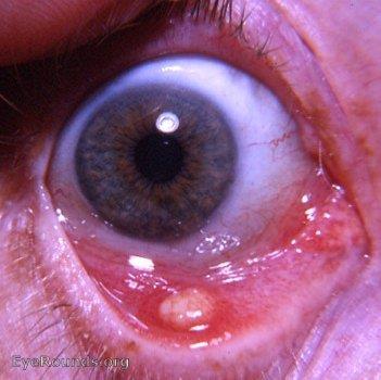
9 Hordeolum vs. Chalazion Hordeolum is acute with active infection Associated with soreness and redness Chalazion is chronic granulomatous inflammation TREAT: hordeolum with WARM compress and oral minocycline NOT topical antibiotic TREAT: chalazion with surgery, WARM compress, and oral minocycline NOT topical antibiotic
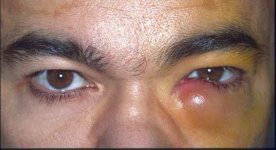
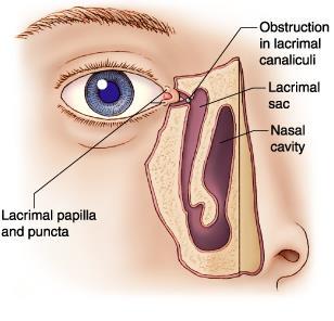
10 Dacryocystitis
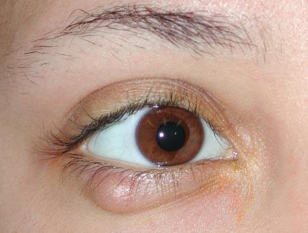
11 Dacryocystitis When this is an isolated case, oral cephalosporin or Augmentin work well. This should NOT be the first treatment for infants up to one year of age For infants: do medial massage, warm compress, erythromycin ointment. Surgery is an option at the one year old point. In adults, recurrence will often require surgery with an orbital specialist
12 Cellulitis


13 Cellulitis: Main Types Preseptal Cellulitis Orbital Cellulitis
14 Cellulitis: Main Types Preseptal Cellulitis Orbital Cellulitis
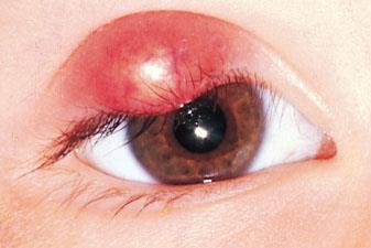
15 Cellulitis: Main Types Preseptal Cellulitis Eyelid chemosis and redness, discharge Usually secondary to an enlarged internal hordeolum No change in vision, no fever Treated with oral antibiotics (usually cephalosporins) Orbital Cellulitis Pain on eye movement, double vision, conjunctival chemosis, eyelid chemosis and redness, fever, proptosis Usually secondary to a sinus infection OCULAR EMERGENCY! Death can occur in 1-2% of cases if left untreated MUST ORDER A CT SCAN STAT Treated with IV antibiotics (usually vancomycin)


16 Cellulitis: Main Types Preseptal Cellulitis Orbital Cellulitis

17


18 Blepharitis Staph blepharitis Caused by the Staph aureus bacteria Flakes at the base of the lashes Demodex blepharitis Caused by the Demodex mite Sleeves at the base of the lashes Meibomian gland dysfunction Stopped up oil glands
19 Blepharitis Treatment Staph blepharitis Lid scrubs with baby shampoo Demodex blepharitis Lid scrubs with baby shampoo Tea tree oil Meibomian gland dysfunction Warm compresses

20 Herpes Zoster Ophthalmicus Caused by the herpes zoster virus (shingles) Respects the midline Usually found in patients older than 50 If it manifests in the upper 1/3 of the face, it can manifest in the eye Treated with antivirals

21 Subconjunctival Hemorrhage Hemorrhage in blood vessels under conjunctiva, can spread over eye and appear to worsen Usually not painful, does not affect vision, may be irritating Trauma, blood thinners, Valsalva maneuver No treatment, will typically resolve in 2-3 weeks Can try cold compresses initially than switch to warm Remote possibility of leukemia, blood disorder

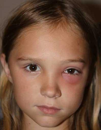
22 Allergic Red Eye Allergy to some agent Seasonal allergies Giant papillary conjunctivitis Vernal conjunctivitis These are all fancy words but what is the give away Treatment topical antihistamine/mast cell stabilizer, oral anti histamine, cold compresses


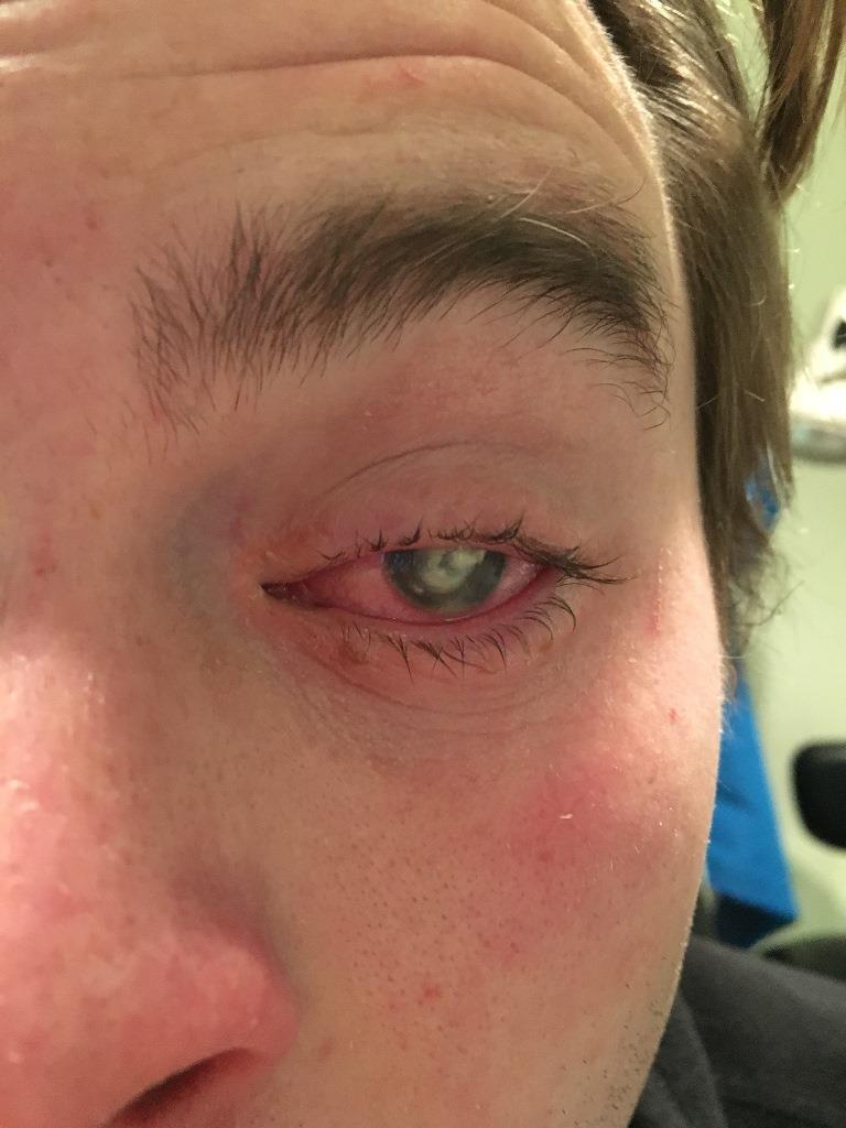
23 Viral Red Eye History of recent URI or contact with someone with a red eye and/or URI Feel for pre-auricular nodes (PAN) or submandibular nodes Eye is watery, very very very red, blurry, might be a little itchy, foreign body sensation, burning Usually starts in one eye and a few days later the other eye becomes involved Supportive treatment

24 Bacterial Red eye Not common in the healthy adult More common in children and the immunocompromised elderly ONE WAY TO tell if it s bacterial? Culture if you must

25 Contact Lens Related Red Eye Soft contact lens or rigid gas permeable lens (RGP)? Dirty little things get under soft lenses, so always always think of fluoroquinolones This patient MUST be seen by Optometry Let s talk Pseudomonas Contact lens abuse and misuse Patient should stop contact lens use until resolved

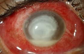
26 Corneal Ulcer Ulcers should always be seen by an eye care provider Generally have true photophobia and lots of inflammation If concomitant contact lens wear, which is usual, see a specialist ASAP Reminder: discontinue CL wear Start with ofloxacin q2h
27

28 Dry Eye Why the oxymoron? Menopause? Treatment ladder Contact lenses
29 Corneal Abrasions Be mindful of a history that might indicate a possibility of penetrating injury (flying arrows, bullets, BB s, grinding materials etc.) There MUST be something in the history that accounts for the abrasion, or it is not just an abrasion If the offending object was dirty, like a dog s toenail, use ofloxacin, possibly with acular or voltaren for pain control If caused by something clean, like cotton swab, can consider maxitrol steroid and antibiotic combo Be careful about Rxing steroids long term


30 Corneal Abrasions It is often important to look for something stuck to the underside of the upper lid If there is no precipitating history, it is likely an erosion
 can be tried as preventative There is an inflammatory component so should see eye care for anti-inflammatory management")
31 Recurrent Corneal Erosion History of trauma Hallmark is Lots of tearing, pain, foreign body sensation Treatment artificial tears, hypertonic solution such as 5% NaCl solution or ointment to dehydrate the cornea to reduce edma and prevent FreshKote (OTC) can be tried as preventative There is an inflammatory component so should see eye care for anti-inflammatory management


32 Episcleritis/Scleritis Engorgement of vessels Mild to severe pain Sectorial to diffuse redness Ocular manifestations of systemic conditions Treated with oral NSAIDs


33 Iritis Almost always truly photophobic MUST see cells to make the diagnosis MUST be treated with topical prednisolone Severity determines the dosage Usually treated with topical cycloplegic for pain relief Consult same day

34 Uveitis Uveitis involves more than just the front of the eye Uvea: iris, ciliary body, and choroid When granulomatous, it is critical to look for systemic association Often idiopathic and unilateral Look for systemic associations if recurrent or bilateral

35

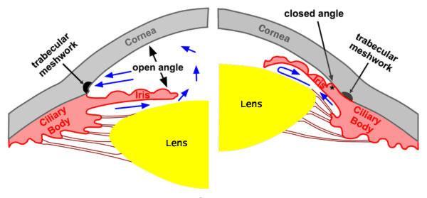
36 Acute Angle Closure Characterized by nausea, red eye on the affected side, blurred vision A TRUE EYE EMERGENCY Check intraocular pressures! Not common Treat with oral acetazolamide, lots of topical antihypertensive drops Consult an eye specialist right away
37 Acute Angle Closure Can present with steamy or cloudy cornea and thus blurred vision Pain, usually and very deep ache around and in the eye. They do not usually experience a pressure sensation Nausea is so predominant for most that it is often misdiagnosed as flu or gastric distress; look for the unilateral red eye Symptoms can be quite minimal; tonometry, slit lamp examination, and gonioscopy essential to diagnosis
38
39 Red flags 39 Sudden onset (within 24h) painless, monocular vision loss Especially in the setting of a known hypertensive and/or diabetic Sudden eye pain while hammering metal Trauma Pain while wearing soft contacts Flashing lights in one eye Curtain or veil obstructing vision in one eye True photophobia New onset diplopia See an eye doctor within 24 hours

40
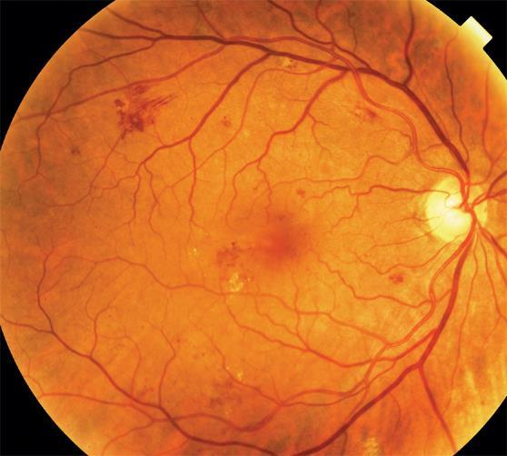
41 Diabetic Retinopathy 41 A disease of the capillaries Most common cause of vision loss in the population <65yo Two main components Retinopathy Non-proliferative Proliferative Findings: hemorrhages, cotton wool spots, growth of new lacy blood vessels Macular edema Clinically significant Not clinically significant Findings: Blunting of the macular reflex, hard exudates, decreased vision Risk factors for development Time since onset Control of blood sugar
42 Diabetic Retinopathy

43 Diabetic Retinopathy 43 Detection and diagnosis Retinal photo screenings Dilated eye exams Symptoms Nothing New floaters Blurred vision Treatment Monitoring Blood glucose control Retinal photocoagulation Anti-VEGF injections Steroid injections Differential: hypertensive retinopathy

44 44 Vitreous Hemorrhage Causes Most commonly: proliferative diabetic retinopathy Posterior vitreous detachment Retinal tear or detachment Wet macular degeneration Symptoms No symptoms Blurred vision New floaters Treatment Monitoring Vitrectomy
 nicking Cotton wool spots, flame hemorrhages, hard exudates Papilledema Risk factors: control of")
45 Hypertensive Retinopathy 45 A disease of the retinal arteries Narrowing of arteriolar lumen compresses the veins Arteriolosclerotic retinopathy vs hypertensive retinopathy Findings Change in arteriolar light reflex Arteriolar narrowing arteriolar/venous (A/V) nicking Cotton wool spots, flame hemorrhages, hard exudates Papilledema Risk factors: control of hypertension

46 Hypertensive Retinopathy 46 Detection and diagnosis: routine dilated eye exams Symptoms Nothing Blurred vision Painful headaches Treatment Blood pressure control Differential: diabetic retinopathy Complications Retinal vein occlusions

47 Retinal vein occlusions Branch retinal vs central retinal Symptoms Nothing Blurred vision Etiology: arteriolar compression A/V nick Blood and thunder Multiple flame-shaped hemorrhages Macular edema Treatment Anti-VEGF Intravitreal steroids Retinal photocoagulation 47 Underlying cause

48 Branch retinal artery occlusion Branch retinal artery occlusion vs central retinal artery occlusion Symptoms Transient monocular vision loss Loss of visual field in one eye Etiology: thrombus, embolus, infection Findings Source, if you re lucky Retinal infarct Treatment: underlying cause Prevent stroke 48


49 Macular Degeneration Leading cause of blindness in the US for people over 65 Drusen accumulate can cause atrophy, tissue loss Neovascularization from the underlying choroid can break through retina and cause bleeding Wet AMD

50 Macular Degeneration Treatment AREDS 2 vitamins for intermediate severe stage, in hopes of slowing progression of geographic atrophy of neovascularization For WET AMD, anti veg-f intra-vitreal injections, stop bleeding and prevent angiogenesis Need close monitoring Amsler Grid for selfmonitoring

 Macula On RD vs.")
51 Flashes and Floaters Differentials: Posterior vitreous detachment, retinal detachment, retinal tear or break Should have a dilated eye exam within 24 hours More ominous if complains of peripheral vision distortion ( curtain/veil ) Macula On RD vs. Macula Off RD

52 52
53 Case from Patient c/o new onset diplopia Also noted unilateral headache Not queried about claudication Treated with prism (common treatment for double vision) Noted to have severe vision loss 2 weeks later Diagnosed with GCA by ESR and CRP No light perception OD No recovery possible due to total optic atrophy 53

54 Giant cell arteritis Inflammation of the medium and large arterioles in the body Most commonly affects the temporal artery AKA temporal arteritis In the eye, causes any number of symptoms due to inflammation of the ophthalmic artery Transient monocular vision loss due to arteritic anterior ischemic optic neuropathy Diplopia due to loss of blood flow to the nerves 54

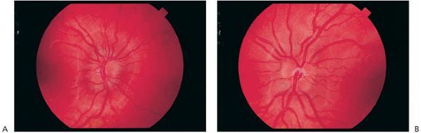
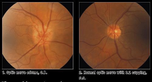
55 Giant cell arteritis Why is it an emergency? Save the other eye Irreversible vision loss in the affected eye If vision loss, death is imminent Typically found in 50+ yo Caucasian males Symptoms and signs Significantly decreased visual acuity + relative afferent pupillary defect Decreased color vision Optic nerve edema General malaise Headaches Pain on jaw claudication Pain along the ipsilateral temple 55
56 Giant cell arteritis Differential diagnosis Swollen nerve and/or decrease in color vision nonarteritic ischemic optic neuropathy Diplopia diabetes, hypertension, trauma Diagnosis Symptoms alone ESR, CRP Gold standard: temporal artery biopsy Treatment 56 Skip lesions Long course of oral steroids
57 Pseudo-tumor Cerebri Characterized by headache, not necessarily intense but often unrelenting Diplopia Transient visual obscuration nausea Optic nerve edema (r/o pseudo-papilledema) more than usually young, usually heavy, not necessarily morbidly so Can be caused by tetracyclines
58
59 Pseudo-papilledema or Idiopathic Intracranial Hypertension Perform neuroimaging Eye care consultation, visual field Traditionally perform lumbar puncture, measure opening pressure, less favored with imaging studies available Treat with Diamox / oral diuretics Sensitive presentation of weight loss strategy Consider offending medications and remove them
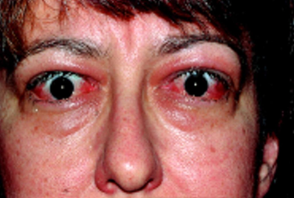
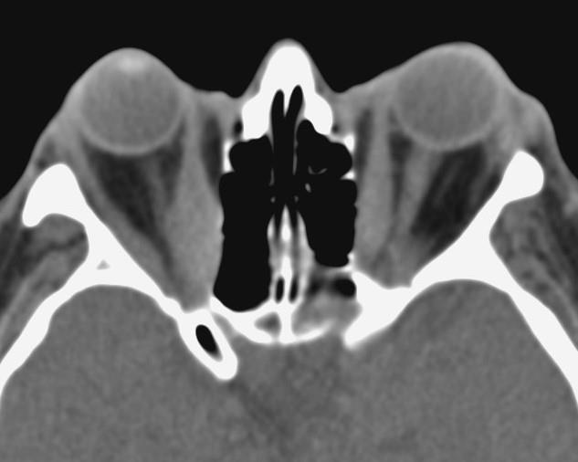
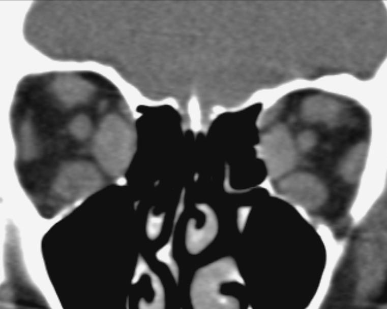
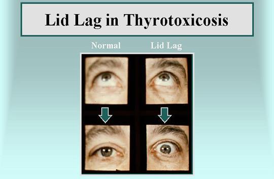
60 Thyroid Eye Disease Usually associated with hyperthyroidism It is an autoimmune disorder with a highly variable course of development, so potential treatments are hard to assess Characterized by proptosis and subsequent exposure complications Lid lag and lid retraction are often readily apparent Imaging will reveal thickening of the extraocular muscles and orbital contents Can result in diplopia Can be sight threatening
61
62
63
64
65 Thyroid Ophthalmolpathy The orbital contents can become so thickened that the optic nerve can be compressed, and vision can be lost. Decompression can necessary to prevent further vision loss Exposure keratitis is a common problem due to proptosis; this can be relieved by canthotomy, cutting the ligament at the outer corner of the eye Lots of lubricants are needed

66
Phone Triage for Optometric Staff ???????? CHEMICAL BURN CHEMICAL BURN
 Phone Triage for Optometric Staff There are very few ocular emergencies that you will have to deal with in practice, but it is imperative that you be able to Michelle Welch, O.D. NSU Oklahoma College of
Acute Eyes for ED. Enis Kocak. The Alfred Ophthalmology
 Acute Eyes for ED Enis Kocak The Alfred Ophthalmology The problem with eyes Things to cover Ocular anatomy Basic assessment Common presentations Eye first aid and procedures Ophthalmic emergencies What
EYE TRAUMA: INCIDENCE
 Introduction EYE TRAUMA: INCIDENCE 2.5 million eye injuries per year in U.S. 40,000 60,000 of eye injuries lead to visual loss Introduction Final visual outcome of many ocular emergencies depends on prompt,
Ocular Urgencies and Emergencies
 Ocular Urgencies and Emergencies Pam Boyce, O.D., F.A.A.O. Boyce Family Eye Care, Ltd. 528 Devon Ave. Park Ridge, IL 60068 847-518-0303 Somebody s going to lose an eye Epidemiology 2.4 million ocular and
OPHTHALMOLOGY REFERRAL GUIDE FOR GPS
 OPHTHALMOLOGY REFERRAL GUIDE FOR GPS A guidebook to support general practitioners in the management and referral of a range of common eye problems. Contents 3 Introduction 4 Ophthalmic Workup 6 Acute Visual
10/4/2013. Bruce K.Williams, MSN, RN,ACNP-BC Sisters of Charity Providence Hospitals. What is the worst thing that can go wrong with an eye?
 Red Eyes, Red Alert! Bruce K.Williams, MSN, RN,ACNP-BC Sisters of Charity Providence Hospitals Red Eyes, Red Alert! Red Eyes, Red Alert! What is the worst thing that can go wrong with an eye? 1 Red Eyes,
Mild NPDR. Moderate NPDR. Severe NPDR
 Diabetic retinopathy Diabetic retinopathy is the most common cause of blindness in adults aged 35-65 years-old. Hyperglycaemia is thought to cause increased retinal blood flow and abnormal metabolism in
5/2/2016 EYE EMERGENCIES. Nathaniel Pelsor, O.D., FAAO Talley Medical-Surgical Eye Care Associates. Anatomy. Tools
 EYE EMERGENCIES Nathaniel Pelsor, O.D., FAAO Talley Medical-Surgical Eye Care Associates Anatomy Tools 1 Contact dermatitis Blepharitis HSV Preseptal Cellulitis Anterior Chamber Subconjunctival hemorrhage
PAINFUL PAINLESS Contact lens user BOV
 Common Causes Allergies Infections Ocular Cornea, uveitis, endophthalmitis Orbital Orbital cellulitis Inflammation Uveitis Scleritis / episcleritis Glaucomas Trauma Foreign bodies Chemical injuries History
10 EYE EMERGENCIES. Who goes, who you better not send! Brant Slomovic, MD, FRCPC University Health Network
 10 EYE EMERGENCIES Who goes, who you better not send! Brant Slomovic, MD, FRCPC University Health Network DISCLOSURES I have none PVD CASE 1 WHAT IS A PVD? a process of aging (45-55) liquefaction of vitreous
Sepideh Tara Rousta, MD FAAO Robert Wood Johnson University Hospital Saint Peter s University Hospital Wills Eye Hospital
 Sepideh Tara Rousta, MD FAAO Robert Wood Johnson University Hospital Saint Peter s University Hospital Wills Eye Hospital 14 mo old w R eye cross (parents) 9 mo old R eye crossing getting worse for past
9/23/2014. Emily Thomas, O.D. MOA Paraoptometric Education October 5, 2014
 Emily Thomas, O.D. MOA Paraoptometric Education October 5, 2014 Anterior toward the front of the body Posterior toward the rear of the body Unilateral only one eye involved Bilateral both eyes involved
OPHTHALMOLOGIC PEARLS FOR THE NON- OPHTHALMOLOGIST. David G. Gross D.O. Deen-Gross Eye Centers Merrillville-Hobart Deengrosseye.
 OPHTHALMOLOGIC PEARLS FOR THE NON- OPHTHALMOLOGIST David G. Gross D.O. Deen-Gross Eye Centers Merrillville-Hobart Deengrosseye.com A FEW OF THE AREAS WE WILL DISCUSS Red Eye Glaucoma Neuro ophthalmic tid
Work Sheet And Course Hand Out
 Work Sheet And Course Hand Out This course provides the primary care health professional with a basic understanding of the eye, its function and the assessment of common sight- and non-sight threatening
Dr Jo-Anne Pon. Dr Sean Every. 8:30-9:25 WS #70: Eye Essentials for GPs 9:35-10:30 WS #80: Eye Essentials for GPs (Repeated)
") Dr Sean Every Ophthalmologist Southern Eye Specialists Christchurch Dr Jo-Anne Pon Ophthalmologist Southern Eye Specialists, Christchurch Hospital, Christchurch 8:30-9:25 WS #70: Eye Essentials for GPs
Dr Sean Every Ophthalmologist Southern Eye Specialists Christchurch Dr Jo-Anne Pon Ophthalmologist Southern Eye Specialists, Christchurch Hospital, Christchurch 8:30-9:25 WS #70: Eye Essentials for GPs
Scrub In. What is the function of vitreous humor? What does the pupil do when exposed to bright light? a. Maintain eye shape and provide color vision
 Scrub In What is the function of vitreous humor? a. Maintain eye shape and provide color vision b. Maintain eye shape and refract light rays c. Provide night vision and color vision d. Provide night vision
EYE INJURIES OBJECTIVES COMMON EYE EMERGENCIES 7/19/2017 IMPROVE ASSESSMENT OF EYE INJURIES
 EYE INJURIES BRITTA ANDERSON D.O. DMC PRIMARY CARE SPORTS MEDICINE ASSOCIATE TEAM PHYSICIAN DETROIT TIGERS OBJECTIVES IMPROVE ASSESSMENT OF EYE INJURIES UNDERSTAND WHAT IS CONSIDERED AN EMERGENCY DEVELOP
The Emergent Eye in the Acute Setting
 The Emergent Eye in the Acute Setting Todd P. Margolis MD, PhD Professor of Ophthalmology & Director of the F.I. Proctor Foundation UCSF Physical Exam-- Visual Acuity Essential Corrected visual acuity
OCCLUSIVE VASCULAR DISORDERS OF THE RETINA
 OCCLUSIVE VASCULAR DISORDERS OF THE RETINA Learning outcomes By the end of this lecture the students would be able to Classify occlusive vascular disorders (OVD) of the retina. Correlate the clinical features
Identify the choice that best completes the statement or answers the question.
 Chapter 5. The Eye Multiple Choice Identify the choice that best completes the statement or answers the question. 1. The most common type of eye disorder is: A. Refractive errors B. Macular conditions
Faculty Financial Disclosure. Learning Objectives: Office Ophthalmology. Basic Eye Exam: What s in your pocket/office? Office Ophthalmology
 Faculty Financial Disclosure Office Ophthalmology Lynn K. Gordon, MD, PhD, has no financial relationships to disclose. Lynn K. Gordon, MD, PhD Professor and Vernon O Underwood Family Chair Department of
Neuro-Ocular Grand Rounds Anthony B. Litwak,OD, FAAO VA Medical Center Baltimore, Maryland
 Neuro-Ocular Grand Rounds Anthony B. Litwak,OD, FAAO VA Medical Center Baltimore, Maryland Dr. Litwak is on the speaker and advisory boards for Alcon and Zeiss Meditek COMMON OPTIC NEUROPATHIES THAT CAN
Patient Symptoms- What They Might Mean. Sarah Dougherty Wood, OD, MS, FAAO Heart of America, February 2011 Paraoptometric Lecture
 Patient Symptoms- What They Might Mean Sarah Dougherty Wood, OD, MS, FAAO Heart of America, February 2011 Paraoptometric Lecture Basic ocular anatomy and physiology Movie projector analogy Blur at near/eye
Ocular Lecture. Sue Bednar NP Ali Atwater PA-C
 Ocular Lecture Sue Bednar NP Ali Atwater PA-C Triaging Ocular Complaints Painful Eye/Red eye +/-blurry vision +/-visual loss +/-floaters +/-fevers If any of the above findings exist, pt is likely to have
Ophthalmology. Juliette Stenz, MD
 Ophthalmology Juliette Stenz, MD Required Slide Disclosures NO SIGNIFICANT FINANCIAL, GENERAL, OR OBLIGATION INTERESTS TO REPORT Required Slide At the end of this session, students will be able to: 1.
OPHTHALMOLOGY DEPARTMENT Primary care referral guidelines
 OPHTHALMOLOGY DEPARTMENT Primary care referral guidelines Contents REFERRAL CATEGIES... 2 Emergency... 2 Urgent... 2 Semi urgent/routine... 2 Not accepted... 2 OPHTHALMOLOGY CONDITIONS NOT ACCEPTED...
Neuro-Ocular Grand Rounds
 Neuro-Ocular Grand Rounds Anthony B. Litwak,OD, FAAO VA Medical Center Baltimore, Maryland Dr. Litwak is on the speaker and advisory boards for Alcon and Zeiss Meditek COMMON OPTIC NEUROPATHIES THAT CAN
Red Eyes, Red Spots, and Red Flags
 Red Eyes, Red Spots, and Red Flags Essential Knowledge of Eye Disease Andrew F. Calman, MD, PhD Associate Clinical Professor of Ophthalmology and Family & Community Medicine, UCSF Seeing Red Red Eyes Common
Professor Helen Danesh-Meyer. Eye Institute Auckland
 Professor Helen Danesh-Meyer Eye Institute Auckland Bitten by Ophthalmology Emergencies Helen Danesh-Meyer, MBChB, MD, FRANZCO Sir William and Lady Stevenson Professor of Ophthalmology Head of Glaucoma
LECTURE # 7 EYECARE REVIEW: PART III
 LECTURE # 7 EYECARE REVIEW: PART III HOW TO TRIAGE EYE EMERGENCIES STEVE BUTZON, O.D. EYECARE REVIEW: HOW TO TRIAGE EYE EMERGENCIES FOR PRIMARY CARE PHYSICIANS Steve Butzon, O.D. Member Director IDOC President
 measure of your overall performance. An isolated glucose test is helpful to let you know what your sugar level is at one moment, but it doesn t tell you whether or not your diabetes is under adequate control
Age-Related Macular Degeneration (AMD)
") Age-Related Macular Degeneration (AMD) What is the Macula? What is Dry AMD (Age-related Macular Degeneration)? Dry AMD is an aging process that causes accumulation of waste product under the macula leading
Age-Related Macular Degeneration (AMD) What is the Macula? What is Dry AMD (Age-related Macular Degeneration)? Dry AMD is an aging process that causes accumulation of waste product under the macula leading
Andrew J. Hendershot, MD Havener Eye Institute The Ohio State University s Wexner Medical Center
 Ocular Trauma for the Primary Care Physician Andrew J. Hendershot, MD Havener Eye Institute The Ohio State University s Wexner Medical Center Relevance Often those with minor eye injuries will first seek
Focusing on A&E. By Sandy Cooper, (Ophthalmic Nurse Practitioner), Tel
, Tel") Focusing on A&E By Sandy Cooper, (Ophthalmic Nurse Practitioner), Tel 01752 439331 Email sandra.cooper5@nhs.net sandracooper041@btinternet.com THINGS TO WORRY ABOUT WITH ANY EYE PROBLEM CHANGES IN VISION
Focusing on A&E By Sandy Cooper, (Ophthalmic Nurse Practitioner), Tel 01752 439331 Email sandra.cooper5@nhs.net sandracooper041@btinternet.com THINGS TO WORRY ABOUT WITH ANY EYE PROBLEM CHANGES IN VISION
Ophthalmology for Primary Care: Do I Really Need to See It? Jennifer R. Olbum, DO
 Ophthalmology for Primary Care Jennifer Olbum, DO CCOM 1988 (Midwestern U.) Medical Retina subspecialist jenolbum@aol.com Daniel J. Nadler, MD LLC Beaver, PA Everett & Hurite Ophthalmic Assoc. Belle Vernon,
How to Handle the Urgent Need Patient: Telephone Triage/Preparation
 How to Handle the Urgent Need Patient: Telephone Triage/Preparation Pamela A. Lowe, O.D., F.A.A.O. Diplomate, American Board of Optometry Professional Eye Care Center, Inc. Chicago/Niles, Illinois Disclosures
THE 35 GOLDEN EYE RULES
 THE 35 GOLDEN EYE RULES The Sense of Sight, from La Dame a la Licorne, The Lady and the Unicorn Tapestries, Late 15th Century Flemish Tapestry in wool and silk, Musée Nationale du Moyen Age, Paris. 1.
Ophthalmic Trauma Update
 Ophthalmic Trauma Update Richard S. Davidson, M.D. Professor of Ophthalmology Vice Chair for Quality and Clinical Affairs UCHealth Eye Center University of Colorado School of Medicine August 5, 2017 Financial
UC SF. g h. Eye Trauma. Martha Neighbor, MD Emergency Services San Francisco General Hospital University of California
 UC SF Eye Trauma sf g h Martha Neighbor, MD Emergency Services San Francisco General Hospital University of California Goals Recognize vision threatening eye emergencies Treat them when we can Know when
DISCLOSURES. PEDIATRIC RED EYES Rachel M. Smith, OD, FCOVD HISTORY, HISTORY, HISTORY WHY RED EYES? EXAMINE THE EYE RED FLAGS TO REFER 3/25/2019
 DISCLOSURES Consultant/Speakers bureaus Research funding PEDIATRIC RED EYES Rachel M. Smith, OD, FCOVD Pediatric Optometrist Children s Hospital & Medical Center Stock ownership/corporate boards employment
FRANZCO, MD, MBBS. Royal Darwin Hospital
 Diabetes and Eye By Dr. Nishantha Wijesinghe FRANZCO, MD, MBBS Consultant Ophthalmologist Royal Darwin Hospital 98% of Diabetics do not need to suffer from severe visual loss Yet Diabetic eye disease is
2/5/2018. Trauma. Subdivided into two main categories: Closed globe Open Globe
 1 2 3 4 5 Ocular Trauma Guide for Eye Care Office Staff Winter Thaw 2018 Aaron Yatskevich OD Definition A broad term used to describe a physical or chemical wound to the eye or eye socket. Ocular trauma
X-Plain Diabetic Retinopathy Reference Summary
 X-Plain Diabetic Retinopathy Reference Summary Introduction Patients with diabetes are more likely to have eye problems that can lead to blindness. Diabetic retinopathy is a disease of the eye s retina
Diabetic Retinopathy
 Diabetic Retinopathy Introduction People with diabetes are more likely to have eye problems that can lead to blindness. Diabetic retinopathy is a disease of the eye s retina that is caused by diabetes.
OOGZIEKTEN VOOR DE HUISARTS F. GOES, JR.
 OOGZIEKTEN VOOR DE HUISARTS F. GOES, JR. HET RODE OOG F. GOES, JR. Condition Signs Symptoms Causes Conjunctivitis Viral Normal vision, normal pupil size Mild to no pain, diffuse Adenovirus (most common),
Test Bank for Medical Surgical Nursing An Integrated Approach 3rd Edition by White
 Test Bank for Medical Surgical Nursing An Integrated Approach 3rd Edition by White Link full download : http://testbankair.com/download/test-bank-for-medical-surgical-nursing-anintegrated-approach-3rd-edition-by-white/
CORNEAL CONDITIONS CORNEAL TRANSPLANTATION
 GENERAL INFORMATION CORNEAL CONDITIONS CORNEAL TRANSPLANTATION WHAT ARE CORNEAL CONDITIONS? The cornea is the clear outer layer of the eye. Shaped like a dome, it helps to protect the eye from foreign
A Case of Carotid-Cavernous Fistula
 A Case of Carotid-Cavernous Fistula By : Mohamed Elkhawaga 2 nd Year Resident of Ophthalmology Alexandria University A 19 year old male patient came to our outpatient clinic, complaining of : -Severe conjunctival
REFERRAL GUIDELINES: OPHTHALMOLOGY
 Outpatient Referral Guidelines Page 1 1 REFERRAL GUIDELINES: OPHTHALMOLOGY Date of birth Demographic Contact details (including mobile phone) Clinical Reason for referral Duration of symptoms Essential
8/30/2018. Eye Disorders. Patrick Sarte. Anatomy of the Eye Uveitis Scleritis vs. Episcleritis Glaucoma Retinal Findings Eyelids
 Eye Disorders Patrick Sarte Anatomy of the Eye Uveitis Scleritis vs. Episcleritis Glaucoma Retinal Findings Eyelids 1 Anatomy of the Eye Anatomy of the Eye 2 Anatomy of the Eye 3 4 A 26 year old woman
NEPTUNE RED BANK BRICK
 NEPTUNE RED BANK BRICK Diabetes & The Eye Diabetics are more likely to develop Cataracts at a younger age. Diabetics are twice as likely to develop Glaucoma when compared to non-diabetics. The primary
Ocular Pathology. I. Congenital and/or developmental. A. Trisomy 21. Hypertelorism (widely spaced eyes) Keratoconus (cone shaped cornea)
 Keratoconus (cone shaped cornea)") I. Congenital and/or developmental Robbins Pathologic Basis of Disease, 6 th Ed. A. Trisomy 21 Hypertelorism (widely spaced eyes) Keratoconus (cone shaped cornea) Focal hypoplasia of iris Cataracts frequently
I. Congenital and/or developmental Robbins Pathologic Basis of Disease, 6 th Ed. A. Trisomy 21 Hypertelorism (widely spaced eyes) Keratoconus (cone shaped cornea) Focal hypoplasia of iris Cataracts frequently
Rafik Girgis. Consultant Ophthalmic Surgeon ( Cataract & Primary Care)
") Rafik Girgis Consultant Ophthalmic Surgeon ( Cataract & Primary Care) Blepharitis Is a very common condition which usually bilateral & symmetrical. The main types are: Anterior, posterior or mixed Complications:
Rafik Girgis Consultant Ophthalmic Surgeon ( Cataract & Primary Care) Blepharitis Is a very common condition which usually bilateral & symmetrical. The main types are: Anterior, posterior or mixed Complications:
Telephone Triage Urgency or Emergency? Mary E. Schmidt, ABOC, CPO
 Telephone Triage Urgency or Emergency? www.eyesystems.info Mary E. Schmidt, ABOC, CPO mary@eyesystems.info Definition of Triage The sorting of patient and allocation of care or treatment according to the
A Curious Case of Bilateral Optic Disc Edema Brittney Dautremont, DO, MPH
 A Curious Case of Bilateral Optic Disc Edema Brittney Dautremont, DO, MPH PGY2 Ophthalmology Resident Grandview Medical Center Dayton, OH CASE PRESENTATION 51 year old white female presenting with blurred
Brampton Hurontario Street Brampton, ON L6Y 0P6
 Diabetic Retinopathy What is Diabetic Retinopathy Diabetic retinopathy is one of the leading causes of blindness world-wide. Diabetes damages blood vessels in many organs of the body including the eyes.
Differential diagnosis of the red eye. Carol Slight Nurse Practitioner Ophthalmology
 Differential diagnosis of the red eye Carol Slight Nurse Practitioner Ophthalmology The red eye Conjunctivitis HSV Keratitis Acute angle closure glaucoma Anterior Uveitis Red eye Scleritis Subconjunctival
Ophthalmology. Corneal Abrasion. History
 Ophthalmology Corneal Abrasion - Usually clear history of very recent trauma - Foreign Body Sensation - Pain +++ - Lacrimation - Photophobia Fig. 1 Corneal Abrasion - Abrasion stains yellow / green with
Examining Children s Eyes
 Paediatric Ophthalmology What to refer & when? Aims Tips for assessing a child s eyes in general practice Common paediatric ophthalmology symptoms and signs What needs to be referred and when? MISS FARIHA
Index. C Canalicular system, 4 Carbonic anhydrase inhibitors, 29 30
 A Acanthamoeba keratitis (AK), 82, 83 Acute angle-closure crisis, 156 Acute angle-closure glaucoma (AACG), 121, 141, 284 causes of, 122 clinical presentation, 153 evaluation, 156 157 management/treatment,
Bleeding in the anterior chamber, obstructing vision Caused by surgery, injury, coagulopathy, sickle cell or idiopathic Needs urgent care to prevent
 Bleeding in the anterior chamber, obstructing vision Caused by surgery, injury, coagulopathy, sickle cell or idiopathic Needs urgent care to prevent long-term vision loss TX by elevating head of bed, reducing
Diabetic Retinopathy Screening Program in the Cree Region of James Bay of Quebec
 RUIS McGILL VIRTUAL HEALTH AND SOCIAL SERVICES CENTRE (CvSSS) SIMPLIFYING TELEHEALTH! Diabetic Retinopathy Screening Program in the Cree Region of James Bay of Quebec Nurse and Imager Training Prepared
MRI masterfile Part 5 WM Heme Strokes.ppt 1
 Ocular and Orbital Trauma Eye Trauma: Incidence 1.3 million eye injuries in the US per year. 40,000 of these injuries lead to blindness in the US. Patrick Sibony, MD March 23, 2013 Ophthalmic Emergencies
Anatomy: There are 6 muscles that move your eye.
 Thyroid Eye Disease Your doctor thinks you have thyroid orbitopathy. This is an autoimmune condition where your body's immune system is producing factors that stimulate enlargement of the muscles that
Ophthalmology. Ophthalmology Services
 Ophthalmology Ophthalmology Services The Ophthalmology service offers the latest and most comprehensive eye care for patients. With a dedicated team of eye surgeons and consultants, we treat vision problems
For further reading we recommend the following excellent textbooks:
 FURTHER READING Intravitreal Injections Downloaded from www.worldscientific.com For further reading we recommend the following excellent textbooks: Clinical Anatomy of the Eye by Richard S Snell and Michael
Preview. Ophthalmology for Primary Care Providers. Useful references. How the eye works
 Preview Ophthalmology for Primary Care Providers Bob Avery, MD, PhD How the eye works The red eye Acute eye conditions Chronic vision loss Basic eye exam Ophthalmology/Surgery University of New Mexico
Ophthalmology for Primary Care Providers
 Ophthalmology for Primary Care Providers Bob Avery, MD, PhD Ophthalmology/Surgery University of New Mexico School of Medicine bavery@salud.unm.edu Preview How the eye works Basic eye exam The red eye Acute
Uveitis. Pt Info Brochure. Q: What is Uvea?
 Pt Info Brochure Uveitis Q: What is Uvea? A: Uvea is the middle layer of the eye. It is the most vascular structure of the eye. It provides nutrition to the other parts of the eye. The uvea is made up
A Patient s Guide to Diabetic Retinopathy
 Diabetic Retinopathy A Patient s Guide to Diabetic Retinopathy 840 Walnut Street, Philadelphia PA 19107 www.willseye.org Diabetic Retinopathy 1. Definition Diabetic retinopathy is a complication of diabetes
Year 2 MBChB Clinical Skills Session Ophthalmoscopy. Reviewed & ratified by: Mr M Batterbury Consultant Ophthalmologist
 Year 2 MBChB Clinical Skills Session Ophthalmoscopy Reviewed & ratified by: o Mr M Batterbury Consultant Ophthalmologist Learning objectives o To understand the anatomy and physiology of the external and
Mom, There s Something Wrong With My Eye
 Mom, There s Something Wrong With My Eye Veeral Shah MD, PHD Texas Children's Hospital Most Common Issues Seen by the Pediatrician Emergent Ocular Issues Seen by the Pediatrician 1 What does this baby
American Board of Optometry Board Certification Examination DETAILED OUTLINE
 American Board of Optometry Board Certification Examination DETAILED OUTLINE General Practice (160 items) The core of the examination is based in the following ten areas of general practice. 1. Ametropia/Ophthalmic
Intravitreal Injection
 for patients Eye Clinic Ipswich Hospital Tel: 01473 703230 Intravitreal Injection What is an intravitreal injection? An intravitreal injection is the injection of a drug into the vitreous body (the jelly
Ophthalmology PANRE Review. Brock Phillips, PA-C
 Ophthalmology PANRE Review Brock Phillips, PA-C I am not an ophthalmologist, optometrist or certified eye guy of any sort - I am a practicing UC/EM PA-C who frequently evaluates eye/vision complaints,
NORTHEAST OHIO NEIGHBORHOOD HEALTH SERVICES, INC. OPTOMETRIC MEDICINE CLINICAL GUIDELINES: TABLE OF CONTENTS
 NORTHEAST OHIO NEIGHBORHOOD HEALTH SERVICES, INC. OPTOMETRIC MEDICINE CLINICAL GUIDELINES: 2012-2013 TABLE OF CONTENTS CONDITION PAGE(S) Complete Eye and Vision Examination 2 Vision Screening Procedure
Assessment and Management of Ocular Trauma. Disclosure I have no direct financial interests in today s subject matter. 3/25/2019. Normal Eye Anatomy
 Assessment and Management of Ocular Trauma Samiksha Fouzdar Jain, MD,FRCS Department of Ophthalmology & Visual Sciences Truhlsen Eye Institute Disclosure I have no direct financial interests in today s
Around The Globe in 60 Minutes
 Around The Globe in 60 Minutes Around the GLOBE in Sixty Minutes Basic Ocular Anatomy, Examination, and Diagnostic Techniques Introduction Focusing on canine and feline ocular anatomy and basic examination
What THE EYE Case THE RED EYE. Case. Infections of the eye 2/3/2014
 Case THE RED EYE Richard A. Jacobs, M.D.,PhD* *Todd Margolis, M.D.,PhD, Prof of Ophthalmology and Director F. I. Proctor Foundation, UCSF Brian Schwartz, M.D., Assistant Professor of Medicine, Division
VN 122 MODULE F EYE AND VISION DISORDERS OCULAR HISTORY 1. PATIENT PERCEPTION OF PROBLEM 2. DECREASED VISION? 3. BLURRED, DOUBLE, DISTORTED?
 VN 122 MODULE F EYE AND VISION DISORDERS OCULAR HISTORY 1. PATIENT PERCEPTION OF PROBLEM 2. DECREASED VISION? 3. BLURRED, DOUBLE, DISTORTED? 4. PAIN? QUALITY OF PAIN? 5. ITCHING, DRY? 6. BOTH EYES? 7.
Aristotle University Thessaloniki Medical School I. & II. Departments of Ophthalmology 90 DIAGNOSTIC & THERAPEUTIC APPROACHES IN OPHTHALMOLOGY
 Aristotle University Thessaloniki Medical School I. & II. Departments of Ophthalmology 90 DIAGNOSTIC & THERAPEUTIC APPROACHES IN OPHTHALMOLOGY The medical student should be able to... I. Pathophysiology
Case Follow Up. Sepi Jooniani PGY-1
 Case Follow Up Sepi Jooniani PGY-1 Triage 54 year old M Pt presents to prelim states noticed today he had reddness to eyes, states worse in R eye. Pt denies any pain or itching. No further complaints.
Department of Ophthalmology
 Department of Ophthalmology Period : 02/July/18 to 30/August/18 Semester : 7 th Semester Lecture Lesson Plan Sr. Date Topic Lesson plan Name of Faculty No. 1 02.07.18 Lens- Lens-Anatomy, Classification
Sudden Vision Loss. Brendan Girschek, MD, FRCSC, FACS Vitreoretinal Surgery Cedar Valley Medical Specialists
 Sudden Vision Loss Brendan Girschek, MD, FRCSC, FACS Vitreoretinal Surgery Cedar Valley Medical Specialists My Credentials -Residency in Ophthalmology at the LSU Eye Center in New Orleans, LA -Fellowship
Understanding Diabetic Retinopathy
 Understanding Diabetic Retinopathy What Is Diabetic Retinopathy? Diabetes damages blood vessels in the rear of the eye. This condition is called diabetic retinopathy. It can lead to vision loss or blindness.
NANOS Patient Brochure
 NANOS Patient Brochure Transient Visual Loss Copyright 2016. North American Neuro-Ophthalmology Society. All rights reserved. These brochures are produced and made available as is without warranty and
Red Eye & Ocular Emergencies. Zafar Shamoon Director of Emergency Services Dearborn Beaumont Hospital
 Red Eye & Ocular Emergencies Zafar Shamoon Director of Emergency Services Dearborn Beaumont Hospital I have no relevant financial relationship with any manufacturers of any commerical products and or providers
TOP 5 EYE CONDITIONS NOT TO BE MISSED
 TOP 5 EYE CONDITIONS NOT TO BE MISSED Dr Kolin Foo Consultant Ophthalmologist Senior Lecturer in Ophthalmology Wellington Hospital Terrace Eye Centre Financial disclosure No financial interest in the products
NO DISCLOSURES THE RED EYE. Case 1/20/2017
 THE RED EYE Richard A. Jacobs, M.D.,PhD* *Todd Margolis, M.D.,PhD, Prof of Ophthalmology and Director F. I. Proctor Foundation, UCSF (Now Chair of Ophthalmology at Washington University in St. Louis) Brian
The Human Eye. Cornea Iris. Pupil. Lens. Retina
 The Retina Thin layer of light-sensitive tissue at the back of the eye (the film of the camera). Light rays are focused on the retina then transmitted to the brain. The macula is the very small area in
TENTATIVE DIAGNOSES Based on the information provided so far, what are the potential diagnoses?
 Case Study #4 PEDIATRIC CASE STUDY SCENARIO Mary Jennings has brought her son Joe to your office. Joe is a 6-year old Jordanian male. He presents with the complaint of an itchy red eye. Mary states that
Differential Diagnosis of Conjunctivitis and Keratoconjunctivitis
 Differential Diagnosis of Conjunctivitis and Keratoconjunctivitis Dr. Victor Malinovsky 2006 Mechanical-Physical Trauma Corneal Abrasions Abrasions (interpalpebral/variable): a focal loss of epithelium
Objectives. Unexplained Vision Loss: Where Do I Go From Here. History. History. Drug Induced Vision Loss
 Objectives Unexplained Vision Loss: Where Do I Go From Here Denise Goodwin, OD, FAAO Coordinator, Neuro-ophthalmic Disease Clinic Pacific University College of Optometry goodwin@pacificu.edu Know the importance
For details on measurement and recording of visual acuity, refer to Annex 1. VISION INTERPRETING RESULTS ABSTRACT
 management update on functional decline in older adults 2012 Unit No. 5 VISION Dr Au Eong Kah Guan, Ms Yulianti, Ms Fifiana ABSTRACT Among Singaporean adults of Chinese origin aged 40 to 79 years old,
Handbook for Medical Students Learning Ophthalmology
 International Council of Ophthalmology Handbook for Medical Students Learning Ophthalmology 2009 Compiled by the Task Force on Undergraduate Teaching in Ophthalmology of the International Council of Ophthalmology
a.superficial (adenoid layer).contain lymphoid tissue.
.contain lymphoid tissue.") Conjunctiva Dr. saifalshamarti Anatomy Microscopic: 1.Epithelium (non keratinized,includes goblet cell). 2.Epithelial basement membrane. 3.Stroma : a.superficial (adenoid layer).contain lymphoid tissue.
Conjunctiva Dr. saifalshamarti Anatomy Microscopic: 1.Epithelium (non keratinized,includes goblet cell). 2.Epithelial basement membrane. 3.Stroma : a.superficial (adenoid layer).contain lymphoid tissue.
People with eye allergies typically have symptoms that include: Eye Anatomy: What Do Eye Allergies Actually Effect?
 Eye Allergies Eye Allergies People with eye allergies typically have symptoms that include: Itchy watery eyes Eyelid problems Dark circles around eyes Dry eyes Reactions to Contacts Let us help allow you
GENERAL INFORMATION DIABETIC EYE DISEASE
 GENERAL INFORMATION DIABETIC EYE DISEASE WHAT IS DIABETIC EYE DISEASE? Diabetic eye disease is a term used to describe the common eye complications seen in people with diabetes. It includes: Diabetic retinopathy
Dr. D. Y. Patil Medical College, Pimpri, Pune
 Dr. D. Y. Patil Medical College, Pimpri, Pune - 411 018 Period : 04/July/16 to 22/September/16 Semester : 7 th Semester Department : Ophthalmology Lecture Lesson Plan Sr No Date Topic Learning objectives