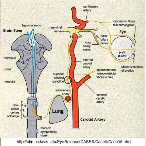
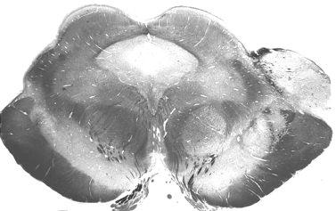
CN V! touch! pain! Touch! P/T!
|
|
|
- Rosanna Little
- 6 years ago
- Views:
Transcription
1 CN V! touch! pain! Touch! P/T!

2 Visual Pathways! L! R! B! A! C! D! LT! E! F! RT! G!

3 hypothalamospinal! and! ALS!
4 Vestibular Pathways! 1. Posture/Balance!!falling! 2. Head Position! 3. Eye-Head Movements nystagmus!!(vestibulo-ocular reflex)! 4. Autonomic Cnt!nausea/vomiting! 5. Perception!!vertigo! MLF! LVST! MVST!
5 Auditory Pathways! of thalamus!
6 FUNCTION! COMPONENT! DEFICITS! Start! Basal Ganglia! Spontaneous Movements! Move! UMN/LMN! Cerebral Cortex! Brainstem, Spinal cord! Roots/peripheral nerves! Plan! Cerebellum! Ataxia! Adjust! Cerebellum! Ataxia! Weak/Paralyzed! Balance/EyeMove! Cerebellum! Loss of balance! Nystagmus!
7 UMN/LMN!
8 Thalamus! Cerebral Cortex! Basal Ganglia! Function: ideation, planning, execution! Cerebellum! 3! head muscles! body muscles! Brainstem! 2! Spinal Cord! 1! Function: cause muscle contraction!
9 Paresis Flaccid, ipsilateral to injury! Decreased tendon reflexes! Decreased muscle tone! Severe muscle atrophy! Fasciculations twitching muscle fibers due to abnormal transmitter release at neuromuscular junction!
10 For Body:!! Corticospinal tract!! Cortico-Bulbo-spinal tracts! Thalamus! Cerebral Cortex! Basal Ganglia! Cerebellum! head muscles! Brainstem! Body muscles! Spinal Cord!
11
12 Lateral Corticospinal T controls limb motoneurons especially for fine movements of hands and feet.! Ventral Corticospinal T (10%) crosses later in spinal cord to influence axial motoneurons, but significance unknown.! Lateral CST! 90%!
13 Post limb of internal capsule! Crus cerebri of midbrain! Basilar pons! Pyramids of medulla! Lateral corticospinal tract!
14 Lateral Corticospinal T provides commands for limb motoneurons - especially for fine movements of hands and feet.! Bulbospinal tracts are controlled by motor cortical areas and cerebellum. They influence posture and muscle tone.!
15 Reticulospinal T! Vestibulospinal T! Rubrospinal T! Ex!t!en!s!or!moton!e!uron!s! o!f! upp!e!r/lo!we!r!limb!s! U!pp!e!r!l!imb!f!lexo!r! moton!e!uron!s!
16 1. Paresis/paralysis! 2. Increased muscle tone- Spasticity! 3. Increased tendon reflexes! 4. Babinski sign a reflex induced by stroking the plantar surface of the foot!

17 Controlled by 2 component system also! UMN- corticobulbar fibers! LMN- motor cranial nerves! Thalamus! Cerebral Cortex! Basal Ganglia! Cranial nerves! head muscles! Cerebellum! Brainstem! Corticobulbar T! Spinal Cord!

18 Cranial Nerve Motoneurons:! midbrain!! CN III, IV extrinsic eye muscles! pons! medulla!! CN V jaw muscles!! CN VI!! CN VII facial muscles!! CN IX, X = Nucleus Ambiguus!for larynx, pharynx!! CN XII tongue! midbrain! pons! medulla!! CN XI trapezius, sternocleidomastoid!
19 Cranial Nerve Lesions ipsilateral effects on muscles!! CN III, IV, VI!paralysis of extrinsic eye muscles!! CN V!jaw deviates to paralyzed side!! CN VII!paralysis of facial muscles!! Nuc Ambiguus!loss of gag reflex/swallowing, hoarseness,!!!uvula deviates to opposite side!! CN XII!tongue deviates to paralyzed side!! CN XI!weakness in shoulder shrug, head turn to!!!!!opposite side! Midline structures often deviate to one side when muscles are weak/paralyzed on one side.!

20 CST! Most corticobulbar fibers synapse on cranial nerve motor nuclei bilaterally.! V! VII! XII! Nuc ambiguus! travel together!
21 1. CN VII - lower facial muscles! 2. CN XII - tongue!
22 Lesions of corticobulbars on one side cause weakness or paralysis of the lower face on the opposite side.! For Lesion on RT! R! L! VII! lower face! working! not working!

23 Corticobulbar fibers supply Hypoglossal Nuc is stronger contralaterally.! RT corticobulbar lesion causes tongue to deviate to opposite side.! R! L! CN XII!
24 Basal Ganglia!
25 Thalamus! Cerebral Cortex! Basal Ganglia! Cerebellum! head muscles! Brainstem! body muscles! Spinal Cord!


26 1. Caudate! } 2. Putamen! Striatum! 3. Globus Pallidus! 4. Substantia Nigra! 5. Subthalamic Nucleus!

27 Output of Basal Ganglia! SN c! VL! dopamine! STN!

28 Physiological Basis of Function/Disease! Cortex motor areas! Putamen! SNc! D2! D1! SNc! GPe! STN! GPi! VL!
29 Symptoms of BG Diseases! Dyskinesias movement disorders!! hemiballismus!!!!! chorea!! dystonia/athetosis!! resting tremor! Hypokinesia / Bradykinesia! Rigidity lead pipe with cogwheeling! Changes in posture!
30 Disease! Lesion Site! Symptoms! Ballism! STN! Wild,flailing movements! Parkinson s! SNc! Resting tremor, movement, rigidity, altered gait/posture! Huntington s! Tardive dyskinesia! Caudate/Putamen! Chorea, dementia! Dopamine and other Receptors! Facial chorea/athetosis! Tourettes s! unknown! Motor and verbal tics! Wilson s Disease, CO poisoning!
31 Cerebellum!
!")
32 1. Controls 3 functions: plan, adjust, balance/eye mov! 2. The 3 functions = 3 regions of cerebellum! 3. How it works: regulates UMNs (cortex and brainstem)! 4. Damage to cerebellum/pathways ataxia (+ vestib symp)!

33 Vestibulocerebellum! nodulus and flocculus! Spinocerebellum! Vermis / Hemisphere! Cerebrocerebellum! Hemisphere!
34 vestibulocerebellum! Lesions - Vestibular symptoms!! - falling, wide-based gait,!!!!nystagmus, nausea, vertigo! nodulus/flocculus!


35 Midline symptoms! Limb symptoms!
36
37 Flocculus/Nodulus! Vestibular Signs! Vermis! Truncal Ataxia! Cerebellar Hemisphere! There are 2 categories of cerebellar symptoms! Limb Ataxia! 1. Vestibular Ataxic Gait! Signs! Falling! Ataxic Gait! Nystagmus!! Nausea,! vertigo!!!2. Ataxia! intention tremor, dysmetria, asynergia, etc! 4th ventricle tumors! Alcohol, MS, tumor! Tumor, infarct, MS!
38 1. Cerebellar symptoms are caused by damage to cerebellum, its peduncles or pathways.! 2. Cerebellar damage produces motor deficits on same side of body.! 3. Cerebellar signs fall into 2 categories:!!! Vestibular - falling/ataxic gait, nystagmus!!! Ataxia!!!! trunk - vermis!!!! limbs - hemisphere, peduncles, pathways!
39
40 Ataxia/Asynergia/! Decomposition of Movement! Dysmetria! Lack of Check! Inappropriate force! Dysdiadochokinesia! Intention/kinetic tremor! Hypotonia! limbs! Dysarthria!
Functional Distinctions
 Functional Distinctions FUNCTION COMPONENT DEFICITS Start Basal Ganglia Spontaneous Movements Move UMN/LMN Cerebral Cortex Brainstem, Spinal cord Roots/peripheral nerves Plan Cerebellum Ataxia Adjust Cerebellum
Upper and Lower Motoneurons for the Head Objectives
 Upper and Lower Motoneurons for the Head Objectives Know the locations of cranial nerve motor nuclei Describe the effects of motor cranial nerve lesions Describe how the corticobulbar tract innervates
Basal ganglia Sujata Sofat, class of 2009
 Basal ganglia Sujata Sofat, class of 2009 Basal ganglia Objectives Describe the function of the Basal Ganglia in movement Define the BG components and their locations Describe the motor loop of the BG
VL VA BASAL GANGLIA. FUNCTIONAl COMPONENTS. Function Component Deficits Start/initiation Basal Ganglia Spontan movements
 BASAL GANGLIA Chris Cohan, Ph.D. Dept. of Pathology/Anat Sci University at Buffalo I) Overview How do Basal Ganglia affect movement Basal ganglia enhance cortical motor activity and facilitate movement.
Spinal Cord: Clinical Applications. Dr. Stuart Inglis
 Spinal Cord: Clinical Applications Dr. Stuart Inglis Tabes dorsalis, also known as syphilitic myelopathy, is a slow degeneration (specifically, demyelination) of the nerves in the dorsal funiculus of the
FUNCTION: It COORDINATES movement HOW IT WORKS
 CEREBELLUM Chris Cohan, Ph.D. Dept. of Pathology/Anat Sci University at Buffalo Objectives: Describe the anatomy of the cerebellum, its 3 functions and associated regions Describe how the cerebellum influences
Motor System Hierarchy
 Motor Pathways Lectures Objectives Define the terms upper and lower motor neurons with examples. Describe the corticospinal (pyramidal) tract and the direct motor pathways from the cortex to the trunk
Unit VIII Problem 5 Physiology: Cerebellum
 Unit VIII Problem 5 Physiology: Cerebellum - The word cerebellum means: the small brain. Note that the cerebellum is not completely separated into 2 hemispheres (they are not clearly demarcated) the vermis
I: To describe the pyramidal and extrapyramidal tracts. II: To discuss the functions of the descending tracts.
 Descending Tracts I: To describe the pyramidal and extrapyramidal tracts. II: To discuss the functions of the descending tracts. III: To define the upper and the lower motor neurons. 1. The corticonuclear
Basal nuclei, cerebellum and movement
 Basal nuclei, cerebellum and movement MSTN121 - Neurophysiology Session 9 Department of Myotherapy Basal Nuclei (Ganglia) Basal Nuclei (Ganglia) Role: Predict the effects of various actions, then make
Biological Bases of Behavior. 8: Control of Movement
 Biological Bases of Behavior 8: Control of Movement m d Skeletal Muscle Movements of our body are accomplished by contraction of the skeletal muscles Flexion: contraction of a flexor muscle draws in a
The Nervous System: Sensory and Motor Tracts of the Spinal Cord
 15 The Nervous System: Sensory and Motor Tracts of the Spinal Cord PowerPoint Lecture Presentations prepared by Steven Bassett Southeast Community College Lincoln, Nebraska Introduction Millions of sensory
DISORDERS OF THE MOTOR SYSTEM. Jeanette J. Norden, Ph.D. Professor Emerita Vanderbilt University School of Medicine
 DISORDERS OF THE MOTOR SYSTEM Jeanette J. Norden, Ph.D. Professor Emerita Vanderbilt University School of Medicine THE MOTOR SYSTEM To understand disorders of the motor system, we need to review how a
Chapter 8. Control of movement
 Chapter 8 Control of movement 1st Type: Skeletal Muscle Skeletal Muscle: Ones that moves us Muscles contract, limb flex Flexion: a movement of a limb that tends to bend its joints, contraction of a flexor
Movement Disorders. Psychology 372 Physiological Psychology. Background. Myasthenia Gravis. Many Types
 Background Movement Disorders Psychology 372 Physiological Psychology Steven E. Meier, Ph.D. Listen to the audio lecture while viewing these slides Early Studies Found some patients with progressive weakness
Connection of the cerebellum
 CEREBELLUM Connection of the cerebellum The cerebellum has external layer of gray matter (cerebellar cortex ), & inner white matter In the white matter, there are 3 deep nuclei : (a) dentate nucleus laterally
Abdullah AlZibdeh. Dr. Maha ElBeltagy. Maha ElBeltagy
 19 Abdullah AlZibdeh Dr. Maha ElBeltagy Maha ElBeltagy Introduction In this sheet, we discuss the cerebellum; its lobes, fissures and deep nuclei. We also go into the tracts and connections in which the
Movement Disorders Will Garrett, M.D Assistant Professor of Neurology
 Movement Disorders Will Garrett, M.D Assistant Professor of Neurology I. The Basal Ganglia The basal ganglia are composed of several structures including the caudate and putamen (collectively called the
Motor tracts Both pyramidal tracts and extrapyramidal both starts from cortex: Area 4 Area 6 Area 312 Pyramidal: mainly from area 4 Extrapyramidal:
 Motor tracts Both pyramidal tracts and extrapyramidal both starts from cortex: Area 4 Area 6 Area 312 Pyramidal: mainly from area 4 Extrapyramidal: mainly from area 6 area 6 Premotorarea: uses external
Organization of Motor Functions 4.
 Organization of Motor Functions 4. Dr. Attila Nagy 2018 Sensory-motor system Limbic cortex Structure Subcortical Motivational sub areas Frontal cortex Task Motivation Sequence Plan Tim e Ascending system
PHYSIOLOHY OF BRAIN STEM
 PHYSIOLOHY OF BRAIN STEM Learning Objectives The brain stem is the lower part of the brain. It is adjoining and structurally continuous with the spinal cord. 1 Mid Brain 2 Pons 3 Medulla Oblongata The
Unit VIII Physiology Review (Problems: 1-6)
") Unit VIII Physiology Review (Problems: 1-6) PROBLEM 1 - What is the difference between receptor potential and generator potential? Receptor potentials: sensory fibers cannot detect the stimuli but must
Unit VIII Physiology Review (Problems: 1-6) PROBLEM 1 - What is the difference between receptor potential and generator potential? Receptor potentials: sensory fibers cannot detect the stimuli but must
Brainstem. Steven McLoon Department of Neuroscience University of Minnesota
 Brainstem Steven McLoon Department of Neuroscience University of Minnesota 1 Course News Change in Lab Sequence Week of Oct 2 Lab 5 Week of Oct 9 Lab 4 2 Goal Today Know the regions of the brainstem. Know
MODULE 6: CEREBELLUM AND BASAL GANGLIA
 MODULE 6: CEREBELLUM AND BASAL GANGLIA This module will summarize the important neuroanatomical and key clinical concepts from Chapters 15 and 16 of the textbook for the course. The first part of this
By Dr. Saeed Vohra & Dr. Sanaa Alshaarawy
 By Dr. Saeed Vohra & Dr. Sanaa Alshaarawy 1 By the end of the lecture, students will be able to : Distinguish the internal structure of the components of the brain stem in different levels and the specific
PETER PAZMANY CATHOLIC UNIVERSITY Consortium members SEMMELWEIS UNIVERSITY, DIALOG CAMPUS PUBLISHER
 PETER PAZMANY CATHOLIC UNIVERSITY SEMMELWEIS UNIVERSITY Development of Complex Curricula for Molecular Bionics and Infobionics Programs within a consortial* framework** Consortium leader PETER PAZMANY
Unit VIII Problem 4 Physiology lab: Brain Stem Lesions
 Unit VIII Problem 4 Physiology lab: Brain Stem Lesions - Motor and sensory somatotopy: Pre-central gyrus: is the motor area. Post-central gyrus: is the sensory area. Somatotopy: there is a map of thee
Cerebellum. Steven McLoon Department of Neuroscience University of Minnesota
 Cerebellum Steven McLoon Department of Neuroscience University of Minnesota 1 Anatomy of the Cerebellum The cerebellum has approximately half of all the neurons in the central nervous system. The cerebellum
A3.1.7 Motor Control. 10 November 2016 Institute of Psychiatry,Psychology and Neuroscience Marinela Vavla
 A3.1.7 Motor Control 10 November 2016 Institute of Psychiatry,Psychology and Neuroscience Marinela Vavla marinela.vavla@kcl.ac.uk Learning objectives Motor systems: components & organization Spinal cord
A. General features of the basal ganglia, one of our 3 major motor control centers:
 Reading: Waxman pp. 141-146 are not very helpful! Computer Resources: HyperBrain, Chapter 12 Dental Neuroanatomy Suzanne S. Stensaas, Ph.D. March 1, 2012 THE BASAL GANGLIA Objectives: 1. What are the main
A. General features of the basal ganglia, one of our 3 major motor control centers:
 Reading: Waxman pp. 141-146 are not very helpful! Computer Resources: HyperBrain, Chapter 12 Dental Neuroanatomy Suzanne S. Stensaas, Ph.D. April 22, 2010 THE BASAL GANGLIA Objectives: 1. What are the
1. The cerebellum coordinates fine movement through interactions with the following motor-associated areas:
 DENT/OBHS 131 2009 Take-home test 4 Week 6: Take-home test (2/11/09 close 2/18/09) 1. The cerebellum coordinates fine movement through interactions with the following motor-associated areas: Hypothalamus
Non-cranial nerve nuclei
 Brainstem Non-cranial nerve nuclei Nucleus Gracile nucleus Cuneate nucleus Infeiro olivary nucleus Pontine nucleus inferior colliculus superior colliculus Red nucleus Substantia nigra Pretectal area Site
2401 : Anatomy/Physiology
 Dr. Chris Doumen Week 7 2401 : Anatomy/Physiology The Cerebrum Central Nervous System TextBook Readings Pages 434-456 and 460-461 Make use of the figures in your textbook ; a picture is worth a thousand
The NIHSS score is 4 (considering 2 pts for the ataxia involving upper and lower limbs.
 Neuroscience case 5 1. Speech comprehension, ability to speak, and word use were normal in Mr. Washburn, indicating that aphasia (cortical language problem) was not involved. However, he did have a problem
Voluntary Movement. Ch. 14: Supplemental Images
 Voluntary Movement Ch. 14: Supplemental Images Skeletal Motor Unit: The basics Upper motor neuron: Neurons that supply input to lower motor neurons. Lower motor neuron: neuron that innervates muscles,
Lecture XIII. Brain Diseases I - Parkinsonism! Brain Diseases I!
 Lecture XIII. Brain Diseases I - Parkinsonism! Bio 3411! Wednesday!! Lecture XIII. Brain Diseases - I.! 1! Brain Diseases I! NEUROSCIENCE 5 th ed! Page!!Figure!!Feature! 408 18.9 A!!Substantia Nigra in
Brainstem. By Dr. Bhushan R. Kavimandan
 Brainstem By Dr. Bhushan R. Kavimandan Development Ventricles in brainstem Mesencephalon cerebral aqueduct Metencephalon 4 th ventricle Mylencephalon 4 th ventricle Corpus callosum Posterior commissure
Making Things Happen 2: Motor Disorders
 Making Things Happen 2: Motor Disorders How Your Brain Works Prof. Jan Schnupp wschnupp@cityu.edu.hk HowYourBrainWorks.net On the Menu in This Lecture In the previous lecture we saw how motor cortex and
The Cerebellum. The Little Brain. Neuroscience Lecture. PhD Candidate Dr. Laura Georgescu
 The Cerebellum The Little Brain Neuroscience Lecture PhD Candidate Dr. Laura Georgescu Learning Objectives 1. Describe functional anatomy of the cerebellum - its lobes, their input and output connections
NS201C Anatomy 1: Sensory and Motor Systems
 NS201C Anatomy 1: Sensory and Motor Systems 25th January 2017 Peter Ohara Department of Anatomy peter.ohara@ucsf.edu The Subdivisions and Components of the Central Nervous System Axes and Anatomical Planes
Role of brainstem in somatomotor (postural) functions
 functions") Role of brainstem in somatomotor (postural) functions (vestibular apparatus) The muscle tone and its regulation VESTIBULAR SYSTEM (Equilibrium) Receptors: Otolith organs Semicircular canals Sensation (information):
Role of brainstem in somatomotor (postural) functions (vestibular apparatus) The muscle tone and its regulation VESTIBULAR SYSTEM (Equilibrium) Receptors: Otolith organs Semicircular canals Sensation (information):
Learning Objectives.
 Learning Objectives 1. Describe the Functions/Components/Deficits of the motor system (table) 2. Explain the difference between upper and lower motoneurons 3. Describe the roles of the Basal Ganglia and
The Motor Systems. What s the motor system? Plan
 The Motor Systems What s the motor system? Parts of CNS and PNS specialized for control of limb, trunk, and eye movements Also holds us together From simple reflexes (knee jerk) to voluntary movements
The Cerebellum. Little Brain. Neuroscience Lecture. Dr. Laura Georgescu
 The Cerebellum Little Brain Neuroscience Lecture Dr. Laura Georgescu Learning Objectives 1. Describe functional anatomy of the cerebellum- its lobes, their input and output connections and their functions.
Located below tentorium cerebelli within posterior cranial fossa. Formed of 2 hemispheres connected by the vermis in midline.
 The Cerebellum Cerebellum Located below tentorium cerebelli within posterior cranial fossa. Formed of 2 hemispheres connected by the vermis in midline. Gray matter is external. White matter is internal,
 Spinal Cord Tracts DESCENDING SPINAL TRACTS: Are concerned with somatic motor function, modification of ms. tone, visceral innervation, segmental reflexes. Main tracts arise form cerebral cortex and others
Dr. Farah Nabil Abbas. MBChB, MSc, PhD
 Dr. Farah Nabil Abbas MBChB, MSc, PhD The Basal Ganglia *Functions in association with motor cortex and corticospinal pathways. *Regarded as accessory motor system besides cerebellum. *Receive most of
CNS consists of brain and spinal cord PNS consists of nerves
 CNS consists of brain and spinal cord PNS consists of nerves 1 As with sensory input, motor output is organized in central nervous system Peripheral Nervous system divides efferent signals somatotopically
COGNITIVE SCIENCE 107A. Motor Systems: Basal Ganglia. Jaime A. Pineda, Ph.D.
 COGNITIVE SCIENCE 107A Motor Systems: Basal Ganglia Jaime A. Pineda, Ph.D. Two major descending s Pyramidal vs. extrapyramidal Motor cortex Pyramidal system Pathway for voluntary movement Most fibers originate
Ch 13: Central Nervous System Part 1: The Brain p 374
 Ch 13: Central Nervous System Part 1: The Brain p 374 Discuss the organization of the brain, including the major structures and how they relate to one another! Review the meninges of the spinal cord and
Cerebellum John T. Povlishock, Ph.D.
 Cerebellum John T. Povlishock, Ph.D. OBJECTIVES 1. To identify the major sources of afferent inputs to the cerebellum 2. To define the pre-cerebellar nuclei from which the mossy and climbing fiber systems
DEVELOPMENT OF BRAIN
 Ahmed Fathalla OBJECTIVES At the end of the lecture, students should: List the components of brain stem. Describe the site of brain stem. Describe the relations between components of brain stem & their
Basal Ganglia. Steven McLoon Department of Neuroscience University of Minnesota
 Basal Ganglia Steven McLoon Department of Neuroscience University of Minnesota 1 Course News Graduate School Discussion Wednesday, Nov 1, 11:00am MoosT 2-690 with Paul Mermelstein (invite your friends)
PHYSIOLOGY OF THE BRAIN STEM
 PHYSIOLOGY OF THE BRAIN STEM Dr Syed Shahid Habib Professor & Consultant Clinical Neurophysiology Dept. of Physiology College of Medicine & KKUH King Saud University OBJECTIVES At the end of this lecture
Biology 218 Human Anatomy
 Chapter 21 Adapted form Tortora 10 th ed. LECTURE OUTLINE A. Overview of Sensations (p. 652) 1. Sensation is the conscious or subconscious awareness of external or internal stimuli. 2. For a sensation
Sheet lab 3. Page 8B Section1 of medulla at pyramidal {motor} decussation:
 Sheet lab 3 Page 8B Section1 of medulla at pyramidal {motor} decussation: This section is at lower third of medulla and is the most close part to spinal cord and it has some characteristic of spinal cord
Faculty of Dental Medicine and Surgery. Sem 4 Cerebellum Dr. Abbas
 Faculty of Dental Medicine and Surgery Sem 4 Cerebellum Dr. Abbas Anatomy of the cerebellum Cerebellum Configurations External - located in posterior cranial fossa - communicate with other structure via
Neurodegenerative Disease. April 12, Cunningham. Department of Neurosciences
 Neurodegenerative Disease April 12, 2017 Cunningham Department of Neurosciences NEURODEGENERATIVE DISEASE Any of a group of hereditary and sporadic conditions characterized by progressive dysfunction,
The Cerebellum. Outline. Lu Chen, Ph.D. MCB, UC Berkeley. Overview Structure Micro-circuitry of the cerebellum The cerebellum and motor learning
 The Cerebellum Lu Chen, Ph.D. MCB, UC Berkeley 1 Outline Overview Structure Micro-circuitry of the cerebellum The cerebellum and motor learning 2 Overview Little brain 10% of the total volume of the brain,
Spinal Interneurons. Control of Movement
 Control of Movement Spinal Interneurons Proprioceptive afferents have a variety of termination patterns in the spinal cord. This can be seen by filling physiologically-identified fibers with HRP, so their
The motor regulator. 1) Basal ganglia/nucleus
 Basal ganglia/nucleus") The motor regulator 1) Basal ganglia/nucleus Neural structures involved in the control of movement Basal Ganglia - Components of the basal ganglia - Function of the basal ganglia - Connection and circuits
The motor regulator 1) Basal ganglia/nucleus Neural structures involved in the control of movement Basal Ganglia - Components of the basal ganglia - Function of the basal ganglia - Connection and circuits
The Neurologic Examination: High-Yield Strategies
 The Neurologic Examination: High-Yield Strategies S. Andrew Josephson, MD Examination Approach Two types of neurologic examinations 1. Screening Examination 2. Testing Hypotheses Select high-yield tests
The Cerebellum. Outline. Overview Structure (external & internal) Micro-circuitry of the cerebellum Cerebellum and motor learning
 Micro-circuitry of the cerebellum Cerebellum and motor learning") The Cerebellum P.T Ji Jun Cheol Rehabilitation Center 1 HansarangAsan Hospital. Outline Overview Structure (external & internal) Micro-circuitry of the cerebellum Cerebellum and motor learning 2 1 Cerebellum
The Cerebellum P.T Ji Jun Cheol Rehabilitation Center 1 HansarangAsan Hospital. Outline Overview Structure (external & internal) Micro-circuitry of the cerebellum Cerebellum and motor learning 2 1 Cerebellum
b. The groove between the two crests is called 2. The neural folds move toward each other & the fuse to create a
 Chapter 13: Brain and Cranial Nerves I. Development of the CNS A. The CNS begins as a flat plate called the B. The process proceeds as: 1. The lateral sides of the become elevated as waves called a. The
Unit VIII Problem 3 Neuroanatomy: Brain Stem, Cranial Nerves and Scalp
 Unit VIII Problem 3 Neuroanatomy: Brain Stem, Cranial Nerves and Scalp - Brain stem: It is connected to the cerebellum and cerebral hemispheres. Rostral end of brain stem: diencephalon is the area which
Copy Right- Hongqi ZHANG-Department of Anatomy-Fudan University. Systematic Anatomy. Nervous system Cerebellum. Dr.Hongqi Zhang ( 张红旗 )
") Systematic Anatomy Nervous system Cerebellum Dr.Hongqi Zhang ( 张红旗 ) Email: zhanghq58@126.com 1 The Cerebellum Cerebellum evolved and developed with the complication of animal movement. Key points about
Systematic Anatomy Nervous system Cerebellum Dr.Hongqi Zhang ( 张红旗 ) Email: zhanghq58@126.com 1 The Cerebellum Cerebellum evolved and developed with the complication of animal movement. Key points about
Approach to a Neurologic Diagnosis
 Approach to a Neurologic Diagnosis Neurologic Diagnosis History Physical & Neurological Examination Ancillary Procedures 3 Questions Asked Focal neurologic deficits Increased intracranial pressure Signs
Brain Stem and cortical control of motor function. Dr Z Akbari
 Brain Stem and cortical control of motor function Dr Z Akbari Brain stem control of movement BS nuclear groups give rise to descending motor tracts that influence motor neurons and their associated interneurons
Damage on one side.. (Notes) Just remember: Unilateral damage to basal ganglia causes contralateral symptoms.
 Just remember: Unilateral damage to basal ganglia causes contralateral symptoms.") Lecture 20 - Basal Ganglia Basal Ganglia (Nolte 5 th Ed pp 464) Damage to the basal ganglia produces involuntary movements. Although the basal ganglia do not influence LMN directly (to cause this involuntary
Lecture 20 - Basal Ganglia Basal Ganglia (Nolte 5 th Ed pp 464) Damage to the basal ganglia produces involuntary movements. Although the basal ganglia do not influence LMN directly (to cause this involuntary
skilled pathways: distal somatic muscles (fingers, hands) (brainstem, cortex) are giving excitatory signals to the descending pathway
 (brainstem, cortex) are giving excitatory signals to the descending pathway") L15 - Motor Cortex General - descending pathways: how we control our body - motor = somatic muscles and movement (it is a descending motor output pathway) - two types of movement: goal-driven/voluntary
L15 - Motor Cortex General - descending pathways: how we control our body - motor = somatic muscles and movement (it is a descending motor output pathway) - two types of movement: goal-driven/voluntary
Strick Lecture 4 March 29, 2006 Page 1
 Strick Lecture 4 March 29, 2006 Page 1 Basal Ganglia OUTLINE- I. Structures included in the basal ganglia II. III. IV. Skeleton diagram of Basal Ganglia Loops with cortex Similarity with Cerebellar Loops
1/2/2019. Basal Ganglia & Cerebellum a quick overview. Outcomes you want to accomplish. MHD-Neuroanatomy Neuroscience Block. Basal ganglia review
 This power point is made available as an educational resource or study aid for your use only. This presentation may not be duplicated for others and should not be redistributed or posted anywhere on the
Cranial Nerves. Steven McLoon Department of Neuroscience University of Minnesota
 Cranial Nerves Steven McLoon Department of Neuroscience University of Minnesota 1 Course News Change in Lab Sequence Week of Oct 2 Lab 5 Week of Oct 9 Lab 4 2 Sensory and Motor Systems Sensory Systems:
Basal Nuclei (Ganglia)
") Doctor said he will not go deep within these slides because we will take them in physiology, so he will explain the anatomical structures, and he will go faster in the functions sheet in yellow Basal Nuclei
Doctor said he will not go deep within these slides because we will take them in physiology, so he will explain the anatomical structures, and he will go faster in the functions sheet in yellow Basal Nuclei
Stanley Pruisinger 1980's
 Neuroanatomy Prion disease cerebellum chapter b/c cerebellar ataxia here as a warning for obvious reasons. Creutzfeldt - Jakob Disease (CJD) "Spongiform" (brain turns to sponge) Jews in Lybia who ate
Spinal cord. We have extension of the pia mater below L1-L2 called filum terminale
 Spinal cord Part of the CNS extend from foramen magnum to the level of L1-L2 (it is shorter than the vertebral column) it is covered by spinal meninges. It is cylindrical in shape. It s lower end become
PSY 315 Lecture 11 (2/23/2011) (Motor Control) Dr. Achtman PSY 215. Lecture 11 Topic: Motor System Chapter 8, pages
 (Motor Control) Dr. Achtman PSY 215. Lecture 11 Topic: Motor System Chapter 8, pages") Corrections: No Corrections Announcements: Exam #2 next Wednesday, March 2, 2011 Monday February 28, 2011 we will be going over the somatosensory system, and there will be time left in class to review
Corrections: No Corrections Announcements: Exam #2 next Wednesday, March 2, 2011 Monday February 28, 2011 we will be going over the somatosensory system, and there will be time left in class to review
Basal Ganglia George R. Leichnetz, Ph.D.
 Basal Ganglia George R. Leichnetz, Ph.D. OBJECTIVES 1. To understand the brain structures which constitute the basal ganglia, and their interconnections 2. To understand the consequences (clinical manifestations)
Somatic Nervous System: Motor Output *
 OpenStax-CNX module: m63208 1 Somatic Nervous System: Motor Output * Steven Telleen Based on Motor Responses by OpenStax This work is produced by OpenStax-CNX and licensed under the Creative Commons Attribution
BASAL GANGLIA. Dr JAMILA EL MEDANY
 BASAL GANGLIA Dr JAMILA EL MEDANY OBJECTIVES At the end of the lecture, the student should be able to: Define basal ganglia and enumerate its components. Enumerate parts of Corpus Striatum and their important
Motor system. Guo-Fang Tseng
 Motor system Guo-Fang Tseng 1 Lower motoneuron: ~ final common path α-motoneuron γ-motoneuron Motor Unit Size principle Muscle spindle: nuclear bag ff. nuclear chain ff. Sensory: 1) from spindle type Ia:
Motor Functions of Cerebral Cortex
 Motor Functions of Cerebral Cortex I: To list the functions of different cortical laminae II: To describe the four motor areas of the cerebral cortex. III: To discuss the functions and dysfunctions of
Internal Organisation of the Brainstem
 Internal Organisation of the Brainstem Major tracts and nuclei of the brainstem (Notes) The brainstem is the major pathway for tracts and houses major nuclei, that contain sensory, motor and autonomics
Basal Ganglia. Today s lecture is about Basal Ganglia and it covers:
 Basal Ganglia Motor system is complex interaction between Lower motor neurons (spinal cord and brainstem circuits) and Upper motor neurons (pyramidal and extrapyramidal tracts) plus two main regulators
Brain Stem. 1. Midbrain 2. Pons 3. Medulla Oblongata
 Brain Stem 1. Midbrain 2. Pons 3. Medulla Oblongata 1 Ext. features Medulla Oblongata *Direct continuation of Spinal Cord *Extend from foramen magnum to lower Pons *More than 2.5 cm in length. *Lower part
Biological Psych Frontal Lobes
 Biological Psych Frontal Lobes Frontal lobe What is it? Home to personality? Lesions: wide variety symptoms More than any part of brain Involved in: motor function problem solving spontaneity memory language
PSY 302: CHAPTER 3 NOTES THE BRAIN (PART II) - 9/5/17. By: Joseline
 - 9/5/17. By: Joseline") PSY 302: CHAPTER 3 NOTES THE BRAIN (PART II) - 9/5/17 By: Joseline Left 3 MAJOR FISSURES : 2HEMISPHERES Right Lateral Ventricle Central Fissure Third Ventricle Sulcus Lateral Fissure Gyros Fissure- Fissures
PSY 302: CHAPTER 3 NOTES THE BRAIN (PART II) - 9/5/17 By: Joseline Left 3 MAJOR FISSURES : 2HEMISPHERES Right Lateral Ventricle Central Fissure Third Ventricle Sulcus Lateral Fissure Gyros Fissure- Fissures
Reflexes. Dr. Baizer
 Reflexes Dr. Baizer 1 Learning objectives: reflexes Students will be able to describe: 1. The clinical importance of testing reflexes. 2. The essential components of spinal reflexes. 3.The stretch reflex.
Developmental sequence of brain
 Cerebellum Developmental sequence of brain Fourth week Fifth week Location of cerebellum Lies above and behind the medullar and pons and occupies posterior cranial fossa Location of cerebellum External
Cranial Nerve VII & VIII
 Cranial Nerve VII & VIII Lecture Objectives Follow up the course of facial nerve from its point of central connections, exit and down to its target areas. Follow up the central connections of the facial
Medial View of Cerebellum
 Meds 5371 System Neuroscience D. L. Oliver CEREBELLUM Anterior lobe (spinal) Posterior lobe (cerebral) Flocculonodular lobe (vestibular) Medial View of Cerebellum 1 Ventral View of Cerebellum Flocculus
Lecturer. Prof. Dr. Ali K. Al-Shalchy MBChB/ FIBMS/ MRCS/ FRCS 2014
 Lecturer Prof. Dr. Ali K. Al-Shalchy MBChB/ FIBMS/ MRCS/ FRCS 2014 Dorsal root: The dorsal root carries both myelinated and unmyelinated afferent fibers to the spinal cord. Posterior gray column: Long
Nsci 2100: Human Neuroanatomy 2017 Examination 3
 Name KEY Lab Section Nsci 2100: Human Neuroanatomy 2017 Examination 3 On this page, write your name and lab section. On your bubble answer sheet, enter your name (last name, space, first name), internet
Course Calendar - Neuroscience
 2006-2007 Course Calendar - Neuroscience Meeting Hours for entire semester: Monday - Friday 1:00-2:20 p.m. Room 1200, COM August 28 August 29 August 30 August 31 September 1 Course introduction, Neurocytology:
The Deconstructed Neurological Examination
 The Deconstructed Neurological Examination Marguerite Knipe, DVM, Diplomate ACVIM (Neurology) I. MENTATION: Normal, Quiet, Obtunded (mild, moderate, severe), Stuporous, Comatose Define stuporous and comatose.
Brainstem. Amadi O. Ihunwo, PhD School of Anatomical Sciences
 Brainstem Amadi O. Ihunwo, PhD School of Anatomical Sciences Lecture Outline Constituents Basic general internal features of brainstem External and Internal features of Midbrain Pons Medulla Constituents
Spinal Cord Organization. January 12, 2011
 Spinal Cord Organization January 12, 2011 Spinal Cord 31 segments terminates at L1-L2 special components - conus medullaris - cauda equina no input from the face Spinal Cord, Roots & Nerves Dorsal root