fmri: Interpretation, Limits and Potential Pitfalls
|
|
|
- Martin Horton
- 6 years ago
- Views:
Transcription
1 fmri: Interpretation, Limits and Potential Pitfalls Seong-Gi Kim
 Pre-synaptic activity")

2 Mapping Brain Functions Stimulation/Task Functional Map (MRI) Pre-synaptic activity Post-synaptic activity Action potentials Neural Activity Blood flow Blood volume Blood oxygenation Vascular Response
3 Vascular Structure Arteries Capillaries Veins Blood oxygenation level ~1.0 ~0.6 Distance
4 Blood Oxygenation Level-dependent Contrast Ogawa et al. Magn. Reson. Med, 1990 HEMOGLOBIN Oxyhemoglobin (Diamagnetic) -O 2 Deoxyhemoglobin (Paramagnetic) Reduce T 2 * -> Reduce signal intensity in T 2 *-weighted images
 (isotropic resolution of 58 μm, 9.")
 Sung-Hong Park et al.")
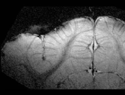
5 T 2 *-weighted images of rat brain (no activation) (isotropic resolution of 58 μm, 9.4 T) 4mm Dark lines venous vessels (>20 micrometer diameters) Sung-Hong Park et al., Magn. Reson. Med., 2008


6 Blood Oxygenation Level-dependent Contrast Ogawa et al. MRM, 1990 Breathing air Breathing 100% O 2 Mouse brain images at 360 MHz
7 Dynamic BOLD MR Measurements in Cats Turner R, Le Bihan D, Moonen CT, Despres D, Frank J Echo-planar time course MRI of cat brain oxygenation changes Magn Reson Med Nov;22(1): Abstract: When deoxygenated, blood behaves as an effective susceptibility contrast agent. Changes in brain oxygenation can be monitored using gradient-echo echo-planar imaging. With this technique, difference images also demonstrate that blood oxygenation is increased during periods of recovery from respiratory challenge.
8 Vascular Structure Arteries Capillaries Veins Blood oxygenation level ~1.0 (Task -> oxygen supply overcompensates oxygen utilization) ~0.6 Distance (Fox et al., 1988)


9 One of First Human fmri Studies Primary Visual Cortex Anatomical Image Functional Image (Visual Stimulation) University of Minnesota/Bell Lab Ogawa et al. Proc Natl Acad Sci USA, 1992
10 Current Status of Functional MRI - Underlying assumption is that fmri signal change is indirectly related to neural activity, and its location is indicative of neural activity site. - Functional MRI with a few millimeter resolution is routinely used for mapping brain functions such as vision, motor, language, cognition, etc.
11 Physiological Changes Biophysical Basis of BOLD fmri Spatial Resolution Interpretation - Quantification Temporal Resolution

12 Vascular Physiological Changes Blood Vessel Dilation Blood Velocity Increase Cerebral Blood Flow Blood Oxygenation Change Costantino Iadecola & Maiken Nedergaard Nature Neurosci, 2007


13 200µm Vessel Imaging of Rat Brain Anatomical Image 1 mm Vazquez et al., High-resolution Anatomical Image 500 µm


14 Vessel Imaging of Rat Brain during Stimulation 200µm 20x Mag., Reverse contrast A Bright: dilation 1 mm V V 4-s forepaw stim Vazquez et al.


")
15 Simultaneous measurements of CBF and P O2 QUANTIFICATION (PO2) Venous PO2 LV MV T SV SA MA Arteries Veins Tissue PO2 LDF (CBF) LA Clark-type oxygen sensor (30 and 4 μm diameter) Vazquez et al., JCBFM, 2010

 Vazquez et al.")
16 CBF and tissue PO2 changes during stimulation Forepaw Stimulation LDF (CBF) Tissue PO2 Time (s) Vazquez et al., JCBFM, 2010


17 Venous Blood PO2 changes during stimulation SO2 Sm. Ven. Med. Ven. PO2 Lar. Ven. Vazquez et al., JCBFM, 2010
18 Physiological Changes Biophysical Basis of BOLD fmri Spatial Resolution Interpretation - Quantification Temporal Resolution
19 Compartmentalization of Water Intravascular water moves freely EV blood vessel (IV) Slow exchange of IV and EV water Intact BBB tight junctions between endothelial cells impede the diffusion of water. (In 50 ms, less than 5% of the capillary water diffuses into the EVS.) Extravascular water moves freely RBC
20 Intravascular Effect -> T 2 Change Reb Blood Cell water Water appears to move freely across the RBC membrane (residence time in RBC ~ 5 ms).
21 Susceptibility effect in Extravascular Pool: Δω out = Δω max (radius of vessel/distance from vessel) 2 Related to deoxyhemoglobin concentration (oxygen saturation level) magnetic field strength 30 μm 300 μm 1% of max at r = 10 a.
22 Susceptibility effect in Extravascular Pool: ΔBout MRI signal at echo time (TE): a summation of all water proton signal within a voxel. Each proton signal decays by T 2 and dephases by local susceptibility effect (i.e., Phase shift) S(TE) = S. exp(-te/t 2 ). e(-iϖte) S. exp(-te/t 2 *) 30 μm 300 μm 1% of max at r = 10 a.
23 Spin Echo (two spins) t = 0 (after 90 pulse) x τ x y y 180 pulse along x Spin-echo x τ x y y
24 Capillary tube (1.4 mm o.d., 1.0 mm i.d.) filled with blood in a saline bath (positioned orthogonal to main magnetic field) SE GE 100% oxyhb 100% deoxyhb Ogawa et al., MRM, 1990
25 Conventional Gradient-echo and Spin-echo BOLD Signal CBV = 2% Δχ = 0.1 ppm Boxerman et al., MRM, 1995
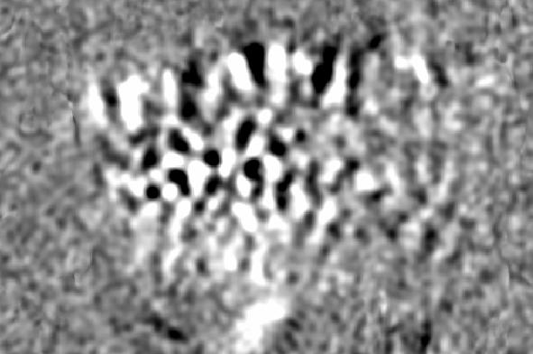
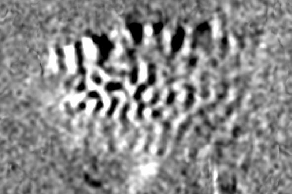
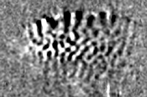
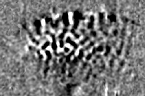
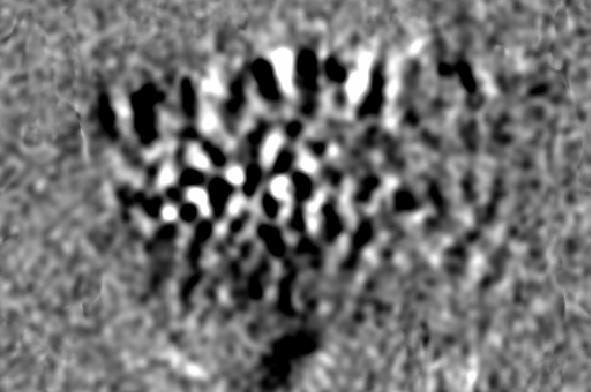
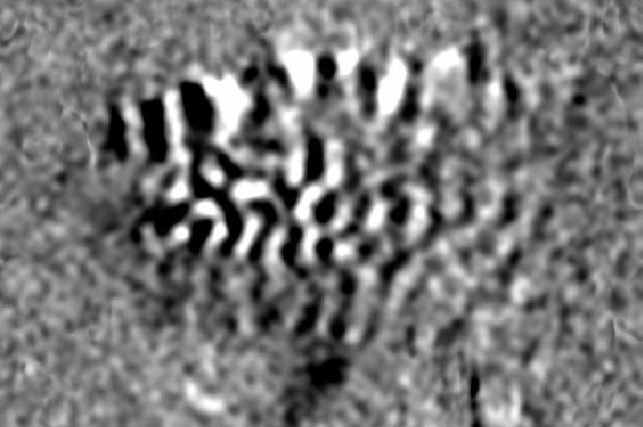
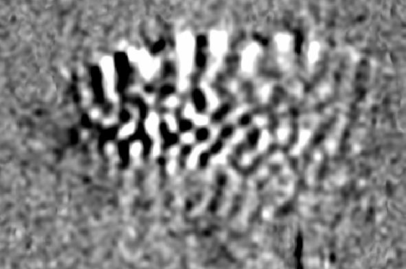
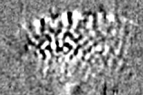
26 Extravascular and Intravascular BOLD Signal Contributions Gradient Echo Spin Echo Extravascular Large vessels Small vessels X X X Intravascular Large Small X X X X
27 Physiological Changes Biophysical Basis of BOLD fmri Spatial Resolution Interpretation - Quantification Temporal Resolution


28 Since all fmri techniques rely on blood signals, it is desirable to detect responses of small vessels which are close to active neurons. Midline Human visual cortex ~ 2 mm White matter Vascular Structures - Histology Duvernoy et al. Brain Research Bulletin, 1981


29 Cortical Layer Model Layer 4 is known to be highest capillary density and metabolic responses. Pia Matter Pia D Gray Matter 3 4 WM 2 mm L 5 White Matter Vascular structure 6 white Cortical cytoarchitecture of cat visual area 18 -Timan et al., Brain Res, Torre et al., Anat Rec 1998
 % Change (TE=20 ms, 9.4T) 6 5 GM WM 4 3 2 1 0-0.5 0 0.5 1 1.5 2 2.")
30 Cortical Depth-Dependent Gradient-echo BOLD fmri (156 x 156 µm 2 in-resolution, 4-shot EPI, 9.4T) % Change (TE=20 ms, 9.4T) 6 5 GM WM Distance from Cortical Surface (mm) 2 mm Zhao et al., NeuroImage, 2006
 mater Torre et al.")
31 Vascular structures vs. fmri resolution Scanning Electron Microscopy Pia (Human cortex) mater Torre et al., 1998 Gray matter BOLD Signal 500 µm Δdeoxyhemoglobin conc. in blood x venous blood volume

32 Gradient-echo vs. Spin-echo BOLD fmri (156 x 156 µm 2 in-resolution, 4-shot EPI, 9.4T) Gradient-Echo Spin-Echo TE=20 ms TE=40 ms 2 mm (%) (%)
33 Spatial Specificity of BOLD Signal to Neural Activity Site - Venous Vascular Structures Pial Venous Vessels: μm diameter Intracortical Veins: μm average diameter 1 2 mm apart

34 Distance between Intracortical Veins artery vein Pia GM WM Distance between emerging venous veins: mm Duvernoy et al. Brain Research Bulletin, 1981


 Orientation columns Hubel")
35 Can you map cortical columns? Neurons with similar properties are clustered as columns Single-neuron Activities Ocular Dominance Columns Color sensitive regions Gray matter (1.5 3 mm) Orientation columns Hubel & Wiesel, 1968
36 Iso-orientation maps in the medial area using fmri (with contrast agent, dilation of small vessels) 5 mm D A P V SPL Signal intensity (arbitrary unit) Fukuda et al., J of Neurosci, 2006


37 Observation of orientation preference maps 5 mm 180 A D V P SPL 90 0 Fukuda et al., J of Neurosci, 2006
38 Left Right Coronal plane mg Marginal gyrus (mg) LS WM Lateral sulcus (LS) BOLD fmri o 90 o 5 mm 0.3 1mm (Kim et al. Nature Neurosci, 3: , 2000)

")
 R 5 mm GE SE")
39 BOLD vs. CBV is-orientation maps (obtained with the differential approach; 0 90 ) GE BOLD SE BOLD CBV 1.0 A -1.0 ΔS (x mean) R 5 mm GE SE CBV 2 mm Moon et al., J of Neurosci, 2007
40 Physiological Basis Biophysical Basis Spatial Resolution Interpretation - Quantification Temporal Resolution
41 BOLD Signals Dependent on Bo, TE, pulse sequence (GE vs SE) Dependent on vessel size, orientation, and density Dependent on hematocrit level Dependent on oxygenation level
42 ΔR 2 * = Δ(1/T 2 *) = percent change/te CBV v (1 ΔS v ) + ΔCBV v (1 S v ) where 1 - S v = CMRO 2 / CBF Cerebral Oxygen Consumption Rate Cerebral Blood Flow Venous Blood Volume
43 Parenchymal Microvessel (<50 μm diameter) Region Blood volume Occipital cortex 1% Corpus callosum 0.4% Cerebellar nuclei 1.3% Rat; Fenstermacher et al.
44 Task/stimulation Neural activity CBF CMRo 2 CBV v dhb T 2 * T 2 * BOLD Signal BOLD Signal
45 1.6 CBV vs. CBF during Hypercapnia (α-chloralose anesthetized rats) rcbv (arbitrary unit) ml/100 g/min rcbv = 0.975rCBF 0.40 rcbv = 0.31 rcbf ( r = 0.85 ) (100% CBF -> 31% CBV) rcbf (arbitrary unit) Lee et al., MRM, 2001
46 rcbv (arbitrary unit) CBF vs. Arterial and Venous CBV rcbv (vein) rcbv (artery) rcbf (arbitrary unit) Lee et al., MRM, 2001
47 Task/stimulation Neural activity CBF CMRo 2 CBV dhb T 2 * T 2 *

48 fmri Signal Change is related to Neural Activity LOGOTHETIS et al. Nature, 412, , 2001

49 fmri Signal Change is related to Neural Activity LOGOTHETIS et al. Nature, 412, , 2001
50 Visual Stimulation under different baseline conditions Normalized BOLD Signal hypercapnia ETCO2 (m m Hg) Time (seconds) n = 6 subjects for each study 25 hypocapnia Cohen et al. JCBFM, 2002
51 Average BOLD Change (%) Average BOLD Change (%) Time (seconds) hypocapnia normocapnia hypercapnia Time (seconds)
52 Interpretation of fmri signals - fmri signal is an index of ensemble of neural activity (presumably monotonic relation) - Neural source of BOLD signal is not clear spiking activities vs. synaptic activity, excitatory vs. inhibitory - Difficulty to compare fmri signals across cortical regions and subjects due to BOLD signal dependencies on vascular structure and volume. - Excellent non-invasive tool to map whole brain functions with relatively high spatial (a few millimeters in humans) and temporal resolution (~a few seconds).
53 Physiological Basis Biophysical Basis Spatial Resolution Interpretation - Quantification Temporal Resolution
 0-2 -1.5-1 -0.5 0 0.5 1 1.5 2 Relative Delay (sec) Provided by P.A.")
54 Heterogeneity of fmri changes in humans: response times (Bilateral finger movements) Relative Delay Time + 2 sec 0 sec - 2 sec # of pixels Time (sec) Relative Delay (sec) Provided by P.A. Bandettini

55 Task Execution Task Execution 2 sec BOLD response BOLD response Time to peak Inter-epoch delay time (1 10 sec)
56 fmri Signal vs. Finger Movements BOLD change (%) finger pressure white matter motor area Delay time= s time (s)
57 mental rotation experiment displayed until decision is made time Presentation Contemplation Decision Richter et al. J. Cogn. Neurosci, 1999


58 Functional Maps of Mental Rotation Supplementary Motor Area Central Sulcu Lateral Premotor Area Superior Parietal Area
59 Response Time-locked Time Courses in M1 and SMA Relative fmri intensity 1% Primary motor Supplementary motor Time from button press (sec)
Daniel Bulte. Centre for Functional Magnetic Resonance Imaging of the Brain. University of Oxford
 Daniel Bulte Centre for Functional Magnetic Resonance Imaging of the Brain University of Oxford Overview Signal Sources BOLD Contrast Mechanism of MR signal change FMRI Modelling Scan design details Factors
BOLD signal dependence on blood flow and metabolism. Outline
 BOLD signal dependence on blood flow and metabolism R. Hoge, MGH NMR Center Outline physiological events accompanying neuronal activation factors affecting BOLD signal sensitivity BOLD response dynamics
PHYSICS OF MRI ACQUISITION. Alternatives to BOLD for fmri
 PHYSICS OF MRI ACQUISITION Quick Review for fmri HST-583, Fall 2002 HST.583: Functional Magnetic Resonance Imaging: Data Acquisition and Analysis Harvard-MIT Division of Health Sciences and Technology
Biennial SPM course The BOLD signal. Cyril Pernet. Centre for Clinical Brain Sciences (CCBS) Neuroimaging Sciences
 Neuroimaging Sciences") Biennial SPM course 2017 The BOLD signal Cyril Pernet Centre for Clinical Brain Sciences (CCBS) Neuroimaging Sciences Overview 1. MRI physics 2. Neurovascular coupling 3. Neural activity and BOLD 4. Experimental
Biennial SPM course 2017 The BOLD signal Cyril Pernet Centre for Clinical Brain Sciences (CCBS) Neuroimaging Sciences Overview 1. MRI physics 2. Neurovascular coupling 3. Neural activity and BOLD 4. Experimental
Titelmaster The physics of functional magnetic resonance imaging (fmri)
") Titelmaster The physics of functional magnetic resonance imaging (fmri) Outline 1.Introduction 2.The fmri experiment 2 3.The physics basis of fmri 4.Application Outline 3 1.Introduction Introduction Phrenology
Titelmaster The physics of functional magnetic resonance imaging (fmri) Outline 1.Introduction 2.The fmri experiment 2 3.The physics basis of fmri 4.Application Outline 3 1.Introduction Introduction Phrenology
Hemodynamics and fmri Signals
 Cerebral Blood Flow and Brain Activation UCLA NITP July 2011 Hemodynamics and fmri Signals Richard B. Buxton University of California, San Diego rbuxton@ucsd.edu... The subject to be observed lay on a
Biophysical and physiological bases of fmri signals: challenges of interpretation and methodological concerns
 Biophysical and physiological bases of fmri signals: challenges of interpretation and methodological concerns Antonio Ferretti aferretti@itab.unich.it Institute for Advanced Biomedical Technologies, University
PERFUSION MRI CONTRAST BASED TECHNIQUES
 PERFUSION MRI CONTRAST BASED TECHNIQUES by Kenny K Israni Mar 28, 2006 PERFUSION - MRI Dynamic Susceptibility contrast Dynamic Relaxivity contrast STEADY-STATE STATE TECHNIQUES Steady-state Susceptibility
Biophysical and physiological origins of blood oxygenation level-dependent fmri signals
 & 2012 ISCBFM All rights reserved 0271-678X/12 $32.00 Review Article Biophysical and physiological origins of blood oxygenation level-dependent fmri signals Seong-Gi Kim 1 and Seiji Ogawa 2,3 www.jcbfm.com
PETER PAZMANY CATHOLIC UNIVERSITY Consortium members SEMMELWEIS UNIVERSITY, DIALOG CAMPUS PUBLISHER
 PETER PAZMANY CATHOLIC UNIVERSITY SEMMELWEIS UNIVERSITY Development of Complex Curricula for Molecular Bionics and Infobionics Programs within a consortial* framework** Consortium leader PETER PAZMANY
P2 Visual - Perception
 P2 Visual - Perception 2014 SOSE Neuroimaging of high-level visual functions gyula.kovacs@uni-jena.de 11/09/06 Functional magnetic resonance imaging (fmri) The very basics What is fmri? What is MRI? The
Perfusion-Based fmri. Thomas T. Liu Center for Functional MRI University of California San Diego May 7, Goal
 Perfusion-Based fmri Thomas T. Liu Center for Functional MRI University of California San Diego May 7, 2006 Goal To provide a basic understanding of the theory and application of arterial spin labeling
The physiology of the BOLD signal What do we measure with fmri?
 The physiology of the BOLD signal What do we measure with fmri? Methods and Models in fmri, 10.11.2012 Jakob Heinzle Translational Neuromodeling Unit (TNU) Institute for Biomedical Engineering (IBT) University
Hemodynamics and fmri Signals
 Cerebral Blood Flow and Brain Activation UCLA NITP July 2010 Hemodynamics and fmri Signals Richard B. Buxton University of California, San Diego rbuxton@ucsd.edu... The subject to be observed lay on a
(This is a sample cover image for this issue. The actual cover is not yet available at this time.)
") (This is a sample cover image for this issue. The actual cover is not yet available at this time.) This article appeared in a journal published by Elsevier. The attached copy is furnished to the author
(This is a sample cover image for this issue. The actual cover is not yet available at this time.) This article appeared in a journal published by Elsevier. The attached copy is furnished to the author
Cortical layer-dependent BOLD and CBV responses measured by spin-echo and gradient-echo fmri: Insights into hemodynamic regulation
 www.elsevier.com/locate/ynimg NeuroImage 30 (2006) 1149 1160 Cortical layer-dependent BOLD and CBV responses measured by spin-echo and gradient-echo fmri: Insights into hemodynamic regulation Fuqiang Zhao,
HST.583 Functional Magnetic Resonance Imaging: Data Acquisition and Analysis Fall 2008
 MIT OpenCourseWare http://ocw.mit.edu HST.583 Functional Magnetic Resonance Imaging: Data Acquisition and Analysis Fall 2008 For information about citing these materials or our Terms of Use, visit: http://ocw.mit.edu/terms.
INTRO TO BOLD FMRI FRANZ JOSEPH GALL ( ) OUTLINE. MRI & Fast MRI Observations Models Statistical Detection
 OUTLINE. MRI & Fast MRI Observations Models Statistical Detection") INTRO TO BOLD FMRI 2014 M.S. Cohen all rights reserved mscohen@g.ucla.edu OUTLINE FRANZ JOSEPH GALL (1758-1828) MRI & Fast MRI Observations Models Statistical Detection PAUL BROCA (1824-1880) WILLIAM JAMES
INTRO TO BOLD FMRI 2014 M.S. Cohen all rights reserved mscohen@g.ucla.edu OUTLINE FRANZ JOSEPH GALL (1758-1828) MRI & Fast MRI Observations Models Statistical Detection PAUL BROCA (1824-1880) WILLIAM JAMES
Perfusion MRI. Youngkyoo Jung, PhD Associate Professor Radiology, Biomedical Engineering, and Clinical & Translational Science Institute
 Perfusion MRI Youngkyoo Jung, PhD Associate Professor Radiology, Biomedical Engineering, and Clinical & Translational Science Institute Perfusion The delivery of blood to a capillary bed in tissue Perfusion
HST.583 Functional Magnetic Resonance Imaging: Data Acquisition and Analysis Fall 2008
 MIT OpenCourseWare http://ocw.mit.edu HST.583 Functional Magnetic Resonance Imaging: Data Acquisition and Analysis Fall 2008 For information about citing these materials or our Terms of Use, visit: http://ocw.mit.edu/terms.
MRI qbold Based Evaluation. Renal Oxidative Metabolism. Department of Radiology and Hernando Gomez, MD Critical Care Medicine
 MRI qbold Based Evaluation of Renal Oxidative Metabolism Xiang He, PhD Department of Radiology and Hernando Gomez, MD Critical Care Medicine Background High oxygen-demand and lower medullary blood flow
Arterial versus total blood volume changes during neural activity-induced cerebral blood flow change: implication for BOLD fmri
 & 2007 ISCBFM All rights reserved 0271-678X/07 $30.00 www.jcbfm.com Arterial versus total blood volume changes during neural activity-induced cerebral blood flow change: implication for BOLD fmri Tae Kim
Introduction to Functional MRI
 Introduction to Functional MRI Douglas C. Noll Department of Biomedical Engineering Functional MRI Laboratory University of Michigan Outline Brief overview of physiology and physics of BOLD fmri Background
Neurovascular Physiology and Pathophysiology
 Neurovascular Physiology and Pathophysiology The physiological questions aim at understanding the molecular and biochemical mechanisms, by which the brain adapts local blood flow to neuronal activity and
This article appeared in a journal published by Elsevier. The attached copy is furnished to the author for internal non-commercial research and
 This article appeared in a journal published by Elsevier. The attached copy is furnished to the author for internal non-commercial research and education use, including for instruction at the authors institution
Table 1. Summary of PET and fmri Methods. What is imaged PET fmri BOLD (T2*) Regional brain activation. Blood flow ( 15 O) Arterial spin tagging (AST)
 Regional brain activation. Blood flow ( 15 O) Arterial spin tagging (AST)") Table 1 Summary of PET and fmri Methods What is imaged PET fmri Brain structure Regional brain activation Anatomical connectivity Receptor binding and regional chemical distribution Blood flow ( 15 O)
Table 1 Summary of PET and fmri Methods What is imaged PET fmri Brain structure Regional brain activation Anatomical connectivity Receptor binding and regional chemical distribution Blood flow ( 15 O)
BOLD signal compartmentalization based on the apparent diffusion coefficient
 Magnetic Resonance Imaging 20 (2002) 521 525 BOLD signal compartmentalization based on the apparent diffusion coefficient Allen W. Song a,b *, Harlan Fichtenholtz b, Marty Woldorff b a Brain Imaging and
Intra-renal Oxygenation. in Human Subjects
 MRI-based Mapping of Intra-renal Oxygenation BOLD in Human Subjects OEF Xiang He, PhD Department of Radiology Background Cortex Brain CBF ~ 1.0 ml/min/g Brain PO 2 ~ 25-35 mm Hg Medullary hypoxia is an
The neurolinguistic toolbox Jonathan R. Brennan. Introduction to Neurolinguistics, LSA2017 1
 The neurolinguistic toolbox Jonathan R. Brennan Introduction to Neurolinguistics, LSA2017 1 Psycholinguistics / Neurolinguistics Happy Hour!!! Tuesdays 7/11, 7/18, 7/25 5:30-6:30 PM @ the Boone Center
Define functional MRI. Briefly describe fmri image acquisition. Discuss relative functional neuroanatomy. Review clinical applications.
 Dr. Peter J. Fiester November 14, 2012 Define functional MRI. Briefly describe fmri image acquisition. Discuss relative functional neuroanatomy. Review clinical applications. Briefly discuss a few examples
Functional MRI at High Fields: Practice and Utility
 FUNCTIONAL MRI AT HIGH FIELDS: PRACTICE AND UTILITY 1 Functional MRI at High Fields: Practice and Utility Kamil Ugurbil, Wei Chen, Xiaoping Hu, Seong-Gi Kim, Xiao-Hung Zhu Center for Magnetic Resonance
Functional MRI with Magnetization Transfer Effects: Determination of BOLD and Arterial Blood Volume Changes
 Functional MRI with Magnetization Transfer Effects: Determination of BOLD and Arterial Blood Volume Changes Tae Kim, 1 * Kristy Hendrich, 1 and Seong-Gi Kim 1,2 Magnetic Resonance in Medicine 60:1518 1523
Pre-surgical planning for brain tumor resection using functional MRI
 June 2011 Divya S Bolar, HMSIV Pre-surgical planning for brain tumor resection using functional MRI Divya S. Bolar,, HMS IV 1 Our patient: clinical history 85-year year-old right-handed handed woman presents
MEDICAL REVIEW Functional Magnetic Resonance Imaging: From Acquisition to Application
 Functional Magnetic Resonance Imaging: From Acquisition to Application Gail Yarmish * and Michael L. Lipton *, Departments of Radiology *, and Neuroscience Albert Einstein College of Medicine Bronx, New
Neural Interpretation of Blood Oxygenation Level-Dependent fmri Maps at Submillimeter Columnar Resolution
 6892 The Journal of Neuroscience, June 27, 2007 27(26):6892 6902 Behavioral/Systems/Cognitive Neural Interpretation of Blood Oxygenation Level-Dependent fmri Maps at Submillimeter Columnar Resolution Chan-Hong
CISC 3250 Systems Neuroscience
 CISC 3250 Systems Neuroscience Levels of organization Central Nervous System 1m 10 11 neurons Neural systems and neuroanatomy Systems 10cm Networks 1mm Neurons 100μm 10 8 neurons Professor Daniel Leeds
Functional MRI and Diffusion Tensor Imaging
 Functional MRI and Diffusion Tensor Imaging Andrew Steven March 23, 2018 Ochsner Neuroscience Symposium None Disclosure 1 Objectives Review basic principles of BOLD fmri and DTI. Discuss indications and
Sung Hong Park. M.S. in Electrical Engineering, KAIST, South Korea, Submitted to the Graduate Faculty of
 NONINVASIVE IMAGING OF BRAIN VASCULATURE WITH HIGH RESOLUTION BLOOD OXYGENATION LEVEL DEPENDENT VENOGRAPHY IN MAGNETIC RESONANCE IMAGING: APPLICATIONS TO FUNCTIONAL AND CLINICAL STUDIES by Sung Hong Park
Methods to examine brain activity associated with emotional states and traits
 Methods to examine brain activity associated with emotional states and traits Brain electrical activity methods description and explanation of method state effects trait effects Positron emission tomography
Principles of Functional MRI
 1 Principles of Functional MRI Seong-Gi Kim and Peter A. Bandettini Introduction The idea that regional cerebral blood flow (CBF) could reflect neuronal activity began with experiments of Roy and Sherrington
Allen W. Song, Marty G. Woldorff, Stacey Gangstead, George R. Mangun, and Gregory McCarthy
 NeuroImage 17, 742 750 (2002) doi:10.1006/nimg.2002.1217 Enhanced Spatial Localization of Neuronal Activation Using Simultaneous Apparent-Diffusion-Coefficient and Blood-Oxygenation Functional Magnetic
CORTICAL LAYER-DEPENDENT HEMODYNAMIC REGULATION INVESTIGATED BY FUNCTIONAL MAGNETIC RESONANCE IMAGING. Cecil Chern-Chyi Yen
 CORTICAL LAYER-DEPENDENT HEMODYNAMIC REGULATION INVESTIGATED BY FUNCTIONAL MAGNETIC RESONANCE IMAGING by Cecil Chern-Chyi Yen B.S. in Physics, National Tsing-Hua University, Taiwan, 1999 M.S. in Eletrical
Concurrent near-infrared spectroscopy (NIRS) and functional magnetic resonance imaging (fmri) of the brain
 and functional magnetic resonance imaging (fmri) of the brain") Motor cortex activation fmri Near-infrared imaging Concurrent near-infrared spectroscopy (NIRS) and functional magnetic resonance imaging (fmri) of the brain Sergio Fantini s group, Department of Biomedical
Motor cortex activation fmri Near-infrared imaging Concurrent near-infrared spectroscopy (NIRS) and functional magnetic resonance imaging (fmri) of the brain Sergio Fantini s group, Department of Biomedical
Temporal dynamics and spatial specificity of arterial and venous blood volume changes during visual stimulation: implication for BOLD quantification
 Temporal dynamics and spatial specificity of arterial and venous blood volume changes during visual stimulation: implication for BOLD quantification Tae Kim 1 and Seong-Gi Kim 1,2 & 2011 ISCBFM All rights
Neuroimaging and Assessment Methods
 Psych 2200, Lecture 5 Experimental Design and Brain Imaging Methods Tues Sept 15, 2015 Revised TA office hours (Sam), today 4-5p, and wed 11:30-1:30. I will not have office hours this thurs but you should
A Primer on Functional Magnetic Resonance Imaging
 DOI 10.1007/s11065-007-908-8 ORIGINAL PAPER A Primer on Functional Magnetic Resonance Imaging Gregory G. Brown Joanna E. Perthen Thomas T. Liu Richard B. Buxton Received: March 007 / Accepted: 4 March
APPLICATIONS OF ASL IN NEUROSCIENCE
 APPLICATIONS OF ASL IN NEUROSCIENCE Luis Hernandez-Garcia, Ph.D. Functional MRI laboratory University of Michigan 1 OVERVIEW Quick review of ASL The niche for ASL Examples of practical applications in
Turbo ASL: Arterial Spin Labeling With Higher SNR and Temporal Resolution
 COMMUNICATIONS Magnetic Resonance in Medicine 44:511 515 (2000) Turbo ASL: Arterial Spin Labeling With Higher SNR and Temporal Resolution Eric C. Wong,* Wen-Ming Luh, and Thomas T. Liu A modified pulsed
Magnetic Resonance Angiography
 Magnetic Resonance Angiography 1 Magnetic Resonance Angiography exploits flow enhancement of GR sequences saturation of venous flow allows arterial visualization saturation of arterial flow allows venous
Introduction to Brain Imaging
 Introduction to Brain Imaging Human Brain Imaging NEUR 570 & BIC lecture series September 9, 2013 Petra Schweinhardt, MD PhD Montreal Neurological Institute McGill University Montreal, Canada Various techniques
MR Advance Techniques. Vascular Imaging. Class II
 MR Advance Techniques Vascular Imaging Class II 1 Vascular Imaging There are several methods that can be used to evaluate the cardiovascular systems with the use of MRI. MRI will aloud to evaluate morphology
Functional Magnetic Resonance Imaging with Arterial Spin Labeling: Techniques and Potential Clinical and Research Applications
 pissn 2384-1095 eissn 2384-1109 imri 2017;21:91-96 https://doi.org/10.13104/imri.2017.21.2.91 Functional Magnetic Resonance Imaging with Arterial Spin Labeling: Techniques and Potential Clinical and Research
Nature Neuroscience: doi: /nn Supplementary Figure 1
 Supplementary Figure 1 Relative expression of K IR2.1 transcript to enos was reduced 29-fold in capillaries from knockout animals. Relative expression of K IR2.1 transcript to enos was reduced 29-fold
Announcement. Danny to schedule a time if you are interested.
 Announcement If you need more experiments to participate in, contact Danny Sanchez (dsanchez@ucsd.edu) make sure to tell him that you are from LIGN171, so he will let me know about your credit (1 point).
NeuroImage 43 (2008) 1 9. Contents lists available at ScienceDirect. NeuroImage. journal homepage:
 1 9. Contents lists available at ScienceDirect. NeuroImage. journal homepage:") NeuroImage 43 (2008) 1 9 Contents lists available at ScienceDirect NeuroImage journal homepage: www.elsevier.com/locate/ynimg Cortical layer-dependent dynamic blood oxygenation, cerebral blood flow and
NeuroImage 43 (2008) 1 9 Contents lists available at ScienceDirect NeuroImage journal homepage: www.elsevier.com/locate/ynimg Cortical layer-dependent dynamic blood oxygenation, cerebral blood flow and
ASSUMPTION OF COGNITIVE UNIFORMITY
 The Human Brain cerebral hemispheres: two most important divisions of the brain, separated by the longitudinal fissure corpus callosum: a large bundle of axons that constitutes the major connection between
Vascular Filters of Functional MRI: Spatial Localization Using BOLD and CBV Contrast
 Vascular Filters of Functional MRI: Spatial Localization Using BOLD and CBV Contrast Joseph B. Mandeville 1,2 * and John J.A. Marota 1,3 Magnetic Resonance in Medicine 42:591 598 (1999) The spatial distributions
HST.583 Functional Magnetic Resonance Imaging: Data Acquisition and Analysis Fall 2006
 MIT OpenCourseWare http://ocw.mit.edu HST.583 Functional Magnetic Resonance Imaging: Data Acquisition and Analysis Fall 2006 For information about citing these materials or our Terms of Use, visit: http://ocw.mit.edu/terms.
Advances in functional MRI of the human brain
 Progress in Nuclear Magnetic Resonance Spectroscopy 44 (2004) 1 32 www.elsevier.com/locate/pnmrs Advances in functional MRI of the human brain J. Frahm*, P. Dechent, J. Baudewig, K.D. Merboldt Biomedizinische
Why high-field MRI? Benefits of high-field MRI. Signal-to-noise ratio (SNR) Contrast (anatomical & functional) 8 x 8 x 8 mm 3 4 x 4 x 4 mm 3
 Contrast (anatomical & functional) 8 x 8 x 8 mm 3 4 x 4 x 4 mm 3") Why high-field MRI? 8 x 8 x 8 mm 3 4 x 4 x 4 mm 3 2 x 2 x 2 mm 3 1 x 1 x 1 mm 3 Voxel volume 2 x 2 x 2 mm 3 = 8 Voxel volume 1 x 1 x 1 mm 3 = 1 Benefits of high-field MRI Signal-to-noise ratio (SNR) Contrast
Why high-field MRI? 8 x 8 x 8 mm 3 4 x 4 x 4 mm 3 2 x 2 x 2 mm 3 1 x 1 x 1 mm 3 Voxel volume 2 x 2 x 2 mm 3 = 8 Voxel volume 1 x 1 x 1 mm 3 = 1 Benefits of high-field MRI Signal-to-noise ratio (SNR) Contrast
Event-Related fmri and the Hemodynamic Response
 Human Brain Mapping 6:373 377(1998) Event-Related fmri and the Hemodynamic Response Randy L. Buckner 1,2,3 * 1 Departments of Psychology, Anatomy and Neurobiology, and Radiology, Washington University,
Use of Multimodal Neuroimaging Techniques to Examine Age, Sex, and Alcohol-Related Changes in Brain Structure Through Adolescence and Young Adulthood
 American Psychiatric Association San Diego, CA 24 May 2017 Use of Multimodal Neuroimaging Techniques to Examine Age, Sex, and Alcohol-Related Changes in Brain Structure Through Adolescence and Young Adulthood
ORIGINAL ARTICLE. Jack A Wells 1, Bernard Siow 1,2, Mark F Lythgoe 1,4 and David L Thomas 3,4
 Journal of Cerebral Blood Flow & Metabolism (2013) 33, 215 224 & 2013 ISCBFM All rights reserved 0271-678X/13 $32.00 www.jcbfm.com ORIGINAL ARTICLE Measuring biexponential transverse relaxation of the
Neurovascular Coupling
 Cerebral Blood Flow and Brain Activation UCLA NITP July 2012 Neurovascular Coupling Richard B. Buxton University of California, San Diego rbuxton@ucsd.edu... The subject to be observed lay on a delicately
Outline. Why Image Animals?
 Small Animal Magnetic Resonance Imaging: Current Trends, Challenges and Perspectives for Pathological Imaging C. Chad Quarles Vanderbilt University Institute of Imaging Science Outline Why image animals?
Nature Neuroscience doi: /nn Supplementary Figure 1. Characterization of viral injections.
 Supplementary Figure 1 Characterization of viral injections. (a) Dorsal view of a mouse brain (dashed white outline) after receiving a large, unilateral thalamic injection (~100 nl); demonstrating that
Combining tdcs and fmri. OHMB Teaching Course, Hamburg June 8, Andrea Antal
 Andrea Antal Department of Clinical Neurophysiology Georg-August University Goettingen Combining tdcs and fmri OHMB Teaching Course, Hamburg June 8, 2014 Classical Biomarkers for measuring human neuroplasticity
Prof. Greg Francis 1/2/19
 Brain scans PSY 200 Greg Francis Lecture 03 How to study the brain without killing someone. Scanning Technology provides insight into brain processes w EEG recordings w MRI w Non-invasive Maps of brain
Supplementary Information Methods Subjects The study was comprised of 84 chronic pain patients with either chronic back pain (CBP) or osteoarthritis
 or osteoarthritis") Supplementary Information Methods Subjects The study was comprised of 84 chronic pain patients with either chronic back pain (CBP) or osteoarthritis (OA). All subjects provided informed consent to procedures
Supplementary Information Methods Subjects The study was comprised of 84 chronic pain patients with either chronic back pain (CBP) or osteoarthritis (OA). All subjects provided informed consent to procedures
Cerebral Cortex 1. Sarah Heilbronner
 Cerebral Cortex 1 Sarah Heilbronner heilb028@umn.edu Want to meet? Coffee hour 10-11am Tuesday 11/27 Surdyk s Overview and organization of the cerebral cortex What is the cerebral cortex? Where is each
Principles of Haemodynamic Coupling for fmri
 Principles of Haemodynamic Coupling for fmri Paul M. Matthews Head, Global Imaging Unit, GlaxoSmithKline and Professor of Clinical Neurosciences, Imperial College paul.m.matthews@gsk.com Regulation of
Outline. Biological Psychology: Research Methods. Dr. Katherine Mickley Steinmetz
 Biological Psychology: Research Methods Dr. Katherine Mickley Steinmetz Outline Neuroscience Methods Histology Electrophysiological Recordings Lesion Neuroimaging Neuroanatomy Histology: Brain structure
Simultaneous Acquisition of Cerebral Blood Volume-, Blood Flow-, and Blood Oxygenation-Weighted MRI Signals at Ultra-High Magnetic Field
 NOTE Magnetic Resonance in Medicine 00:00 00 (2014) Simultaneous Acquisition of Cerebral Blood Volume-, Blood Flow-, and Blood Oxygenation-Weighted MRI Signals at Ultra-High Magnetic Field Steffen N. Krieger,
Improved spatial localization of post-stimulus BOLD undershoot relative to positive BOLD
 www.elsevier.com/locate/ynimg NeuroImage 34 (2007) 1084 1092 Improved spatial localization of post-stimulus BOLD undershoot relative to positive BOLD Fuqiang Zhao, Tao Jin, Ping Wang, and Seong-Gi Kim
ASL BASICS II. Learning Objectives. Outline. Acquisition. M. A. Fernández-Seara, Ph. D. Arterial spin labeled perfusion MRI: basic theory
 Acquisition ASL BASICS II M. A. Fernández-Seara, Ph. D. Neuroimaging Laboratory Center for Applied Medical Research University of Navarra Pamplona, Spain Outline Arterial spin labeled perfusion MRI: basic
Temporal decoupling of oxy- and deoxy-hemoglobin hemodynamic responses detected by functional near-infrared spectroscopy (fnirs)
") Temporal decoupling of oxy- and deoxy-hemoglobin hemodynamic responses detected by functional near-infrared spectroscopy (fnirs) Nicoladie D. Tam a, and George Zouridakis b a Department of Biological Sciences,
Temporal decoupling of oxy- and deoxy-hemoglobin hemodynamic responses detected by functional near-infrared spectroscopy (fnirs) Nicoladie D. Tam a, and George Zouridakis b a Department of Biological Sciences,
Supporting Information
 Supporting Information Lingnau et al. 10.1073/pnas.0902262106 Fig. S1. Material presented during motor act observation (A) and execution (B). Each row shows one of the 8 different motor acts. Columns in
Echo-Time and Field Strength Dependence of BOLD Reactivity in Veins and Parenchyma Using Flow- Normalized Hypercapnic Manipulation
 Echo-Time and Field Strength Dependence of BOLD Reactivity in Veins and Parenchyma Using Flow- Normalized Hypercapnic Manipulation Christina Triantafyllou 1,2 *, Lawrence L. Wald 2,3, Richard D. Hoge 4,5
Brain and Cognition. Cognitive Neuroscience. If the brain were simple enough to understand, we would be too stupid to understand it
 Brain and Cognition Cognitive Neuroscience If the brain were simple enough to understand, we would be too stupid to understand it 1 The Chemical Synapse 2 Chemical Neurotransmission At rest, the synapse
Procedia - Social and Behavioral Sciences 159 ( 2014 ) WCPCG 2014
 WCPCG 2014") Available online at www.sciencedirect.com ScienceDirect Procedia - Social and Behavioral Sciences 159 ( 2014 ) 743 748 WCPCG 2014 Differences in Visuospatial Cognition Performance and Regional Brain Activation
Available online at www.sciencedirect.com ScienceDirect Procedia - Social and Behavioral Sciences 159 ( 2014 ) 743 748 WCPCG 2014 Differences in Visuospatial Cognition Performance and Regional Brain Activation
Vascular Origins of BOLD and CBV fmri Signals: Statistical Mapping and Histological Sections Compared
 The Open Neuroimaging Journal, 2010, 4, 1-8 1 Open Access Vascular Origins of BOLD and CBV fmri Signals: Statistical Mapping and Histological Sections Compared Aneurin J Kennerley*, John E Mayhew, Peter
Physiological and Physical Basis of Functional Brain Imaging 6. EEG/MEG. Kâmil Uludağ, 20. November 2007
 Physiological and Physical Basis of Functional Brain Imaging 6. EEG/MEG Kâmil Uludağ, 20. November 2007 Course schedule 1. Overview 2. fmri (Spin dynamics, Image formation) 3. fmri (physiology) 4. fmri
Frontiers of Brain Mapping Using MRI
 JOURNAL OF MAGNETIC RESONANCE IMAGING 23:945 957 (2006) Invited Review Frontiers of Brain Mapping Using MRI Noam Harel, PhD, 1 Kâmil Uǧurbil, PhD, 1,2 * Kâmil Uludaǧ, PhD, 2 and Essa Yacoub, PhD 1 Over
functional MRI everything you always wanted to know, but never dared to MD PhD
 functional MRI everything you always wanted to know, but never dared to ask @MarionSmits, MD PhD Associate Professor of Neuroradiology Dept. of Radiology, Erasmus MC, Rotterdam (NL) Honorary Consultant
Thalamo-Cortical Relationships Ultrastructure of Thalamic Synaptic Glomerulus
 Central Visual Pathways V1/2 NEUR 3001 dvanced Visual Neuroscience The Lateral Geniculate Nucleus () is more than a relay station LP SC Professor Tom Salt UCL Institute of Ophthalmology Retina t.salt@ucl.ac.uk
Stuttering Research. Vincent Gracco, PhD Haskins Laboratories
 Stuttering Research Vincent Gracco, PhD Haskins Laboratories Stuttering Developmental disorder occurs in 5% of children Spontaneous remission in approximately 70% of cases Approximately 1% of adults with
Gross Organization I The Brain. Reading: BCP Chapter 7
 Gross Organization I The Brain Reading: BCP Chapter 7 Layout of the Nervous System Central Nervous System (CNS) Located inside of bone Includes the brain (in the skull) and the spinal cord (in the backbone)
Introduction. Cardiac Imaging Modalities MRI. Overview. MRI (Continued) MRI (Continued) Arnaud Bistoquet 12/19/03
 MRI (Continued) Arnaud Bistoquet 12/19/03") Introduction Cardiac Imaging Modalities Arnaud Bistoquet 12/19/03 Coronary heart disease: the vessels that supply oxygen-carrying blood to the heart, become narrowed and unable to carry a normal amount
Introduction Cardiac Imaging Modalities Arnaud Bistoquet 12/19/03 Coronary heart disease: the vessels that supply oxygen-carrying blood to the heart, become narrowed and unable to carry a normal amount
LESSON 1.3 WORKBOOK. How can we study the behaving brain?
 LESSON 1.3 WORKBOOK How can we study the behaving brain? We are in the middle of a technological revolution when it comes to how closely we can look at the behaving brain. Scientists and doctors now have
MR Advance Techniques. Cardiac Imaging. Class III
 MR Advance Techniques Cardiac Imaging Class III Black Blood Imaging & IR Blue= O2 poor blood Red=O2 rich blood Inversion pulses can produce black blood imaging in GRE pulse sequences. Specially on the
The Visual System. Cortical Architecture Casagrande February 23, 2004
 The Visual System Cortical Architecture Casagrande February 23, 2004 Phone: 343-4538 Email: vivien.casagrande@mcmail.vanderbilt.edu Office: T2302 MCN Required Reading Adler s Physiology of the Eye Chapters
Neural Correlates of Human Cognitive Function:
 Neural Correlates of Human Cognitive Function: A Comparison of Electrophysiological and Other Neuroimaging Approaches Leun J. Otten Institute of Cognitive Neuroscience & Department of Psychology University
Abstract. Introduction. Material and Methods. Results. Conclusion
 Dynamic susceptibility contrast MRI calibrated using T1-based steadystate CBV and vascular space occupancy (VASO): Comparison with model-free arterial spin labelling Emelie Lindgren Supervisors: Linda
The discovery by Roy and Sherrington (1) that regional
 that regional") Localized cerebral blood flow response at submillimeter columnar resolution Timothy Q. Duong, Dae-Shik Kim, Kâmil Uğurbil, and Seong-Gi Kim* Center for Magnetic Resonance Research, Departments of Radiology
Localized cerebral blood flow response at submillimeter columnar resolution Timothy Q. Duong, Dae-Shik Kim, Kâmil Uğurbil, and Seong-Gi Kim* Center for Magnetic Resonance Research, Departments of Radiology
Does stimulus quality affect the physiologic MRI responses to brief visual activation?
 Brain Imaging 0, 277±28 (999) WE studied the effect of stimulus quality on the basic physiological response characteristics of oxygenationsensitive MRI signals. Paradigms comprised a contrastreversing
Improved cortical-layer specificity of vascular space occupancy fmri with slab inversion relative to spin-echo BOLD at 9.4 T
 www.elsevier.com/locate/ynimg NeuroImage 40 (2008) 59 67 Improved cortical-layer specificity of vascular space occupancy fmri with slab inversion relative to spin-echo BOLD at 9.4 T Tao Jin a, and Seong-Gi
Supplementary Online Content
 Supplementary Online Content Gregg NM, Kim AE, Gurol ME, et al. Incidental cerebral microbleeds and cerebral blood flow in elderly individuals. JAMA Neurol. Published online July 13, 2015. doi:10.1001/jamaneurol.2015.1359.
Contact: Course outline: Contact for other times.
 Contact: kdelaney@uvic.ca Course outline: http://web.uvic.ca/~kdelaney/b367 Scheduled office hours: 1:00-3:00, M&Th Cunn. 259A Contact kdelaney@uvic.ca for other times. Quiz (0.5 hrs) midterm (1.4 hrs)
Homework Week 2. PreLab 2 HW #2 Synapses (Page 1 in the HW Section)
") Homework Week 2 Due in Lab PreLab 2 HW #2 Synapses (Page 1 in the HW Section) Reminders No class next Monday Quiz 1 is @ 5:30pm on Tuesday, 1/22/13 Study guide posted under Study Aids section of website
Homework Week 2 Due in Lab PreLab 2 HW #2 Synapses (Page 1 in the HW Section) Reminders No class next Monday Quiz 1 is @ 5:30pm on Tuesday, 1/22/13 Study guide posted under Study Aids section of website