Brain AVM with Accompanying Venous Aneurysm with Intracerebral and Intraventricular Hemorrhage
|
|
|
- Blake Walker
- 5 years ago
- Views:
Transcription
1 Cronicon OPEN ACCESS EC PAEDIATRICS Case Report Brain AVM with Accompanying Venous Aneurysm with Intracerebral and Intraventricular Hemorrhage Dimitrios Panagopoulos* Neurosurgical Department, University Hospital of Alexandroupoli, Thrace, Greece *Corresponding Author: Dimitrios Panagopoulos, Assistant Professor, Neurosurgical Department, University Hospital of Alexandroupoli, Thrace, Greece. Received: June 18, 2018; Published: June 28, 2018 Abstract Background: Arteriovenous malformations (AVM s) and intracranial aneurysm are collectively the most common causes of spontaneous subarachnoid and intracerebral and/or intraventricular hemorrhage. In children, saccular aneurysms can be attendant to the AVM in 29% of children with AVMs. The majority of aneurysms are arterial in location (37%), with a similar percentage of intranidal (30%) and venous (33%) locations. Case Presentation: A 13 year s old male was admitted to our hospital due to gradually deteriorating level of consciousness. CT and MRI revealed intra-parenchymal and intraventricular hemorrhage. Work up included a DSA which revealed an AVM with an associated venous aneurysm. Endovascular embolization of the AVM was executed and the postoperative course of the patient was benign. Conclusions: Contrary to many other pediatric centers, our clinical practice has been to perform urgent cerebral angiography in children with suspected AVMs to identify and potentially treat AVM-associated aneurysms. We have done this because of our hypothesis that AVM- associated aneurysms have a higher risk of re-hemorrhage than isolated AVMs. Keywords: Arteriovenous Malformation; Associated Venous Aneurysm; Rupture; Embolization Abbreviations CT: Computed Tomography; MRI: Magnetic Resonance Imaging; DSA: Digital Subtraction Angiography; AVM: Arteriovenous Malformation; AV: Arteriovenous; T2 W: T2 Weighted; T1W: T1 Weighted; T2 GRE: Gradient Echo Sequence; FLAIR: Fluid-Attenuated Inversion Recovery Introduction Arteriovenous malformations are one of the major causes of stroke in children [1,2]. Because intracerebral hemorrhage is the most devastating as well as the most common clinical presentation (46% - 87%) in children with AVMs; there is a strong interest in establishing treatment strategies that could prevent or reduce both the initial hemorrhage and possible re-bleeding. Even though AVMs are rare in kids, estimated to represent 3% of all AVMs [3,4], they tend to rupture more frequently than in adults [4,5].
 [6].")
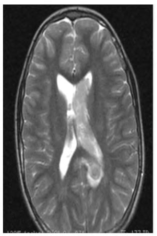
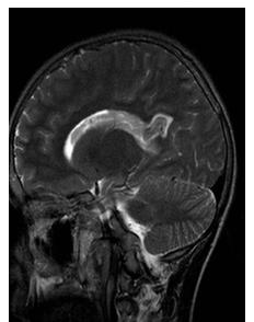
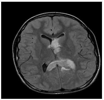
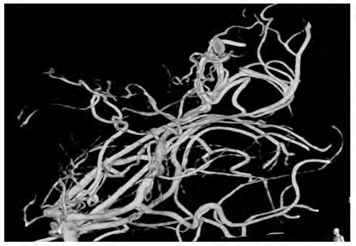
2 716 An enormous diversity of brain vascular malformations occurs in children. These include vein of Galen malformations, dural AV fistulas, non-galenic pial AV fistulas, and nidal arteriovenous malformations (AVMs) [6]. AVMs are defined by a group of vessels with an abnormal low-resistance connection between arteries and veins occurring in a focal geographic area of the brain parenchyma - the nidus. Aneurysms separate and distinct from the circle of Willis are commonly found located adjacent to or within AVMs, and are typically classified as either arterial, intranidal, or venous in location [1]. The nature of these AVM-associated aneurysms has been partially investigated in adults. Although the data are mixed, the preponderance of data suggests that the presence of an aneurysm may be an independent risk factor for hemorrhage in adults with AVMs. For this reason, most adults with ruptured AVMs undergo prompt cerebral angiography with endovascular treatment of AVM associated aneurysms. In children, however, conventional cerebral angiography and treatment are often performed in a delayed fashion, in part because the nature of AVM associated aneurysms is generally unknown. Because pediatric and adult aneurysms may have different causes, extrapolation of data from adult studies may not be applicable. In this study, we describe our experience regarding the clinical behavior and treatment of AVM-associated aneurysm in a child with AVM. Case Report A thirteen year s old male was admitted to the emergency department of our hospital due to progressive loss of consciousness with a rapid clinical deterioration. Upon admission, CT and MRI were performed, which revealed intraparenchymal hemorrhage in the region of the splenium of the corpus callosum with accompanying intraventricular hemorrhage in the left lateral ventricle (Figure 1a-1j). Figure 1a

3 717 Figure 1b Figure 1c

4 718 Figure 1d Figure 1e

5 719 Figure 1f Figure 1g

6 720 Figure 1h Figure 1i


7 721 Figure 1j Figure 1a-1j: Initial MRI scan, T2 W, Τ2*GRE, T1W before and after gadolinium enhancement and FLAIR, revealing intra-parenchymal hemorrhage in the anatomic territory of the splenium of the corpus callosum and the isthmus of the cingulate gyrus. An accompanying intraventricular hemorrhage in the left lateral and third ventricle is visualized. After gadolinium administration, there is moderate contrast enhancement. The long arrow at figure 1h depicts the location of the nidus in the territory of the splenium of the corpus callosum. DSA was performed, which revealed an underlying AVM with an accompanying venous aneurysm in the vicinity of the splenium of the corpus callosum. Arterial feeders were originating from the left side of the posterior cerebral circulation, whereas the draining venous channels were terminating to the left transverse venous sinus (Figure 2a-2d). Figure 2a

8 722 Figure 2b Figure 2c


9 723 Figure 2d Figure 2a-2d: Initial DSA of posterior cerebral circulation, revealing the underlying AVM with the associated venous aneurysm. Arterial feeders of the nidus are branches of the left posterior cerebral artery. Subsequently, the patient underwent trans-arterial embolization of the AVM through elective catheterization of the left posterior cerebral artery. After embolization, a small residual remnant of the nidus of the AVM remained with delayed and slow opacification of the draining vein of the malformation (Figure 3a-3c). Figure 3a


10 724 Figure 3b Figure 3c Figure 3a-3c: Elective DSA of the posterior cerebral circulation, after selective catheterization of the left posterior cerebral artery. It reveals a small residual portion of the nidus remaining patent and thus opacified, with concurrent slow, delayed filling of the draining vein.
. The patient s clinical course was uneventful, with gradual recovery of the neurological and cognitive function.")

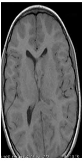
11 After the embolization procedure, a CT scan was performed, which verified the embolization result and that no adverse consequences, especially hemorrhage and infarction in the territory, were incurred (Figure 3d). The patient s clinical course was uneventful, with gradual recovery of the neurological and cognitive function. 725 Figure 3d Figure 3d: CT scan performed immediately after the embolization procedure. It delineates the location of the occluded nidus of the AVM as a hyperdense area. No adverse effects, such as hemorrhage or infarction, are noted. A follow-up DSA was performed four months after the ictus, which revealed the existence of an early draining vein in the vicinity of the known AVM, without residual opacification of the nidus of the AVM (Figure 4a-4b). Figure 4a

12 726 Figure 4b Figure 4a-4b: DSA performed four months after the embolization procedure, revealing complete occlusion of the nidus, which is not opacified. It only depicts the existence of an early draining vein. Results and Discussion In a large study reporting the overall incidence of AVM associated aneurysms in children, as well as their association with hemorrhage, an AVM-associated aneurysm was found in 29% of children with AVMs [1]. The majority of aneurysms were arterial in location (37%), with a similar percentage of intranidal (30%) and venous (33%) locations. The presence of an aneurysm could not be predicted based on patient age, sex, or location of the AVM. AVM-associated aneurysms in venous and intra-nidal locations (48% of patients) did not appear to be associated with hemorrhage when separated from arterial aneurysms. AVMs and their draining veins were often located deep within the brain in children, raising the possibility that centrally-located AVMs may arise earlier in development or be more likely to come to clinical attention early in life than more peripherally located AVMs. Regarding clinical status, most studies that demonstrate that children with AVMs overall improve clinically over time [1,7]. Comparison of clinical status at initial presentation, discharge, and most recent follow-up all suggested that while children with AVM associated aneurysms have worse neurological status at discharge, there was no significant difference between children with isolated AVMs and those with AVM-associated aneurysms at long-term follow-up.
13 727 It has been routine clinical practice to perform urgent cerebral angiography in children with suspected AVMs to identify and potentially treat AVM-associated aneurysms [1]. This is based on the hypothesis that AVM-associated aneurysms have a higher risk of rehemorrhage than isolated AVMs. Regarding treatment of pediatric AVM s, endovascular embolization is feasible and safe in treating pediatric AVMs; it will continue to evolve and improve and should be incorporated into the treatment paradigm for adjunctive treatment in the pediatric population [3]. Conclusion Some researchers advocate that microsurgical resection remains the gold standard for the treatment of all accessible pediatric AVMs, especially in cases where urgent intervention is needed such as in acute intracranial hemorrhage [3]. We illustrate a case with a not easily accessible AVM with an acute cerebral hemorrhage in an eloquent brain region that could not be evacuated operatively, so we did not prefer this strategy. On the contrary, other researchers have stated that early angiography with endovascular treatment of arterial-based aneurysms in children with AVMs may be indicated [1] this strategy was the one followed by us, with excellent clinical outcome. The optimal management for pediatric AVMs remains controversial. Lifelong risks of bleeding and potential deficits are relatively high compared to the adult population. The technology for the management of these lesions is still evolving. A multidisciplinary approach using multimodality therapy if needed has been proved to be beneficial in approaching these lesions in all age groups [3]. Conflict of Interest I declare that no financial interest or any conflict of interest exists. Bibliography 1. Anderson R, et al. Arteriovenous malformation-associated aneurysms in the pediatric population. Journal of Neurosurgery: Pediatrics 9.1 (2012): Fullerton HJ., et al. Long-term hemorrhage risk in children versus adults with brain arteriovenous malformations. Stroke (2005): Ghanem M., et al. Arteriovenous Malformations in the Pediatric Population: Review of the Existing Literature. Interventional Neurology (2016): Di Rocco C., et al. Cerebral arteriovenous malformations in children. Acta Neurochirurgica (2000): Jankowitz BT., et al. Treatment of pediatric intracranial vascular malformations using Onyx-18. Journal of Neurosurgery: Pediatrics 2.3 (2008): Hetts S., et al. Influence of Patient Age on Angioarchitecture of Brain Arteriovenous Malformations. American Journal of Neuroradiology 35.7 (2014): Sanchez-Mejia RO., et al. Superior outcomes in children compared with adults after microsurgical resection of brain arteriovenous malformations. Journal of Neurosurgery (2006): Volume 7 Issue 7 July 2018 All rights reserved by Dimitrios Panagopoulos.
Endovascular Treatment of Cerebral Arteriovenous Malformations. Bs. Nguyễn Ngọc Pi Doanh- Bs Đặng Ngọc Dũng Khoa Ngoại Thần Kinh
 Endovascular Treatment of Cerebral Arteriovenous Malformations Bs. Nguyễn Ngọc Pi Doanh- Bs Đặng Ngọc Dũng Khoa Ngoại Thần Kinh Stroke Vascular Malformations of the Brain Epidemiology: - Incidence: 0.1%,
Spontaneous occlusion of a cerebral arteriovenous malformation after subtotal endovascular embolisation
 206 Chiriac et al Spontaneous occlusion of a cerebral arteriovenous malformation Spontaneous occlusion of a cerebral arteriovenous malformation after subtotal endovascular embolisation A. Chiriac, N. Dobrin*,
Neurosurgical decision making in structural lesions causing stroke. Dr Rakesh Ranjan MS, MCh, Dip NB (Neurosurgery)
") Neurosurgical decision making in structural lesions causing stroke Dr Rakesh Ranjan MS, MCh, Dip NB (Neurosurgery) Subarachnoid Hemorrhage Every year, an estimated 30,000 people in the United States experience
Neurosurgical decision making in structural lesions causing stroke Dr Rakesh Ranjan MS, MCh, Dip NB (Neurosurgery) Subarachnoid Hemorrhage Every year, an estimated 30,000 people in the United States experience
Vascular Malformations of the Brain: A Review of Imaging Features and Risks
 Vascular Malformations of the Brain: A Review of Imaging Features and Risks Comprehensive Neuroradiology: Best Practices October 27-30, 2016 Sudhakar R. Satti, MD Associate Director Neurointerventional
Brain Arteriovenous Malformations Endovascular Therapy and Associated Therapeutic Protocols Jorge Guedes Cabral de Campos
 Endovascular Therapy and Associated Therapeutic Protocols Jorge Guedes Cabral de Campos Neuroradiology Department Hospital de Santa Maria University of Lisbon CEREBRAL AVM CLINICAL / EPIDEMIOLOGY Brain
Vascular Malformations
 Vascular Malformations LTC Robert Shih Chief of Neuroradiology Walter Reed Medical Center Special thanks to LTC Alice Smith (retired) Disclosures: None. This presentation reflects the personal views of
Supratentorial cerebral arteriovenous malformations : a clinical analysis
 Original article: Supratentorial cerebral arteriovenous malformations : a clinical analysis Dr. Rajneesh Gour 1, Dr. S. N. Ghosh 2, Dr. Sumit Deb 3 1Dept.Of Surgery,Chirayu Medical College & Research Centre,
Occlusive hyperemia: a theory for the hemodynamic complications following resection of intracerebral arteriovenous malformations
 J Neurosurg 78: 167-175, 1993 Occlusive hyperemia: a theory for the hemodynamic complications following resection of intracerebral arteriovenous malformations NAYEF R. F. AL-RODHAN, M.D., PH.D., THORALF
7/5/2016. Neonatal high-output cardiac failure. Case 1 POSTNATAL STRATEGIES FOR CEREBRAL ATERIOVENOUS MALFORMATIONS
 John Deveikis, M.D. POSTNATAL STRATEGIES FOR CEREBRAL ATERIOVENOUS MALFORMATIONS JULY, 2016 Neonatal high-output cardiac failure Tachypnea, tachycardia, hypotension, failure to thrive When congenital heart
The outcome of treatment for arteriovenous malformations of the brain: A five-year retrospective series from the Philippines
 Neurology Asia 2006; 11 : 91 96 ORIGINAL ARTICLES The outcome of treatment for arteriovenous malformations of the brain: A five-year retrospective series from the Philippines Roland Mark M GIGATARAS MD,
Role of Three-Dimensional Rotational Angiography in the Treatment of Spinal Dural Arteriovenous Fistulas
 Open Access Case Report DOI: 10.7759/cureus.1932 Role of Three-Dimensional Rotational Angiography in the Treatment of Spinal Dural Arteriovenous Fistulas Yigit Ozpeynirci 1, Bernd Schmitz 2, Melanie Schick
Moyamoya Syndrome with contra lateral DACA aneurysm: First Case report with review of literature
 Romanian Neurosurgery Volume XXXI Number 3 2017 July-September Article Moyamoya Syndrome with contra lateral DACA aneurysm: First Case report with review of literature Ashish Kumar Dwivedi, Pradeep Kumar,
EMBOLIZATION OF ARTERIOVENOUS FISTULA AFTER RADIOSURGERY FOR MULTIPLE CEREBRAL ARTERIOVENOUS MALFORMATIONS
 Arteriovenous fistula after radiosurgery for multiple CAVM EMBOLIZATION OF ARTERIOVENOUS FISTULA AFTER RADIOSURGERY FOR MULTIPLE CEREBRAL ARTERIOVENOUS MALFORMATIONS Chao-Bao Luo, Wan-Yuo Guo, Michael
Imaging of Cerebrovascular Disease
 Imaging of Cerebrovascular Disease A Practical Guide Val M. Runge, MD Editor-in-Chief of Investigative Radiology Institute for Diagnostic, Interventional, and Pediatric Radiology Inselspital, University
Pediatric Neurointervention: Vein of Galen Malformations
 Pediatric Neurointervention: Vein of Galen Malformations Johanna T. Fifi, M.D. Assistant Professor of Neurology, Neurosurgery, and Radiology Icahn School of Medicine at Mount Sinai November 9 th, 2014
Spontaneous Obliteration of Pial Arteriovenous Malformations: A Review of 27 Cases
 AJNR Am J Neuroradiol :, March 00 Spontaneous Obliteration of Pial Arteriovenous Malformations: A Review of ases Maneesh. Patel, Timothy J. Hodgson, Andras A. Kemeny, and David M. Forster BAKGROUND AND
Dural Arteriovenous Malformations and Fistulae (DAVM S DAVF S)
") Jorge Guedes Campos NEUROIMAGING DEPARTMENT HOSPITAL SANTA MARIA UNIVERSITY OF LISBON PORTUGAL DEFINITION region of arteriovenous shunting confined to a leaflet of packymeninges often adjacent to a major
Jorge Guedes Campos NEUROIMAGING DEPARTMENT HOSPITAL SANTA MARIA UNIVERSITY OF LISBON PORTUGAL DEFINITION region of arteriovenous shunting confined to a leaflet of packymeninges often adjacent to a major
UPSTATE Comprehensive Stroke Center. Neurosurgical Interventions Satish Krishnamurthy MD, MCh
 UPSTATE Comprehensive Stroke Center Neurosurgical Interventions Satish Krishnamurthy MD, MCh Regional cerebral blood flow is important Some essential facts Neurons are obligatory glucose users Under anerobic
CLEAR III TRIAL : UPDATE ON SURGICAL MATTERS THAT MATTER
 CLEAR III TRIAL : UPDATE ON SURGICAL MATTERS THAT MATTER CLEAR Surgical Center Team July 2011 Trial Enrollment Status Updates Insert latest enrollment update chart from most recent CLEAR newsletter Imaging
Arteriovenous Malformations in the Pediatric Population: Review of the Existing Literature
 Published online: September 1, 2016 1664 9737/16/0054 0218$39.50/0 Review Arteriovenous Malformations in the Pediatric Population: Review of the Existing Literature Mohammad El-Ghanem a Tareq Kass-Hout
CT angiography and its role in the investigation of intracranial haemorrhage
 CT angiography and its role in the investigation of intracranial haemorrhage RD Magazine, 39, 458, 29-30 Dr M Igra Radiology SPR Leeds General Infirmary Dr I Djoukhadar Research fellow Wolfson Molecular
Endovascular treatment of intracranial arteriovenous malformations
 Endovascular treatment of intracranial arteriovenous malformations Tomaž Šeruga Department of Radiology, Teaching Hospital Maribor, Maribor, Slovenia Background. The aim of the study was the introduction
Radiographic and statistical analysis of Brain Arteriovenous Malformations.
 Radiographic and statistical analysis of Brain Arteriovenous Malformations. Poster No.: C-0996 Congress: ECR 2017 Type: Educational Exhibit Authors: C. E. Rodriguez 1, A. Lopez Moreno 1, D. Sánchez Paré
Cerebral arteriovenous malformations in children: radiology assesment
 Cerebral arteriovenous malformations in children: radiology assesment Poster No.: C-1588 Congress: ECR 2015 Type: Scientific Exhibit Authors: J. S. Gaete, A. Sanchez-Montanez Garcia-Carpintero, E. Vasquez,
NEURORADIOLOGY Part I
 NEURORADIOLOGY Part I Vörös Erika University of Szeged Department of Radiology SZEGED BRAIN IMAGING METHODS Plain film radiography Ultrasonography (US) Computer tomography (CT) Magnetic resonance imaging
Distal anterior cerebral artery (DACA) aneurysms are. Case Report
 aneurysms are. Case Report") 248 Formos J Surg 2010;43:248-252 Distal Anterior Cerebral Artery Aneurysm: an Infrequent Cause of Transient Ischemic Attack Followed by Diffuse Subarachnoid Hemorrhage: Report of a Case Che-Chuan Wang
248 Formos J Surg 2010;43:248-252 Distal Anterior Cerebral Artery Aneurysm: an Infrequent Cause of Transient Ischemic Attack Followed by Diffuse Subarachnoid Hemorrhage: Report of a Case Che-Chuan Wang
Transverse-Sigmoid Sinus Dural Arteriovenous Malformations
 Transverse-Sigmoid Sinus Dural Arteriovenous Malformations Kenan I. Amautovic, M.D., and Ali F. Krisht, M.D. '-...--- Learning Objectives: After reading this article, the participant should: 1. Have an
Epidemiology And Treatment Of Cerebral Aneurysms At An Australian Tertiary Level Hospital
 ISPUB.COM The Internet Journal of Neurosurgery Volume 9 Number 2 Epidemiology And Treatment Of Cerebral Aneurysms At An Australian Tertiary Level Hospital A Granger, R Laherty Citation A Granger, R Laherty.
Dilemma in Imaging Diagnosis, Endovascular Management and Complications
 Vascular anomaly at the craniocervical junction presenting with sub arachnoid hemorrhage: Dilemma in Imaging Diagnosis, Endovascular Management and Complications Ajeet 1* 1. Department of Radiology, St
neuroradiology solutions
 neuroradiology solutions www.abmedica.it ab medica s.p.a. Via nerviano, 31-20020 Lainate (MI) tel +39 02 933051 - fax +39 02 93305400 abmedica@abmedica.it Original Article Spontaneous thrombosis of the
PTA 106 Unit 1 Lecture 3
 PTA 106 Unit 1 Lecture 3 The Basics Arteries: Carry blood away from the heart toward tissues. They typically have thicker vessels walls to handle increased pressure. Contain internal and external elastic
l' ".'"` va" Fig. 1 Patient 1. Precontrast computed tomographic scans demonstrating areas of increased attenuation
 136 -. i 'sit'' -k tz#. / e, = r + -e l' ".'"` va" "t 'hua th ;] fteqhiv.r'" ' Fig. 1 Patient 1. Precontrast computed tomographic scans demonstrating areas of increased attenuation in the region of the
I ntracranial haemorrhage is the main cause of morbidity and
 294 PAPER Concurrent arterial aneurysms in brain arteriovenous malformations with haemorrhagic presentation C Stapf, J P Mohr, J Pile-Spellman, R R Sciacca, A Hartmann, H C Schumacher, H Mast... See end
Principles Arteries & Veins of the CNS LO14
 Principles Arteries & Veins of the CNS LO14 14. Identify (on cadaver specimens, models and diagrams) and name the principal arteries and veins of the CNS: Why is it important to understand blood supply
Journal of Radiology Case Reports
 Pediatric Holohemispheric Developmental Venous Anomaly: Definitive characterization by 3D Susceptibility Weighted Magnetic Resonance Angiography Michael A. Casey 1, Sourabh Lahoti 2, Ajeet Gordhan 2* 1.
MASSIVE EPISTAXIS IN A NEONATE: A SYMPTOM OF VEIN OF GALEN MALFORMATION!
 CASE REPORT MASSIVE EPISTAXIS IN A NEONATE: A SYMPTOM OF VEIN OF GALEN MALFORMATION! Shagufta Wahab 1, Rizwan Ahmad Khan 2, Manjari Thapa Manger 3 1. Radiodiagnosis, Aligarh Muslim University, Aligarh,
What Is an Arteriovenous malformation (AVM)?
?") American Society of Neuroradiology What Is an Arteriovenous malformation (AVM)? From the Cerebrovascular Imaging and Intervention Committee of the American Heart Association Cardiovascular Council Randall
American Society of Neuroradiology What Is an Arteriovenous malformation (AVM)? From the Cerebrovascular Imaging and Intervention Committee of the American Heart Association Cardiovascular Council Randall
Microsurgery for ruptured cerebellar arteriovenous malformations
 European Review for Medical and Pharmacological Sciences Microsurgery for ruptured cerebellar arteriovenous malformations S.-F. GONG 1,2, X.-B. WANG 1,3, Y.-Q. LIAO 1,2, T.-P. JIANG 1,2, J.-B. HE 1,2,
Overview of Cerebrovascular Malformations
 Overview of Cerebrovascular Malformations Pursuit of Neurovascular Excellence 8 th annual Barbara Albani, MD Chief, Neurointerventional Surgery Christiana Care Health Systems Newark, DE Financial Disclosures
Cerebrovascular Malformations in the Elderly Indications for Treatment
 Cerebrovascular Malformations in the Elderly Indications for Treatment Johanna T. Fifi, MD, FAHA, FSVIN Director of Endovascular Ischemic Stroke Assistant Professor of Neurology, Neurosurgery, and Radiology
General Data. Gender: Female Birthday and age: 1932/11/03, 73 y/o Occupation: house keeper Date of Admission: 2005/03/30
 General Data Gender: Female Birthday and age: 1932/11/03, 73 y/o Occupation: house keeper Date of Admission: 2005/03/30 Chief Complain Dizziness and light headache for recent 1 year. Present illness Hypertension
Neurosurgical Management of Stroke
 Overview Hemorrhagic Stroke Ischemic Stroke Aneurysmal Subarachnoid hemorrhage Neurosurgical Management of Stroke Jesse Liu, MD Instructor, Neurological Surgery Initial management In hospital management
Pearls and Pitfalls in Neuroradiology of Cerebrovascular Disease The Essentials with MR and CT
 Pearls and Pitfalls in Neuroradiology of Cerebrovascular Disease The Essentials with MR and CT Val M. Runge, MD Wendy R. K. Smoker, MD Anton Valavanis, MD Control # 823 Purpose The focus of this educational
Intracranial spontaneous hemorrhage mechanisms, imaging and management
 Intracranial spontaneous hemorrhage mechanisms, imaging and management Dora Zlatareva Department of Diagnostic Imaging Medical University, Sofia, Bulgaria Intracranial hemorrhage (ICH) ICH 15% of strokes
The standard examination to evaluate for a source of subarachnoid
 Published April 11, 2013 as 10.3174/ajnr.A3478 ORIGINAL RESEARCH INTERVENTIONAL Use of CT Angiography and Digital Subtraction Angiography in Patients with Ruptured Cerebral Aneurysm: Evaluation of a Large
Spinal Vascular Lesions
 Spinal Vascular Lesions Spinal Vascular Lesions Spinal cord infarction Hemangioblastoma Cavernous malformation Vascular malformations (Type 1-4) Spinal artery aneurysm Troy Hutchins, MD Assistant Professor
A.J. Hauer Intracranial dural arteriovenous fistulae
 A.J. Hauer 27-06-2018 Intracranial dural arteriovenous fistulae Dural arteriovenous fistulae (davfs) epidemiology Pathological anastomoses (within the dural leaflets) between meningeal arteries and dural
lek Magdalena Puławska-Stalmach
 lek Magdalena Puławska-Stalmach tytuł pracy: Kliniczne i radiologiczne aspekty tętniaków wewnątrzczaszkowych a wybór metody leczenia Summary An aneurysm is a localized, abnormal distended lumen of the
A Case of Carotid-Cavernous Fistula
 A Case of Carotid-Cavernous Fistula By : Mohamed Elkhawaga 2 nd Year Resident of Ophthalmology Alexandria University A 19 year old male patient came to our outpatient clinic, complaining of : -Severe conjunctival
Marc Norman, Ph.D. - Do Not Use without Permission 1. Cerebrovascular Accidents. Marc Norman, Ph.D. Department of Psychiatry
 Cerebrovascular Accidents Marc Norman, Ph.D. Department of Psychiatry Neuropsychiatry and Behavioral Medicine Neuropsychology Clinical Training Seminar 1 5 http://www.nlm.nih.gov/medlineplus/ency/images/ency/fullsize/18009.jpg
Angioarchitecture of Brain Arteriovenous Malformations and the Risk of Bleeding: An Analysis of Patients in Northeastern Malaysia
 Brief Communication Angioarchitecture of Brain Arteriovenous Malformations and the Risk of Bleeding: An Analysis of Patients in Northeastern Malaysia Shibani KanDai 1, Mohd Shafie abdullah 1, Nyi Nyi naing
Modern treatment of brain arteriovenous malformation
 ORIGINAL RESEARCH W.J. van Rooij M. Sluzewski G.N. Beute Brain AVM Embolization with Onyx BACKGROUND AND PURPOSE: To report the initial experience by using a new liquid embolic agent (Onyx) for embolization
Diagnosis and Management of AVM in the Pregnant Patient
 Diagnosis and Management of AVM in the Pregnant Patient Wade Cooper, D.O. University of Michigan Assistant Professor Departments of Neurology & Anesthesiology Disclosures Wade Cooper - None Developmental
Pial arteriovenous fistula of the spine in a child with hemiplegia
 CASE REPORT Pial arteriovenous fistula of the spine in a child with hemiplegia Kazuki Hatayama 1, Shinichiro Goto 1, Ayumi Nishida 2 & Masaru Inoue 1 1 Department of Pediatrics, Okayama Red-Cross Hospital,
Background. Recommendations for Imaging of Acute Ischemic Stroke: A Scientific Statement From the American Heart Association
 for Imaging of Acute Ischemic Stroke: A Scientific Statement From the American Heart Association An Scientific Statement from the Stroke Council, American Heart Association and American Stroke Association
Cryptogenic Enlargement Of Bilateral Superior Ophthalmic Veins
 ISPUB.COM The Internet Journal of Radiology Volume 18 Number 1 Cryptogenic Enlargement Of Bilateral Superior Ophthalmic Veins K Kragha Citation K Kragha. Cryptogenic Enlargement Of Bilateral Superior Ophthalmic
Treatment of brain AVMs includes different modalities
 ORIGINAL RESEARCH W.J. van Rooij S. Jacobs M. Sluzewski B. van der Pol G.N. Beute M.E. Sprengers Curative Embolization of Brain Arteriovenous Malformations with Onyx: Patient Selection, Embolization Technique,
Prevalence of Arteriovenous Malformation (AVM) in Idiopathic Generalized Seizures (IGS) Using Digital Subtraction Angiography (DSA)
 in Idiopathic Generalized Seizures (IGS) Using Digital Subtraction Angiography (DSA)") Research Article imedpub Journals www.imedpub.com Prevalence of Arteriovenous Malformation (AVM) in Idiopathic Generalized Seizures (IGS) Using Digital Subtraction Angiography (DSA) Abstract Objectives:
Research Article imedpub Journals www.imedpub.com Prevalence of Arteriovenous Malformation (AVM) in Idiopathic Generalized Seizures (IGS) Using Digital Subtraction Angiography (DSA) Abstract Objectives:
NEURO IMAGING 2. Dr. Said Huwaijah Chairman of radiology Dep, Damascus Univercity
 NEURO IMAGING 2 Dr. Said Huwaijah Chairman of radiology Dep, Damascus Univercity I. EPIDURAL HEMATOMA (EDH) LOCATION Seventy to seventy-five percent occur in temporoparietal region. CAUSE Most likely caused
ANALYSIS OF TREATMENT OUTCOMES WITH LINAC BASED STEREOTACTIC RADIOSURGERY IN INTRACRANIAL ARTERIOVENOUS MALFORMATIONS
 ANALYSIS OF TREATMENT OUTCOMES WITH LINAC BASED STEREOTACTIC RADIOSURGERY IN INTRACRANIAL ARTERIOVENOUS MALFORMATIONS Dr. Maitri P Gandhi 1, Dr. Chandni P Shah 2 1 Junior resident, Gujarat Cancer & Research
Vascular Malformations of the Brain. William A. Cox, M.D. Forensic Pathologist/Neuropathologist. September 8, 2014
 Vascular Malformations of the Brain William A. Cox, M.D. Forensic Pathologist/Neuropathologist September 8, 2014 Vascular malformations of the brain are classified into four principal groups: arteriovenous
Onyx in Brain Arteriovenous Malformation Embolisation
 Original Article Submitted: 4 Jan 2016 Accepted: 9 May 2016 Online: 30 June 2016 Onyx in Brain Arteriovenous Malformation Embolisation Hilwati Hashim 1, A Sobri Muda 2, Aida Abdul Aziz 3, Zuhanis Abdul
Case Report Endoport-Assisted Microsurgical Treatment of a Ruptured Periventricular Aneurysm
 Case Reports in Neurological Medicine Volume 2016, Article ID 8654262, 4 pages http://dx.doi.org/10.1155/2016/8654262 Case Report Endoport-Assisted Microsurgical Treatment of a Ruptured Periventricular
Intra-arterial nimodipine for the treatment of vasospasm due to aneurysmal subarachnoid hemorrhage
 Romanian Neurosurgery (2016) XXX 4: 461 466 461 DOI: 10.1515/romneu-2016-0074 Intra-arterial nimodipine for the treatment of vasospasm due to aneurysmal subarachnoid hemorrhage A. Chiriac, Georgiana Ion*,
A Guide to the Radiologic Evaluation of Extra-Axial Hemorrhage
 July 2013 A Guide to the Radiologic Evaluation of Extra-Axial Hemorrhage John Dickson, Harvard Medical School Year III Agenda 1. Define extra-axial hemorrhage and introduce its subtypes 2. Review coup
Diagnosis of Subarachnoid Hemorrhage (SAH) and Non- Aneurysmal Causes
 and Non- Aneurysmal Causes") Diagnosis of Subarachnoid Hemorrhage (SAH) and Non- Aneurysmal Causes By Sheila Smith, MD Swedish Medical Center 1 Disclosures I have no disclosures 2 Course Objectives Review significance and differential
Diagnosis of Subarachnoid Hemorrhage (SAH) and Non- Aneurysmal Causes By Sheila Smith, MD Swedish Medical Center 1 Disclosures I have no disclosures 2 Course Objectives Review significance and differential
Angel J. Lacerda MD PhD, Daisy Abreu MD, Julio A. Díaz MD, Sandro Perez MD, Julio C Martin MD, Daiyan Martin MD.
 Angel J. Lacerda MD PhD, Daisy Abreu MD, Julio A. Díaz MD, Sandro Perez MD, Julio C Martin MD, Daiyan Martin MD. Introduction: Spontaneous intracerebral haemorrhage (SICH) represents one of the most severe
Supplementary material 1. Definitions of study endpoints (extracted from the Endpoint Validation Committee Charter) 1.
 1.") Rationale, design, and baseline characteristics of the SIGNIFY trial: a randomized, double-blind, placebo-controlled trial of ivabradine in patients with stable coronary artery disease without clinical
Rationale, design, and baseline characteristics of the SIGNIFY trial: a randomized, double-blind, placebo-controlled trial of ivabradine in patients with stable coronary artery disease without clinical
Methods. Treatment options for intracranial arteriovenous malformations
 AJNR Am J Neuroradiol 25:1139 1143, August 2004 Complete Obliteration of Intracranial Arteriovenous Malformation with Endovascular Cyanoacrylate Embolization: Initial Success and Rate of Permanent Cure
VASCULAR MALFORMATIONS. Owen Samuels, MD Adam Webb, MD Emory University
 VASCULAR MALFORMATIONS Owen Samuels, MD Adam Webb, MD Emory University Introduction Brain and spinal cord vascular malformations can be separated into five main categories: 1) Arteriovenous malformation,
Overview of imaging modalities for cerebral aneurysms
 Overview of imaging modalities for cerebral aneurysms Soroush Zaghi BIDMC PCE: Radiology August 2008 (Images from BIDMC, PACS.) Our Patient: Presentation Our patient is a 57 y/o woman who reports blowing
Interventions in the Management of Acute Stroke. Dr Md Shafiqul Islam Associate Professor Neurosurgery Dhaka Medical College Hospital
 Interventions in the Management of Acute Stroke Dr Md Shafiqul Islam Associate Professor Neurosurgery Dhaka Medical College Hospital Acute stroke intervention Number of stroke patients increasing day by
Published February 7, 2013 as /ajnr.A3409
 Published February 7, 2013 as 10.3174/ajnr.A3409 ORIGINAL RESEARCH INTERVENTIONAL Combined Treatment of Brain AVMs with Use of Onyx Embolization Followed by Radiosurgery L. Pierot, K. Kadziolka, F. Litré,
Assessment of Vasospasm and Delayed Cerebral Ischemia after Subarachnoid Hemorrhage: Current concepts and Value of CT Perfusion and CT Angiography
 Assessment of Vasospasm and Delayed Cerebral Ischemia after Subarachnoid Hemorrhage: Current concepts and Value of CT Perfusion and CT Angiography Poster No.: C-2563 Congress: ECR 2012 Type: Educational
/ / / / / / Hospital Abstraction: Stroke/TIA. Participant ID: Hospital Code: Multi-Ethnic Study of Atherosclerosis
 Multi-Ethnic Study of Atherosclerosis Participant ID: Hospital Code: Hospital Abstraction: Stroke/TIA History and Hospital Record 1. Was the participant hospitalized as an immediate consequence of this
North Oaks Trauma Symposium Friday, November 3, 2017
 Traumatic Intracranial Hemorrhage Aaron C. Sigler, DO, MS Neurosurgery Tulane Neurosciences None Disclosures Overview Anatomy Epidural hematoma Subdural hematoma Cerebral contusions Outline Traumatic ICH
Intracranial dural arteriovenous fistulas (DAVFs) with retrograde
 with retrograde") ORIGINAL RESEARCH W.J. van Rooij M. Sluzewski G.N. Beute Dural Arteriovenous Fistulas with Cortical Venous Drainage: Incidence, Clinical Presentation, and Treatment BACKGROUND AND PURPOSE: Our purpose
ORIGINAL RESEARCH W.J. van Rooij M. Sluzewski G.N. Beute Dural Arteriovenous Fistulas with Cortical Venous Drainage: Incidence, Clinical Presentation, and Treatment BACKGROUND AND PURPOSE: Our purpose
CENTRAL NERVOUS SYSTEM TRAUMA and Subarachnoid Hemorrhage. By: Shifaa AlQa qa
 CENTRAL NERVOUS SYSTEM TRAUMA and Subarachnoid Hemorrhage By: Shifaa AlQa qa Subarachnoid Hemorrhage Causes: Rupture of a saccular (berry) aneurysm Vascular malformation Trauma Hematologic disturbances
Evaluation and treatment of intracranial aneurysms using Dual Energy CT Angiography (DECTA) and rotational Digital Subtraction Angiography (DSA).
 and rotational Digital Subtraction Angiography (DSA).") Evaluation and treatment of intracranial aneurysms using Dual Energy CT Angiography (DECTA) and rotational Digital Subtraction Angiography (DSA). L. Testaverde, G. Pelle, A. Saltarelli, P. Rabuffi, M.
Evaluation and treatment of intracranial aneurysms using Dual Energy CT Angiography (DECTA) and rotational Digital Subtraction Angiography (DSA). L. Testaverde, G. Pelle, A. Saltarelli, P. Rabuffi, M.
Vein of Galen Aneurysms
 Interventional Neuroradiology 7 (Suppll): 99103, 2001 Vein of Galen Aneurysms Experience with Eleven Cases. KOIYAA, H. NAKAJIA,. NISHIKAWA, K. YAANAKA, Y. IWAI, T. YASUI, T. ORIKAWA*, S. KITANO*, H. SAKAOTO*,A.
Presentation, imaging features, and endovascular treatment of vein of Galen aneurysmal malformations in the neonatal period and early infancy.
 Presentation, imaging features, and endovascular treatment of vein of Galen aneurysmal malformations in the neonatal period and early infancy. Poster No.: C-2153 Congress: ECR 2012 Type: Educational Exhibit
An Introduc+on to Stroke
 An Introduc+on to Stroke Elizabeth Huntoon MS, MD Assistant Professor Department of Physical Medicine and Rehabilita>on Vanderbilt University School of Medicine Defini+on Sudden focal neurologic deficit
Summary of some of the landmark articles:
 Summary of some of the landmark articles: The significance of unruptured intracranial saccular aneurysms: Weibers et al Mayo clinic. 1987 1. 131 patients with 161 aneurysms were followed up at until death,
T HE blood supply of cerebral arteriovenous malformations is often extensive
 NOVEMBER, 1974 ROENTGENOGRAPHIC ANALYSIS OF ARTERIOVENOUS MALFORMATIONS OF THE OCCIPITAL LOBE* By B. TODD TROOST, M.D.,t and THOMAS H. NEWTON, M.D4 T HE blood supply of cerebral arteriovenous malformations
Carotid artery stenting for long CTO and pseudo occlusion of carotid artery -2 case reports-
 Carotid artery stenting for long CTO and pseudo occlusion of carotid artery -2 case reports- Katsutoshi Takayama, MD, Ph.D Department of Radiology and Interventional Neuroradiology Ishinkai Yao General
Complex dural arteriovenous fistulas. Results of combined endovascular and neurosurgical treatment in 16 patients
 J Neurosurg 71:352-358,1989 Complex dural arteriovenous fistulas Results of combined endovascular and neurosurgical treatment in 16 patients STANLEY L. BARNWELL, M.D., PH.D., VAN V. HALBACH, M.D., RANDALL
Intracranial Pial Arteriovenous Fistula Mimicking a Vein of Galen Aneurysm with Hydrocephalus Managed with Endovascular Method: Case Report
 Intracranial Pial Arteriovenous Fistula Mimicking a Vein of Galen Aneurysm with Hydrocephalus Managed with Endovascular Method: Case Report Jorge Valderrama 1, Ricardo Garcia 1, Guru Satyarthee 2, Willem
Spontaneous Recanalization after Complete Occlusion of the Common Carotid Artery with Subsequent Embolic Ischemic Stroke
 Original Contribution Spontaneous Recanalization after Complete Occlusion of the Common Carotid Artery with Subsequent Embolic Ischemic Stroke Abstract Introduction: Acute carotid artery occlusion carries
Dr. Shakir Husain MD, DM, FINR Consultant & Chief of Services Department of NeuroEndoVascular Therapy & Stroke. Program Director
 EGAS MUNIZ FELLOWSHIP INTERVENTIONAL NEUROLOGY & STROKE Neurointervention is fast becoming an important subspecialty of neurosciences. There are many unexplored dimensions of these techniques, which may
Retrospective analytical six months study of vascular abnormalities of brain
 International Journal of Advances in Medicine http://www.ijmedicine.com pissn 2349-3925 eissn 2349-3933 Research Article DOI: http://dx.doi.org/10.18203/2349-3933.ijam20160185 Retrospective analytical
Advances in Neuro-Endovascular Care for Acute Stroke
 Advances in Neuro-Endovascular Care for Acute Stroke Ciarán J. Powers, MD, PhD, FAANS Associate Professor Program Director Department of Neurological Surgery Surgical Director Comprehensive Stroke Center
Cerebral haemorrhage from a remote varix in the venous outflow of an arteriovenous malformation treated successfully by embolisation
 The British Journal of Radiology, 83 (2010), e129 e134 CASE REPORT Cerebral haemorrhage from a remote varix in the venous outflow of an arteriovenous malformation treated successfully by embolisation 1
Differences between CS-DAVF and TCCF to reveal and redefine CS-DAVF
 Pan et al. Chinese Neurosurgical Journal (2018) 4:26 https://doi.org/10.1186/s41016-018-0121-z CHINESE MEDICAL ASSOCIATION COMMENTARY Differences between CS-DAVF and TCCF to reveal and redefine CS-DAVF
NEURORADIOLOGY DIL part 3
 NEURORADIOLOGY DIL part 3 Bleeds and hemorrhages K. Agyem MD, G. Hall MD, D. Palathinkal MD, Alexandre Menard March/April 2015 OVERVIEW Introduction to Neuroimaging - DIL part 1 Basic Brain Anatomy - DIL
Historical perspective
 SPINAL AVM Introduction Vascular malformations of spinal cord are a rare clinical entity, representing 5% of all primary spinal cord lesions, with arteriovenous malformations(avm) & cavernous malformations
Untangling Cerebral Dural Arteriovenous Fistulas
 Untangling Cerebral Dural Arteriovenous Fistulas Bradley A. Gross, MD Assistant Professor, Dept of Neurosurgery, University of Pittsburgh September 2017 davfs Definition Clinical Presentation Natural History
Dynamic 3D MR Angiography of Intra- and Extracranial Vascular Malformations at 3T: A Technical Note
 AJNR Am J Neuroradiol 26:630 634, March 2005 Technical Note Dynamic 3D MR Angiography of Intra- and Extracranial Vascular Malformations at 3T: A Technical Note S. Ziyeh, R. Strecker, A. Berlis, J. Weber,
Vascular malformations: Venous malformations anomalous veins drain normal brain tissue for 65% of all cases 2.5%. was 0, 3% per year
 Vascular malformations: 1. Venous malformations: congenital venous anomalies pathologically characterised by anomalous veins (thickened and hyalinised walls) separated by normal brain. These anatomically
The treatment of brain arteriovenous malformations. Neurologic Complications of Arteriovenous Malformation Embolization Using Liquid Embolic Agents
 ORIGINAL RESEARCH M.V. Jayaraman M.L. Marcellus S. Hamilton H.M. Do D. Campbell S.D. Chang G.K. Steinberg M.P. Marks Neurologic Complications of Arteriovenous Malformation Embolization Using Liquid Embolic