Overview of Cerebrovascular Malformations
|
|
|
- Dorthy Lewis
- 5 years ago
- Views:
Transcription

1 Overview of Cerebrovascular Malformations Pursuit of Neurovascular Excellence 8 th annual Barbara Albani, MD Chief, Neurointerventional Surgery Christiana Care Health Systems Newark, DE
2 Financial Disclosures None
3 Objectives Present an overview of vascular malformations of the brain Review the pathophysiology and natural history of cerebrovascular malformations Explore current management strategies Describe treatment options for these lesions
 www.")
4 Cerebrovascular Malformations Primarily congenital lesions of cerebral vessels 0.1-4% population Two classifications Histopathologic Functional (high/low flow)

5 Histopathology Arteriovenous malformations (AVMs) Arterial nidus (nest) Draining vein No intervening capillary bed Venous angiomas (DVA) Dilated WM veins Normal intervening brain Capillary telangiectasias Dilated capillaries Normal brain Cavernous malformations. Blood lakes No normal brain Pathological section cavernous malformation
 Seen angiographically Endovascular treatment considered Arteriovenous")
6 Functional Non Shunting lesions (low flow) Occult angiographically Microsurgical /XRT or no treatment Shunting lesions (high flow) Seen angiographically Endovascular treatment considered Arteriovenous malformation

7 Low Flow NON-SHUNTING LESIONS
 Developmental venous")
8 Non shunting lesions Types Developmental venous anomaly Capillary telangictasia Cavernous malformation Sinus Peri Cranii (will not review) Developmental venous anomaly

9 Developmental venous anomalies (aka venous angiomas, venous malformations) Incidence Most common CVM (60%) 2% Population Pathology Functional vein Persistent fetal drainage Relationship with cav mal Usually solitary Rare Multiple Blue rubber bleb nevus syn Incidental / do not touch Rare to bleed (0.15%/yr) Contrast MRI
")


10 Developmental venous anomalies CT- often negative MRI and CTA (choice) Radial pattern of vessels converging on an enlarged central trunk May occasionally be confused for an AVM Angiography Visible on angiography as a normal variant of venous drainage without arteriovenous shunting Caput medusa Imaging Angiogram Medusa
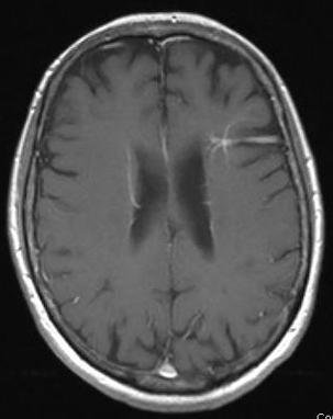
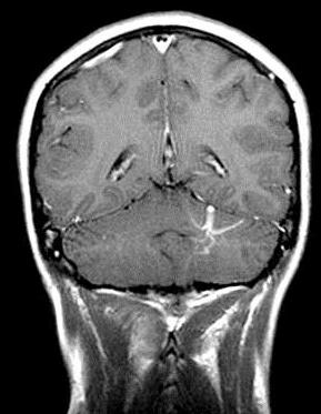
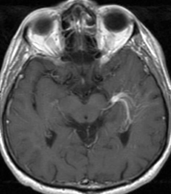
11 Developmental venous anomalies MRI: T1 with contrast

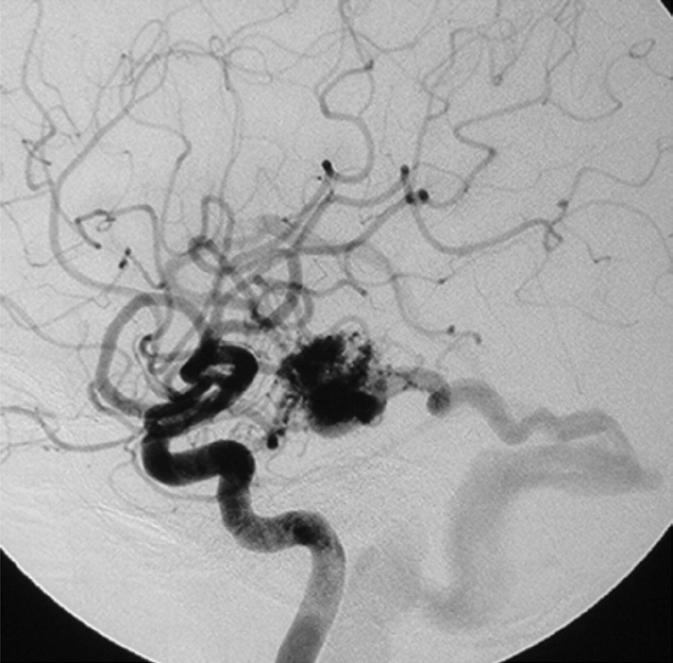
12 Developmental venous anomalies Angiography
 Incidental and asymptomatic Essentially do not hemorrhage Can be solitary or multiple Angiographically occult Do not touch, no follow")
13 Capillary telangiectasias (aka capillary angiomas, capillary malformations, captels) 2 nd most common CVM (10-20%) Pathology Dilated capillaries-normal intervening brain Brainstem most common (medulla / pons) Incidental and asymptomatic Essentially do not hemorrhage Can be solitary or multiple Angiographically occult Do not touch, no follow up

14 Capillary telangiectasias Imaging CT Usually negative May show mild hyperdensity, calcification, or enhancement MRI (choice) Unenhanced images often negative May have mild hyperintensity on T2/FLAIR images Signal loss on gradient echo or susceptibility-weighted MRI Mild contrast enhancement Angiography Negative No role for endovascular intervention MRI T1 without contrast MRI T1 with contrast MRI T2 MRI SWI
 Pathology Collections of blood filled caverns, no normal")

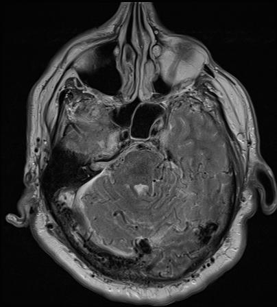
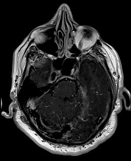
15 Cavernous malformations (aka cavernous angiomas, cavernomas, cavernous hemangiomas) Pathology Collections of blood filled caverns, no normal brain 50% calcify %/yr bleed risk 80% supratentorial 25-40% associated DVA Present 40-60yrs 0.5% population Gross appearance resembles a mulberry
 Familial 20-30% of cases More likely to have multiple")

16 Cavernous malformations Two forms: Sporadic 70-80% of cases More likely to have solitary lesions (~65-70%) Familial 20-30% of cases More likely to have multiple lesions (~70-75%) Lesions more likely to increase in size and number over time Highest incidence in families of Mexican descent 40% remain asymptomatic despite multiple lesions

17 Cavernous malformations Presentation and clinical features Asymptomatic (30-50%) Seizures (60%) Focal neurological deficits (up to 50%) Headaches (25-50%) Natural history May demonstrate spontaneous enlargement, regression, de novo formation, or remain unchanged with time Hemorrhage All lesions believed to undergo chronic microhemorrhages Typically bleed in small amounts, with episodes separated by months or years Symptoms may wax and wane with each bleed and resorption of blood products
 Gradient echo sequences detect lesions with greater sensitivity Angiography Negative No")
18 Cavernous malformations Imaging CT Could be negative in small lesions May show hyperdensity related to calcification or hemorrhage May demonstrate mild or prominent enhancement MRI Imaging study of choice Mixed signal intensity on T1/T2 images surrounded by a black rim of hemosiderin ( popcorn appearance) Gradient echo sequences detect lesions with greater sensitivity Angiography Negative No role for endovascular intervention May be useful to exclude other vascular malformations if imaging is unclear CT FLAIR T2

19 Cavernous malformations Treatment options Conservative management Surgery If asymptomatic or only mild symptoms Surveillance imaging is an option Medical management geared towards seizure control Recurrent symptomatic hemorrhages Intractable seizures or other progressive neurologic deficits Surgically accessible lesions Stereotactic Radiosurgery Controversial, still under evaluation, but several small series and reviews have shown a benefit in rebleeding rates and seizure control May be an option for symptomatic lesions that are not accessible to surgery
 Arteriovenous malformations Arteriovenous")
20 Intermission Half way there Non- shunting (low flow) Developmental venous anomalies Capillary telangiectasias Cavernous malformations Shunting (high flow) Arteriovenous malformations Arteriovenous fistulas

21 High Flow SHUNTING LESIONS

22 Shunting lesions Types Arteriovenous Malformations Arteriovenous fistula Arteriovenous fistula

23 Arteriovenous malformations vs fistulas Both are abnormal connections between arteries and veins without an intervening capillary bed Essentially a short circuit in the vasculature, resulting in artery to vein shunting, or rapid flow of arterial blood into the venous system


24 What s the difference between an AV malformation and AV fistula? AVM: the transition from artery to vein takes place through an abnormal tangle of blood vessels called a nidus (Latin for nest ) AVF: direct connection between artery and vein without an intervening nidus

25 Arteriovenous malformations Pathology Gross appearance described as a snarl of tangled vessels, or a bag of worms Nidus may be compact or diffuse, and range in size from a few mm to entire hemisphere Functional brain tissue usually not present within a compact nidus, but may be present in diffuse lesions Gliosis, fibrosis, and calcification may be present in the adjacent brain parenchyma

26 Arteriovenous malformations Histopathology Arteries Abnormally dilated Degenerative changes due to high flow Prone to aneurysm formation There may be single or multiple feeding arteries Nidus Vessels may resemble arteries, veins, or be dysplastic May contain aneurysms and islands of sclerotic tissue Veins Arterialized and thickened due to high flow Venous aneurysms or varices may develop There may be single or multiple draining veins

27 Arteriovenous malformations Etiology Unclear, but felt to be congenital/developmental not genetic Felt to be dynamic lesions over lifespan Epidemiology Estimates of prevalence in the general population range from to 0.6% Slight male preponderance (55%) Mean age at diagnosis: 31 years Most AVMs are sporadic Related syndromes Familial intracranial AVMs (rare) Hereditary hemorrhagic telangiectasia (Rendu Osler Weber Syndrome) Wyburn-Mason Syndrome Sturge-Weber Syndrome
 Risk decreases to baseline after 3-5")
28 Arteriovenous malformations Natural history- Risk of AVM hemorrhage varies based on prior hemorrhage Without prior hemorrhage: 2-4% per year Not well understood With prior hemorrhage: Approximately 7% in first year after hemorrhage (although some studies report up to 17% in first year) Risk decreases to baseline after 3-5 years
 Presence of aneurysms (15-25% have aneurysms) Deep or posterior fossa")
29 Arteriovenous malformations Peak age for hemorrhage:15-20 yrs With each bleed: Mortality: 10% (5-30%) Morbidity: 20-30% Risk factors for hemorrhage Prior hemorrhage Smaller size Venous drainage Deep Single draining vein Impaired drainage (venous stenosis) Presence of aneurysms (15-25% have aneurysms) Deep or posterior fossa location
30 Arteriovenous malformations Clinical presentation Hemorrhage Sx- Seizures, HA, focality ICH 1-2 % of all ICH 3% in young adults SAH 9% all SAH IVH IVH Seizures (20-25%) Headaches (15%) Focal deficits / LD (Shunting<5%)


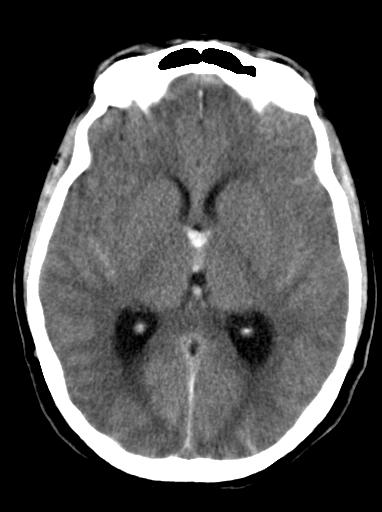
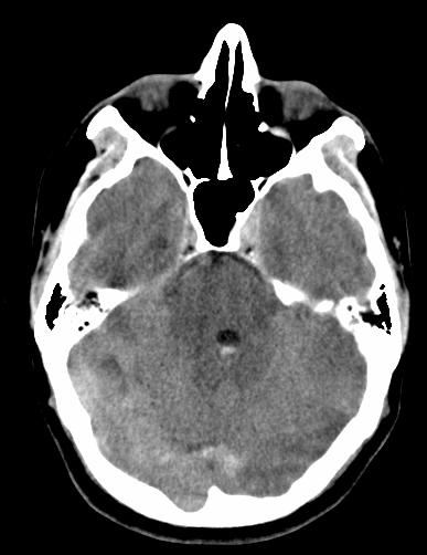
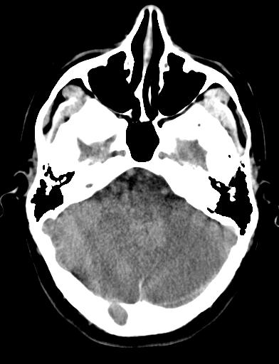
31 CT Arteriovenous malformations Imaging Non contrast CT MRI Will show acute hemorrhage May be negative for small, unruptured AVMs CT with contrast or CTA may show serpentine, enlarged veins and increased vascularity More sensitive than CT for subtle lesions Precise anatomic localization of lesions Post contrast CT Angiography Gold standard Greater sensitivity for small or subtle AVMs Defines anatomy of feeding arteries and draining veins Most sensitive study for detection of aneurysms
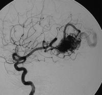
32 Arteriovenous malformations Angiogram

33 Arteriovenous malformations Management options Conservative management Surgery Stereotactic radiosurgery Embolization Combination Who to intervene on w/ hemorrhage clear Asymptomatic lesions less clear AVMs are complex lesions Need to take into account multiple variables before deciding on best treatment option for an individual patient *AVM Management Equipoise Survey: physician opinions regarding the management of brain arteriovenous malformations Kevin M Cockroft1,2,3, Ki-Eun Chang4, Erik B Lehman3, Robert E Harbaugh- J NeuroIntervent Surg ;2013

34 Arteriovenous malformations Who to treat. Multidisciplinary international randomized, controlled trial 800 adult patients with unruptured brain AVM Randomly assigned to prophylactic invasive therapy vs medical management Invasive therapy: endovascular, neurosurgical, and/or radiosurgery (alone or in combo) Follow for 5-10 years Primary endpoint Death from any cause Stroke (sx with imaging findings) Secondary endpoint: long term clinical status Mohr JP et al., Lancet Feb 15;383(9917):614-21
 in medical management group vs 35 (30.")
35 Arteriovenous malformation-aruba Randomization from April 2007 April 2013 Study stopped on recommendation of data and safety monitoring board appointed by NINDS/NIH because of superiority of medical management group 223 patients enrolled Mean follow-up 33.3 months Primary endpoint reached by 11 (10.1%) in medical management group vs 35 (30.7%) in interventional group
36 Arteriovenous malformation ARUBA-Limitations Length of follow up not long enough Unfairly biased toward procedural complications Not detect long-term benefits of prophylactic intervention Lack of standardization of treatment arm high risk lesions were selected out Did not control how patients treatments were selected Treatments should be tested individually to see merit Heterogeneous group Primary outcome of any sx stroke with imaging findings should be better defined (ie mrs>2)

37 Arteriovenous malformations Surgery Gold standard for treatment of small, accessible lesions Risk stratification using Spetzler- Martin grading system Timing of surgery after a bleed Early: Late: Clot has significant mass effect Lesion is surgically accessible Smaller clot burden If patient is a poor surgical candidate soon after the bleed Early imaging studies may not show AVM clearly / in total
38 Spetzler-Martin Grading Scale for AVMs Predicts surgical risk of AVMs

 Overall: 8.")
39 Surgical outcomes of AVMs Obliteration rates Spetzler-Martin Grades I-III: % Grades I-III account for 60-80% of all AVMs Grades IV-V: little data Complications Post-op mortality (death) Overall: 3.3% Grades I-III: 0-3.9% Grades IV-V: 11-38% Post-op morbidity(deficit) Overall: 8.6% Grades I-III: 0-5% Grades IV-V: 12-22%

40 Arteriovenous malformations Stereotactic radiosurgery Cyberknife Suite, Christiana Care, Newark, DE Involves delivering multiple beams of radiation, each from a different location All beams converge on the target, or iso-center Radiation dose high to the iso-center, but low to nontargeted structures
 Staged treatment can be considered for some larger lesions Latency period of about 2 years for AVM")
41 Arteriovenous malformations Stereotactic radiosurgery Advantages Minimally invasive Relatively low-risk Useful for treatment of surgically inaccessible lesions Disadvantages Most effective for smaller lesions (< 3 cm) Staged treatment can be considered for some larger lesions Latency period of about 2 years for AVM obliteration

42 Arteriovenous malformations Stereotactic radiosurgery outcomes Obliteration rates Lesions diameter < 3 cm: 75-95% Lesion diameter > 3 cm: </= 70% Complications Overall rate of neurological complications: 8% Radiation injury to brain: 6% Permanent neurologic deficits: 4.8% Radiation necrosis
43 Arteriovenous malformation Radiosurgery 28 year old female with severe headaches Worsening over time Intermittent left sided weakness


44 Arteriovenous malformation Radiosurgery MRI pre treatment Angiogram pre treatment AP image Lateral image


45 Arteriovenous malformation Radiosurgery 2yr Post Treatment MRI 2yr Post Treatment Angio


46 Treatment goals Risk Arteriovenous malformations Embolization Cure 5-10% cases Targeted therapy Hemorrhage risk / tx high risk features Adjunct to surgery or SRS Pre-surgical embolization Reduce volume of nidus Shorten operative time and blood loss Pre-SRS embolization Reduce volume of nidus If SRS does not obliterate AVM, repeat embolization can be performed Permanent morbidity: 2-14% Mortality: 1-3.7%
47 Arteriovenous malformation Endovascular Case 43 year old female with intractable headaches and medically refractory seizures No evidence of hemorrhage on NCCT MRI suggested AVM Diagnostic cerebral angiogram confirmed Treatment with Onyx Embolization


48 Arteriovenous malformation Endovascular Case- cure Pre Embolization Post Embolization Embolic Cast
49 Arteriovenous malformation Endovascular Case 55 year old male with sudden onset headache Evidence of hemorrhage on NCCT MRI suggested AVM Diagnostic cerebral angiogram confirmed high risk features Treatment with Onyx Embolization
50 Arteriovenous malformation Endovascular Targeted treatment


51 Arteriovenous malformation Endovascular Targeted treatment MRI without contrast


52 Arteriovenous malformation Endovascular Targeted treatment Angiogram


53 Arteriovenous malformations Conservative management Asymptomatic AVMs Large or difficult to treat lesions Patients at high risk of complications Sometimes okay to accept the risk of hemorrhage when the risk of treatment is too high

54 Arteriovenous fistula Occur anywhere along dura Direct communication Acquired lesions Trauma Surgery Post-partum Sinus thrombosis
 Presentation depends on location and venous drainage Kiyosue; RadioGraphics 2004; 24:1637")
55 Arteriovenous fistula 10-15% of all intracranial vascular malformations Peak presentation (M=F) Presentation depends on location and venous drainage Kiyosue; RadioGraphics 2004; 24:
56 Arteriovenous fistula- Risk

57 Arteriovenous fistual Poorly understood Prognosis depends on venous drainage 98% w/o CVD benign course With CVD aggressive course Hemorrhage Neurological symptoms Natural history


58 Arteriovenous fistula Treatment Who to treat Benign Pre treatment Only severe symptoms Malignant Secondary to high risk of bleed Embolization Mainstay of treatment Surgery adjunctive Cast Post treatment
59 Arteriovenous fistula Case 70 year old male with rapidly progressive dementia and Parkinson-like tremors Multiple trips to the ER Last ER visit became comatose (GCS 3) and was transferred as possible ETOH withdrawal
60 Arteriovenous fistula T2 Malignant T1 post gad

61 Arteriovenous fistula Angiogram Multiple bilateral feeders Occluded R transverse-sigmoid Retrograde filling of deep and superficial veins Embolized multiple branches Anterograde venous flow Resolution of symptoms Malignant
62 Summary Reviewed and classified the vascular malformations of the brain and discussed potential treatments Cerebral AVMs are complex, congenital lesions with a significant hemorrhage risk Cerebral AVMs may require a combination of embolization, surgery, and/or SRS for treatment Cerebral arteriovenous fistulas are acquired lesions that have risk from hemorrhage related to venous drainage and are treated with embolization primarily

63 Thank you!
Vascular Malformations of the Brain: A Review of Imaging Features and Risks
 Vascular Malformations of the Brain: A Review of Imaging Features and Risks Comprehensive Neuroradiology: Best Practices October 27-30, 2016 Sudhakar R. Satti, MD Associate Director Neurointerventional
Vascular Malformations
 Vascular Malformations LTC Robert Shih Chief of Neuroradiology Walter Reed Medical Center Special thanks to LTC Alice Smith (retired) Disclosures: None. This presentation reflects the personal views of
VASCULAR MALFORMATIONS. Owen Samuels, MD Adam Webb, MD Emory University
 VASCULAR MALFORMATIONS Owen Samuels, MD Adam Webb, MD Emory University Introduction Brain and spinal cord vascular malformations can be separated into five main categories: 1) Arteriovenous malformation,
Vascular malformations: Venous malformations anomalous veins drain normal brain tissue for 65% of all cases 2.5%. was 0, 3% per year
 Vascular malformations: 1. Venous malformations: congenital venous anomalies pathologically characterised by anomalous veins (thickened and hyalinised walls) separated by normal brain. These anatomically
What Is an Arteriovenous malformation (AVM)?
?") American Society of Neuroradiology What Is an Arteriovenous malformation (AVM)? From the Cerebrovascular Imaging and Intervention Committee of the American Heart Association Cardiovascular Council Randall
American Society of Neuroradiology What Is an Arteriovenous malformation (AVM)? From the Cerebrovascular Imaging and Intervention Committee of the American Heart Association Cardiovascular Council Randall
Life after ARUBA: Management of Unruptured Brain Arteriovenous Malformations (AVMs)
") Life after ARUBA: Management of Unruptured Brain Arteriovenous Malformations (AVMs) Eric L. Zager, MD University of Pennsylvania Department of Neurosurgery No Disclosures Brain AVMs Incidence ~1 in 100,000
Life after ARUBA: Management of Unruptured Brain Arteriovenous Malformations (AVMs) Eric L. Zager, MD University of Pennsylvania Department of Neurosurgery No Disclosures Brain AVMs Incidence ~1 in 100,000
Endovascular Treatment of Cerebral Arteriovenous Malformations. Bs. Nguyễn Ngọc Pi Doanh- Bs Đặng Ngọc Dũng Khoa Ngoại Thần Kinh
 Endovascular Treatment of Cerebral Arteriovenous Malformations Bs. Nguyễn Ngọc Pi Doanh- Bs Đặng Ngọc Dũng Khoa Ngoại Thần Kinh Stroke Vascular Malformations of the Brain Epidemiology: - Incidence: 0.1%,
Radiographic and statistical analysis of Brain Arteriovenous Malformations.
 Radiographic and statistical analysis of Brain Arteriovenous Malformations. Poster No.: C-0996 Congress: ECR 2017 Type: Educational Exhibit Authors: C. E. Rodriguez 1, A. Lopez Moreno 1, D. Sánchez Paré
Diagnosis and Management of AVM in the Pregnant Patient
 Diagnosis and Management of AVM in the Pregnant Patient Wade Cooper, D.O. University of Michigan Assistant Professor Departments of Neurology & Anesthesiology Disclosures Wade Cooper - None Developmental
Vascular Malformations of the Brain. William A. Cox, M.D. Forensic Pathologist/Neuropathologist. September 8, 2014
 Vascular Malformations of the Brain William A. Cox, M.D. Forensic Pathologist/Neuropathologist September 8, 2014 Vascular malformations of the brain are classified into four principal groups: arteriovenous
Neurosurgical decision making in structural lesions causing stroke. Dr Rakesh Ranjan MS, MCh, Dip NB (Neurosurgery)
") Neurosurgical decision making in structural lesions causing stroke Dr Rakesh Ranjan MS, MCh, Dip NB (Neurosurgery) Subarachnoid Hemorrhage Every year, an estimated 30,000 people in the United States experience
Neurosurgical decision making in structural lesions causing stroke Dr Rakesh Ranjan MS, MCh, Dip NB (Neurosurgery) Subarachnoid Hemorrhage Every year, an estimated 30,000 people in the United States experience
Brain AVM with Accompanying Venous Aneurysm with Intracerebral and Intraventricular Hemorrhage
 Cronicon OPEN ACCESS EC PAEDIATRICS Case Report Brain AVM with Accompanying Venous Aneurysm with Intracerebral and Intraventricular Hemorrhage Dimitrios Panagopoulos* Neurosurgical Department, University
Dural Arteriovenous Malformations and Fistulae (DAVM S DAVF S)
") Jorge Guedes Campos NEUROIMAGING DEPARTMENT HOSPITAL SANTA MARIA UNIVERSITY OF LISBON PORTUGAL DEFINITION region of arteriovenous shunting confined to a leaflet of packymeninges often adjacent to a major
Jorge Guedes Campos NEUROIMAGING DEPARTMENT HOSPITAL SANTA MARIA UNIVERSITY OF LISBON PORTUGAL DEFINITION region of arteriovenous shunting confined to a leaflet of packymeninges often adjacent to a major
Supratentorial cerebral arteriovenous malformations : a clinical analysis
 Original article: Supratentorial cerebral arteriovenous malformations : a clinical analysis Dr. Rajneesh Gour 1, Dr. S. N. Ghosh 2, Dr. Sumit Deb 3 1Dept.Of Surgery,Chirayu Medical College & Research Centre,
General Data. Gender: Female Birthday and age: 1932/11/03, 73 y/o Occupation: house keeper Date of Admission: 2005/03/30
 General Data Gender: Female Birthday and age: 1932/11/03, 73 y/o Occupation: house keeper Date of Admission: 2005/03/30 Chief Complain Dizziness and light headache for recent 1 year. Present illness Hypertension
Cerebrovascular Malformations in the Elderly Indications for Treatment
 Cerebrovascular Malformations in the Elderly Indications for Treatment Johanna T. Fifi, MD, FAHA, FSVIN Director of Endovascular Ischemic Stroke Assistant Professor of Neurology, Neurosurgery, and Radiology
Historical perspective
 SPINAL AVM Introduction Vascular malformations of spinal cord are a rare clinical entity, representing 5% of all primary spinal cord lesions, with arteriovenous malformations(avm) & cavernous malformations
Untangling Cerebral Dural Arteriovenous Fistulas
 Untangling Cerebral Dural Arteriovenous Fistulas Bradley A. Gross, MD Assistant Professor, Dept of Neurosurgery, University of Pittsburgh September 2017 davfs Definition Clinical Presentation Natural History
Diffuse Proliferative Cerebral Angiopathy: A case report and review of the literature
 Diffuse Proliferative Cerebral Angiopathy: A case report and review of the literature Rohit 1*, Poh Sun Goh 1 1. Department of Radiology, National University hospital, Singapore * Correspondence: Dr. Rohit,
Clinical Commissioning Policy: Arteriovenous Malformations. December Reference : NHSCB/D5/4
 Clinical Commissioning Policy: Arteriovenous Malformations December 2012 Reference : NHSCB/D5/4 NHS Commissioning Board Clinical Commissioning Policy: Arteriovenous Malformations First published: December
Marc Norman, Ph.D. - Do Not Use without Permission 1. Cerebrovascular Accidents. Marc Norman, Ph.D. Department of Psychiatry
 Cerebrovascular Accidents Marc Norman, Ph.D. Department of Psychiatry Neuropsychiatry and Behavioral Medicine Neuropsychology Clinical Training Seminar 1 5 http://www.nlm.nih.gov/medlineplus/ency/images/ency/fullsize/18009.jpg
Modern Management of ICH
 Modern Management of ICH Bradley A. Gross, MD Assistant Professor, Dept of Neurosurgery, University of Pittsburgh October 2018 ICH Background Assessment & Diagnosis Medical Management Surgical Management
Brain Arteriovenous Malformations Endovascular Therapy and Associated Therapeutic Protocols Jorge Guedes Cabral de Campos
 Endovascular Therapy and Associated Therapeutic Protocols Jorge Guedes Cabral de Campos Neuroradiology Department Hospital de Santa Maria University of Lisbon CEREBRAL AVM CLINICAL / EPIDEMIOLOGY Brain
Explaining All of the Options for AVM: Cerebral Arteriovenous Malformation
 Explaining All of the Options for AVM: Cerebral Arteriovenous Malformation Recorded on: November 19, 2012 Bernard Bendok, M.D. Director of the Neurointerventional Program Northwestern Memorial Hospital
Contents. 1 Embryological and Anatomical Introduction... 1
 1 Embryological and Anatomical Introduction.... 1 1.1 Preliminary Remarks.................... 1 1.2 Leptomeninges....................... 21 1.3 Subpial Space........................ 22 1.3.1 Anatomy...........................
Intracranial spontaneous hemorrhage mechanisms, imaging and management
 Intracranial spontaneous hemorrhage mechanisms, imaging and management Dora Zlatareva Department of Diagnostic Imaging Medical University, Sofia, Bulgaria Intracranial hemorrhage (ICH) ICH 15% of strokes
EMBOLIZATION OF ARTERIOVENOUS FISTULA AFTER RADIOSURGERY FOR MULTIPLE CEREBRAL ARTERIOVENOUS MALFORMATIONS
 Arteriovenous fistula after radiosurgery for multiple CAVM EMBOLIZATION OF ARTERIOVENOUS FISTULA AFTER RADIOSURGERY FOR MULTIPLE CEREBRAL ARTERIOVENOUS MALFORMATIONS Chao-Bao Luo, Wan-Yuo Guo, Michael
INTRACRANIAL CAVERNOMA
 INTRACRANIAL CAVERNOMA INTRODUCTION CEREBRAL CAVERNOUS MALFORMATION (CCM), CAVERNOUS HEMANGIOMA, CAVERNOUS ANGIOMA, CRYPTIC VASCULAR MALFORMATION,OCCULT VASCULAR MALFORMATION, HEMORRHOID OF BRAIN. Developmental
CENTRAL NERVOUS SYSTEM TRAUMA and Subarachnoid Hemorrhage. By: Shifaa AlQa qa
 CENTRAL NERVOUS SYSTEM TRAUMA and Subarachnoid Hemorrhage By: Shifaa AlQa qa Subarachnoid Hemorrhage Causes: Rupture of a saccular (berry) aneurysm Vascular malformation Trauma Hematologic disturbances
CLEAR III TRIAL : UPDATE ON SURGICAL MATTERS THAT MATTER
 CLEAR III TRIAL : UPDATE ON SURGICAL MATTERS THAT MATTER CLEAR Surgical Center Team July 2011 Trial Enrollment Status Updates Insert latest enrollment update chart from most recent CLEAR newsletter Imaging
7/5/2016. Neonatal high-output cardiac failure. Case 1 POSTNATAL STRATEGIES FOR CEREBRAL ATERIOVENOUS MALFORMATIONS
 John Deveikis, M.D. POSTNATAL STRATEGIES FOR CEREBRAL ATERIOVENOUS MALFORMATIONS JULY, 2016 Neonatal high-output cardiac failure Tachypnea, tachycardia, hypotension, failure to thrive When congenital heart
DOWNLOAD PDF RADIOSURGERY FOR CAVERNOUS MALFORMATIONS IN BASAL GANGLIA, THALAMUS AND BRAINSTEM KIDA, Y
 Chapter 1 : Stereotactic radiosurgery for cavernous malformations â Mayo Clinic Most of the lesions were located in the brainstem, followed by the lobar region, cerebellum, thalamus, and basal ganglia
[(PHY-3a) Initials of MD reviewing films] [(PHY-3b) Initials of 2 nd opinion MD]
![[(PHY-3a) Initials of MD reviewing films] [(PHY-3b) Initials of 2 nd opinion MD]](/thumbs/89/98619893.jpg "[(PHY-3a) Initials of MD reviewing films] [(PHY-3b) Initials of 2 nd opinion MD]") 2015 PHYSICIAN SIGN-OFF (1) STUDY NO (PHY-1) CASE, PER PHYSICIAN REVIEW 1=yes 2=no [strictly meets case definition] (PHY-1a) CASE, IN PHYSICIAN S OPINION 1=yes 2=no (PHY-2) (PHY-3) [based on all available
2015 PHYSICIAN SIGN-OFF (1) STUDY NO (PHY-1) CASE, PER PHYSICIAN REVIEW 1=yes 2=no [strictly meets case definition] (PHY-1a) CASE, IN PHYSICIAN S OPINION 1=yes 2=no (PHY-2) (PHY-3) [based on all available
2. Subarachnoid Hemorrhage
 Causes: 2. Subarachnoid Hemorrhage A. Saccular (berry) aneurysm - Is the most frequent cause of clinically significant subarachnoid hemorrhage is rupture of a saccular (berry) aneurysm. B. Vascular malformation
Skull radiographs... 5 CT... 5 MRI... 15
 INTRACRANIAL VASCULAR MALFORMATIONS Vas30 (1) Intracranial Vascular Malformations Last updated: September 5, 2017 ARTERIOVENOUS MALFORMATIONS (AVM)... 2 PATHOLOGY, PATHOPHYSIOLOGY... 2 Hemodynamics...
Sonography of soft-tissue vascular lesions
 Sonography of soft-tissue vascular lesions Oscar M. Navarro Associate Professor, University of Toronto Dept. of Diagnostic Imaging, The Hospital for Sick Children Toronto, Canada Declaration of Disclosure
Journal of Radiology Case Reports
 Pediatric Holohemispheric Developmental Venous Anomaly: Definitive characterization by 3D Susceptibility Weighted Magnetic Resonance Angiography Michael A. Casey 1, Sourabh Lahoti 2, Ajeet Gordhan 2* 1.
Summary of some of the landmark articles:
 Summary of some of the landmark articles: The significance of unruptured intracranial saccular aneurysms: Weibers et al Mayo clinic. 1987 1. 131 patients with 161 aneurysms were followed up at until death,
Diagnosis of Subarachnoid Hemorrhage (SAH) and Non- Aneurysmal Causes
 and Non- Aneurysmal Causes") Diagnosis of Subarachnoid Hemorrhage (SAH) and Non- Aneurysmal Causes By Sheila Smith, MD Swedish Medical Center 1 Disclosures I have no disclosures 2 Course Objectives Review significance and differential
Diagnosis of Subarachnoid Hemorrhage (SAH) and Non- Aneurysmal Causes By Sheila Smith, MD Swedish Medical Center 1 Disclosures I have no disclosures 2 Course Objectives Review significance and differential
Biomedical Research 2017; 28 (2):
:") Biomedical Research 2017; 28 (2): 957-962 ISSN 0970-938X www.biomedres.info Analysis on the effect and prognostic factors of cerebral arteriovenous malformations (AVM) after endovascular embolization combined
Biomedical Research 2017; 28 (2): 957-962 ISSN 0970-938X www.biomedres.info Analysis on the effect and prognostic factors of cerebral arteriovenous malformations (AVM) after endovascular embolization combined
A.J. Hauer Intracranial dural arteriovenous fistulae
 A.J. Hauer 27-06-2018 Intracranial dural arteriovenous fistulae Dural arteriovenous fistulae (davfs) epidemiology Pathological anastomoses (within the dural leaflets) between meningeal arteries and dural
TCD AND VASOSPASM SAH
 CURRENT TREATMENT FOR CEREBRAL ANEURYSMS TCD AND VASOSPASM SAH Michigan Sonographers Society 2 Nd Annual Fall Vascular Conference Larry N. Raber RVT-RDMS Clinical Manager General Ultrasound-Neurovascular
INTRACRANIAL CAVERNOMA
 INTRACRANIAL CAVERNOMA PRESENTER : Dr Shameem Ahmed MODERATOR : Dr P.S. Chandra Dr G.D. Satyarthee DEPT OF NEUROSUGERY, AIIMS INTRODUCTION CEREBRAL CAVERNOUS MALFORMATION (CCM), CAVERNOUS HEMANGIOMA, CAVERNOUS
Neurosurgical Management of Stroke
 Overview Hemorrhagic Stroke Ischemic Stroke Aneurysmal Subarachnoid hemorrhage Neurosurgical Management of Stroke Jesse Liu, MD Instructor, Neurological Surgery Initial management In hospital management
Retrospective analytical six months study of vascular abnormalities of brain
 International Journal of Advances in Medicine http://www.ijmedicine.com pissn 2349-3925 eissn 2349-3933 Research Article DOI: http://dx.doi.org/10.18203/2349-3933.ijam20160185 Retrospective analytical
Transverse-Sigmoid Sinus Dural Arteriovenous Malformations
 Transverse-Sigmoid Sinus Dural Arteriovenous Malformations Kenan I. Amautovic, M.D., and Ali F. Krisht, M.D. '-...--- Learning Objectives: After reading this article, the participant should: 1. Have an
Cryptogenic Enlargement Of Bilateral Superior Ophthalmic Veins
 ISPUB.COM The Internet Journal of Radiology Volume 18 Number 1 Cryptogenic Enlargement Of Bilateral Superior Ophthalmic Veins K Kragha Citation K Kragha. Cryptogenic Enlargement Of Bilateral Superior Ophthalmic
Occlusive hyperemia: a theory for the hemodynamic complications following resection of intracerebral arteriovenous malformations
 J Neurosurg 78: 167-175, 1993 Occlusive hyperemia: a theory for the hemodynamic complications following resection of intracerebral arteriovenous malformations NAYEF R. F. AL-RODHAN, M.D., PH.D., THORALF
Ultrasound imaging of vascular anomalies: pearls and pitfalls
 Ultrasound imaging of vascular anomalies: pearls and pitfalls Oscar Navarro, MD Dept. of Medical Imaging, University of Toronto Dept. of Diagnostic Imaging, The Hospital for Sick Children Declaration of
NEURORADIOLOGY Part I
 NEURORADIOLOGY Part I Vörös Erika University of Szeged Department of Radiology SZEGED BRAIN IMAGING METHODS Plain film radiography Ultrasonography (US) Computer tomography (CT) Magnetic resonance imaging
Pediatric Neurointervention: Vein of Galen Malformations
 Pediatric Neurointervention: Vein of Galen Malformations Johanna T. Fifi, M.D. Assistant Professor of Neurology, Neurosurgery, and Radiology Icahn School of Medicine at Mount Sinai November 9 th, 2014
Selective disconnection of cortical venous reflux as treatment for cranial dural arteriovenous fistulas
 J Neurosurg 101:31 35, 2004 Selective disconnection of cortical venous reflux as treatment for cranial dural arteriovenous fistulas J. MARC C. VAN DIJK, M.D., PH.D., KAREL G. TERBRUGGE, M.D., ROBERT A.
Spinal Vascular Lesions
 Spinal Vascular Lesions Spinal Vascular Lesions Spinal cord infarction Hemangioblastoma Cavernous malformation Vascular malformations (Type 1-4) Spinal artery aneurysm Troy Hutchins, MD Assistant Professor
Uncommon Symptomatic Cerebral Vascular Malformations
 Uncommon Symptomatic Cerebral Vascular Malformations Mauro Bergui and Gianni Boris Bradac Summary: We describe three cases of unusual vascular malformations in which the most relevant angiographic findings
North Oaks Trauma Symposium Friday, November 3, 2017
 Traumatic Intracranial Hemorrhage Aaron C. Sigler, DO, MS Neurosurgery Tulane Neurosciences None Disclosures Overview Anatomy Epidural hematoma Subdural hematoma Cerebral contusions Outline Traumatic ICH
Identifying Cerebrovascular Disorders. Wengui Yu, MD, PhD Department of Neurology, University of California, Irvine
 Identifying Cerebrovascular Disorders Wengui Yu, MD, PhD Department of Neurology, University of California, Irvine Objectives Review different types of cerebrovascular disorders. Briefly discuss etiology,
Intracranial dural arteriovenous fistulas (DAVFs) with retrograde
 with retrograde") ORIGINAL RESEARCH W.J. van Rooij M. Sluzewski G.N. Beute Dural Arteriovenous Fistulas with Cortical Venous Drainage: Incidence, Clinical Presentation, and Treatment BACKGROUND AND PURPOSE: Our purpose
ORIGINAL RESEARCH W.J. van Rooij M. Sluzewski G.N. Beute Dural Arteriovenous Fistulas with Cortical Venous Drainage: Incidence, Clinical Presentation, and Treatment BACKGROUND AND PURPOSE: Our purpose
Spontaneous occlusion of a cerebral arteriovenous malformation after subtotal endovascular embolisation
 206 Chiriac et al Spontaneous occlusion of a cerebral arteriovenous malformation Spontaneous occlusion of a cerebral arteriovenous malformation after subtotal endovascular embolisation A. Chiriac, N. Dobrin*,
Cerebrovascular Disorders. Blood, Brain, and Energy. Blood Supply to the Brain 2/14/11
 Cerebrovascular Disorders Blood, Brain, and Energy 20% of body s oxygen usage No oxygen/glucose reserves Hypoxia - reduced oxygen Anoxia - Absence of oxygen supply Cell death can occur in as little as
ANALYSIS OF TREATMENT OUTCOMES WITH LINAC BASED STEREOTACTIC RADIOSURGERY IN INTRACRANIAL ARTERIOVENOUS MALFORMATIONS
 ANALYSIS OF TREATMENT OUTCOMES WITH LINAC BASED STEREOTACTIC RADIOSURGERY IN INTRACRANIAL ARTERIOVENOUS MALFORMATIONS Dr. Maitri P Gandhi 1, Dr. Chandni P Shah 2 1 Junior resident, Gujarat Cancer & Research
a. Ischemic stroke An acute focal infarction of the brain or retina (and does not include anterior ischemic optic neuropathy (AION)).
).") 12.0 Outcomes 12.1 Definitions 12.1.1 Neurologic Outcome Events a. Ischemic stroke An acute focal infarction of the brain or retina (and does not include anterior ischemic optic neuropathy (AION)). Criteria:
12.0 Outcomes 12.1 Definitions 12.1.1 Neurologic Outcome Events a. Ischemic stroke An acute focal infarction of the brain or retina (and does not include anterior ischemic optic neuropathy (AION)). Criteria:
Spontaneous Obliteration of Pial Arteriovenous Malformations: A Review of 27 Cases
 AJNR Am J Neuroradiol :, March 00 Spontaneous Obliteration of Pial Arteriovenous Malformations: A Review of ases Maneesh. Patel, Timothy J. Hodgson, Andras A. Kemeny, and David M. Forster BAKGROUND AND
NIH Public Access Author Manuscript J Am Coll Radiol. Author manuscript; available in PMC 2013 June 24.
 NIH Public Access Author Manuscript Published in final edited form as: J Am Coll Radiol. 2010 January ; 7(1): 73 76. doi:10.1016/j.jacr.2009.06.015. Cerebral Aneurysms Janet C. Miller, DPhil, Joshua A.
Definition พ.ญ.ส ธ ดา เย นจ นทร. Epidemiology. Definition 5/25/2016. Seizures after stroke Can we predict? Poststroke seizure
 Seizures after stroke Can we predict? พ.ญ.ส ธ ดา เย นจ นทร PMK Epilepsy Annual Meeting 2016 Definition Poststroke seizure : single or multiple convulsive episode(s) after stroke and thought to be related
SPINAL EPIDURAL ARTERIOVENOUS MALFORMATIONS: REPORT OF A CASE WITH DISCUSSION OF CLASSIFICATION AND TREATMENT
 SPINAL EPIDURAL ARTERIOVENOUS MALFORMATIONS: REPORT OF A CASE WITH DISCUSSION OF CLASSIFICATION AND TREATMENT Caitlin M. Clark, B.A. and W. Craig Clark, M.D., Ph.D., FAANS, FACS, FICS INTRODUCTION True
This article appeared in a journal published by Elsevier. The attached copy is furnished to the author for internal non-commercial research and
 This article appeared in a journal published by Elsevier. The attached copy is furnished to the author for internal non-commercial research and education use, including for instruction at the authors institution
The outcome of treatment for arteriovenous malformations of the brain: A five-year retrospective series from the Philippines
 Neurology Asia 2006; 11 : 91 96 ORIGINAL ARTICLES The outcome of treatment for arteriovenous malformations of the brain: A five-year retrospective series from the Philippines Roland Mark M GIGATARAS MD,
A Case of Carotid-Cavernous Fistula
 A Case of Carotid-Cavernous Fistula By : Mohamed Elkhawaga 2 nd Year Resident of Ophthalmology Alexandria University A 19 year old male patient came to our outpatient clinic, complaining of : -Severe conjunctival
The treatment of brain arteriovenous malformations. Neurologic Complications of Arteriovenous Malformation Embolization Using Liquid Embolic Agents
 ORIGINAL RESEARCH M.V. Jayaraman M.L. Marcellus S. Hamilton H.M. Do D. Campbell S.D. Chang G.K. Steinberg M.P. Marks Neurologic Complications of Arteriovenous Malformation Embolization Using Liquid Embolic
PTA 106 Unit 1 Lecture 3
 PTA 106 Unit 1 Lecture 3 The Basics Arteries: Carry blood away from the heart toward tissues. They typically have thicker vessels walls to handle increased pressure. Contain internal and external elastic
Arteriovenous malformations
 Arteriovenous malformations What are arteriovenous malformations? Arteriovenous malformations (AVMs) are abnormal, snarled tangles of blood vessels that cause multiple irregular connections between the
Vivek R. Deshmukh, MD Director, Cerebrovascular and Endovascular Neurosurgery Chairman, Department of Neurosurgery Providence Brain and Spine
 Vivek R. Deshmukh, MD Director, Cerebrovascular and Endovascular Neurosurgery Chairman, Department of Neurosurgery Providence Brain and Spine Institute The Oregon Clinic Disclosure I declare that neither
Dural arteriovenous fistulas (DAVFs) are abnormal. Long-term angiographic results of endovascularly cured intracranial dural arteriovenous fistulas
 are abnormal. Long-term angiographic results of endovascularly cured intracranial dural arteriovenous fistulas") clinical article J Neurosurg 124:1123 1127, 2016 Long-term angiographic results of endovascularly cured intracranial dural arteriovenous fistulas Sudheer Ambekar, MD, Brandon G. Gaynor, MD, Eric C. Peterson,
clinical article J Neurosurg 124:1123 1127, 2016 Long-term angiographic results of endovascularly cured intracranial dural arteriovenous fistulas Sudheer Ambekar, MD, Brandon G. Gaynor, MD, Eric C. Peterson,
Contents I MEDICAL RADIOLOGY. Diagnostic Imaging. Editors: A. L. Baert, Leuven M. Knauth, Göttingen K. Sartor, Heidelberg
 Contents I MEDICAL RADIOLOGY Diagnostic Imaging Editors: A. L. Baert, Leuven M. Knauth, Göttingen K. Sartor, Heidelberg Contents III M. Forsting I. Wanke (Eds.) Intracranial Vascular Malformations and
MASSIVE EPISTAXIS IN A NEONATE: A SYMPTOM OF VEIN OF GALEN MALFORMATION!
 CASE REPORT MASSIVE EPISTAXIS IN A NEONATE: A SYMPTOM OF VEIN OF GALEN MALFORMATION! Shagufta Wahab 1, Rizwan Ahmad Khan 2, Manjari Thapa Manger 3 1. Radiodiagnosis, Aligarh Muslim University, Aligarh,
C. Douglas Phillips, MD FACR Director of Head and Neck Imaging Weill Cornell Medical College NewYork-Presbyterian Hospital
 C. Douglas Phillips, MD FACR Director of Head and Neck Imaging Weill Cornell Medical College NewYork-Presbyterian Hospital I have no financial disclosures Understand range of pathology that may present
Imaging of Cerebrovascular Disease
 Imaging of Cerebrovascular Disease A Practical Guide Val M. Runge, MD Editor-in-Chief of Investigative Radiology Institute for Diagnostic, Interventional, and Pediatric Radiology Inselspital, University
Onyx in Brain Arteriovenous Malformation Embolisation
 Original Article Submitted: 4 Jan 2016 Accepted: 9 May 2016 Online: 30 June 2016 Onyx in Brain Arteriovenous Malformation Embolisation Hilwati Hashim 1, A Sobri Muda 2, Aida Abdul Aziz 3, Zuhanis Abdul
Khalil Zahra, M.D Neuro-interventional radiology
 Khalil Zahra, M.D Neuro-interventional radiology 1 Disclosure None 2 Outline Etiology and pathogensis Imaging techniques and Features Literature review Treatment modalities Endovascular techniques Long
Acute stroke imaging
 Acute stroke imaging Aims Imaging modalities and differences Why image acute stroke Clinical correlation to imaging appearance What is stroke Classic definition: acute focal injury to the central nervous
Coronary Arteriovenous Malformation presenting as Acute Myocardial Infarction. Choon Ta NG, Aaron WONG, Foong-Koon CHEAH, Chi Keong CHING
 Coronary Arteriovenous Malformation presenting as Acute Myocardial Infarction Choon Ta NG, Aaron WONG, Foong-Koon CHEAH, Chi Keong CHING The patient 49 year old Male presented with Chest tightness x 1
24. An infant with recurrent pneumonia underwent a frontal chest radiograph (Fig 24-A) followed by
 followed by") 24. An infant with recurrent pneumonia underwent a frontal chest radiograph (Fig 24-A) followed by diagnosis? ndings, what is the most likely A. Pulmonary sequestration B. Congenital pulmonary airway malformation
24. An infant with recurrent pneumonia underwent a frontal chest radiograph (Fig 24-A) followed by diagnosis? ndings, what is the most likely A. Pulmonary sequestration B. Congenital pulmonary airway malformation
ARTERIOVENOUS MALFORMATION OR CONTUSION : A DIAGNOSTIC DILEMMA. Yong Pei Yee, Ibrahim Lutfi Shuaib, Jafri Malin Abdullah*
 Malaysian Journal of Medical Sciences, Vol. 8, No. 2, July 2001 (47-51) CASE REPORT ARTERIOVENOUS MALFORMATION OR CONTUSION : A DIAGNOSTIC DILEMMA Yong Pei Yee, Ibrahim Lutfi Shuaib, Jafri Malin Abdullah*
with susceptibility-weighted imaging and computed tomography perfusion abnormalities in diagnosis of classic migraine
 Emerg Radiol (2012) 19:565 569 DOI 10.1007/s10140-012-1051-2 CASE REPORT Susceptibility-weighted imaging and computed tomography perfusion abnormalities in diagnosis of classic migraine Christopher Miller
Dr H. Gharebaghian MD Neurologist Department of Neurology Kermanshah Faculty of Medicine
 Dr H. Gharebaghian MD Neurologist Department of Neurology Kermanshah Faculty of Medicine Definitions Seizures are transient events that include symptoms and/or signs of abnormal excessive hypersynchronous
Non-Invasive Follow-up Evaluation of Post-Embolized AVM with Time-Resolved MRA: A Case Report
 Non-Invasive Follow-up Evaluation of Post-Embolized AVM with Time-Resolved MRA: A Case Report Yong Woon Shim, MD 1 Tae-Sub Chung, MD 1 Won-Suk Kang, MD 1 Jin-Yang Joo, MD 2 Ralph Strecker, MD 3 Juergen
Sinus Venous Thrombosis
 Sinus Venous Thrombosis Joseph J Gemmete, MD FACR, FSIR, FAHA Professor Departments of Radiology and Neurosurgery University of Michigan Hospitals Ann Arbor, MI Outline Introduction Medical Treatment Options
SPINAL AVM: CLASSIFICATION AND MANAGEMENT STRATEGIES. Presented by : Anuj K Tripathi
 SPINAL AVM: CLASSIFICATION AND MANAGEMENT STRATEGIES Presented by : Anuj K Tripathi SPINAL AVM Spinal vascular malformations represent a rare and insufficiently studied pathological entity Great difficulties
SURGICAL TREATMENT OF SPINAL ARTERIOVENOUS MALFORMATIONS: VASCULAR ANATOMY AND SURGICAL OUTCOME
 Spinal Arteriovenous Malformations SURGICAL TREATMENT OF SPINAL ARTERIOVENOUS MALFORMATIONS: VASCULAR ANATOMY AND SURGICAL OUTCOME Po-An Tai, Yong-Kwang Tu, and Hon-Man Liu 1 Background and purpose: Spinal
Chapter 4 Section 20.1
 Surgery Chapter 4 Section 20.1 Issue Date: August 29, 1985 Authority: 32 CFR 199.4(c)(2) and (c)(3) Copyright: CPT only 2006 American Medical Association (or such other date of publication of CPT). All
Modern treatment of brain arteriovenous malformation
 ORIGINAL RESEARCH W.J. van Rooij M. Sluzewski G.N. Beute Brain AVM Embolization with Onyx BACKGROUND AND PURPOSE: To report the initial experience by using a new liquid embolic agent (Onyx) for embolization
Index. C Capillary telangiectasia, intracerebral hemorrhage in, 295 Carbon monoxide, formation of, in intracerebral hemorrhage, edema due to,
 Neurosurg Clin N Am 13 (2002) 395 399 Index Note: Page numbers of article titles are in boldface type. A Age factors, in intracerebral hemorrhage outcome, 344 Albumin, for intracerebral hemorrhage, 336
Case 9511 Hypertensive microangiopathy
 Case 9511 Hypertensive microangiopathy Schepers S, Barthels C Section: Neuroradiology Published: 2011, Nov. 3 Patient: 67 year(s), male Authors' Institution Department of Radiology, Jessa ziekenhuis campus
Magnetic Resonance Imaging. Basics of MRI in practice. Generation of MR signal. Generation of MR signal. Spin echo imaging. Generation of MR signal
 Magnetic Resonance Imaging Protons aligned with B0 magnetic filed Longitudinal magnetization - T1 relaxation Transverse magnetization - T2 relaxation Signal measured in the transverse plane Basics of MRI
Cerebral Aneurysms. U.S. DEPARTMENT OF HEALTH AND HUMAN SERVICES Public Health Service National Institutes of Health
 Cerebral Aneurysms U.S. DEPARTMENT OF HEALTH AND HUMAN SERVICES Public Health Service National Institutes of Health Cerebral Aneurysms What is a cerebral aneurysm? cerebral aneurysm (also known as an
Microsurgery for ruptured cerebellar arteriovenous malformations
 European Review for Medical and Pharmacological Sciences Microsurgery for ruptured cerebellar arteriovenous malformations S.-F. GONG 1,2, X.-B. WANG 1,3, Y.-Q. LIAO 1,2, T.-P. JIANG 1,2, J.-B. HE 1,2,
Multi-modality management of intracranial aneurysms
 Multi-modality management of intracranial aneurysms Christopher Koebbe, Maj, USAF, MC Staff Neurosurgeon San Antonio Military Medical Consortium Clinical Assistant Professor Department of Neurological
Chapter 4 Section 20.1
 Surgery Chapter 4 Section 20.1 Issue Date: August 29, 1985 Authority: 32 CFR 199.4(c)(2) and (c)(3) 1.0 CPT 1 PROCEDURE CODES 61000-61626, 61680-62264, 62268-62284, 62290-63048, 63055-64484, 64505-64595,
Chapter 4 Section 20.1
 Surgery Chapter 4 Section 20.1 Issue Date: August 29, 1985 Authority: 32 CFR 199.4(c)(2) and (c)(3) Copyright: CPT only 2006 American Medical Association (or such other date of publication of CPT). All
CEREBRO VASCULAR ACCIDENTS
 CEREBRO VASCULAR S MICHAEL OPONG-KUSI, DO MBA MORTON CLINIC, TULSA, OK, USA 8/9/2012 1 Cerebrovascular Accident Third Leading cause of deaths (USA) 750,000 strokes in USA per year. 150,000 deaths in USA