Concepts of Imaging and Knobology
|
|
|
- Gervais Freeman
- 5 years ago
- Views:
Transcription

1 Concepts of Imaging and Knobology Pravin Patil, MD FACC FASE Associate Professor of Medicine Director, Cardiovascular Disease Training Program Lewis Katz School of Medicine at Temple University Disclosures No relevant financial disclosures 1


2 Echocardiography Tomographic Imaging Echo is a thin slice imaging tool like cardiac CT, MRI and nuclear imaging However, images are not automatically acquired Achieved by probe manipulation, acoustic windows, patient positioning, balancing of artifacts and image processing The ability to optimize the image acquisition and processing is part of competency in echocardiography Knobology Source: Philips, GE, Siemens, Esaote, Toshiba 2


3 Ultrasound Probe Source: WikiRadiography.net Phased Array Garbi, M. The EAE Textbook of Echocardiography


4 Machines and Knobs TGC LGC Zoom Depth Focus Comp Auto Gain Gain 4
5 4/23/2018 Focus Comp LGC Gain Zoom Depth Auto Gain TGC ` Comp Zoom Gain Depth TGC Auto Gain 5


6 Rise of the Touchscreen and Trackpad Source: Philips Healthcare, GE Healthcare Echo Screen Anatomy 6

7 Echo Screen Anatomy 7


8 Frame Rate Temporal Resolution Limited by line density and time to scan Improves with narrow sector width Unless line density decreases Appearance FR <15 Hz appear choppy FR >15 Hz appear smooth Echo Screen Anatomy 8


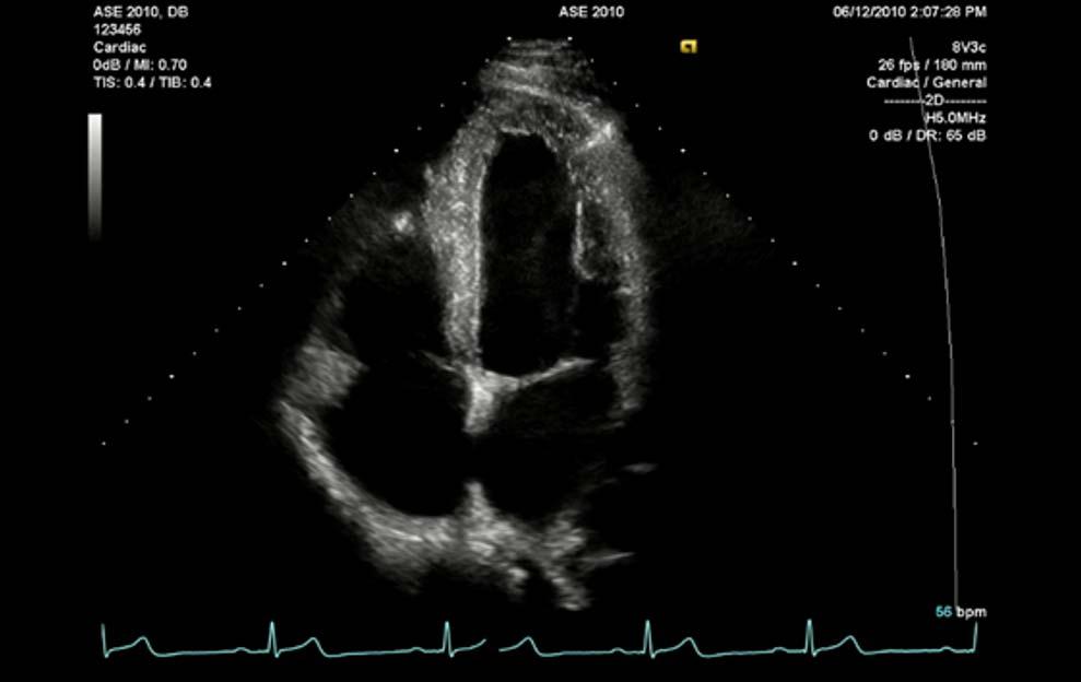
9 Imaging Depth Imaging Depth Start with larger than needed Adjust to place ROI approximately ¾ Leave small area behind to observe useful artifacts like shadowing Depth 9

10 Depth 12 cm Depth 20 cm Depth LPE Echo Screen Anatomy 10

11 Frequency 11


12 Axial Resolution Frequency Zoom Larger Pixels, Higher Frame Rate Not Higher Axial Resolution 12
13 Harmonic Imaging Improves signal-to-noise ratio Contrast Non-linear resonance of bubbles to compressions and rarefactions of ultrasound wave Tissue (incidental discovery) Related to propagation of sound through the myocardium Non-linear response due to higher speed during compression than in rarefaction. McCulloch, et al. JASE 2000 Tissue Harmonic Imaging Non-linear distortion of acoustic signal in tissue generates harmonics SNR 1.5 Tissue Noise Noise/artifacts generate no significant harmonic Signal Amplitude SNR 3 Tissue Harmonic Imaging takes advantage of increased SNR f 2f 13


14 Fundamental Second Harmonic Garbi, M. The EAE Textbook of Echocardiography 2011 Tissue Harmonic Imaging Fundamental Tissue Harmonic 14


15 Tissue Harmonic Imaging Bubbles Have Harmonics too.. Harmonics 1.3/2.6 MHz Fundamental 1.6 MHz 15

16 Echo Screen Anatomy Lateral Resolution Focal Zone / Focus Increased ability to discern two separate objects at the FZ Apical Wall Motion Abnormality, Concern for LV thrombus 16


17 Echo Screen Anatomy Gain Compression Adapted from Garbi, M. The EAE Textbook of Echocardiography


18 Auto Gain 2D Gain Decrease Doppler Gain 18

 19")
19 Time Gain Compensation (TGC) Lateral Gain Compensation (LGC) 19
20 Compression Color 20

21 Human Grayscale Humans perceive approximately 30 shades of gray Cones are high bandwidth and allow humans to see at least 10 million shades of color Echo Screen Anatomy 21
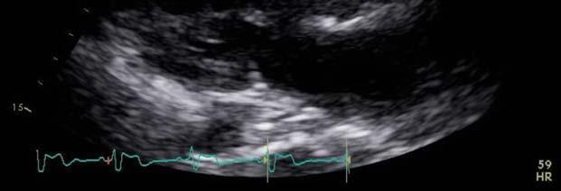
22 Ultrasound Bioeffects Ultrasound is mechanical energy Thermal effects Mechanical effects Background No evidence that diagnostic ultrasound produces harm Subtle or transient effects not well understood ALARA As Low As Reasonably Achievable Mechanical Index (MI) Quantification of US acoustic intensity MI = P(Pascals)/ Frequency(MHz) Non-thermal (Mechanical) Bioeffects MI expresses the acoustic pressure of US beam on insonated structures Lower MI induces increased bubble resonance and harmonics 22

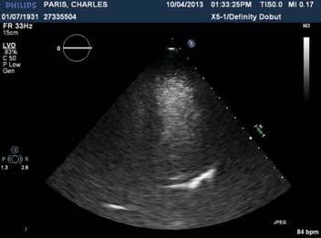
23 Low MI Example 23
24 Thermal Index (TI) Quantification of potential for tissue heating As ultrasound travels through tissue energy is absorbed by tissue and converted to heat Frequency and intensity dependent Recommendation is to keep tissue heating < 1.5 C Caution with the febrile patient Thermal Bioeffects Doppler Echocardiography Optimal 2D images when ultrasound beam is perpendicular to structures Optimal Doppler imaging when ultrasound beam is parallel to flow Apical views allow alignment with most cardiac flows (i.e. aortic, mitral and tricuspid valves) 24

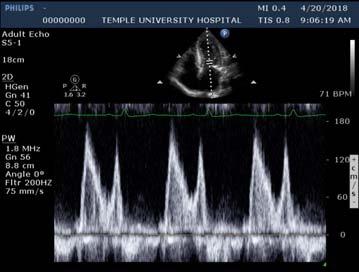
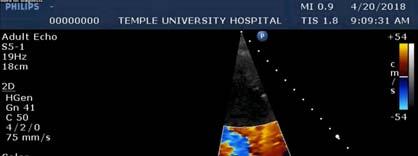
25 Doppler Echocardiography Color Flow Doppler Pulse wave modality that cannot resolve high velocities Turbulence/variance maps can help define jet, direction and turbulence Pulse Wave Spectral Doppler Range specific Subject to aliasing at high velocities like CFD Continuous Wave Spectral Doppler Able to resolve high velocities Range ambiguous Color Flow Doppler Pay attention to the baseline Make note of the Nyquist limit Color scales vary Variance maps (see example) Optimize size of box and sector for frame rate 25


26 Variance Map s/p Mitral ViV S3 26 mm Spectral Doppler Scale Too Small Too Large Scale Optimized 26


27 Spectral Doppler Baseline Baseline Adjusted Doppler Gain 27


28 Doppler Gain Nyquist Limit 28


29 Doppler Sweep 75 mm/s 150 mm/s ASCeXAM Focus Image optimization Knobology is part of achieving competency in echocardiography Key components of image optimization 2D Gain Resolution Compression Depth Mechanical index Doppler Gain Harmonic Imaging Doppler Sweep/Scale Frame Rate 29
30 Thank You! 30
The Normal Echocardiogram
 The Normal Echocardiogram Pravin V. Patil, MD FACC Lewis Katz School of Medicine at Temple University Acknowledgments Dr. Susan Wiegers Dr. Martin Keane Temple Cardiac Sonographers Disclosures No relevant
Physical Principles of Ultrasound
 Physical Principles of Ultrasound Grateful appreciation to Richard A. Lopchinsky, MD, FACS and Nancy H. Van Name, RDMS, RTR, and MarleneKattaron, RDMS 2000 UIC All Rights Reserved. Course Objectives Identify
Ultrasound Physics & Terminology
 Ultrasound Physics & Terminology This module includes the following: Basic physics terms Basic principles of ultrasound Ultrasound terminology and terms Common artifacts seen Doppler principles Terms for
ULTRASOUND. OB/Gyn (Core) Ultrasound PIEZOELECTRIC EFFECT. Principles of Ultrasound Physics and Instrumentation. Nathan Pinkney, BS, CDOS
 Ultrasound PIEZOELECTRIC EFFECT. Principles of Ultrasound Physics and Instrumentation. Nathan Pinkney, BS, CDOS") 1 OB/Gyn (Core) Ultrasound Principles of Ultrasound Physics and Instrumentation Nathan Pinkney, BS, CDOS Philadelphia College of Osteopathic Medicine 2016 ULTRASOUND CATEGORIES OF SOUND INFRASOUND = below
1 OB/Gyn (Core) Ultrasound Principles of Ultrasound Physics and Instrumentation Nathan Pinkney, BS, CDOS Philadelphia College of Osteopathic Medicine 2016 ULTRASOUND CATEGORIES OF SOUND INFRASOUND = below
Diploma of Medical Ultrasonography (DMU) Physical Principles of Ultrasound and Instrumentation Syllabus
 Physical Principles of Ultrasound and Instrumentation Syllabus") Diploma of Medical Ultrasonography (DMU) Physical Principles of Ultrasound and Instrumentation Syllabus Page 1 of 7 11/18 Candidates are expected to cover all of the content of this syllabus when preparing
Diploma of Medical Ultrasonography (DMU) Physical Principles of Ultrasound and Instrumentation Syllabus Page 1 of 7 11/18 Candidates are expected to cover all of the content of this syllabus when preparing
ULTRASOUND IMAGING EE 472 F2018. Prof. Yasser Mostafa Kadah
 ULTRASOUND IMAGING EE 472 F2018 Prof. Yasser Mostafa Kadah www.k-space.org Recommended Textbook Diagnostic Ultrasound: Physics and Equipment, 2nd ed., by Peter R. Hoskins (Editor), Kevin Martin (Editor),
Diagnostic Ultrasound. Sutiporn Khampunnip, M.D.
 Diagnostic Ultrasound Sutiporn Khampunnip, M.D. Definition of Ultrasound Ultrasound is simply sound waves, like audible sound. High-frequency sound and refers to mechanical vibrations above 20 khz. Human
Introduction to Biomedical Imaging
 Alejandro Frangi, PhD Computational Imaging Lab Department of Information & Communication Technology Pompeu Fabra University www.cilab.upf.edu Basic principles. Comparison to X-rays Ultrasound > 20kHz
Ultrasound Principles cycle Frequency Wavelength Period Velocity
 ! Teresa S. Wu, MD, FACEP Director, EM Ultrasound Program & Fellowship Co-Director, Simulation Based Training Program & Fellowship Associate Program Director, EM Residency Program Maricopa Medical Center
Diagnostic approach to heart disease
 Diagnostic approach to heart disease Initial work up History Physical exam Chest radiographs ECG Special studies Echocardiography Cardiac catheterization Echocardiography principles Technique of producing
Image optimization for critical care US
 Image optimization for critical care US 1 Although we assume you are already familiar with focused US in the ED, it might not hurt to revise the basics: Machines & transducers US appearance of normal tissues
ASCeXAM / ReASCE. Practice Board Exam Questions Monday Morning
 ASCeXAM / ReASCE Practice Board Exam Questions Monday Morning Ultrasound Physics Artifacts Doppler Physics Imaging, Knobology, and Artifacts Echocardiographic Evaluation of the RV Tricuspid and Pulmonary
Ultrasound Applied Physics
 Ultrasound Applied Physics University of Toronto Department of Medical Imaging Applied Physics Mini-Course #3 2016 Ultrasound Laboratory Manual and Examination Booklet 1/21/2016 Ultrasound Applied Physics
Introduction. Cardiac Imaging Modalities MRI. Overview. MRI (Continued) MRI (Continued) Arnaud Bistoquet 12/19/03
 MRI (Continued) Arnaud Bistoquet 12/19/03") Introduction Cardiac Imaging Modalities Arnaud Bistoquet 12/19/03 Coronary heart disease: the vessels that supply oxygen-carrying blood to the heart, become narrowed and unable to carry a normal amount
Introduction Cardiac Imaging Modalities Arnaud Bistoquet 12/19/03 Coronary heart disease: the vessels that supply oxygen-carrying blood to the heart, become narrowed and unable to carry a normal amount
Lesson 07: Ultrasound Transducers. This lesson contains 73 slides plus 16 multiple-choice questions.
 Lesson 07: Ultrasound Transducers This lesson contains 73 slides plus 16 multiple-choice questions. This lesson was derived from pages 33 through 42 in the textbook: Ultrasound Transducers Ultrasound Transducers
Ultrasound 10/1/2014. Basic Echocardiography for the Internist. Mechanical (sector) transducer Piezoelectric crystal moved through a sector sweep
 transducer Piezoelectric crystal moved through a sector sweep") Ultrasound Basic Echocardiography for the Internist Carol Gruver, MD, FACC UT Erlanger Cardiology Mechanical wave of compression and rarefaction Requires a medium for transmission Ultrasound frequency
Ultrasound Basic Echocardiography for the Internist Carol Gruver, MD, FACC UT Erlanger Cardiology Mechanical wave of compression and rarefaction Requires a medium for transmission Ultrasound frequency
Principles of Ultrasound. Cara C. Prideaux, M.D. University of Utah PM&R Sports Medicine Fellow March 14, 2012
 Principles of Ultrasound Cara C. Prideaux, M.D. University of Utah PM&R Sports Medicine Fellow March 14, 2012 None Disclosures Outline Introduction Benefits and Limitations of US Ultrasound (US) Physics
WELCOME! Introduction to Bedside Ultrasound
 WELCOME! Introduction to Bedside Ultrasound TEACHERS University of California-Irvine School of Medicine Nathan Molina nathan.d.molina@gmail.com Trevor Plescia taplescia90@gmail.com Jack Silva jpsilva42@gmail.com
RPVI Exam Review ecourse
 RPVI Exam Review ecourse The RPVI Exam Review ecourse consists of ten Vascular Physics Modules and fourteen Vascular Specialty Modules. Detailed descriptions of module content are listed below. During
Ultrasound: Past and Present. Lecturer: Dr. John M Hudson, PhD
 Ultrasound: Past and Present Lecturer: Dr. John M Hudson, PhD Disclosures 2 No conflicts of interest to declare Course Outline 3 1. Survey of ultrasound physics & applications 2. (Sep 21) 3. (Sep 28) 4.
Basic Ultrasound Physics Board Review Questions
 Basic Ultrasound Physics Board Review Questions Sidney K. Edelman, PhD ESP Ultrasound The Woodlands, TX Question 1 What is the wavelength of 2 MHz sound in soft tissue? 1. 1.54 mm 2. 0.75 mm 3. 0.75 cm
This brief introduction considers the physics needed to get basic ultrasound images from a typical point of care machine.
 AAGBI seminar on Focused echocardiography for anaesthesia Craig Morris Physics This brief introduction considers the physics needed to get basic ultrasound images from a typical point of care machine.
Normal TTE Examination, Doppler Echocardiography and Normal Antegrade Flow Patterns
 Normal TTE Examination, Doppler Echocardiography and Normal Antegrade Flow Patterns Pravin Patil, MD FACC FASE Associate Professor of Medicine Director, Cardiovascular Disease Training Program Lewis Katz
1 Fundamentals. Basic Definitions and Physics Principles. Fundamentals
 1 To become versed in the language of ultrasonography, it is necessary to review some of the basic principles of physics. The wave physics principles of ordinary (i.e., audible) sound apply to ultrasound
Doppler Basic & Hemodynamic Calculations
 Doppler Basic & Hemodynamic Calculations August 19, 2017 Smonporn Boonyaratavej MD Division of Cardiology, Department of Medicine Chulalongkorn University Cardiac Center, King Chulalongkorn Memorial Hospital
Basic Physics of Ultrasound and Knobology
 WELCOME TO UTMB Basic Physics of Ultrasound and Knobology By Daneshvari Solanki, FRCA Laura B. McDaniel Distinguished Professor Anesthesiology and Pain Medicine University of Texas Medical Branch Galveston,
Tissue Strain Analytics Virtual Touch Tissue Imaging and Quantification
 Whitepaper Tissue Strain Analytics Virtual Touch Tissue Imaging and Quantification ACUSON S2000 Ultrasound System Answers for life. Page 1 Tissue Strain Analytics: Virtual Touch Tissue Imaging and Quantification
Principles of echocardiography for the anesthesiologist.
 Principles of echocardiography for the anesthesiologist. Nikolaos Skubas, MD PhD Abstract Ultrasound-based diagnostic techniques are now part of the cardiological patients chart, while echocardiography
The Physics of Ultrasound. The Physics of Ultrasound. Claus G. Roehrborn. Professor and Chairman. Ultrasound Physics
 The Physics of Ultrasound Pipe Organ 10-8000 Emission Dog 452-1080 Man 85-1100 Spectrum Bat 10,000-120,000 Porpoise 7000-120,000 Claus G. Roehrborn Professor and Chairman 10 20 Cycles per second Reception
Ultrasound Physics & Doppler
 Ultrasound Physics & Doppler Endocrine University 2018 Mark Lupo, MD, FACE, ECNU Objectives Review the essential components of ultrasound physics in neck sonography Demonstrate the importance of ultrasound
Optimising your Doppler settings for an accurate PI. Alison McGuinness Mid Yorks Hospitals
 Optimising your Doppler settings for an accurate PI Alison McGuinness Mid Yorks Hospitals Applications Both maternal uterine and fetal circulations can be studied with doppler sonography Uterine arteries
What is Ultrasound? Resolution Image production Attenuation Imaging modes Ultrasound artifacts... 7
 What is Ultrasound?... 1 Resolution... 3 Image production... 3 Attenuation... 4 Imaging modes... 5 Ultrasound artifacts... 7 0 What is Ultrasound? High frequency sound of frequencies 2-50 MHz is used in
New 3D Quantification of Mitral Regurgitation Severity. Judy Hung, MD Cardiac Ultrasound Laboratory Massachusetts General Hospital Boston, MA
 New 3D Quantification of Mitral Regurgitation Severity Judy Hung, MD Cardiac Ultrasound Laboratory Massachusetts General Hospital Boston, MA No Financial Disclosures No off label discussion of devices
Basic Physics of Ultrasound in Transesophageal Echocardiography
 SPECIAL ARTICLE IJUTPC Basic Physics of Ultrasound in Transesophageal Echocardiography Basic Physics of Ultrasound in Transesophageal Echocardiography 1 Mary Korula, 2 Ravi Hebballi 1 Senior Consultant,
Strain Assessment in Echo
 Strain Assessment in Echo Joe M. Moody, Jr, MD UTHSCSA and STVHCS 2010 Acknowledging many illustrations from Weyman s text and others. Echo-Doppler Basic Principles Background: Ultrasound physics (resolution,
Dr Emma Chung. Safety first - Physical principles for excellent imaging
 Safety first - Physical principles for excellent imaging Dr Emma Chung Lecturer in Medical Physics, University of Leicester Clinical Scientist, University Hospitals of Leicester NHS Trust Thanks to Caroline
2/2/2011. Strain and Strain Rate Imaging How, Why and When? Movement vs Deformation. Doppler Myocardial Velocities. Movement. Deformation.
 Strain and Strain Rate Imaging How, Why and When? João L. Cavalcante, MD Advanced Cardiac Imaging Fellow Cleveland Clinic Foundation Disclosures: No conflicts of interest Movement vs Deformation Movement
Supplement (videos)
") Supplement (videos) Ruben s tube (sound): http://www.youtube.com/watch?v=gpcquuwqayw Doppler US (diagnostic use): http://www.youtube.com/watch?v=fgxzg-j_hfw http://www.youtube.com/watch?v=upsmenyoju8 High
Supplement (videos) Ruben s tube (sound): http://www.youtube.com/watch?v=gpcquuwqayw Doppler US (diagnostic use): http://www.youtube.com/watch?v=fgxzg-j_hfw http://www.youtube.com/watch?v=upsmenyoju8 High
Ultrasound. Principles of Medical Imaging. Contents. Prof. Dr. Philippe Cattin. MIAC, University of Basel. Oct 17th, 2016
 Ultrasound Principles of Medical Imaging Prof. Dr. Philippe Cattin MIAC, University of Basel Contents Abstract 1 Image Generation Echography A-Mode B-Mode M-Mode 2.5D Ultrasound 3D Ultrasound 4D Ultrasound
Preamble (disclaimer)
") Preamble (disclaimer) PHYSICS AND PRINCIPLES OF HEAD/NECK ULTRASOUND Joseph C. Sniezek, MD FACS LTC, MC, USA Otolaryngology/H&N Surgery Tripler Army Medical Center 1. I am not a physicist 2. ACS has recommended
Preamble (disclaimer) PHYSICS AND PRINCIPLES OF HEAD/NECK ULTRASOUND Joseph C. Sniezek, MD FACS LTC, MC, USA Otolaryngology/H&N Surgery Tripler Army Medical Center 1. I am not a physicist 2. ACS has recommended
The 2 nd Cambridge Advanced Emergency Ultrasound Course
 The 2 nd Cambridge Advanced Emergency Ultrasound Course Addenbrooke s Hospital Cambridge Sept 2008 1 2 Faculty! UK! USA! Australia! Toshiba! Emergency Medicine! Radiology 3 Programme! Day 1 Introduction
What is Ultrasound? What is Ultrasound? B A. Basic Principles of Ultrasound. Basic Principles of Ultrasound. Basic Principles of Ultrasound
 Introduction to Ultrasound Principles Mani Montazemi, RDMS Baylor College of Medicine Division of Maternal-Fetal Medicine Department of Obstetrics and Gynecology Manager, Maternal Fetal Center Imaging
2015 ARDMS Sonography Principles & Instrumentation Job Task Analysis Summary Report
 P a g e 1 2015 ARDMS Sonography Principles & Instrumentation Job Task Analysis Summary Report American Registry for Diagnostic Medical Sonography (ARDMS) P a g e 2 Table of Contents ABOUT THE REPORT...
Physics. Norman McDicken Tom Anderson CHAPTER ULTRASOUND. Ultrasound Propagation
 CHPTER 2 Physics Norman McDicken Tom nderson This chapter provides an introduction to the physics of medical ultrasound (US). Several books exist that can be consulted to extend the material presented
CONTENTS. Test Number cpd Tanya Reynolds (Nat. Dip. Diag. Rad., B. Tech. Diag. Rad., B. Tech. Ultrasound)
") CONTENTS page 1-15 page 16 BASIC 2-DIMENSIONAL ULTRASOUND PRINCIPLES Multiple Choice Test Test Number cpd 41640 Tanya Reynolds (Nat. Dip. Diag. Rad., B. Tech. Diag. Rad., B. Tech. Ultrasound) Tanya is
CONTENTS page 1-15 page 16 BASIC 2-DIMENSIONAL ULTRASOUND PRINCIPLES Multiple Choice Test Test Number cpd 41640 Tanya Reynolds (Nat. Dip. Diag. Rad., B. Tech. Diag. Rad., B. Tech. Ultrasound) Tanya is
DIGITAL IMAGE PROCESSING IN ULTRASOUND IMAGES
 DIGITAL IMAGE PROCESSING IN ULTRASOUND IMAGES Kamaljeet Kaur Computer Science & Engineering Department Guru Nanak Dev Engg. College, Ludhiana. Punjab-India meetk.89@gmail.com ABSTRACT-- Image processing
Critical Care Ultrasound Study Notes
 Critical Care Ultrasound Study Notes Compiled by David Tripp October 2014 Ultrasound Physics 2 Ultrasound in Tissue 2 Ultrasound Interaction with Tissue 2 Pulsed Ultrasound and Imaging 3 Image Formation
Ultrasound Physics and Knobology Alan Macfarlane. Consultant Anaesthetist Glasgow Royal Infirmary
 Ultrasound Physics and Knobology Alan Macfarlane Consultant Anaesthetist Glasgow Royal Infirmary RAPM 2009; 34: 40-46 Ultrasound Proficiency Understanding US image generation and device operation Image
Emergency Medicine Interest Group (EMIG) 2016
 2016") Emergency Medicine Interest Group (EMIG) 2016 Welcome to the flipped classroom (learning objectives summary) for the 2016 Emergency Medicine Interest Group (EMIG) Procedures Workshop. Overview - Tuesday
Emergency Medicine Interest Group (EMIG) 2016 Welcome to the flipped classroom (learning objectives summary) for the 2016 Emergency Medicine Interest Group (EMIG) Procedures Workshop. Overview - Tuesday
Pulse-Echo Ultrasound Imaging. Resolution in Ultrasound Imaging. Doppler Ultrasound. Resolution vs Penetration. Medical Imaging (EL582/BE620/GA4426)
") Medical Imaging (EL582/BE620/GA4426) Pulse-Echo Ultrasound Imaging Ultrasound Imaging Lecture 2 Daniel (Dan) Turnbull, Ph.D. Skirball Institute and Dept of Radiology NYU School of Medicine (daniel.turnbull@med.nyu.edu)
Medical Imaging (EL582/BE620/GA4426) Pulse-Echo Ultrasound Imaging Ultrasound Imaging Lecture 2 Daniel (Dan) Turnbull, Ph.D. Skirball Institute and Dept of Radiology NYU School of Medicine (daniel.turnbull@med.nyu.edu)
Point-of-Care Ultrasound: An Introduction
 Point-of-Care Ultrasound: An Introduction Delegation Teaching Package for Registered Respiratory Therapists and Anesthesia Assistants Developed by: Rob Bryan RRT, AA Edited by: Kelly Hassall RRT, FCSRT,
Introduction & Physics of ED Ultrasound. Objectives. What? - Limited Studies. Who? - ED Docs
 Introduction & Physics of ED Ultrasound Martine Sargent, MD Ultrasound Director, Assistant Professor UCSF Department of Emergency Medicine San Francisco General Hospital & Trauma Center Objectives Who?
DC-6. Diagnostic Ultrasound System
 DC-6 Diagnostic Ultrasound System MINDRAY is proud to introduce DC-6, a color Doppler ultrasound system for general applications. DC-6 incorporates the latest digital ultrasound image processing technology
Ultrasound in Medicine
 Ultrasound in Medicine Experimental Equipment for Medical Education Universities Colleges Medical Schools Medical and Med-Technical Training Education can befun! WELCOME TO GAMPT Devices and accessories
Welcome, Intro & Goals
 Welcome, Intro & Goals PP16 Imaging Conference Bicol Hospital, Legaspi City, Philippines July 2016 David Adams, ACS, RCS, RDCS, FASE Duke University Medical Center Echocardiography The Anatomy Lesson of
Hands-on. Physics Workshop. 2-day Workshop June 1-2, 2018 Ann Arbor, MI. Program Director: Brian Fowlkes, PhD
 Hands-on Ultrasound Physics Workshop 2-day Workshop June 1-2, 2018 Ann Arbor, MI Program Director: Brian Fowlkes, PhD Faculty: Paul Carson, PhD Mitch Goodsitt, PhD Oliver Kripfgans, PhD Sandra Larson,
Application of Phased Array Radar Theory to Ultrasonic Linear Array Medical Imaging System
 Application of Phased Array Radar Theory to Ultrasonic Linear Array Medical Imaging System R. K. Saha, S. Karmakar, S. Saha, M. Roy, S. Sarkar and S.K. Sen Microelectronics Division, Saha Institute of
Lesson 03: Sound Wave Propagation and Reflection. This lesson contains 15 slides plus 14 multiple-choice questions.
 Lesson 03: Sound Wave Propagation and Reflection This lesson contains 15 slides plus 14 multiple-choice questions. Accompanying text for the slides in this lesson can be found on pages 8 through 14 in
Introduction to Ultrasound Guided Region Anesthesia
 Introduction to Ultrasound Guided Region Anesthesia Brian D. Sites, MD Dept of Anesthesiology Dartmouth-Hitchcock Medical Center INTRODUCTION Welcome to Introduction to Ultrasound Guided Regional Anesthesia.
DISCLOSURE. Myocardial Mechanics. Relevant Financial Relationship(s) Off Label Usage
 Off Label Usage") 7th Annual Team Echocardiography: The Heart of Cardiovascular Medicine Tissue Doppler, Strain, Speckle: What? How? Christopher J Kramer RDCS Aurora Medical Group Advanced Cardiovascular Services, Aurora
7th Annual Team Echocardiography: The Heart of Cardiovascular Medicine Tissue Doppler, Strain, Speckle: What? How? Christopher J Kramer RDCS Aurora Medical Group Advanced Cardiovascular Services, Aurora
DOW-RAD, DOW DIAGNOSTIC COMPLEX, DUHS TRAINING PROGRAM HANDBOOK 2013
 DOW-RAD, DOW DIAGNOSTIC COMPLEX, DUHS TRAINING PROGRAM HANDBOOK 2013 CERTIFICATE COURSE INVASCULAR/DOPPLER ULTRASOUND: Introduction: Ultrasound is an evolving technology with wide spectrum application
Aixplorer MultiWave Ultrasound System An Innovation in Breast Ultrasound Imaging
 b r e a s t i m a g i n g S O L U T I O N S Aixplorer MultiWave Ultrasound System An Innovation in Breast Ultrasound Imaging The new wave in breast ultrasound Aixplorer is a next-generation ultrasound
Velocity, strain and strain rate: Doppler and Non-Doppler methods. Thoraxcentre, Erasmus MC,Rotterdam
 Velocity, strain and strain rate: Doppler and Non-Doppler methods J Roelandt J. Roelandt Thoraxcentre, Erasmus MC,Rotterdam Basics of tissue Doppler imaging Instantaneous annular velocity profiles IVCT
Tissue Doppler and Strain Imaging. Steven J. Lester MD, FRCP(C), FACC, FASE
, FACC, FASE") Tissue Doppler and Strain Imaging Steven J. Lester MD, FRCP(C), FACC, FASE Relevant Financial Relationship(s) None Off Label Usage None a. Turn the wall filters on and turn down the receiver gain. b. Turn
Tissue Doppler and Strain Imaging Steven J. Lester MD, FRCP(C), FACC, FASE Relevant Financial Relationship(s) None Off Label Usage None a. Turn the wall filters on and turn down the receiver gain. b. Turn
Tissue Doppler Imaging
 Cronicon OPEN ACCESS Hesham Rashid* Tissue Doppler Imaging CARDIOLOGY Editorial Department of Cardiology, Benha University, Egypt *Corresponding Author: Hesham Rashid, Department of Cardiology, Benha University,
ARTIFACTS: THEORY AND ILLUSTRATIVE EXAMPLES
 ARTIFACTS: THEORY AND ILLUSTRATIVE EXAMPLES Robert A. Levine, M.D. Marielle Scherrer-Crosbie, M.D. Eric M. Isselbacher, M.D. No conflicts of interest Philippe Bertrand, Pieter Vendervoort, Hasselt and
Ultrasound Knobology
 Ultrasound Knobology Raj Dasgupta MD, FACP, FCCP, FASSM Assistant Professor of Clinical Medicine Pulmonary / Critical Care / Sleep Medicine University of Southern California (USC) Objectives Physics of
Development of Ultrasound Based Techniques for Measuring Skeletal Muscle Motion
 Development of Ultrasound Based Techniques for Measuring Skeletal Muscle Motion Jason Silver August 26, 2009 Presentation Outline Introduction Thesis Objectives Mathematical Model and Principles Methods
CONTRAST ECHOCARDIOGRAPHY
 CONTRAST ECHOCARDIOGRAPHY How Should it Be Administered and How Do I Optimize My Machine Settings? Keith Collins, MS RDCS FASE Monday, Feb. 15, 2016 State of the Art Tscc.exe Contrast Is Needed When Poor
Basic of Ultrasound Physics E FAST & Renal Examination. Dr Muhammad Umer Ihsan MBBS,MD, DCH CCPU,DDU1,FACEM
 Basic of Ultrasound Physics E FAST & Renal Examination Dr Muhammad Umer Ihsan MBBS,MD, DCH CCPU,DDU1,FACEM What is Sound? Sound is Mechanical pressure waves What is Ultrasound? Ultrasounds are sound waves
Goals. Access flow and renal artery stenosis evaluation by Doppler ultrasound. Reimbursement. WHY use of Doppler Ultrasound
 Access flow and renal artery stenosis evaluation by Doppler ultrasound Adina Voiculescu, MD Interventional Nephrology Brigham and Women s Hospital Boston Instructor at Harvard Medical School Understand
ARTIFACTS: THEORY AND ILLUSTRATIVE EXAMPLES
 ARTIFACTS: THEORY AND ILLUSTRATIVE EXAMPLES Robert A. Levine, M.D. Marielle Scherrer-Crosbie, M.D. Eric M. Isselbacher, M.D. 60 year old man Cardiac source of embolus? NAME THAT MASS! 1 NAME THAT MASS!
Ultrasound guidance in regional anesthesia has
 Ultrasound and Regional Anesthesia Artifacts and Pitfall Errors Associated With Ultrasound-Guided Regional Anesthesia. Part I: Understanding the Basic Principles of Ultrasound Physics and Machine Operations
Underwater Acoustic Measurements in Megahertz Frequency Range.
 Underwater Acoustic Measurements in Megahertz Frequency Range. Current State and Prospects of Development in Russia Alexander M. Enyakov,, Many medical applications of underwater acoustic measurements
Terminology Tissue Appearance
 By Marc Nielsen, MD Advantages/Disadvantages Generation of Image Ultrasound Machine/Transducer selection Modes of Ultrasound Terminology Tissue Appearance Scanning Technique Real-time Portable No ionizing
Chapter 3 Physical Principles of Ultrasound of the Male Genitalia
 Chapter 3 Physical Principles of Ultrasound of the Male Genitalia Bruce R. Gilbert and Pat Fox Fulgham Introduction The value of ultrasound evaluation of the male genitalia depends, in large part, on the
Qualitative and Quantitative Assessment of Perfusion
 APCDE 2011 Qualitative and Quantitative Assessment of Perfusion Hyun Ju Yoon Chonnam National University Hospital Gwangju, Korea ISCHEMIC CASCADE Blood flow mismatch Perfusion defects on nuclear imaging,
Focus on your needs. Ultrasound system HS60 SAMSUNG MEDISON CO., LTD. CT-HS60 V1.0-GI-FT EN
 Samsung Medison, an affiliate of Samsung Electronics, is a global medical company founded in 1985. With a mission to bring health and well-being to people's lives, the company manufactures diagnostic ultrasound
3/20/2017. Disclosures. Ultrasound Fundamentals. Ultrasound Fundamentals. Bone Anatomy. Tissue Characteristics
 Disclosures Images of ultrasound equipment in this presentation are not an endorsement Fundamentals of Musculoskeletal Ultrasound Physics and Knobology Shane A. Shapiro, M.D. Assistant Professor Orthopedic
Back to Basics: Common Errors In Quantitation In Everyday Practice
 Back to Basics: Common Errors In Quantitation In Everyday Practice Deborah Agler, ACS, RDCS, FASE October 9, 2017 ASE: Echo Florida Rebecca T. Hahn, MD Director of Interventional Echocardiography Professor
ULTRASOUND NOMENCLATURE
 Chapter 1: Ultrasound Nomenclature, Image Orientation, and Basic Instrumentation CYNTHIA SIKOWSKI Ultrasound waves are sound waves that have a frequency exceeding 20,000 Hz. When sound waves are transmitted
ACUSON Sequoia C512 Echocardiography System The ultimate in imaging
 ACUSON Sequoia C512 Echocardiography System The ultimate in imaging Optimum imaging for every patient ACUSON Sequoia C512 Patient Specific Imaging Programmable Waveform Generator Coherent Pulse Formation
GE Healthcare. LOGIQ E9 XDclear 2.0. Complete Ultrasound HEAD TO TOE OBESE TO THIN NEONATE TO GERIATRIC
 GE Healthcare LOGIQ E9 XDclear 2.0 Complete Ultrasound HEAD TO TOE OBESE TO THIN NEONATE TO GERIATRIC In addition to extraordinary image quality, the system has been further enhanced to meet the needs
Features and Benefits
 Features and Benefits DC7 Significant Features HIPAA Compliant Frequency Compound Imaging Standard i-clear Standard i-beam Standard SCI Digital Processing Channels 2048 Dynamic Range > 160 Db 10 Bit A/D
Tissue Doppler and Strain Imaging
 Tissue Doppler and Strain Imaging Steven J. Lester MD, FRCP(C), FACC, FASE Relevant Financial Relationship(s) None Off Label Usage None 1 Objective way with which to quantify the minor amplitude and temporal
: Biomedical Signal Processing
 : Biomedical Signal Processing 0. Introduction: Biomedical signal processing refers to the applications of signal processing methods, such as Fourier transform, spectral estimation and wavelet transform,
Objectives 8/17/2011. Challenges in Cardiac Imaging. Challenges in Cardiac Imaging. Basic Cardiac MRI Sequences
 8/17/2011 Traditional Protocol Model for Tomographic Imaging Cardiac MRI Sequences and Protocols Frandics Chan, M.D., Ph.D. Stanford University Medical Center Interpretation Lucile Packard Children s Hospital
Disclosures Rebecca T. Hahn, MD, FASE
 The New ASE Guidelines for Native Valvular Regurgitation Mitral Regurgitation The New ASE Guidelines: Role of 2D/3D and CMR (With caveats and comments from R. Hahn) William A. Zoghbi MD, FASE, MACC Professor
The Effect and Mechanisms of Ultrasound Transducer Degradation on the Quality and Clinical Efficacy of Diagnostic Ultrasound Examinations
 The Effect and Mechanisms of Ultrasound Transducer Degradation on the Quality and Clinical Efficacy of Diagnostic Ultrasound Examinations In diagnostic ultrasound studies the health of the transducer (also
Application of Time Reversal Technique for the Inspection of Composite Structures
 More info ab Application of Time Reversal Technique for the Inspection of Composite Structures Daniel RICHARD, Guy MAES ASNT NDT of Composites 2017 Quebec, 6-8 June 2017 NDT in Canada 2017 Conference (June
Tissue Doppler and Strain Imaging
 Tissue Doppler and Strain Imaging Steven J. Lester MD, FRCP(C), FACC, FASE Relevant Financial Relationship(s) None Off Label Usage None 1 Objective way with which to quantify the minor amplitude and temporal
GE Healthcare. LOGIQ E9 XDclear 2.0. Complete Ultrasound HEAD TO TOE OBESE TO THIN NEONATE TO GERIATRIC
 GE Healthcare LOGIQ E9 XDclear 2.0 Complete Ultrasound HEAD TO TOE OBESE TO THIN NEONATE TO GERIATRIC Complete Ultrasound HEAD TO TOE OBESE TO THIN NEONATE TO GERIATRIC OUR BEST JUST GOT BETTER. With the
Steven J. Lester, MD, FACC, FRCP(C), FASE Mayo Clinic. Relevant Financial Relationship(s) None. Off Label Usage
, FASE Mayo Clinic. Relevant Financial Relationship(s) None. Off Label Usage") Steven J. Lester, MD, FACC, FRCP(C), FASE Mayo Clinic Relevant Financial Relationship(s) None Off Label Usage 1 2 1. Define ultrasound contrast? 2. Recognize the interaction of the bubbles with ultrasound
Steven J. Lester, MD, FACC, FRCP(C), FASE Mayo Clinic Relevant Financial Relationship(s) None Off Label Usage 1 2 1. Define ultrasound contrast? 2. Recognize the interaction of the bubbles with ultrasound
DMS-2350: SONOGRAPHIC INSTRUMENT/PHYSICS
 DMS-2350: Sonographic Instrument/Physics 1 DMS-2350: SONOGRAPHIC INSTRUMENT/PHYSICS Cuyahoga Community College Viewing:DMS-2350 : Sonographic Instrument/Physics Board of Trustees: 2014-03-20 Academic Term:
Byung Ihn Choi, M.D. Department of Radiology Seoul National University Hospital
 Byung Ihn Choi, M.D. Department of Radiology Seoul National University Hospital CEUS & US Elastography : Contents CEUS Introduction Contrast agents & imaging Clinical application US Video WS Summary US
Thyroid Ultrasound Physics and Doppler
 Thyroid Ultrasound Physics and Doppler Advanced AACE-ACE US training course 2017 Dev Abraham MD, MRCP(UK), ECNU, FACE Professor of Medicine, University of Utah No Disclosures Natural Ability to see with
iu22 Liver Shear Wave ElastPQ
 iu22 Liver Shear Wave ElastPQ Clinical Case Study Lucy Wang Clinical Application Specialist ASEAN Case Study: History: 58-year-old male patient, hepatitis B virus (HBV) carrier, with non clinical symptoms
Debbie Childs RDMS, RVT Sonographer Murphy Medical Center Murphy, NC
 Debbie Childs RDMS, RVT Sonographer Murphy Medical Center Murphy, NC Worked at Murphy Medical Center as a sonographer for 18 years Registered in Abdomen, OB/GYN, Breast, & Vascular Ultrasound ACR Accredited
Aortic Stenosis: Spectrum of Disease, Low Flow/Low Gradient and Variants
 Aortic Stenosis: Spectrum of Disease, Low Flow/Low Gradient and Variants Martin G. Keane, MD, FASE Professor of Medicine Lewis Katz School of Medicine at Temple University Basic root structure Parasternal
Hemodynamic Assessment. Assessment of Systolic Function Doppler Hemodynamics
 Hemodynamic Assessment Matt M. Umland, RDCS, FASE Aurora Medical Group Milwaukee, WI Assessment of Systolic Function Doppler Hemodynamics Stroke Volume Cardiac Output Cardiac Index Tei Index/Index of myocardial