ASCeXAM / ReASCE. Practice Board Exam Questions Monday Morning
|
|
|
- Pamela Parrish
- 5 years ago
- Views:
Transcription
1 ASCeXAM / ReASCE Practice Board Exam Questions Monday Morning Ultrasound Physics Artifacts Doppler Physics Imaging, Knobology, and Artifacts Echocardiographic Evaluation of the RV Tricuspid and Pulmonary Valve Disease Artifacts: Theory and Illustrative Examples Robert A. Levine, MD 1
2 This patient shows: 1. Biventricular Wires 2. Reverberation 3. Mirror Image 4. Side Lobes This patient shows: 1. Biventricular Wires 2. Reverberation 3. Mirror Image 4. Side Lobes 2
3 Echo Map of True Object and Side Lobe Artifacts 3
4 Apical TEE: What type of occluder device is in the atrial septum? 1. Helical 2. Figure of 8 3. Circular 4. Conical Apical TEE: What type of occluder device is in the atrial septum? 1. Helical 2. Figure of 8 3. Circular 4. Conical 4

5 AMPLATZER DEVICE AND ECHO This change in apparent shape is caused by which physical effect? 1. Scattering 2. Refraction 3. Reflection 4. Acoustic Shadowing 5

6 This change in apparent shape is caused by which physical effect? 1. Scattering 2. Refraction 3. Reflection 4. Acoustic Shadowing JASE 2014; 27:

7 Physics principle: Angle of reflection = angle of incidence for a specular reflector Result: Figure-of-8 artifact 7

8 Result: Figure-of-8 artifact versus true shape when beam views device en face Artifactual echoes : 1. Always Move Parallel To A Real Object 2. Create Turbulent Color Flow In Their Vicinity 3. Are Infrequent By TEE 4. Can Appear To Pass Through Real Objects 8
9 Artifactual echoes : 1. Always Move Parallel To A Real Object 2. Create Turbulent Color Flow In Their Vicinity 3. Are Infrequent By TEE 4. Can Appear To Pass Through Real Objects Concepts of Imaging, Knobology, and Artifacts David B. Adams, RCS, RDCS, FASE 9
10 Which standard 2D TTE view typically allows viewing of the LAA? 1. Parasternal Long Axis 2. Apical 4 Chamber 3. Subcostal 4 Chamber 4. Apical 2 Chamber Which standard 2D TTE view typically allows viewing of the LAA? 1. Parasternal Long Axis 2. Apical 4 Chamber 3. Subcostal 4 Chamber 4. Apical 2 Chamber 10
11 The problem with this image can be corrected using: 1. Overall Gain 2. TGC Controls 3. LGC Controls 4. Another View DUKE: Adams The problem with this image can be corrected using: 1. Overall Gain 2. TGC Controls 3. LGC Controls 4. Another View DUKE: Adams 11
12 To correct this view the transducer beam should be angled: 1. Laterally 2. Medially 3. Cranially-up an interspace 4. Caudally-down an interspace To correct this view the transducer beam should be angled: 1. Laterally 2. Medially 3. Cranially-up an interspace 4. Caudally-down an interspace 12
13 Where should you position the pulsed wave Doppler sample volume for mitral inflow? 1. At the mitral valve annulus 2. In the middle of the mitral leaflets 3. At the tips of the mitral valve in systole 4. At the tips of the mitral valve in diastole Where should you position the pulsed wave Doppler sample volume for mitral inflow? 1. At the mitral valve annulus 2. In the middle of the mitral leaflets 3. At the tips of the mitral valve in systole 4. At the tips of the mitral valve in diastole 13
14 The best view to measure the RVOT, pulmonic valve and PA flow is: 1. Right sternal border 2. Subcostal short axis 3. Parasternal long axis 4. Parasternal short axis The best view to measure the RVOT, pulmonic valve and PA flow is: 1. Right sternal border 2. Subcostal short axis 3. Parasternal long axis 4. Parasternal short axis 14
15 What is an advantage of continuous wave Doppler over pulsed wave Doppler? 1. Aliasing 2. Range resolution 3. Detection of high velocities 4. Assessing the severity of regurgitation What is an advantage of continuous wave Doppler over pulsed wave Doppler? 1. Aliasing 2. Range resolution 3. Detection of high velocities 4. Assessing the severity of regurgitation 15
16 When evaluating which of the following is it best to use a low wall filter? 1. Aortic Stenosis 2. Pulmonary Veins 3. Mitral Regurgitation 4. Tricuspid Regurgitation DUKE: Adams When evaluating which of the following is it best to use a low wall filter? 1. Aortic Stenosis 2. Pulmonary Veins 3. Mitral Regurgitation 4. Tricuspid Regurgitation DUKE: Adams 16
17 What should be done in order to measure the E wave duration? 1. Increase the gain 2. Find a better window 3. Decrease the wall filter 4. Increase the wall filter DUKE: Adams What should be done in order to measure the E wave duration? 1. Increase the gain 2. Find a better window 3. Decrease the wall filter 4. Increase the wall filter DUKE: Adams 17


18 Duke ER The End DUKE: Adams Echocardiography Evaluation of the Right Ventricle Lawrence G. Rudski, MD, FASE 18
19 Question 1 A 35 year old woman is referred for an echocardiogram to evaluate progressive dyspnea on exertion. There is insufficient tricuspid and pulmonic regurgitation to estimate pulmonary artery pressure. Which of the following echocardiographic signs is most suggestive of pulmonary arterial hypertension: 1) IVC measuring 25 mm with 30% collapse with respiration 2) D-shaped interventricular septum configuration in systole 3) D-shaped interventricular septum configuration in diastole 4) RV measuring 50 mm at base, with TAPSE of 23 mm Question 1 A 35 year old woman is referred for an echocardiogram to evaluate progressive dyspnea on exertion. There is insufficient tricuspid and pulmonic regurgitation to estimate pulmonary artery pressure. Which of the following echocardiographic signs is most suggestive of pulmonary arterial hypertension: 1) IVC measuring 25 mm with 30% collapse with respiration 2) D-shaped interventricular septum configuration in systole 3) D-shaped interventricular septum configuration in diastole 4) RV measuring 50 mm at base, with TAPSE of 23 mm 19
 Normal size and systolic function 2) Normal size with reduced systolic function 3) Increased size with normal systolic function 4)")
20 Question 2 Regarding the following echo images. The RV size and systolic function can best be described as: 1) Normal size and systolic function 2) Normal size with reduced systolic function 3) Increased size with normal systolic function 4) Increased size with reduced systolic function Question 2 Regarding the following echo images. The RV size and systolic function can best be described as: 1) Normal size and systolic function 2) Normal size with reduced systolic function 3) Increased size with normal systolic function 4) Increased size with reduced systolic function 20
21 Question 3 Which view is used to determine RV size in the described condition? 1) Apical 4-chamber view to measure RV basal dimension in Normal Subjects 2) RV focused view to determine relative RV:LV size in patients with pulmonary hypertension 3) Subcostal view to determine RV size in the setting of pulmonary embolism 4) RVOT dimension in parasternal long-axis view to determine RV size in Arrhythmogenic Right Ventricular Cardiomyopathy Question 3 Which view is used to determine RV size in the described condition? 1) Apical 4-chamber view to measure RV basal dimension in Normal Subjects 2) RV focused view to determine relative RV:LV size in patients with pulmonary hypertension 3) Subcostal view to determine RV size in the setting of pulmonary embolism 4) RVOT dimension in parasternal long-axis view to determine RV size in Arrhythmogenic Right Ventricular Cardiomyopathy 21
22 Question 4 A 50 year old patient presents with to the ER 2 weeks after a knee replacement. The following image is obtained. What additional echo findings would you expect? 1)Moderate tricuspid regurgitation, spap 60 mmhg, dilated IVC 2)Moderate TR, clot in transit, small IVC 3)Mild to moderate TR, spap 47 mmhg, LVEF 70% 4)Mild to moderate TR, Pulmonary Acceleration time 70 msec, Dilated IVC Question 4 A 50 year old patient presents with to the ER 2 weeks after a knee replacement. The following image is obtained. What additional echo findings would you expect? 1)Moderate tricuspid regurgitation, spap 60 mmhg, dilated IVC 2)Moderate TR, clot in transit, small IVC 3)Mild to moderate TR, spap 47 mmhg, LVEF 70% 4)Mild to moderate TR, Pulmonary Acceleration time 70 msec, Dilated IVC 22
23 Question 5: A patient presents with dilated neck veins and peripheral edema. His echo images demonstrate the following: From the information provided, please calculate the EOA Aliaising Velocity: 35 cm/s TR Vmax: 3.5 m/s PISA height 1 cm Blood pressure 120/80 mmhg Heart Rate 60 1) < 0.2 cm2 2) cm2 3) cm2 4) > 0.6 cm2 Question 5: A patient presents with dilated neck veins and peripheral edema. His echo images demonstrate the following: From the information provided, please calculate the EOA Aliaising Velocity: 35 cm/s TR Vmax: 3.5 m/s PISA height 1 cm Blood pressure 120/80 mmhg Heart Rate 60 1) < 0.2 cm2 2) cm2 3) cm2 4) > 0.6 cm2 23

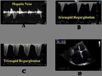
24 Question 6: Which of the following images best suggests severe TR? Question 6: Which of the following images best suggests severe TR?
25 Question 7: Which technical parameters are, in general, important in considering affecting quantification of TR severity and not MR severity? 1. Eccentricity of jet and Coanda effect 2. Color gain and scale setting 3. Compliance of the atrium 4. Phase of respiratory cycle Question 7: Which technical parameters are, in general, important in considering affecting quantification of TR severity and not MR severity? 1. Eccentricity of jet and Coanda effect 2. Color gain and scale setting 3. Compliance of the atrium 4. Phase of respiratory cycle 25


26 Question 8: A 78 year old patient presents with syncope and is found to have transient complete heart block. He is referred for an echocardiogram prepacemaker insertion. You obtain the following images: Question 8: What is the most appropriate next procedure? 1. Transesophageal Echocardiogram 2. Repeat image after ingestion of carbonated beverage 3. Agitated saline contrast through left arm 4. Agitated saline contrast through right arm 26

27 Answer : C agitated saline contrast through left arm showing persistent LSVC. Important pre pacemaker implantation Question 9: A patient has the following quantitative measures of RV function. Which is NOT recommended to diagnose RV systolic dysfunction ASE guidelines? 1. Fractional area change of 30% 2. TAPSE 15mm 3. S 8 cm/s 4. Free wall strain by speckle tracking -17% 5. MPI by pulsed Doppler of


28 Answer - 4 All of the above indicate significant RV systolic dysfunction. Answer 4 2D strain by speckle tracking is not one of the recommended methods to use in the routine exam owing to significant inter-vendor variability Question 10: Of the clips below, which diagnosis is least likely to be represented? Acute Pulmonary Embolism 2. Pulmonary Arteria Hypertension Atrial Septal Defect 4. RV infarction 5. Takotsubo Cardiomyopathy 28
29 Answer- 3 Choice 3 ASD usually demonstrates RV volume overload seen in multiple views, usually with preserved systolic function. An exception is the rare form associated with abnormal BMPR2 haplotype and PH representing fewer than 5% of ASDs, or those that present VERY late. 1 RV infarct associated with inferior MI 2 PAH 3 - Takotsubo 4 Acute PE Question 11: All of the following are compatible with severe tricuspid regurgitation except: 1. RV basal dimension measuring 50mm 2. Flow reversal in the hepatic veins after the P wave 3. PISA radius of 10 mm at an aliasing velocity of 30 cm/s 4. Non-parabolic TR CW Doppler signal with an early peak 29

30 Answer - 2 The flow reversal representing severe TR is after the QRS i.e. In systole Question 12: All of the following are true except: 1. The simplified bernoulli equation is not valid 2. The septum is d-shaped in diastole 3. The PAP may be overestimated due to the presence of laminar flow 4. The RA pressure is likely > 15 mmhg 30

31 Answer - 3 Laminar flow is likely to underestimate severity of TR. The simplified bernoulli equation is predicated on turbulent flow Question 13 Which of the following is least likely? 1. A diastolic rumble will be heard over the apex 2. The IVC will be markedly dilated 3. Mitral inflow pulsed doppler will demonstrate E:A reversal 4. The RV systolic pressure will be significantly elevated 31
32 Answer - 3 This patient has rheumatic TS, and therefore likely also has rheumatic mitral valve disease There is severe RA enlargement and therefore likely marked elevation of RA pressure The patient is in atrial fibrillation, so there will not be much of an A wave present Basic Ultrasound Physics Sidney K. Edelman, PhD 32
33 What would you do to decrease aliasing? 1. image by decreasing the Nyquist limit 2. image with a higher frequency transducer 3. use pulsed rather than CW Doppler 4. image in a view with a shallower gate 5. decrease the gain What would you do to decrease aliasing? 1. image by decreasing the Nyquist limit 2. image with a higher frequency transducer 3. use pulsed rather than CW Doppler 4. image in a view with a shallower gate 5. decrease the gain 33
34 A patient is evaluated with TEE for severe mitral regurgitation. Which color Doppler setting will decrease the jet area on TEE from the initial image? 1. lower the pulse repetition frequency 2. increase the Nyquist limit 3. increasing the Doppler gain 4. increase compression 5. decrease the reject A patient is evaluated with TEE for severe mitral regurgitation. Which color Doppler setting will decrease the jet area on TEE from the initial image? 1. lower the pulse repetition frequency 2. increase the Nyquist limit 3. increasing the Doppler gain 4. increase compression 5. decrease the reject 34
35 What is the wavelength of 2 MHz sound in soft tissue? m mm mm us What is the wavelength of 2 MHz sound in soft tissue? m mm mm us 35
36 The major source of ultrasound information used to create a two-dimensional image is: 1. specular reflections 2. Raleigh scattering 3. scattering 4. backscatter The major source of ultrasound information used to create a two-dimensional image is: 1. specular reflections 2. Raleigh scattering 3. scattering 4. backscatter 36
37 Which of the following forms of resolution varies within the depth of an image? 1. axial 2. contrast 3. lateral 4. harmonic Which of the following forms of resolution varies within the depth of an image? 1. axial 2. contrast 3. lateral 4. harmonic 37
Tricuspid and Pulmonary Valve Disease
 Tricuspid and Pulmonary Valve Disease Lawrence Rudski MD FRCPC FACC FASE Professor of Medicine Director, Division of Cardiology Jewish General Hospital McGill University Question 1 All of the following
Tricuspid and Pulmonary Valve Disease
 Tricuspid and Pulmonary Valve Disease Lawrence Rudski MD FRCPC FACC FASE Professor of Medicine Director, Division of Cardiology Jewish General Hospital McGill University Right Sided Failure Edema Gut congestion
Hemodynamic Assessment. Assessment of Systolic Function Doppler Hemodynamics
 Hemodynamic Assessment Matt M. Umland, RDCS, FASE Aurora Medical Group Milwaukee, WI Assessment of Systolic Function Doppler Hemodynamics Stroke Volume Cardiac Output Cardiac Index Tei Index/Index of myocardial
Basic Ultrasound Physics Board Review Questions
 Basic Ultrasound Physics Board Review Questions Sidney K. Edelman, PhD ESP Ultrasound The Woodlands, TX Question 1 What is the wavelength of 2 MHz sound in soft tissue? 1. 1.54 mm 2. 0.75 mm 3. 0.75 cm
Doppler Basic & Hemodynamic Calculations
 Doppler Basic & Hemodynamic Calculations August 19, 2017 Smonporn Boonyaratavej MD Division of Cardiology, Department of Medicine Chulalongkorn University Cardiac Center, King Chulalongkorn Memorial Hospital
The Normal Echocardiogram
 The Normal Echocardiogram Pravin V. Patil, MD FACC Lewis Katz School of Medicine at Temple University Acknowledgments Dr. Susan Wiegers Dr. Martin Keane Temple Cardiac Sonographers Disclosures No relevant
Right Heart Evaluation ASE Guidelines Review. Chris Mann RDCS, RCS, FASE Faculty, Echocardiography Pitt Community College Greenville, NC
 Right Heart Evaluation ASE Guidelines Review Chris Mann RDCS, RCS, FASE Faculty, Echocardiography Pitt Community College Greenville, NC Objectives Briefly review right atrial and right ventricular anatomy
Appendix II: ECHOCARDIOGRAPHY ANALYSIS
 Appendix II: ECHOCARDIOGRAPHY ANALYSIS Two-Dimensional (2D) imaging was performed using the Vivid 7 Advantage cardiovascular ultrasound system (GE Medical Systems, Milwaukee) with a frame rate of 400 frames
The Doppler Examination. Katie Twomley, MD Wake Forest Baptist Health - Lexington
 The Doppler Examination Katie Twomley, MD Wake Forest Baptist Health - Lexington OUTLINE Principles/Physics Use in valvular assessment Aortic stenosis (continuity equation) Aortic regurgitation (pressure
Adult Echocardiography Examination Content Outline
 Adult Echocardiography Examination Content Outline (Outline Summary) # Domain Subdomain Percentage 1 2 3 4 5 Anatomy and Physiology Pathology Clinical Care and Safety Measurement Techniques, Maneuvers,
ARTIFACTS: THEORY AND ILLUSTRATIVE EXAMPLES
 ARTIFACTS: THEORY AND ILLUSTRATIVE EXAMPLES Robert A. Levine, M.D. Marielle Scherrer-Crosbie, M.D. Eric M. Isselbacher, M.D. No conflicts of interest Philippe Bertrand, Pieter Vendervoort, Hasselt and
Pulmonary Hypertension. Echocardiography: Pearls & Pitfalls
 Pulmonary Hypertension Echocardiography: Pearls & Pitfalls Αθανάσιος Γ. Κουτσάκης Ειδικευόμενος Καρδιολογίας Α Καρδιολογική Κλινική ΑΠΘ Σεμινάρια Ομάδων Εργασίας Ελληνικής Καρδιολογικής Εταιρείας Ιωάννινα,
PART II ECHOCARDIOGRAPHY LABORATORY OPERATIONS ADULT TRANSTHORACIC ECHOCARDIOGRAPHY TESTING
 PART II ECHOCARDIOGRAPHY LABORATORY OPERATIONS ADULT TRANSTHORACIC ECHOCARDIOGRAPHY TESTING STANDARD - Primary Instrumentation 1.1 Cardiac Ultrasound Systems SECTION 1 Instrumentation Ultrasound instruments
PROSTHETIC VALVE BOARD REVIEW
 PROSTHETIC VALVE BOARD REVIEW The correct answer D This two chamber view shows a porcine mitral prosthesis with the typical appearance of the struts although the leaflets are not well seen. The valve
Diagnostic approach to heart disease
 Diagnostic approach to heart disease Initial work up History Physical exam Chest radiographs ECG Special studies Echocardiography Cardiac catheterization Echocardiography principles Technique of producing
Normal TTE/TEE Examinations
 Normal TTE/TEE Examinations Geoffrey A. Rose, MD FACC FASE Sanger Heart & Vascular Institute Before you begin imaging... Obtain the patient s Height Weight BP PLAX View PLAX View Is apex @ 9-10 o clock?
RIGHT VENTRICULAR SIZE AND FUNCTION
 RIGHT VENTRICULAR SIZE AND FUNCTION Edwin S. Tucay, MD, FPCC, FPCC, FPSE Philippine Society of Echocardiography Quezon City, Philippines Echo Mission, BRTTH, Legaspi City, July 1-2, 2016 NO DISCLOSURE
Fig.1 Normal appearance of RV in SAX:
 Tutorial 7 - Assessment of the right heart Assessment of the Right heart The right heart assessment clinically and echocardiographically is not a very important part of mainstream cardiology. In the ICU,
Right Ventricle Steven J. Lester MD, FACC, FRCP(C), FASE Mayo Clinic, Arizona
, FASE Mayo Clinic, Arizona") Right Ventricle Steven J. Lester MD, FACC, FRCP(C), FASE Mayo Clinic, Arizona 1. In which scenario will applying the simplified Bernoulli equation to the peak tricuspid regurgitation velocity and adding
Right Ventricle Steven J. Lester MD, FACC, FRCP(C), FASE Mayo Clinic, Arizona 1. In which scenario will applying the simplified Bernoulli equation to the peak tricuspid regurgitation velocity and adding
AIMI-HF PROCEDURE MANUAL TECHNICAL GUIDE FOR ECHOCARDIOGRAPHY. MHI Core Laboratory E. O Meara - J.C. Tardif J. Vincent, G. Grenier, C.
 AIMI-HF PROCEDURE MANUAL TECHNICAL GUIDE FOR ECHOCARDIOGRAPHY MHI Core Laboratory E. O Meara - J.C. Tardif J. Vincent, G. Grenier, C. Roy February 2016 Montreal Heart Institute HF Research Aude Turgeon,
Adel Hasanin Ahmed 1
 Adel Hasanin Ahmed 1 PERICARDIAL DISEASE The pericardial effusion ends anteriorly to the descending aorta and is best visualised in the PLAX. PSAX is actually very useful sometimes for looking at posterior
New 3D Quantification of Mitral Regurgitation Severity. Judy Hung, MD Cardiac Ultrasound Laboratory Massachusetts General Hospital Boston, MA
 New 3D Quantification of Mitral Regurgitation Severity Judy Hung, MD Cardiac Ultrasound Laboratory Massachusetts General Hospital Boston, MA No Financial Disclosures No off label discussion of devices
Echocardiography: Guidelines for Valve Quantification
 Echocardiography: Guidelines for Echocardiography: Guidelines for Chamber Quantification British Society of Echocardiography Education Committee Richard Steeds (Chair), Gill Wharton (Lead Author), Jane
Diastolic Function: What the Sonographer Needs to Know. Echocardiographic Assessment of Diastolic Function: Basic Concepts 2/8/2012
 Diastolic Function: What the Sonographer Needs to Know Pat Bailey, RDCS, FASE Technical Director Beaumont Health System Echocardiographic Assessment of Diastolic Function: Basic Concepts Practical Hints
Echo Doppler Assessment of Right and Left Ventricular Hemodynamics.
 Echo Doppler Assessment of Right and Left Ventricular Hemodynamics. Itzhak Kronzon, MD, FASE, FACC, FESC, FAHA, FACP, FCCP Northwell, Lenox Hill Hospital, New York Professor of Cardiology Hofstra University
ARTIFACTS: THEORY AND ILLUSTRATIVE EXAMPLES
 ARTIFACTS: THEORY AND ILLUSTRATIVE EXAMPLES Robert A. Levine, M.D. Marielle Scherrer-Crosbie, M.D. Eric M. Isselbacher, M.D. 60 year old man Cardiac source of embolus? NAME THAT MASS! 1 NAME THAT MASS!
MITRAL STENOSIS. Joanne Cusack
 MITRAL STENOSIS Joanne Cusack BSE Breakdown Recognition of rheumatic mitral stenosis Qualitative description of valve and sub-valve calcification and fibrosis Measurement of orifice area by planimetry
P = 4V 2. IVC Dimensions 10/20/2014. Comprehensive Hemodynamic Evaluation by Doppler Echocardiography. The Simplified Bernoulli Equation
 Comprehensive Hemodynamic Evaluation by Doppler Echocardiography Itzhak Kronzon, MD North Shore LIJ/ Lenox Hill Hospital New York, NY Disclosure: Philips Healthcare St. Jude Medical The Simplified Bernoulli
DOPPLER HEMODYNAMICS (1) QUANTIFICATION OF PRESSURE GRADIENTS and INTRACARDIAC PRESSURES
 QUANTIFICATION OF PRESSURE GRADIENTS and INTRACARDIAC PRESSURES") THORAXCENTRE DOPPLER HEMODYNAMICS (1) QUANTIFICATION OF PRESSURE GRADIENTS and INTRACARDIAC PRESSURES J. Roelandt DOPPLER HEMODYNAMICS Intracardiac pressures and pressure gradients Volumetric measurement
THORAXCENTRE DOPPLER HEMODYNAMICS (1) QUANTIFICATION OF PRESSURE GRADIENTS and INTRACARDIAC PRESSURES J. Roelandt DOPPLER HEMODYNAMICS Intracardiac pressures and pressure gradients Volumetric measurement
Index. K Knobology, TTE artifact, image resolution, ultrasound, 14
 A Acute aortic regurgitation (AR), 124 128 Acute aortic syndrome (AAS) classic aortic dissection diagnosis, 251 263 evolutive patterns, 253 255 pathology, 250 251 classifications, 247 248 incomplete aortic
Echocardiography as a diagnostic and management tool in medical emergencies
 Echocardiography as a diagnostic and management tool in medical emergencies Frank van der Heusen MD Department of Anesthesia and perioperative Care UCSF Medical Center Objective of this presentation Indications
British Society of Echocardiography
 British Society of Echocardiography Affiliated to the British Cardiac Society A Minimum Dataset for a Standard Adult Transthoracic Echocardiogram From the British Society of Echocardiography Education
Valvular Regurgitation: Can We Do Better Than Colour Doppler?
 Valvular Regurgitation: Can We Do Better Than Colour Doppler? A/Prof David Prior St Vincent s Hospital Melbourne Sports Cardiology Valvular Regurgitation Valve regurgitation volume loads the ventricles
Prof. JL Zamorano Hospital Universitario Ramón y Cajal
 Prof. JL Zamorano Hospital Universitario Ramón y Cajal Should we forget TR? Nath J et al. Impact of tricuspid regurgitation on long-term survival. J Am Coll Cardiol. 2004; 43:405-409 Why is it difficult
Echocardiography. Guidelines for Valve and Chamber Quantification. In partnership with
 Echocardiography Guidelines for Valve and Chamber Quantification In partnership with Explanatory note & references These guidelines have been developed by the Education Committee of the British Society
Case # 1. Page: 8. DUKE: Adams
 Case # 1 Page: 8 1. The cardiac output in this patient is reduced because of: O a) tamponade physiology O b) restrictive physiology O c) coronary artery disease O d) left bundle branch block Page: 8 1.
Evaluation of Left Ventricular Diastolic Dysfunction by Doppler and 2D Speckle-tracking Imaging in Patients with Primary Pulmonary Hypertension
 ESC Congress 2011.No 85975 Evaluation of Left Ventricular Diastolic Dysfunction by Doppler and 2D Speckle-tracking Imaging in Patients with Primary Pulmonary Hypertension Second Department of Internal
Diastolic Heart Function: Applying the New Guidelines Case Studies
 Diastolic Heart Function: Applying the New Guidelines Case Studies Mitral Regurgitation The New ASE William Guidelines: A. Zoghbi Role MD, of FASE, 2D/3D MACCand CMR Professor and Chairman, Department
Quantification of Mitral Stenosis: Planimetry, pressure Half time, Continuity Common Errors
 Quantification of Mitral Stenosis: Planimetry, pressure Half time, Continuity Common Errors Christopher J Kramer RDCS Advanced Cardiovascular Services Aurora Health Care Milwaukee, WI No Disclosures Baumgartner,
Comprehensive Hemodynamics By Doppler Echocardiography. The Echocardiographic Swan-Ganz Catheter.
 Comprehensive Hemodynamics By Doppler Echocardiography. The Echocardiographic Swan-Ganz Catheter. Itzhak Kronzon, MD, FASE, FACC, FESC, FAHA, FACP, FCCP North Shore HS, LIJ/Lenox Hill Hospital, New York
2/4/2011. Nathan Kerner, M.D.
 Nathan Kerner, M.D. Definition Elevated pressures - cut off usually >40 mmhg pulmonary artery systolic pressure (PASP) Usually associated with elevated pulmonary vascular resistance (PVR) measured in dynessec/cm
Uncommon Doppler Echocardiographic Findings of Severe Pulmonic Insufficiency
 Uncommon Doppler Echocardiographic Findings of Severe Pulmonic Insufficiency Rahul R. Jhaveri, MD, Muhamed Saric, MD, PhD, FASE, and Itzhak Kronzon, MD, FASE, New York, New York Background: Two-dimensional
Echocardiographic Cardiovascular Risk Stratification: Beyond Ejection Fraction
 Echocardiographic Cardiovascular Risk Stratification: Beyond Ejection Fraction October 4, 2014 James S. Lee, M.D., F.A.C.C. Associates in Cardiology, P.A. Silver Spring, M.D. Disclosures Financial none
Concepts of Imaging and Knobology
 Concepts of Imaging and Knobology Pravin Patil, MD FACC FASE Associate Professor of Medicine Director, Cardiovascular Disease Training Program Lewis Katz School of Medicine at Temple University Disclosures
Cardiac ultrasound protocols
 Cardiac ultrasound protocols IDEXX Telemedicine Consultants Two-dimensional and M-mode imaging planes Right parasternal long axis four chamber Obtained from the right side Displays the relative proportions
ECHOCARDIOGRAPHY DATA REPORT FORM
 Patient ID Patient Study ID AVM - - Date of form completion / / 20 Initials of person completing the form mm dd yyyy Study period Preoperative Postoperative Operative 6-month f/u 1-year f/u 2-year f/u
Certificate in Clinician Performed Ultrasound (CCPU) Syllabus. Rapid Cardiac Echo (RCE)
 Syllabus. Rapid Cardiac Echo (RCE)") Certificate in Clinician Performed Ultrasound (CCPU) Syllabus Rapid Cardiac Echo (RCE) Purpose: Rapid Cardiac Echocardiography (RCE) This unit is designed to cover the theoretical and practical curriculum
Certificate in Clinician Performed Ultrasound (CCPU) Syllabus Rapid Cardiac Echo (RCE) Purpose: Rapid Cardiac Echocardiography (RCE) This unit is designed to cover the theoretical and practical curriculum
Disclosures Rebecca T. Hahn, MD, FASE
 The New ASE Guidelines for Native Valvular Regurgitation Mitral Regurgitation The New ASE Guidelines: Role of 2D/3D and CMR (With caveats and comments from R. Hahn) William A. Zoghbi MD, FASE, MACC Professor
Congenital. Unicuspid Bicuspid Quadricuspid
 David Letterman s Top 10 Aortic Stenosis The victim can be anyone: Echo is the question and the answer!!!! Hilton Head Island Echocardiography Conference 2012 Timothy E. Paterick, MD, JD, MBA Christopher
LV FUNCTION ASSESSMENT: WHAT IS BEYOND EJECTION FRACTION
 LV FUNCTION ASSESSMENT: WHAT IS BEYOND EJECTION FRACTION Jamilah S AlRahimi Assistant Professor, KSU-HS Consultant Noninvasive Cardiology KFCC, MNGHA-WR Introduction LV function assessment in Heart Failure:
Normal TTE Examination, Doppler Echocardiography and Normal Antegrade Flow Patterns
 Normal TTE Examination, Doppler Echocardiography and Normal Antegrade Flow Patterns Pravin Patil, MD FACC FASE Associate Professor of Medicine Director, Cardiovascular Disease Training Program Lewis Katz
Prosthetic valve dysfunction: stenosis or regurgitation
 Prosthetic valve dysfunction: stenosis or regurgitation Jean G. Dumesnil MD, FRCP(C), FACC, FASE(Hon) Quebec Heart and Lung Institute, Québec, Québec No disclosures Possible Causes of High Gradients in
Back to Basics: Common Errors In Quantitation In Everyday Practice
 Back to Basics: Common Errors In Quantitation In Everyday Practice Deborah Agler, ACS, RDCS, FASE October 9, 2017 ASE: Echo Florida Rebecca T. Hahn, MD Director of Interventional Echocardiography Professor
M-Mode Echocardiography Is it still Alive? Itzhak Kronzon, MD,FASE. Sampling Rate M-Mode: 1800 / sec 2D: 30 / sec
 M-Mode Echocardiography Is it still Alive? Itzhak Kronzon, MD,FASE Honoraria: Philips Classical M-mode Echocardiography M-Mode offers better time and image resolution. Sampling Rate M-Mode: 1800 / sec
Pulsed Wave Doppler and Color Flow Doppler Evaluation in Healthy Dogs and Dogs with Cardiac Disease
 Cloud Publications International Journal of Advanced Veterinary Science and Technology 2016, Volume 5, Issue 2, pp. 256-265, Article ID Sci-446 ISSN 2320-3595 Research Article Open Access Pulsed Wave Doppler
Atrial Septal Defects
 Supplementary ACHD Echo Acquisition Protocol for Atrial Septal Defects The following protocol for echo in adult patients with atrial septal defects (ASDs) is a guide for performing a comprehensive assessment
TAVR: Echo Measurements Pre, Post And Intra Procedure
 2017 ASE Florida, Orlando, FL October 10, 2017 8:00 8:25 AM 25 min TAVR: Echo Measurements Pre, Post And Intra Procedure Muhamed Sarić MD, PhD, MPA Director of Noninvasive Cardiology Echo Lab Associate
ECHO HAWAII. My home. Pulmonary Hypertension and Pulmonary Embolism: Role of Echo U.S.A. Japan. Hawaii Island 1/9/2018
 Pulmonary Hypertension and Pulmonary Embolism: Role of Echo ECHO HAWAII January 15 19, 2018 Kenya Kusunose, MD, PhD, FASE Tokushima University Hospital Japan My home Japan U.S.A Hawaii Island 1 Economy
Transthoracic echocardiography in the evaluation of pediatric pulmonary hypertension and ventricular dysfunction
 REVIEW ARTICLE Transthoracic echocardiography in the evaluation of pediatric pulmonary hypertension and ventricular dysfunction Martin Koestenberger, 1 Mark K. Friedberg, 2 Eirik Nestaas, 3 Ina Michel-Behnke,
Chamber Quantitation Guidelines - Update II
 Chamber Quantitation Guidelines - Update II Right Heart Measurements Steven A. Goldstein MD FACC FASE Professor of Medicine Georgetown University Medical Center MedStar Heart Institute Washington Hospital
Pediatric Echocardiography Examination Content Outline
 Pediatric Echocardiography Examination Content Outline (Outline Summary) # Domain Subdomain Percentage 1 Anatomy and Physiology Normal Anatomy and Physiology 10% 2 Abnormal Pathology and Pathophysiology
Interventions in Adult Congenital Heart Disease: Role of CV Imaging. Associate Professor. ACHD mortality. Pillutla. Am Heart J 2009;158:874-9
 Interventions in Adult Congenital Heart Disease: Role of CV Imaging Sangeeta Shah MD, FACC, FASE Associate Professor ACHD mortality Pillutla. Am Heart J 2009;158:874-9 Adult Congenital Heart Disease Heterogenity
The background of the Cardiac Sonographer Network News masthead is a diagnostic image:
 Number 5 Welcome Number 5 Welcome to the newsletter created just for you: sonographers who perform pediatric echocardiograms in primarily adult echo labs. Each issue features tips on echocardiography of
Echo in Pulmonary HTN
 Echo in Pulmonary HTN Steven A. Goldstein MD FACC FASE Professor of Medicine Georgetown University Medical Center MedStar Heart Institute Washington Hospital Center Monday, October 10, 2017 Pulmonary Artery
Assessment of LV systolic function
 Tutorial 5 - Assessment of LV systolic function Assessment of LV systolic function A knowledge of the LV systolic function is crucial in the undertanding of and management of unstable hemodynamics or a
Doppler Color Flow Imaging #4
 Doppler Color Flow Imaging #4 Joseph A. Kisslo, MD David B. Adams, RDCS INTRODUCTION Doppler color flow imaging is a method for noninvasively imaging blood flow through the heart by displaying flow data
Evaluation of the Right Ventricle in Candidates for Right Ventricular Assist Device Implantation.
 Evaluation of the Right Ventricle in Candidates for Right Ventricular Assist Device Implantation. Evaluation of RVAD Function. Ioannis A Paraskevaidis Attikon University Hospital Historical Perspective
Quantification of MR
 Valvular Regurgitation: Putting the New Guidelines into Practice James D. Thomas, MD, FACC, FASE, FESC Director, Center for Heart Valve Disease Bluhm Cardiovascular Institute Professor of Medicine, Feinberg
Quantitation of right ventricular dimensions and function
 SCCS Basics of cardiac assessment Quantitation of right ventricular dimensions and function Tomasz Kukulski, MD PhD Dept of Cardiology, Congenital Heart Disease and Electrotherapy Silesian Medical University
Echocardiography Conference
 Echocardiography Conference David Stultz, MD Cardiology Fellow, PGY-6 September 20, 2005 Atrial Septal Aneurysm Bulging of Fossa Ovalis Associated commonly with Atrial septal defect or small perforations
What are the best diagnostic tools to quantify aortic regurgitation?
 What are the best diagnostic tools to quantify aortic regurgitation? Agnès Pasquet, MD, PhD Pôle de Recherche Cardiovasculaire Institut de Recherche Expérimentale et Clinique Université catholique de Louvain
LUST trial. Echocardiography USER S MANUAL
 LUST trial Echocardiography USER S MANUAL Rosa Sicari, Luna Gargani Ins1tute of Clinical Physiology Na1onal Council of Research, Pisa, Italy Parameters required (1) Aortic root Measurement of aortic root
TEE Outside of the Cardiac OR
 TEE Outside of the Cardiac OR STEVE GIBSON MD PHD OU DEPARTMENT OF ANESTHEIOLOGY I have no financial relationships or conflicts of interest to disclose TRANSESOPHAGEAL ECHOCARDIOGRAPHY Basic principles
Incorporating the New Echo Guidelines Into Everyday Practice
 Incorporating the New Echo Guidelines Into Everyday Practice Clinical Case RIGHT VENTRICULAR FAILURE Gustavo Restrepo MD President Elect Interamerican Society of Cardiology Director Fellowship Training
JOINT MEETING 2 Tricuspid club Chairpersons: G. Athanassopoulos, A. Avgeropoulou, M. Khoury, G. Stavridis
 JOINT MEETING 2 Tricuspid club Chairpersons: G. Athanassopoulos, A. Avgeropoulou, M. Khoury, G. Stavridis Similarities and differences in Tricuspid vs. Mitral Valve Anatomy and Imaging. Echo evaluation
Right Heart Hemodynamics: Echo-Cath Discrepancies
 Department of cardiac, thoracic and vascular sciences University of Padua, School of Medicine Padua, Italy Right Heart Hemodynamics: Echo-Cath Discrepancies Luigi P. Badano, MD, PhD, FESC, FACC **Dr. Badano
Three-dimensional Wall Motion Tracking:
 Three-dimensional Wall Motion Tracking: A Novel Echocardiographic Method for the Assessment of Ventricular Volumes, Strain and Dyssynchrony Jeffrey C. Hill, BS, RDCS, FASE Jennifer L. Kane, RCS Gerard
25 different brand names >44 different models Sizes mm
 Types of Prosthetic Valves BIOLOGIC STENTED Porcine xenograft Pericardial xenograft STENTLESS Porcine xenograft Pericardial xenograft Homograft (allograft) Autograft PERCUTANEOUS MECHANICAL Bileaflet Single
Right Heart Catheterization. Franz R. Eberli MD Chief of Cardiology Stadtspital Triemli, Zurich
 Right Heart Catheterization Franz R. Eberli MD Chief of Cardiology Stadtspital Triemli, Zurich Right Heart Catheterization Pressure measurements Oxygen saturation measurements Cardiac output, Vascular
Bogdan A. Popescu. University of Medicine and Pharmacy Bucharest, Romania. EAE Course, Bucharest, April 2010
 Bogdan A. Popescu University of Medicine and Pharmacy Bucharest, Romania EAE Course, Bucharest, April 2010 This is how it started Mitral stenosis at a glance 2D echo narrow diastolic opening of MV leaflets
Basic Approach to the Echocardiographic Evaluation of Ventricular Diastolic Function
 Basic Approach to the Echocardiographic Evaluation of Ventricular Diastolic Function J A F E R A L I, M D U N I V E R S I T Y H O S P I T A L S C A S E M E D I C A L C E N T E R S T A F F C A R D I O T
Aortic Stenosis: Spectrum of Disease, Low Flow/Low Gradient and Variants
 Aortic Stenosis: Spectrum of Disease, Low Flow/Low Gradient and Variants Martin G. Keane, MD, FASE Professor of Medicine Lewis Katz School of Medicine at Temple University Basic root structure Parasternal
Marti McCulloch, BS, MBA, RDCS, FASE Houston, Texas
 Marti McCulloch, BS, MBA, RDCS, FASE Houston, Texas Mitral Regurgitation What to Expect Review Specific Signs of Severity Supportive Signs of Severity Qualitative Parameters Structural Doppler Quantitative
Revealing new insights. irotate electronic rotation and xplane adjustable biplane imaging. Ultrasound cardiology. irotate and xplane
 Ultrasound cardiology irotate and xplane Revealing new insights irotate electronic rotation and xplane adjustable biplane imaging Annemien van den Bosch and Jackie McGhie Department of Cardiology, Erasmus
SONOGRAPHER & NURSE LED VALVE CLINICS
 SONOGRAPHER & NURSE LED VALVE CLINICS Frequency of visits and alerts AORTIC STENOSIS V max > 4.0 m/s or EOA < 1.0 cm 2 V max 3.5 4.0 m/s + Ca+ V max 3.0 4.0 m/s or EOA 1.0-1.5 cm 2 V max 2.5 3.0 m/s every
Echocardiographic Evaluation of the Cardiomyopathies. Stephanie Coulter, MD, FACC, FASE April, 2016
 Echocardiographic Evaluation of the Cardiomyopathies Stephanie Coulter, MD, FACC, FASE April, 2016 Cardiomyopathies (CMP) primary disease intrinsic to cardiac muscle Dilated CMP Hypertrophic CMP Infiltrative
Quantification of Cardiac Chamber Size
 2017 KSE 2017-11-25 Quantification of Cardiac Chamber Size Division of Cardiology Keimyung University Dongsan Medical Center In-Cheol Kim M.D., Ph.D. LV size and function Internal linear dimensions PLX
The Doppler echocardiographic assessment of valvular regurgitation in normal children

Advanced Applica,on of Point- of- Care Echocardiography in Cri,cal Care. Dr. Mark Tutschka Dr. Rob ArnAield
 Advanced Applica,on of Point- of- Care Echocardiography in Cri,cal Care Dr. Mark Tutschka Dr. Rob ArnAield OBJECTIVES Provide an overview of common advanced echocardiographic techniques suitable for use
Pericardial Diseases. Smonporn Boonyaratavej, MD. Division of Cardiology, Department of Medicine Chulalongkorn University
 Pericardial Diseases Smonporn Boonyaratavej, MD Division of Cardiology, Department of Medicine Chulalongkorn University Cardiac Center, King Chulalongkorn Memorial Hospital 21 AUGUST 2016 Pericardial
MITRAL REGURGITATION ECHO PARAMETERS TOOL
 Comprehensive assessment of qualitative and quantitative parameters, along with the use of standardized nomenclature when reporting echocardiographic findings, helps to better define a patient s MR and
ASCeXAM / ReASCE. Practice Board Exam Questions. Monday Afternoon
 ASCeXAM / ReASCE Practice Board Exam Questions Monday Afternoon Aortic Valve Prostheses Aortic Regurgitation Stress Echocardiography Ischemic Heart Disease and Myocardial Infarction 3D Echocardiography
Cardiac Ausculation in the Elderly
 Cardiac Ausculation in the Elderly 박성하 신촌세브란스병원심장혈관병원심장내과 Anatomy Surface projection of the Heart and Great Vessels Evaluating pulsation Superior vena cava Rt. pulmonary artery Right atrium Right ventricle
CASE REPORT: DOUBLE ORIFICE MITRAL VALVE WITH CLEFT IN ANTERIOR LEAFLET OF DOMINANT VALVE IN AN AFRO-CARIBBEAN
 CASE REPORT: DOUBLE ORIFICE MITL VAE WITH CLEFT IN ANTERIOR LEAFLET OF DOMINANT VAE IN AN AFRO-CARIBBEAN Disclosure: No potential conflict of interest. Received: 27.08.13 Accepted: 23.06.14 Citation: EMJ
Presenter: Steven Brust, HCS-D, HCS-H Product Manager, Home Health Coding Center
 Presenter: Steven Brust, HCS-D, HCS-H Product Manager, Home Health Coding Center Pinpoint & properly assign the appropriate heart failure codes Left- vs. Right-sided Left ventricular failure (LVF) may
OPTIMIZING ECHO ACQUISTION FOR STRAIN AND DIASTOLOGY
 OPTIMIZING ECHO ACQUISTION FOR STRAIN AND DIASTOLOGY October 8, 2017 Deborah Agler, ACS, RDCS, FASE Coordinator of Education and Training Cleveland Clinic General Principles Diastology Clinical Data Heart
Emergency department bedside echocardiography diagnosis of massive pulmonary embolism with direct visualization of thrombus in the pulmonary artery
 Crit Ultrasound J (2011) 3:155 160 DOI 10.1007/s13089-011-0081-4 CASE REPORT Emergency department bedside echocardiography diagnosis of massive pulmonary embolism with direct visualization of thrombus
JFICMI Basic Critical Care Echocardiography (BCCE)
") JFICMI Basic Critical Care Echocardiography (BCCE) 2017 Introduction The International expert statement on training standards for critical care ultrasonography position paper published in Intensive Care
JFICMI Basic Critical Care Echocardiography (BCCE) 2017 Introduction The International expert statement on training standards for critical care ultrasonography position paper published in Intensive Care
좌심실수축기능평가 Cardiac Function
 Basic Echo Review Course 좌심실수축기능평가 Cardiac Function Seonghoon Choi Cardiology Hallym university LV systolic function Systolic function 좌심실수축기능 - 심근의수축으로심실에서혈액을대동맥으로박출하는기능 실제임상에서 LV function 의의미 1Diagnosis
Adopted by Council March 2006, Revised March 2012, September 2015
 Guidelines, Policies and Statements E4 Education Protocol: Competences Required Of Cardiac Sonographers Who Practice Adult Transthoracic Cardiac Ultrasound Examinations Adopted by Council March 2006, Revised