MR Imaging of Atherosclerotic Plaques
|
|
|
- Mildred Tucker
- 5 years ago
- Views:
Transcription


1 MR Imaging of Atherosclerotic Plaques Yeon Hyeon Choe, MD Department of Radiology, Samsung Medical Center, Sungkyunkwan University, Seoul
2 MRI for Carotid Atheroma Excellent tissue contrast (fat, fibrous tissue, collagen, thrombus) Noninvasive More accurate and objective estimation of stenosis degree than US
3 Carotid MRI at 1.5T Experiences for a decade MRI has shown capability to detect vulnerable plaques with large lipid/ necrotic cores, intraplaque hemorrhage and plaque cap rupture. 3.0 T MRI is expected to show higher resolution enough for smaller arteries.
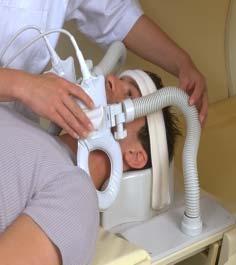

4 SENS-Flex-M Coil
5 MRI Appearance of Plaque Components on Various Imaging Sequences Plaque Components T2WI T1WI Proton WI TOF T1WI, Fat- CE on Post- Suppr. Contrast Recent Hemorrhage Variable H-to-M H Variable H No Lipid-rich Necrotic Core Variable (L) H M-to-H H M No Fibrous Tissue Variable M M H M-to-L Yes Calcification L L L L L No Tissue contrast is relative to SI of muscle


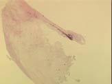
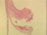
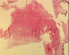
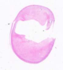
6 Stable Plaque T2WI Pathology
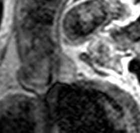
7 Lipid-rich Plaque FISSURE T1WI Proton-WI T2WI TOF

8 Fibrous Cap on MRI


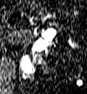
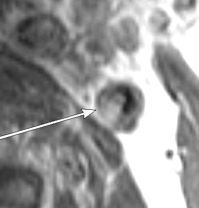
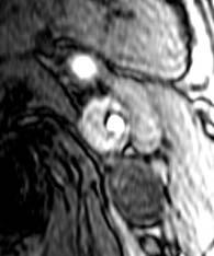
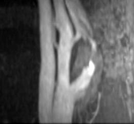
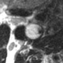
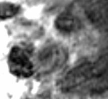
9 Fresh Intraplaque Hemorrhage with Fibrous Cap Rupture Cap rupture Fresh hemorrhage TOF


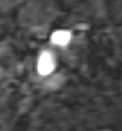
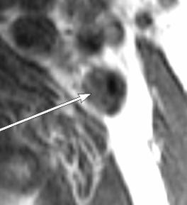
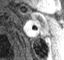
10 Intact Plaque Cap with Intraplaque Hemorrhage (fibrin) TOF Cap T1FS Fibrin Hemorrhage in 31% of 153 endarterectomy specimens
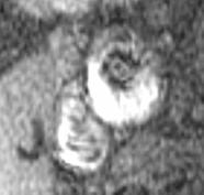
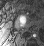
11 Contrast-enhancement in Fibrous Component Pre Post
12 Increased neovascularity within plaque may contribute to plaque instability. Contrast-enhancement represents neovasculature in the histopathology (sensitivity 76%, specificity 79% by Yuan). Contrast-enhancement improves differentiation of tissue types (fibrous tissue, necrotic core, calcifications).

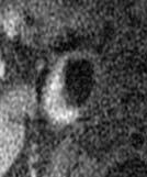
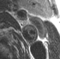
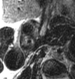
13 Large Necrotic Core without Significant Contrast Enhancement T2WI T1WI, PC

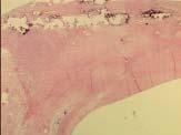
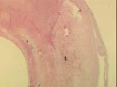
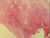
14 Hydrophilic Necrosis A T1WI T2WI Specimen
15 MRI Accuracy Ex vivo T2WI: Accurate for fibrocalcific plaques (90% sensitivity, 100% specificity) -Serfaty et al In vivo multispectral MRI for lipid-rich cores and intraplaque hemorrahge: overall accuracy 87%, sensitivity 85%, specificity 92% -Yuan et al
16 Fibrous Cap Rupture at MRI and Recent TIA or Stroke (Yuan, Circulation 2002) Intact thick FC (n = 11) % Symptomatic 9 Likelihood to have TIA/stroke 1X Odds Ratio Intact thin FC 50 10X 6 (n = 12) (95% CI = 1.0, 104) Ruptured FC 70 23X 18 (n = 30) (95% CI = 3, 210)
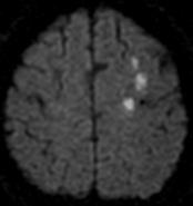
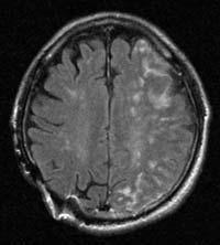
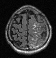
17 Brain MR imaging in Pts with Unstable Carotid Plaques In 153 patients, carotid MRI-MRA and carotid endarterectomy were performed. 97 atheromas (72%): histologically unstable plaque Good agreement between MR imaging and histologically vulnerable plaque 59 (61%) showed acute ischemic lesions in the brain on diffusion MR imaging.

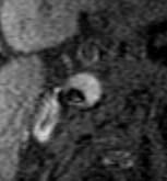
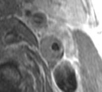
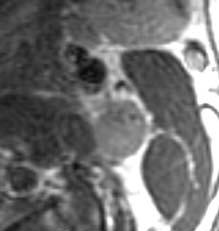
18 Case 1, Ulcerative Plaque with Hemorrhage TOF T1FS T2 MIP


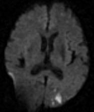
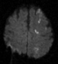
19 Multifocal Recent Infarctions on Diffusion-weighted MRI
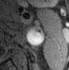
20 Case 2, Hemorrhagic Plaque TOF T2 T1FS T1CE

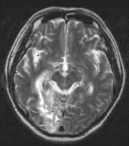
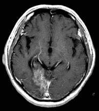
21 T2 FLAIR T1CE
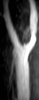
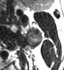
22 Case 3, Hemorrhagic Plaque TOF T1FS T2 T1CE MIP

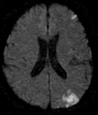
23 Multiple New Infarctions
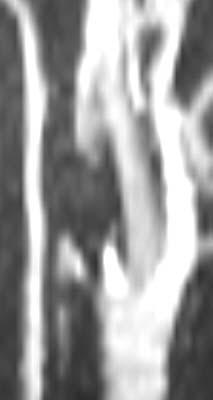
24 MRA of Intraplaque Hemorrhage
25 Materials & Methods 153 consecutive patients who underwent MRI and carotid endarterectomy were included in this study. Performed high-resolution MR imaging before carotid surgery. Findings on surgery, MRI (in vivo, specimen), and pathology were correlated. Analysis of carotid bifurcation and proximal ICA
26 Results 42 atheromas (31%) had intraplaque hemorrhage on surgicopathological findings. 38 (90.5%) showed high signal intensity halo around the carotid artery on TOF MR angiography. Additional sequence of T1-weighted fatsaturated spin echo images also revealed evidence of intraplaque hemorrhage by showing unsaturated high signal intensity in the plaque in 71% of patients.
27 Results Correlated with surgical and histopathological findings, MIP images of TOF MR angiography demonstrated sensitivity = 91%, specificity = 91%, negative predictive value = 77%, positive predictive value = 93%. The combination of high signal intensity areas on T1- WI with fat suppression, low signal intensity areas on T2-WI, and high SI areas on source images of the 3 D time-of-flight MRA sequence also can suggest intraplaque hemorrhage.

28
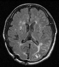
29 FLAIR DWI

30 A T1WI T2WI TOF T1CE
31 Conclusions High-resolution MR imaging can help identify intraplaque hemorrhage noninvasively. High SI on source images of 3D-TOF MRA and fat-suppressed T1WI can suggest intraplaque hemorrhage. Maximum intensity projection images of TOF MR angiography are useful in noninvasive detection of carotid intraplaque hemorrhage.
32 Changes in Carotid Plaques after Statin Therapy: MRI Evaluation 45 pts with 30-70% stenosis PreTx (Simbastatin 20 mg) and F-up MRI (19 ± 7.5 m) Decreased mean total chol., LDL, & CRP compared with control group: Total Chol = 188 ± 26 vs 158 ± 50, p<.001 LDL = 125 ± 23 vs 110± 42, p <.001 CRP = 0.41 ± 0.59 vs 0.10 ± 1.3, p<0.01
33 Results Stabilization of plaque components on MRI in 16% (7/45) Decreased plaque thickness: 4.2 mm± 1.0 vs 3.4 mm ± 1.3, p < 0.001


34 Increased Fibrotic Component on T2WI at 20 m f-up


35 Improved Wall Thickness, at 1 y (0.5 mm)
36 3.0 T for Atherosclerotic Plaque Imaging Higher signal-to-noise ratio Better background suppression Shorter scan time Higher resolution
37 Carotid MRI Techniques at 3.0 T T1-weighed spin echo imaging (precontrast axial, again with fat-saturation, postcontrast axial) T2-weighted spin echo imaging (axial) Proton-WI (axial) 3D time-of-flight MRA (source and 3D images) Two 15-cm phased-array coils, bilateral application Field of view: cm x cm Slice thickness = 2 mm, matrix, NEX = 1
38 SMC Experience of 3.0 T MRI in Carotid Plaque Imaging Image quality is generally compatible with 1.5 T s. Higher resolution and tissue contrast with 3.0 T MRI


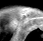
39 R ICA Plaque, 1.5T vs 3.0T T1WI, T vs 3.0 T


40 MIP of 3D-TOF T vs 3.0 T

41 T vs 3.0 T
42 Coronary Artery Plaque MRI
43 High-resolution of 3.0 T system is most beneficial in coronary artery plaque imaging. Can knowledge of carotid plaque MRI be applied to coronary artery imaging?
44 Imaging Protocol Coronary MRA using 3-point technique Black-blood plaque imaging on the stenotic segments T1WI, precontrast (DBIR) T2WI T1WI, postcontrast TFE Slice thickness, mm; image matrix, 256x256, FOV 35 cm, en = 1

45 Coronary Artery Plaque Imaging: LAD Stenosis on CAG


46 Coronary Plaque Imaging at 3.0T T2WI T1WI-preContrast T1WI-postContrast
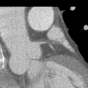

47 Fibrofatty Plaques (M/48 with Acute MI)


48 Intravascular US Shows Noncalcified Plaques without Large Hypoechoic Area Left main LAD-LCX (9 o clock) bifurcation site (4-6 o clock site plaque)

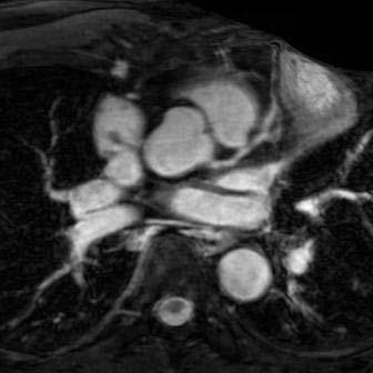
49 Coronary MRA


50 L Main T2WI, Fat-sat T2WI, 1.5 mm ST PostContrast
51 Contrast Agents for MR Imaging of Atherosclerosis Ultrasmall superparamagnetic particles of iron oxide (USPIO, Combidex ) Fibrin-specific polypeptide (EPIX) Plaque detection by Gadofluorine (Schering)

52 USPIO in Watanabe Hyperlipidemic Rabbits (5 ds) Watanabe Control Watanabe USPIO USPIO No USPIO Ruehm at al. Circulation 2001


53 Fe in Macrophages of Atheroma Prussian Blue staining EM

54 Gadofluorine M Accumulation in necrotic, edematous, lipid rich areas and macrophages (?) In vitro staining in human coronary artery


55 2 ds after Gadofluorine M injection in Cholesterol-fed diabetic Rabbit, 14-T MR Microscopy vs Specimen Courtesy of Dr Whal Lee, Seoul National University
 Botnar, Circulation")
56 Fibrin-specific polypeptide (20 h post EP-1873, EPIX) Botnar, Circulation 2004
57 Alpha v beta 3 -Integrin-targeted Imaging: significances Plaque angiogenesis and expansion of vasa vasorum: critical process for initiation of vascular lesions Site-specific drug delivery for early atheroslcerotic disease

58 Alpha v beta 3 -Integrin-targeted paramagnetic nanoparticles in atherosclerotic rabbits Winter, Circulation 2003
59 Conclusions High-resolution MRI can identify vulnerable carotid plaques and allows early intervention on unstable plaques. 3.0 T MRI allows characterization of atherosclerotic plaques in coronary arteries.
Non-invasive Imaging of Carotid Artery Atherosclerosis
 Non-invasive Imaging of Carotid Artery Atherosclerosis 최연현 성균관의대삼성서울병원영상의학과 Noninvasive Techniques US with Doppler CT MRI Ultrasonography Techniques of Carotid US US Anatomy (ICA vs ECA) Gray scale and
Atherosclerosis is a chronic disease that affects medium
 Classification of Human Carotid Atherosclerotic Lesions With In Vivo Multicontrast Magnetic Resonance Imaging Jian-Ming Cai, MD, PhD; Thomas S. Hatsukami, MD; Marina S. Ferguson, BS; Randy Small, BS; Nayak
OCT. molecular imaging J Jpn Coll Angiol, 2008, 48: molecular imaging MRI positron-emission tomography PET IMT
 48 6 CT MRI PET OCT molecular imaging J Jpn Coll Angiol, 2008, 48: 456 461 atherosclerosis, imaging gold standard computed tomography CT magnetic resonance imaging MRI CT B intima media thickness IMT B
Pathology of Coronary Artery Disease
 Pathology of Coronary Artery Disease Seth J. Kligerman, MD Pathology of Coronary Artery Disease Seth Kligerman, MD Assistant Professor Medical Director of MRI University of Maryland Department of Radiology
Vulnerable Plaque Pathophysiology, Detection, and Intervention. VP: A Local Problem or Systemic Disease. Erling Falk, Denmark
 Vulnerable Plaque Pathophysiology, Detection, and Intervention VP: A Local Problem or Systemic Disease Erling Falk, Denmark Vulnerable Plaque Pathophysiology, Detection, and Intervention VP: A Local Problem
Pathology of Vulnerable Plaque Angioplasty Summit 2005 TCT Asia Pacific, Seoul, April 28-30, 2005
 Pathology of Vulnerable Plaque Angioplasty Summit 25 TCT Asia Pacific, Seoul, April 28-3, 25 Renu Virmani, MD CVPath, A Research Service of the International Registry of Pathology Gaithersburg, MD Plaque
Invasive Coronary Imaging Modalities for Vulnerable Plaque Detection
 Invasive Coronary Imaging Modalities for Vulnerable Plaque Detection Gary S. Mintz, MD Cardiovascular Research Foundation New York, NY Greyscale IVUS studies have shown Plaque ruptures do not occur randomly
Imaging Atheroma The quest for the Vulnerable Plaque
 Imaging Atheroma The quest for the Vulnerable Plaque P.J. de Feijter 1. Department of Cardiology 2. Department of Radiology Coronary Heart Disease Remains the Leading Cause of Death in the U.S, Causing
Plaque Imaging: What It Can Tell Us. Kenneth Snyder, MD, PhD L Nelson Hopkins MD FACS Elad Levy MD MBA FAHA FACS Adnan Siddiqui MD PhD
 Plaque Imaging: What It Can Tell Us Kenneth Snyder, MD, PhD L Nelson Hopkins MD FACS Elad Levy MD MBA FAHA FACS Adnan Siddiqui MD PhD Buffalo Disclosure Information FINANCIAL DISCLOSURE: Research and consultant
Imaging Overview for Vulnerable Plaque: Data from IVUS Trial and An Introduction to VH-IVUS Imgaging
 Imaging Overview for Vulnerable Plaque: Data from IVUS Trial and An Introduction to VH-IVUS Imgaging Gary S. Mintz,, MD Cardiovascular Research Foundation New York, NY Today, in reality, almost everything
Noninvasive Imaging of Atherosclerotic Plaques Using MRI and CT
 Original Review rticle Korean Circulation J 2005;35:1-14 ISSN 1738-5520 c 2005, The Korean Society of Circulation Noninvasive Imaging of therosclerotic Plaques Using MRI and CT Yeon Hyeon Choe, MD Department
Department of Radiology University of California San Diego. MR Angiography. Techniques & Applications. John R. Hesselink, M.D.
 Department of Radiology University of California San Diego MR Angiography Techniques & Applications John R. Hesselink, M.D. Vascular Imaging Arterial flow void Flow enhancement Gadolinium enhancement Vascular
Contemporary Carotid Imaging and Approach to Treatment: Course Notes Thursday, June 22, 2017 David M. Pelz, MD, FRCPC
 CNSF Meeting, Victoria, BC. June 2017 Contemporary Carotid Imaging and Approach to Treatment: Course Notes Thursday, June 22, 2017 David M. Pelz, MD, FRCPC A. Objectives 1. To understand the current imaging
Contemporary carotid imaging: from degree of stenosis to plaque vulnerability
 literature review J Neurosurg 124:27 42, 2016 Contemporary carotid imaging: from degree of stenosis to plaque vulnerability Waleed Brinjikji, MD, 1 John Huston III, MD, 1 Alejandro A. Rabinstein, MD, 2
Neuroradiology MR Protocols
 Neuroradiology MR Protocols Brain protocols N 1: Brain MRI without contrast N 2: Pre- and post-contrast brain MRI N 3 is deleted N 4: Brain MRI without or pre-/post-contrast (seizure protocol) N 5: Pre-
頸動脈の MRI (A) はじめに 狭窄の評価 特集 : 脈管疾患診断における非侵襲的画像診断 : 進歩と現状. MRA MRI MRI J Jpn Coll Angiol :
 はじめに 狭窄の評価 特集 : 脈管疾患診断における非侵襲的画像診断 : 進歩と現状. MRA MRI MRI J Jpn Coll Angiol :") Online publication January 22, 2010 総 説 特集 : 脈管疾患診断における非侵襲的画像診断 : 進歩と現状 頸動脈の MRI (A) 1 2 3 要旨 : MRA MRI MRI J Jpn Coll Angiol 2009 49: 459 464 Key words: carotid artery, magnetic resonance (MR), atherosclerosis,
Online publication January 22, 2010 総 説 特集 : 脈管疾患診断における非侵襲的画像診断 : 進歩と現状 頸動脈の MRI (A) 1 2 3 要旨 : MRA MRI MRI J Jpn Coll Angiol 2009 49: 459 464 Key words: carotid artery, magnetic resonance (MR), atherosclerosis,
Coronary Artery Thermography
 Coronary Artery Thermography The 10th Anniversary, Interventional Vascular Therapeutics Angioplasty Summit 2005 TCT Asia Pacific Christodoulos Stefanadis Professor of Cardiology Athens Medical School In
Plaque protrusion during carotid artery stenting: risk factors determined by MR plaque imaging
 Plaque protrusion during carotid artery stenting: risk factors determined by MR plaque imaging Katsutoshi Takayama, M.D., Ph.D. Department of Radiology and Interventional neuroradiology, Ishinkai Yao General
Cardiovascular Research Foundation and Columbia University Medical Center, New York.
 Virtual Histology Intravascular Ultrasound Analysis of Non-culprit Attenuated Plaques Detected by Grayscale Intravascular Ultrasound in Patients with Acute Coronary Syndromes Xiaofan Wu, Akiko Maehara,
Carotid intraplaque hemorrhage in patients with greater than fifty percent carotid stenosis was associated an acute focal cerebral infarction
 Neurology Asia 2018; 23(3) : 209 216 Carotid intraplaque hemorrhage in patients with greater than fifty percent carotid stenosis was associated an acute focal cerebral infarction Sangheon Kim MD, Hyo Sung
CLINICAL APPLICATIONS OF OPTICAL COHERENCE TOMOGRAPHY. Konstantina P. Bouki, FESC 2 nd Department of Cardiology General Hospital Of Nikea, Pireaus
 CLINICAL APPLICATIONS OF OPTICAL COHERENCE TOMOGRAPHY Konstantina P. Bouki, FESC 2 nd Department of Cardiology General Hospital Of Nikea, Pireaus OPTICAL COHERENCE TOMOGRAPHY (OCT) IVUS and OCT IVUS OCT
MRI of carotid atherosclerotid plaques
 MRI of carotid atherosclerotid plaques Poster No.: C-0208 Congress: ECR 2015 Type: Scientific Exhibit Authors: N. Ananyeva, T. Rostovtseva, R. Ezhova, O. Zheleznyakova, K. Laptev; Saint-Petersburg/RU Keywords:
Detection of carotid plaque neovascularization with Superb Micro-Vascular Imaging
 Detection of carotid plaque neovascularization with Superb Micro-Vascular Imaging Yong Qiang Professor Ultrasonic Department, Beijing An-Zhen Hospital, Capital Medical University, China 1. Background Conventional
Essentials of Clinical MR, 2 nd edition. 99. MRA Principles and Carotid MRA
 99. MRA Principles and Carotid MRA As described in Chapter 12, time of flight (TOF) magnetic resonance angiography (MRA) is commonly utilized in the evaluation of the circle of Willis. TOF MRA allows depiction
CT Imaging of Atherosclerotic Plaque. William Stanford MD Professor-Emeritus Radiology University of Iowa College of Medicine Iowa City, IA
 CT Imaging of Atherosclerotic Plaque William Stanford MD Professor-Emeritus Radiology University of Iowa College of Medicine Iowa City, IA PREVALENCE OF CARDIOVASCULAR DISEASE In 2006 there were 80 million
Chapter 43 Noninvasive Coronary Plaque Imaging
 hapter 43 Noninvasive oronary Plaque Imaging NIRUDH KOHLI The goal of coronary imaging is to define the extent of luminal narrowing as well as composition of an atherosclerotic plaque to facilitate appropriate
Optical Coherence Tomography (OCT): A New Imaging Tool During Carotid Artery Stenting
: A New Imaging Tool During Carotid Artery Stenting") Chapter 6 Optical Coherence Tomography (OCT): A New Imaging Tool During Carotid Artery Stenting Shinichi Yoshimura, Masanori Kawasaki, Kiyofumi Yamada, Arihiro Hattori, Kazuhiko Nishigaki, Shinya Minatoguchi
Chapter 6 Optical Coherence Tomography (OCT): A New Imaging Tool During Carotid Artery Stenting Shinichi Yoshimura, Masanori Kawasaki, Kiyofumi Yamada, Arihiro Hattori, Kazuhiko Nishigaki, Shinya Minatoguchi
Identification of Fibrous Cap Rupture With Magnetic Resonance Imaging Is Highly Associated With Recent Transient Ischemic Attack or Stroke
 Identification of Fibrous Cap Rupture With Magnetic Resonance Imaging Is Highly Associated With Recent Transient Ischemic Attack or Stroke Chun Yuan, PhD; Shao-xiong Zhang, MD, PhD; Nayak L. Polissar,
The 10 th International & 15 th National Congress on Quality Improvement in Clinical Laboratories
 The 10 th International & 15 th National Congress on Quality Improvement in Clinical Laboratories Cardiac biomarkers in atherosclerosis Najma Asadi MD-APCP Ross and Colleagues in 1973: Response to Injury
Quantitative Imaging of Transmural Vasa Vasorum Distribution in Aortas of ApoE -/- /LDL -/- Double Knockout Mice using Nano-CT
 Quantitative Imaging of Transmural Vasa Vasorum Distribution in Aortas of ApoE -/- /LDL -/- Double Knockout Mice using Nano-CT M. Kampschulte 1, M.D.; A. Brinkmann 1, M.D.; P. Stieger 4, M.D.; D.G. Sedding
Carotid Artery Wall Imaging: Perspective and. Guidelines from the ASNR Vessel Wall. Imaging Study Group and Expert Consensus
 Carotid Artery Wall Imaging: Perspective and Guidelines from the ASNR Vessel Wall Imaging Study Group and Expert Consensus Recommendations of the American Society of Neuroradiology Type of manuscript:
Noninvasive Coronary Imaging: Plaque Imaging by MDCT
 Coronary Physiology & Imaging Summit 2007 Noninvasive Coronary Imaging: Plaque Imaging by MDCT Byoung Wook Choi Department of Radiology Yonsei University, Seoul, Korea Stary, H. C. et al. Circulation
State of the Art. Advances in Cardiovascular Imaging. ESC Congres Stockholm September 1, 2010 Frank E. Rademakers, MD, PhD, FESC
 State of the Art Advances in Cardiovascular Imaging ESC Congres Stockholm September 1, 2010 Frank E. Rademakers, MD, PhD, FESC Coronary Artery Disease Content Patho Physiology Imaging requirements Economical
Journal of the American College of Cardiology Vol. 51, No. 10, by the American College of Cardiology Foundation ISSN /08/$34.
 Journal of the American College of Cardiology Vol. 51, No. 10, 2008 2008 by the American College of Cardiology Foundation ISSN 0735-1097/08/$34.00 Published by Elsevier Inc. doi:10.1016/j.jacc.2007.10.054
Assessment of plaque morphology by OCT in patients with ACS
 Assessment of plaque morphology by OCT in patients with ACS Takashi Akasaka, M.D. Department of Cardiovascular Medicine Wakayama, Japan Unstable plaque Intima Lipid core Plaque rupture and coronary events
Η Πυρηνική Καρδιολογία Το 2017 ΟΜΑΔΑ ΕΡΓΑΣΙΑΣ ΑΠΕΙΚΟΝΙΣΤΙΚΩΝ ΤΕΧΝΙΚΩΝ
 Η Πυρηνική Καρδιολογία Το 2017 ΟΜΑΔΑ ΕΡΓΑΣΙΑΣ ΑΠΕΙΚΟΝΙΣΤΙΚΩΝ ΤΕΧΝΙΚΩΝ huma human n Setting diagnosis of the early stages of chronic diseases (i.e cancer, neuropsychiatric, cardiovascular disorders), in
Can IVUS Define Plaque Features that Impact Patient Care?
 Can IVUS Define Plaque Features that Impact Patient Care? A Pichard L Satler, K Kent, R Waksman, W Suddath, N Bernardo, N Weissman, M Angelo, D Harrington, J Lindsay, J Panza. Washington Hospital Center
OCT; Comparative Imaging Results with IVUS, VH and Angioscopy
 OCT; Comparative Imaging Results with IVUS, VH and Angioscopy Takashi Akasaka, M.D. Department of Cardiovascular Medicine Wakayama, Japan Comparison among coronary imaging techniques OCT IVUS MRI CAG Angioscopy
Gary S. Mintz,, MD. IVUS Observations in Acute (vs Chronic) Coronary Artery Disease: Structure vs Function
 Coronary Artery Disease: Structure vs Function") Gary S. Mintz,, MD IVUS Observations in Acute (vs Chronic) Coronary Artery Disease: Structure vs Function Important IVUS Observations: Remodeling Originally used (first by Glagov) ) to explain atherosclerosis
Gary S. Mintz,, MD IVUS Observations in Acute (vs Chronic) Coronary Artery Disease: Structure vs Function Important IVUS Observations: Remodeling Originally used (first by Glagov) ) to explain atherosclerosis
2yrs 2-6yrs >6yrs BMS 0% 22% 42% DES 29% 41% Nakazawa et al. J Am Coll Cardiol 2011;57:
 Pathology of In-stent Neoatherosclerosis in BMS and DES 197 BMS, 103 SES, and 106 PES with implant duration >30 days The incidence of neoatherosclerosis was significantly greater in DES (31%) than BMS
Ischemic heart disease
 Ischemic heart disease Introduction In > 90% of cases: the cause is: reduced coronary blood flow secondary to: obstructive atherosclerotic vascular disease so most of the time it is called: coronary artery
Plaque Characteristics in Coronary Artery Disease. Chourmouzios Arampatzis MD, PhD, FESC
 Plaque Characteristics in Coronary Artery Disease Chourmouzios Arampatzis MD, PhD, FESC Disclosure Statement of Financial Interest Regarding this Presentation NONE Atherosclerosis Model proposed by Stary
Between Coronary Angiography and Fractional Flow Reserve
 Visual-Functional Mismatch Between Coronary Angiography and Fractional Flow Reserve Seung-Jung Park, MD., PhD. University of Ulsan, College of Medicine Asan Medical Center, Seoul, Korea Visual - Functional
The PROSPECT Trial. A Natural History Study of Atherosclerosis Using Multimodality Intracoronary Imaging to Prospectively Identify Vulnerable Plaque
 The PROSPECT Trial Providing Regional Observations to Study Predictors of Events in the Coronary Tree A Natural History Study of Atherosclerosis Using Multimodality Intracoronary Imaging to Prospectively
MR Advance Techniques. Vascular Imaging. Class II
 MR Advance Techniques Vascular Imaging Class II 1 Vascular Imaging There are several methods that can be used to evaluate the cardiovascular systems with the use of MRI. MRI will aloud to evaluate morphology
Optical Coherence Tomography
 Optical Coherence Tomography Disclosure Information Demetrius Lopes MD The following relationships exist related to this presentation: University Grant/Research Support: Rush University Industry Grant
ARTICLE IN PRESS. Available online at Magnetic Resonance Imaging xx (2008) xxx xxx
 xxx xxx") Available online at www.sciencedirect.com Magnetic Resonance Imaging xx (2008) xxx xxx An optimized 3D inversion recovery prepared fast spoiled gradient recalled sequence for carotid plaque hemorrhage
Available online at www.sciencedirect.com Magnetic Resonance Imaging xx (2008) xxx xxx An optimized 3D inversion recovery prepared fast spoiled gradient recalled sequence for carotid plaque hemorrhage
Assessment of Vulnerable Plaque by IVUS and VH-IVUS
 Assessment of Vulnerable Plaque by IVUS and VH-IVUS Akiko Maehara, MD Director of Intravascular Imaging & Physiology Core Laboratories Associate Director of MRI/MDCT Core Laboratory Cardiovascular Research
Usefulness of Plaque Magnetic Resonance Imaging in Identifying High-Risk Carotid Plaques Irrespective of the Degree of Stenosis
 Journal of Cerebrovascular and Endovascular Neurosurgery pissn 2234-8565, eissn 2287-3139, http://dx.doi.org/10.7461/jcen.2017.19.4.291 Original Article Usefulness of Plaque Magnetic Resonance Imaging
Stroke prevention in asymptomatic carotid stenosis. ΛΙΛΛΗΣ ΛΕΩΝΙΔΑΣ Καρδιολόγος Επιστημονικός Συνεργάτης Α Καρδιολογικής Κλινικής ΑΠΘ ΠΓΝΘ ΑΧΕΠΑ
 Stroke prevention in asymptomatic carotid stenosis ΛΙΛΛΗΣ ΛΕΩΝΙΔΑΣ Καρδιολόγος Επιστημονικός Συνεργάτης Α Καρδιολογικής Κλινικής ΑΠΘ ΠΓΝΘ ΑΧΕΠΑ Σεμινάρια Ομάδων Εργασίας Ελληνικής Καρδιολογικής Εταιρείας
MRI of Carotid Atherosclerosis
 Vascular and Interventional Radiology Review Kerwin et al. MRI of Carotid Atherosclerosis Vascular and Interventional Radiology Review William S. Kerwin 1,2 Thomas Hatsukami 3 Chun Yuan 1 Xue-Qiao Zhao
NEW SUBTRACTION ALGORITHMS FOR EVALUATION OF BREAST LESIONS ON DYNAMIC CONTRAST ENHANCED MR MAMMOGRAPHY
 A-056 NEW SUBTRACTION ALGORITHMS FOR EVALUATION OF BREAST LESIONS ON DYNAMIC CONTRAST ENHANCED MR MAMMOGRAPHY So Hee Cho, M.D., Byung Gil Choi, M.D., Hak Hee Kim, M.D., Euy Neyng Kim, M.D., Bum-soo Kim,
High-Resolution MR Imaging of the Cervical Arterial Wall: What the Radiologist Needs to Know 1
 Note: This copy is for your personal non-commercial use only. To order presentation-ready copies for distribution to your colleagues or clients, contact us at www.rsna.org/rsnarights. EDUCATION EXHIBIT
Vascular disease. Structural evaluation of vascular disease. Goo-Yeong Cho, MD, PhD Seoul National University Bundang Hospital
 Vascular disease. Structural evaluation of vascular disease Goo-Yeong Cho, MD, PhD Seoul National University Bundang Hospital resistance vessels : arteries
High Field MR of the Spine
 Department of Radiology University of California San Diego 3T for MR Applications Advantages High Field MR of the Spine Increased signal-to-noise Better fat suppression Increased enhancement with gadolinium
The PROSPECT Trial. A Natural History Study of Atherosclerosis Using Multimodality Intracoronary Imaging to Prospectively Identify Vulnerable Plaque
 The PROSPECT Trial Providing Regional Observations to Study Predictors of Events in the Coronary Tree A Natural History Study of Atherosclerosis Using Multimodality Intracoronary Imaging to Prospectively
Magnetic Resonance Angiography
 Magnetic Resonance Angiography 1 Magnetic Resonance Angiography exploits flow enhancement of GR sequences saturation of venous flow allows arterial visualization saturation of arterial flow allows venous
Left main coronary artery (LMCA): The proximal segment
: The proximal segment") Anatomy and Pathology of Left main coronary artery G Nakazawa Tokai Univ. Kanagawa, Japan 1 Anatomy Difinition Left main coronary artery (LMCA): The proximal segment RCA AV LAD LM LCX of the left coronary
Anatomy and Pathology of Left main coronary artery G Nakazawa Tokai Univ. Kanagawa, Japan 1 Anatomy Difinition Left main coronary artery (LMCA): The proximal segment RCA AV LAD LM LCX of the left coronary
Arteriosclerosis & Atherosclerosis
 Arteriosclerosis & Atherosclerosis Arteriosclerosis = hardening of arteries = arterial wall thickening + loss of elasticity 3 types: -Arteriolosclerosis -Monckeberg medial sclerosis -Atherosclerosis Arteriosclerosis,
Can We Identify Vulnerable Patients & Vulnerable Plaque?
 Can We Identify Vulnerable Patients & Vulnerable Plaque? We Know Enough to Treat High-Risk Lesions? Takashi Akasaka, MD, PhD Department of Cardiovascular Medicine, Japan Disclosure Statement of Financial
Review Article Carotid Artery Disease and Stroke: Assessing Risk with Vessel Wall MRI
 International Scholarly Research Network ISRN Cardiology Volume 2012, Article ID 180710, 13 pages doi:10.5402/2012/180710 Review Article Carotid Artery Disease and Stroke: Assessing Risk with Vessel Wall
Spotty Calcification as a Marker of Accelerated Progression of Coronary Atherosclerosis : Insights from Serial Intravascular Ultrasound
 Spotty Calcification as a Marker of Accelerated Progression of Coronary Atherosclerosis : Insights from Serial Intravascular Ultrasound Department of Cardiovascular Medicine Heart and Vascular Institute
Magnetic Resonance Imaging. Basics of MRI in practice. Generation of MR signal. Generation of MR signal. Spin echo imaging. Generation of MR signal
 Magnetic Resonance Imaging Protons aligned with B0 magnetic filed Longitudinal magnetization - T1 relaxation Transverse magnetization - T2 relaxation Signal measured in the transverse plane Basics of MRI
Atherosclerosis and its thrombotic complications are the
 Advances in Cardiovascular Imaging Imaging Biomarkers in Atherosclerosis Trials Jean-Claude Tardif, MD; Frédéric Lesage, PhD; François Harel, MD, PhD; Philippe Romeo, MD; Josephine Pressacco, MD, PhD Atherosclerosis
Measurement of T 1 in the Vessel Wall Using MRI
 Measurement of T 1 in the Vessel Wall Using MRI by Rahul Sarkar A thesis submitted in conformity with the requirements for the degree of Master of Science Medical Biophysics University of Toronto Copyright
Although plaque morphology of patients with
 1740 Short Communications Rupture of Atheromatous Plaque as a Cause of Thrombotic Occlusion of Stenotic Internal Carotid Artery Jun Ogata, MD, Junichi Masuda, MD, Chikao Yutani, MD, and Takenori Yamaguchi,
Added Value of Invasive Coronary Imaging for Plaque Rupture and Erosion
 Assessment of Coronary Plaque Rupture and Erosion Added Value of Invasive Coronary Imaging for Plaque Rupture and Erosion Yukio Ozaki, MD, PhD, FACC, FESC Cardiology Dept., Fujita Health Univ. Toyoake,
Coronary Artery Imaging. Suvipaporn Siripornpitak, MD Inter-hospital Conference : Rajavithi Hospital
 Coronary Artery Imaging Suvipaporn Siripornpitak, MD Inter-hospital Conference : Rajavithi Hospital Larger array : cover scan area Detector size : spatial resolution Rotation speed : scan time Retrospective
MRI for Plaque Assessment An overview of the literature.
 MRI for Plaque Assessment An overview of the literature. BY FEDERICO E. MORDINI, MD; STEVEN SMART, MD; AND IOANNIS KOKTZOGLOU, PHD Although assessment of atherosclerosis is routine in clinical practice,
Vessel Wall Imaging of Intracranial Arterial Disease Commercial Interests
 Vessel Wall Imaging of Intracranial Arterial Disease Commercial Interests Disclosures No relevant commercial interests Off Label / Investigational Use No off label / investigational use Daniel Mandell,
The aorta is an integral part of the cardiovascular system and should not be considered as just a conduit for blood supply from the heart to the
 The aorta is an integral part of the cardiovascular system and should not be considered as just a conduit for blood supply from the heart to the limbs and major organs. A range of important pathologies
Basic Mechanisms of Atherosclerosis and Plaque Rupture: Clinical Implications
 12 th Annual Cardiovascular Disease Prevention Symposium February 8, 2013 KEYNOTE ADDRESS Basic Mechanisms of Atherosclerosis and Plaque Rupture: Clinical Implications Ira Tabas, M.D., Ph.D. Richard J.
STABILITY Stabilization of Atherosclerotic plaque By Initiation of darapladib TherapY. Harvey D White on behalf of The STABILITY Investigators
 STABILITY Stabilization of Atherosclerotic plaque By Initiation of darapladib TherapY Harvey D White on behalf of The STABILITY Investigators Lipoprotein- associated Phospholipase A 2 (Lp-PLA 2 ) activity:
Treatment Considerations for Carotid Artery Stenosis. Danielle Zielinski, RN, MSN, ACNP Rush University Neurosurgery
 Treatment Considerations for Carotid Artery Stenosis Danielle Zielinski, RN, MSN, ACNP Rush University Neurosurgery 4.29.2016 There is no actual or potential conflict of interest in regards to this presentation
Surface disruption, defined as the presence of ulceration or
 ORIGINAL RESEARCH H.R. Underhill C. Yuan V.L. Yarnykh B. Chu M. Oikawa L. Dong N.L. Polissar G.A. Garden S.C. Cramer T.S. Hatsukami Predictors of Surface Disruption with MR Imaging in Asymptomatic Carotid
Dr Rodney Itaki Lecturer Anatomical Pathology Discipline. University of Papua New Guinea School of Medicine & Health Sciences Division of Pathology
 Arterial Diseases Dr Rodney Itaki Lecturer Anatomical Pathology Discipline University of Papua New Guinea School of Medicine & Health Sciences Division of Pathology Disease Spectrum Arteriosclerosis Atherosclerosis
Case Report Nonstenotic Culprit Plaque: The Utility of High-Resolution Vessel Wall MRI of Intracranial Vessels after Ischemic Stroke
 Case Reports in Radiology Volume 2015, Article ID 356582, 4 pages http://dx.doi.org/10.1155/2015/356582 Case Report Nonstenotic Culprit Plaque: The Utility of High-Resolution Vessel Wall MRI of Intracranial
CARDIAC IMAGING FOR SUBCLINICAL CAD
 CARDIAC IMAGING FOR SUBCLINICAL CAD WHY DON'T YOU ADOPT MORE SMART TECHNIQUE? Whal Lee, M.D. Seoul National University Hospital Department of Radiology We are talking about Coronary artery Calcium scoring,
Title for Paragraph Format Slide
 Title for Paragraph Format Slide Presentation Title: Month Date, Year Atherosclerosis A Spectrum of Disease: February 12, 2015 Richard Cameron Padgett, MD Executive Medical Director, OHVI Pt RB Age 38
Cardiac CT Angiography
 Cardiac CT Angiography Dr James Chafey, Radiologist Why do we need a better test for C.A.D? 1. CAD is the leading cause of death in the US CAD 31% Cancer 23% Stroke 7% 2. The prevalence of atherosclerosis
Correspondence should be addressed to Kuncheng Li;
 Hindawi BioMed Research International Volume 2017, Article ID 4281629, 7 pages https://doi.org/10.1155/2017/4281629 Research Article Incremental Value of Plaque Enhancement in Patients with Moderate or
Validation of Automatically Classified Magnetic Resonance Images for Carotid Plaque Compositional Analysis
 Validation of Automatically Classified Magnetic Resonance Images for Carotid Plaque Compositional Analysis Sharon E. Clarke, MD, PhD; Vadim Beletsky, MD; Robert R. Hammond, MD; Robert A. Hegele, MD; Brian
In-vivo Cerebral Aneurysm Wall Imaging
 In-vivo Cerebral Aneurysm Wall Imaging David M. Hasan, MD Associate Professor Chief of Vascular Neurosurgery University of Iowa Hospitals and Clinics Disclosures NIH funding Founder and CEO of Advanced
High-risk vulnerable plaques. Kostis Raisakis G.Gennimatas General Hospital of Athens
 High-risk vulnerable plaques. Kostis Raisakis G.Gennimatas General Hospital of Athens Overview: 1 Definition-Pathology 2 3 Diagnostic Strategies Invasive Non Invasive Prognostic Value of Detection 4 Treatment
Assessment of vulnerable plaque by OCT
 Assessment of vulnerable plaque by OCT Comparison with histology and possible clinical applications Takashi Akasaka, M.D. Department of Cardiovascular Medicine Wakayama, Japan Identification of vulnerable
Cottrell Memorial Lecture. Has Reversing Atherosclerosis Become the New Gold Standard in the Treatment of Cardiovascular Disease?
 Cottrell Memorial Lecture Has Reversing Atherosclerosis Become the New Gold Standard in the Treatment of Cardiovascular Disease? Stephen Nicholls MBBS PhD @SAHMRI_Heart Disclosures Research support: AstraZeneca,
Blood Vessels. Dr. Nabila Hamdi MD, PhD
 Blood Vessels Dr. Nabila Hamdi MD, PhD ILOs Understand the structure and function of blood vessels. Discuss the different mechanisms of blood pressure regulation. Compare and contrast the following types
Diagnostic and Prognostic Value of Coronary Ca Score
 Diagnostic and Prognostic Value of Coronary Ca Score Dr. Ghormallah Alzahrani Cardiac imaging division, Adult Cardiology department Prince Sultan Cardiac Center ( PSCC) Madina, June 2 Coronary Calcium
FFR CT : Beyond FFR. Bon-Kwon Koo, MD, PhD. Seoul National University Hospital, Seoul, Korea. Seoul National University Hospital Cardiovascular Center
 FFR CT : Beyond FFR Bon-Kwon Koo, MD, PhD, Seoul, Korea Patient-specific non-invasive coronary hemodynamic assessment Non-invasive, Pt-specific Hemodynamics Pressure Pressure difference Pressure gradient
Carotid Plaque MRI and Stroke Risk A Systematic Review and Meta-analysis
 Carotid Plaque MRI and Stroke Risk A Systematic Review and Meta-analysis Ajay Gupta, MD; Hediyeh Baradaran, MD; Andrew D. Schweitzer, MD; Hooman Kamel, MD; Ankur Pandya, PhD; Diana Delgado, MLS; Allison
PoS(FISBH2006)019. Imaging Vulnerable Plaque A radionuclide approach. H. William Strauss, M.D. 1
019. Imaging Vulnerable Plaque A radionuclide approach. H. William Strauss, M.D. 1") A radionuclide approach H. William Strauss, M.D. 1 Section of Nuclear Medicine, Memorial Sloan Kettering Cancer Center, New York, NY 10021 United States E-mail: straussh@mskcc.org Frontiers in Imaging
A radionuclide approach H. William Strauss, M.D. 1 Section of Nuclear Medicine, Memorial Sloan Kettering Cancer Center, New York, NY 10021 United States E-mail: straussh@mskcc.org Frontiers in Imaging
Journal of the American College of Cardiology Vol. 45, No. 12, by the American College of Cardiology Foundation ISSN /05/$30.
 Journal of the American College of Cardiology Vol. 45, No. 12, 2005 2005 by the American College of Cardiology Foundation ISSN 0735-1097/05/$30.00 Published by Elsevier Inc. doi:10.1016/j.jacc.2004.09.080
Scan-Rescan Reproducibility of Carotid Atherosclerotic Plaque Morphology and Tissue Composition Measurements Using Multicontrast MRI at 3T
 JOURNAL OF MAGNETIC RESONANCE IMAGING 31:168 176 (2010) Original Research Scan-Rescan Reproducibility of Carotid Atherosclerotic Plaque Morphology and Tissue Composition Measurements Using Multicontrast
Intracranial atherosclerotic disease (ICAD) is a significant cause
 is a significant cause") ORIGINAL RESEARCH BRAIN T1 Gadolinium Enhancement of Intracranial Atherosclerotic Plaques Associated with Symptomatic Ischemic Presentations P. Vakil, J. Vranic, M.C. Hurley, R.A. Bernstein, A.W. Korutz,
ORIGINAL RESEARCH BRAIN T1 Gadolinium Enhancement of Intracranial Atherosclerotic Plaques Associated with Symptomatic Ischemic Presentations P. Vakil, J. Vranic, M.C. Hurley, R.A. Bernstein, A.W. Korutz,
Use of high-resolution 3.0-T magnetic resonance imaging to characterize atherosclerotic plaques in patients with cerebral infarction
 2424 Use of high-resolution 3.0-T magnetic resonance imaging to characterize atherosclerotic plaques in patients with cerebral infarction PENG XU 1*, LULU LV 2*, SHAODONG LI 1, HAITAO GE 1, YUTAO RONG
IVUS Analysis. Myeong-Ki. Hong, MD, PhD. Cardiac Center, Asan Medical Center University of Ulsan College of Medicine, Seoul, Korea
 IVUS Analysis Myeong-Ki Hong, MD, PhD Cardiac Center, Asan Medical Center University of Ulsan College of Medicine, Seoul, Korea Intimal disease (plaque) is dense and will appear white Media is made of