Radiation Exposure in Pregnancy. John R. Mayo UNIVERSITY OF BRITISH COLUMBIA
|
|
|
- Griselda Blankenship
- 5 years ago
- Views:
Transcription
1 Radiation Exposure in Pregnancy John R. Mayo UNIVERSITY OF BRITISH COLUMBIA
2 Illustrative Clinical Scenario 32 year old female 34 weeks pregnant with recent onset shortness of breath and central chest pain Query pulmonary embolism Very concerned about radiation exposure to her unborn child Other diagnostic possibilities include: pneumothorax,, cardiac failure, malignancy, normal, etc.
3 Issues PE is a major cause of maternal death Accurate diagnosis important as treatment has specific risks in pregnancy Radiation is a known cause of; fetal malformation, stillbirth, infant malignancy Goal: Accurate diagnosis while minimizing radiation exposure
4 Risk of radiation to fetus Generally agreed that fetal doses greater than 100 mgy or 100 msv require counseling as associated with higher risks These doses essentially never occur in diagnostic imaging practice However, any level of radiation exposure in pregnancy causes anxiety for everyone (mother, family, doctors)
5 Diagnostic Tools for PE D dimer blood test Doppler Ultrasound of the lower limbs Nuclear Medicine Ventilation Perfusion Scintigraphy Contrast enhanced CT pulmonary angiography (CTPA) Pulmonary angiography
6 D Dimer D dimer levels rise through pregnancy By weeks median value is 500 units. However even at 30 weeks, 20% are less than 500, the cut off for PE in our institution Since the is essentially no risk and minimal cost, we routinely obtain it looking for the cases where it excludes PE (<500)
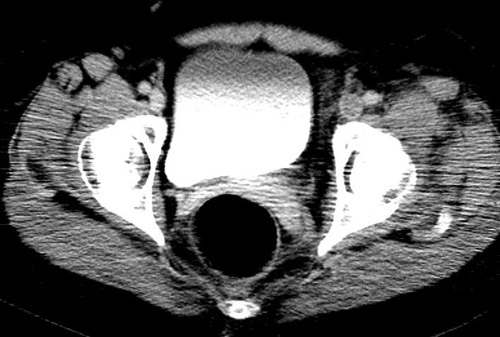
7 Doppler Ultrasound Legs No radiation exposure If leg vein DVT found, anticoagulation will be instituted Simple, easy, fast, accurate No reason not to do it Radiologist intervention may aid timeliness! We require this prior to any exam using radiation in pregnant patients
8 Nuclear Medicine VP Scintigraphy Less sensitive for PE than CTPA Has limited ability to provide alternate diagnosis (e.g. pneumonia, mediastinal mass, cardiac failure, etc) Higher radiation dose to fetus (370 micro G) than CTPA (131 micro G), both tiny doses Far lower dose to maternal breast tissue Radiation decision is; who takes the hit!
9 CTPA Highly accurate for PE and other chest pathologies Delivers a high dose to maternal breast tissue (10-70 mgy) ) compared to 2 view mammogram (3mGy) Minimal dose to fetus since the beam is highly collimated Iodinated contrast media used that requires thyroid testing of baby at delivery
10 Suggested Algorithm D dimer Doppler ultrasound Radiology consult Chest radiograph Abnormal CXR perform CTPA Normal CXR, perform CTPA or VP scintigraphy according to local preference


11 Clinical Case: Results D dimer positive (>500) Doppler ultrasound of the legs, negative CXR:

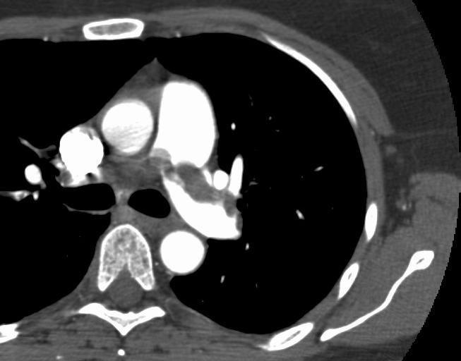
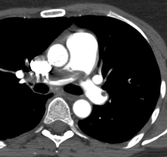
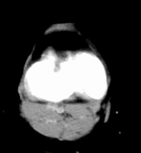
12 CT Pulmonary Angiogram
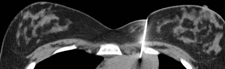
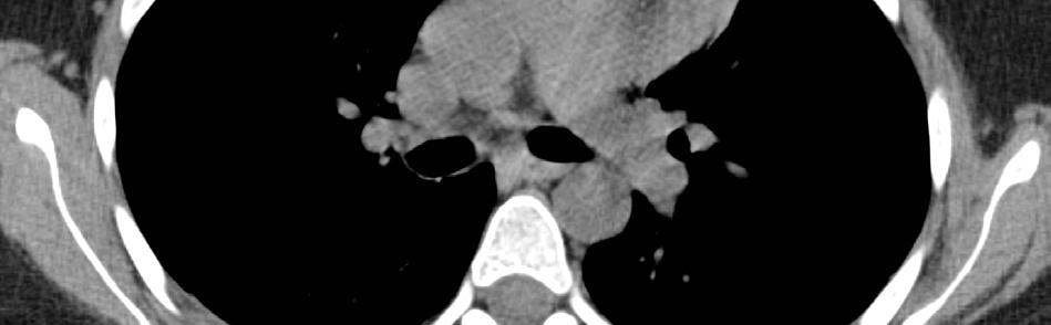
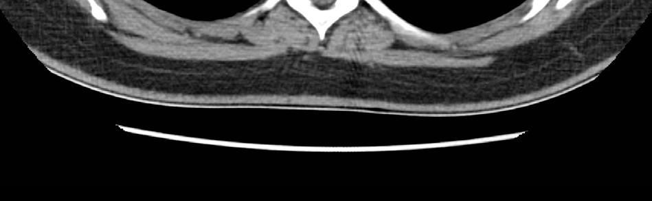
13 Diagnosis: Thymoma & PE
14 Thank you
15 Intra - luminal filling defect: central PA
16 Intra - luminal filling defects: target sign
17 Intra - luminal filling defect: tram track sign
18 Vascular cut off sign
19 Wedge shaped peripheral density Ground glass Consolidation
20 Disclosure of Support Funding received from BC Lung Association Canadian Institutes of Health Research (CIHR) Terry Fox Research Institute
21 Overview CTPA technique Diagnostic findings in acute and chronic PE Artefacts in CTPA Evidence supporting CTPA CTV Radiation dose Further questions
22 CT Vascular Anatomy 3 vascular compartments: Central and segmental PA s Single slice CT (1 track) Sub-segmental PA s Multi- slice CT (4, 8, 16, 64 track) Distal vessels, capillary bed not resolved using CT

23 CT volume averaging effect 1 x 1 x 1 mm
24 Technical Goals of CTPA Thin slices 3 x 1 x 1 mm 1 track 1 x 0.6 x 0.6 mm 16 track 1 x 0.5 x 0.5 mm 64 track Scan large regions of the lungs Dense contrast enhancement of blood Inject intravenous contrast 320 mg/ml at 3-43 ml/sec for a total volume of ml

25 Thin slices improve detection of small subsegmental clots 2.5 mm 1.25 mm Ghaye et al, Radiology 2001;219:
26 Interpretation technique Interpret images using a scrolling technique on a workstation Initially use mediastinal settings (W 450 L 35) Widen window and adjust level to see small peripheral vessels (W 600 L 100) Review parenchyma at lung settings (W 1500 L -750)
27 Acute Pulmonary Embolism: Direct signs Diagnostic Findings Intra-luminal filling defect Vessel cut-off CT equivalent of pulmonary angiographic signs of acute PE

28 Saddle embolus

29 Intra - luminal filling defect: central PA

30 Intra - luminal filling defects: target sign

31 Intra - luminal filling defect: tram track sign

32 Vascular cut off sign
33 Suggestive Findings Indirect signs, suggestive of acute PE 1,2 dilated central pulmonary arteries wedge shaped consolidation dilated right ventricle 1. Coche et al, Radiology 1998;207: Shah et al, Radiology 1999;211:

34 Dilated central arteries


35 Wedge shaped peripheral density Ground glass Consolidation

36 Non enhancing wedge shaped consolidation

37 Dilated right ventricle
38 Chronic PE findings Focal wall thickening Webs and bands Small vessels Hypertrophied bronchial arteries Mosaic perfusion

39 Focal wall thickening

40 Webs and bands

41 Hypertrophied bronchial arteries
42 CTPA Interpretative pitfalls Motion artefact Sub-optimal contrast injection Limited signal to noise Hilar lymph nodes

43 Motion artifact

44 Sub-optimal contrast injection and noise


45 Sub-optimal contrast mixing Yoo et al, RSNA 2003


46 Pseudo vascular cut off secondary to nodes


47 Pseudo vascular cut off secondary to nodes Transverse section Coronal section
48 Sub-optimal examinations Most series report 3-6% 3 rate of sub-optimal examinations 1 Comparable to rate of sub-optimal pulmonary angiograms in PIOPED, 35/1099, 3% 1. Stein et al, Circulation 1992;85:
49 Review of the Evidence Many direct comparisons of spiral CT to: pulmonary angiography VP scintigraphy Systematic reviews Clinical utility studies alternate diagnosis Experimental animal trials
50 Recent Systematic Review Investigated the clinical validity of a negative CT scan in suspected PE 1 Systematic review of studies from 1990 to patients in 15 selected studies 12 single slice, 2 multislice,, 1 EBCT 1. Quiroz R et al, JAMA 295;16:
51 Systematic Review Negative predictive value 99.4% (95% CI 98.7%-99.9%) 99.9%) Negative Likelihood ratio of mortality 0.01 (95% CI ) 0.02) Concluded that the clinical validity of negative CT similar to pulmonary angiography
52 Clinical Utility Trials Increased clinical utility of spiral CT over V-P P scintigraphy 1 diagnosed PE in 23 of 25 cases suggested or confirmed alternate diagnosis in 57 of 85 useful information in 80 of 110, 73% 1. Kim et al, Radiology 1999;210:

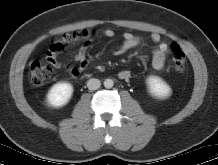
53 Metastatic Bone Lesion
54 Animal Trials How good are spiral CT and pulmonary angiography when compared to an external gold standard? Requires an animal trial
55 Motivation: Pulmonary Angiography may be a flawed gold standard Poor inter observer agreement (70%) for limited subsegmental embolism Wide variation in the rate of limited subsegmental embolism 6% 1-30% 2 1. Stein et al, Circulation 1992;85: Oser et al, Radiology 1996;199:31-35
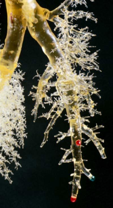
56 True Gold Standard Experiment Spiral CT and pulmonary angiography in 16 anesthetized, ventilated juvenile pigs Embolized subsegmental sized coloured methacrylate beads Spiral CT and pulmonary angiography Methacrylate cast of the pulmonary arteries, providing a true gold standard 1 1. Baile et al, Amer J Resp Crit Care Med 2000;161:

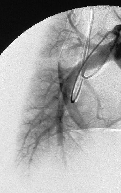
57 C B

58 A 1 2 3
59 RESULTS - CAST 84 of 86 emboli recovered (98%) 5 emboli outside imaged volume 79 emboli for analysis 15 large 18 small 46 combined
60 COMPARISON OF GOLD STANDARDS Sensitivity (%) CT 3 CT 1 Angio Angio Cast
61 COMPARISON OF GOLD STANDARDS Positive Predictive Value (%) CT 3 CT 1 Angio Angio Cast
62 Angiography As Gold Standard Missed 8 emboli (false negative) Incorrectly scored as CT false positives Falsely identified 12 emboli (false positives) Incorrectly scored as CT false negatives Conclusion The use of Pulmonary Angiography as a gold standard can be misleading
63 Outcome Trial Results Both trials showed CTPA acceptable first choice examination for query PE Anderson et al raised the question whether all emboli detected with CTPA require treatment
64 Current Data Suggests 85-90% Sensitive 90-95% 95% Specific Single slice CT, segmental PE Multi-slice CT, segmental and subsegmental PE
65 CT Venography Easy to perform Good patient acceptance Initial reports show good sensitivity and specificity (>90%) compared to ultrasound High gonadal radiation exposure Variable billing strategy
66 CTV Technique 120kVp, 250 mas axial scans 150 ml of 300 mg I per ml non ionic contrast media 5 mm thick slices at 50 mm spacings obtained from pelvis to knees 2 to 4 minutes following CTPA Review using display settings of: width 400, level 40 and narrower width

67 Normal CTV

68 Normal CTV
69 Radiographic Findings Diagnostic findings Intra lumenal non enhancing filling defect Localized non opacification of a vein segment Suggestive findings Enlargment of the vein (venous expansion) Enhancement of the vein wall

70 CTV of DVT

71 CTV of DVT
72 CTV Results CTV added to CTPA identifies more clot Increases the anticoagulation rate by 25% However, the rate of positive PE and DVT in query PE scans is less than 10% Therefore a large number of CTV studies are performed to find a few DVT
73 CTV Controversy Given higher sensitivity of new multidetector row scanners, do we need CTV? If we need CTV, which patients should receive it? Further study is necessary
74 Radiation dose Single slice CT PE dose 3-63 msv Radiation dose mildly increased using 4 and 8 detector row scanners 16, 32, 64 row scanners with dose modulation reduce radiation dose, however image noise is increased More patients are being studied

75 Current issues Significance of isolated subsegmental PE Role of perfusion maps
76 Thank you
77 Clinical Utility Studies Compared spiral CT with V-P V scintigraphy as initial investigation 78 patients outcome: confident diagnosis spiral CT 90%, 35 of 39 V-P scintigraphy 54%, 21 of 39 p<0.001 Cross et al, Clinical Radiology 1998;53:
Pulmonary Embolism. Thoracic radiologist Helena Lauri
 Pulmonary Embolism Thoracic radiologist Helena Lauri 8.5.2017 Statistics 1-2 out of 1000 adults annually are diagnosed with deep vein thrombosis (DVT) and/or pulmonary embolism (PE) About half of patients
Epidermiology Early pulmonary embolism
 Epidermiology Early pulmonary embolism Sitang Nirattisaikul Faculty of Medicine, Prince of Songkla University 3 rd most common cause of cardiovascular death in the United States, following ischemic heart
Imaging of acute pulmonary thromboembolism*
 Silva, Isabela et al. Imaging of acute pulmonary thromboembolism Imaging of acute pulmonary thromboembolism* C. ISABELA S. SILVA, NESTOR L. MÜLLER The diagnosis of acute pulmonary thromboembolism is based
PULMONARY EMBOLISM ANGIOCT (CTA) ASSESSMENT OF VASCULAR OCCLUSION EXTENT AND LOCALIZATION OF EMBOLI 1. BACKGROUND
 ASSESSMENT OF VASCULAR OCCLUSION EXTENT AND LOCALIZATION OF EMBOLI 1. BACKGROUND") JOURNAL OF MEDICAL INFORMATICS & TECHNOLOGIES Vol. 11/2007, ISSN 1642-6037 Damian PTAK * pulmonary embolism, AngioCT, postprocessing techniques, Mastora score PULMONARY EMBOLISM ANGIOCT (CTA) ASSESSMENT
JOURNAL OF MEDICAL INFORMATICS & TECHNOLOGIES Vol. 11/2007, ISSN 1642-6037 Damian PTAK * pulmonary embolism, AngioCT, postprocessing techniques, Mastora score PULMONARY EMBOLISM ANGIOCT (CTA) ASSESSMENT
Cover Page. The handle holds various files of this Leiden University dissertation.
 Cover Page The handle http://hdl.handle.net/1887/19768 holds various files of this Leiden University dissertation. Author: Langevelde, Kirsten van Title: Are pulmonary embolism and deep-vein thrombosis
Pulmonary Embolism. Pulmonary Embolism. Pulmonary Embolism. PE - Clinical
 Pulmonary embolus - a practical approach to investigation and treatment Sam Janes Wellcome Senior Fellow and Respiratory Physician, University College London Background Diagnosis Treatment Common: 50 cases
Clinical Guide - Suspected PE (Reviewed 2006)
") Clinical Guide - Suspected (Reviewed 2006) Principal Developer: B. Geerts Secondary Developers: C. Demers, C. Kearon Background Investigation of patients with suspected pulmonary emboli () remains problematic
Clinical Guide - Suspected (Reviewed 2006) Principal Developer: B. Geerts Secondary Developers: C. Demers, C. Kearon Background Investigation of patients with suspected pulmonary emboli () remains problematic
Patient Dose in the Diagnosis of PE
 Patient Dose in the Diagnosis of PE IZAAZ BADSHAH 2018 CANM-CAMRT Joint Annual Meeting March 22-24, 2018 Disclosure I do not have a financial interest, arrangement or affiliation including receipt of honoraria
Too much medicine and venous thromboembolism: How can we make things Well again?
 Too much medicine and venous thromboembolism: How can we make things Well again? Emily G McDonald MD MSc; Assistant professor of medicine; McGill University Health Centre Canadian Society of Internal Medicine;
Proper Diagnosis of Venous Thromboembolism (VTE)
") Proper Diagnosis of Venous Thromboembolism (VTE) Whal Lee, M.D. Seoul National University Hospital Department of Radiology 2 nd EFORT Asia Symposium, 3 rd November 2010, Taipei DVT - Risk Factors Previous
Proper Diagnosis of Venous Thromboembolism (VTE) Whal Lee, M.D. Seoul National University Hospital Department of Radiology 2 nd EFORT Asia Symposium, 3 rd November 2010, Taipei DVT - Risk Factors Previous
Audit of CT Pulmonary Angiogram in suspected pulmonary embolism patients
 Audit of CT Pulmonary Angiogram in suspected pulmonary embolism patients Poster No.: C-2511 Congress: ECR 2012 Type: Scientific Exhibit Authors: N. D. Gupta, M. K. Heir, P. Bradding; Leicester/UK Keywords:
The Location and Size of Pulmonary Embolism in Antineoplastic Chemotherapy Patients 1
 The Location and Size of Pulmonary Embolism in Antineoplastic Chemotherapy Patients 1 Yun Joo Park, M.D., Woocheol Kwon, M.D., Won-Yeon Lee, M.D. 2, Sang Baek Koh, M.D. 3, Seong Ah Kim, M.D., Myung Soon
Audit of CT Pulmonary Angiogram in suspected pulmonary embolism patients
 Audit of CT Pulmonary Angiogram in suspected pulmonary embolism patients Poster No.: C-2511 Congress: ECR 2012 Type: Scientific Exhibit Authors: N. D. Gupta, M. K. Heir, P. Bradding; Leicester/UK Keywords:
CT Pulmonary Angiography 2010
 2010 Annual Course CT Pulmonary Angiography 2010 Vassilios Raptopoulos, MD Beth Israel Deaconess Medical Center Harvard Medical School CTPA 2010 Utilization Technique DVT Radiation In Pregnancy Large Small
Detectability of subsegmental pulmonary vessels in 64 MDCT-pulmonary angiography.
 ISPUB.COM The Internet Journal of Radiology Volume 11 Number 2 Detectability of subsegmental pulmonary vessels in 64 MDCT-pulmonary angiography. T Niemann, G Bongartz Citation T Niemann, G Bongartz. Detectability
Reporting SPECT-VQ. Alp Notghi
 Reporting SPECT-VQ Alp Notghi 20 year old female 24 weeks pregnant Clinical History : SOB and chest pain for past 3 days.?pe Doppler USS excluded DVT Case 4413041 Normal Case 4413041 CXR report: The heart
MDCT and Pulmonary Embolism. Heber MacMahon The University of Chicago Department of Radiology
 MDCT and Pulmonary Embolism Heber MacMahon The University of Chicago Department of Radiology https://tinyurl.com/hmpe2018 Disclosures Consultant for Riverain Medical Minor stockholder in Hologic, Inc.
Deep Vein Thrombosis and Pulmonary Embolism: Patient Information
 Deep Vein Thrombosis and Pulmonary Embolism: Patient Information A Deep Vein Thrombosis (DVT) and a Pulmonary Embolism (PE) are both disorders of unwanted blood clotting. Unwanted blood clots can occur
Chronic Thromboembolic Pulmonary Hypertension (CTEPH): A Primer
: A Primer") Chronic Thromboembolic Pulmonary Hypertension (CTEPH): A Primer H. Page McAdams, MD Duke University Medical Center Durham, NC 27710 page.mcadams@duke.edu Question Which of the following imaging tests is
Chronic Thromboembolic Pulmonary Hypertension (CTEPH): A Primer H. Page McAdams, MD Duke University Medical Center Durham, NC 27710 page.mcadams@duke.edu Question Which of the following imaging tests is
Dr. Rami M. Adil Al-Hayali Assistant Professor in Medicine
 Dr. Rami M. Adil Al-Hayali Assistant Professor in Medicine Venous thromboembolism: pulmonary embolism (PE) deep vein thrombosis (DVT) 1% of all patients admitted to hospital 5% of in-hospital mortality
Imaging in Pulmonary Embolism. Gamal Rabie Agmy, MD,FCCP Professor of Chest Diseases, Assiut university
 Imaging in Pulmonary Embolism Gamal Rabie Agmy, MD,FCCP Professor of Chest Diseases, Assiut university Background Information Pulmonary embolism is a life-threatening condition that occurs when a clot
New Criteria for Ventilation-Perfusion Lung Scan Interpretation: A Basis for Optimal Interaction with Helical CT Angiography 1
 1206 July-August 2000 RG Volume 20 Number 4 New Criteria for Ventilation-Perfusion Lung Scan Interpretation: A Basis for Optimal Interaction with Helical CT Angiography 1 Alexander Gottschalk, MD Introduction
Cover Page. The handle holds various files of this Leiden University dissertation.
 Cover Page The handle http://hdl.handle.net/1887/21764 holds various files of this Leiden University dissertation. Author: Mos, Inge Christina Maria Title: A more granular view on pulmonary embolism Issue
CT angiography of pulmonary arteries to detect pulmonary embolism with low kv settings
 CT angiography of pulmonary arteries to detect pulmonary embolism with low kv settings Poster No.: C-3289 Congress: ECR 2010 Type: Scientific Exhibit Topic: Chest - Your latest results Authors: M. K. Gill,
Low-dose CT Lung Cancer Screening Guidelines for Pulmonary Nodules Management Version 2
 Low-dose CT Lung Cancer Screening Guidelines for Pulmonary Nodules Management Version 2 The Committee for Management of CT-screening-detected Pulmonary Nodules 2009-2011 The Japanese Society of CT Screening
Computer-assisted detection of pulmonary embolism: evaluation of pulmonary CT angiograms performed in an on-call setting
 Eur Radiol (2010) 20: 801 806 DOI 10.1007/s00330-009-1628-7 CHEST Rianne Wittenberg Joost F. Peters Jeroen J. Sonnemans Mathias Prokop Cornelia M. Schaefer-Prokop Computer-assisted detection of pulmonary
Simplified approach to investigation of suspected VTE
 Simplified approach to investigation of suspected VTE Diagnosis of DVT and PE THSNA 2016, Chicago 15 April 2016 Clive Kearon, McMaster University, Canada Relevant Disclosures Research Support/P.I. Employee
SCINTIGRAPHY OF THE LUNGS THE VQ SCAN
 SCINTIGRAPHY OF THE LUNGS THE VQ SCAN By George N. Sfakianakis, M.D. Professor of Radiology and Pediatrics October 2009 PULMONARY EMBOLISM 94,000 cases annually in US. Not a disease by itself. A potentially
Disclosures. CTA of Acute and Chronic Pulmonary Embolism. Background. Imaging. Which imaging test should be used to evaluate VTE? Objectives.
 CTA of Acute and Chronic Pulmonary Embolism None Disclosures Smita Patel, M.B.B.S., M.R.C.P., F.R.C.R. Professor, Cardiothoracic Radiology Department of Radiology University of Michigan Objectives To assess
Anticoagulation Forum: Management of Tiny Clots
 Anticoagulation Forum: Management of Tiny Clots Casey O Connell, MD FACP Associate Professor Jane Anne Nohl Division of Hematology Keck School of Medicine USC DISCLOSURES None 4/11/2017 Objectives Define
Corresponding Author: Dr. Kishan A Bhgwat
 IOSR Journal of Dental and Medical Sciences (IOSR-JDMS) e-issn: 2279-0853, p-issn: 2279-0861.Volume 17, Issue 2 Ver. 14 February. (2018), PP 06-11 www.iosrjournals.org Hounsfield s Unit (HU) value inthe
Managing Radiation Risk in Pediatric CT Imaging
 Managing Radiation Risk in Pediatric CT Imaging Mahadevappa Mahesh, MS, PhD, FAAPM, FACR, FACMP, FSCCT. Professor of Radiology and Cardiology Johns Hopkins University School of Medicine Chief Physicist
Tests Your Pulmonologist Might Order. Center For Cardiac Fitness Pulmonary Rehab Program The Miriam Hospital
 Tests Your Pulmonologist Might Order Center For Cardiac Fitness Pulmonary Rehab Program The Miriam Hospital BASIC ANATOMY OF THE LUNGS Lobes of Lung 3 lobes on the Right lung 2 lobes on the Left Blood
Acute Management of Pulmonary Embolism
 Acute Management of Pulmonary Embolism Dr Alex West Respiratory Consultant Guy s and St Thomas Hospital London Declarations - none Order of Play Up date in Diagnostic Imaging - CTPA and V:Q SPECT Sub-massive
High density thrombi of pulmonary embolism on precontrast CT scan: Is it dangerous?
 High density thrombi of pulmonary embolism on precontrast CT scan: Is it dangerous? Poster No.: C-1753 Congress: ECR 2013 Type: Authors: Keywords: DOI: Scientific Exhibit B. Y. Lee, H. R. KIM, J. I. Jung,
VTE General Background
 VTE General Background VTE incidence is about 1:1000 persons annually >250,000 admissions for VTE annually >100,000 people die of PE annually >90% of PE s arise from lower limb DVT 50% of DVT at diagnosis
Fundamentals, Techniques, Pitfalls, and Limitations of MDCT Interpretation and Measurement
 Fundamentals, Techniques, Pitfalls, and Limitations of MDCT Interpretation and Measurement 3 rd Annual Imaging & Physiology Summit November 20-21, 21, 2009 Seoul, Korea Wm. Guy Weigold, MD, FACC Cardiovascular
Computed Tomography (CT) - Chest
 - Chest") Scan for mobile link. Computed Tomography (CT) - Chest What is CT Scanning of the Chest? Computed tomography, more commonly known as a CT or CAT scan, is a diagnostic medical test that, like traditional
Scan for mobile link. Computed Tomography (CT) - Chest What is CT Scanning of the Chest? Computed tomography, more commonly known as a CT or CAT scan, is a diagnostic medical test that, like traditional
HI-Res Extremity Sensation 16
 Page 1 Routine Extremity - (2/14/2013) CTDI: ~20 mgy per acquisition Used for evaluation of: Humerus Forearm Femur Knee Tib/Fib Billing: 1. CT Upper/Lower Extremity of concern without contrast, with contrast,
P ulmonary embolism (PE) is a common disease estimated to
 is a common disease estimated to") 53 PULMONARY EMBOLISM Prospective evaluation of unsuspected pulmonary embolism on contrast enhanced multidetector CT (MDCT) scanning Gillian Ritchie, Simon McGurk, Catriona McCreath, Catriona Graham, John
53 PULMONARY EMBOLISM Prospective evaluation of unsuspected pulmonary embolism on contrast enhanced multidetector CT (MDCT) scanning Gillian Ritchie, Simon McGurk, Catriona McCreath, Catriona Graham, John
ED Diagnosis of DVT or tools to rule out DVT in your ED
 ED Diagnosis of DVT or tools to rule out DVT in your ED Ralph Wang UCSF Department of Emergency Medicine 53 yo f c/o left leg swelling recent cholecystectomy its midnight how do you manage this patient?
Having a V/Q scan or CTPA scan of your lungs whilst pregnant
 Having a V/Q scan or CTPA scan of your lungs whilst pregnant Department of Radiology Information for Patients i Radiology Leaflet No. 93 University Hospitals of Leicester NHS Trust Introduction This leaflet
Computed Tomography (CT) - Head
 - Head") Scan for mobile link. Computed Tomography (CT) - Head Computed tomography (CT) of the head uses special x-ray equipment to help assess head injuries, severe headaches, dizziness, and other symptoms of
Scan for mobile link. Computed Tomography (CT) - Head Computed tomography (CT) of the head uses special x-ray equipment to help assess head injuries, severe headaches, dizziness, and other symptoms of
Cover Page. The handle holds various files of this Leiden University dissertation
 Cover Page The handle http://hdl.handle.net/1887/40114 holds various files of this Leiden University dissertation Author: Exter, Paul L. den Title: Diagnosis, management and prognosis of symptomatic and
Ask EuroSafe Imaging Tips & Tricks. CT Working Group. CT in Pregnancy
 Ask EuroSafe Imaging Tips & Tricks CT Working Group CT in Pregnancy Eileen Kelly (Galway University Hospitals, IE) Matthias Stefan May (University Hospital Erlangen, DE) Robert Bujila (Karolinska University
Original articles. Role of spiral volumetric computed tomographic scanning in the assessment of patients with
 Thorax 1996;51:23-28 23 Original articles Department of Diagnostic Radiology A B van Rossum F E E Treumiet G J Kieft R Schepers-Bok Department of Internal Medicine S J Smith Leyenburg Hospital, Leyweg
CT angiography techniques. Boot camp
 CT angiography techniques Boot camp Overview Basic concepts Contrast administration arterial opacification Time scan acquisition during the arterial phase Protocol examples Helical non-gated CTA Pulmonary
Request Card Task ANSWERS
 Request Card Task ANSWERS Medical Student Workbook Author: Dr Sam Leach, SpR Case 1 What differential diagnoses are most likely? Which investigation is most appropriate? Case 1 The most likely diagnosis
Difficulties of timely diagnosis of the Pulmonary Embolism of patients with chronic obstructive lung disease: possibility MSCT.
 Difficulties of timely diagnosis of the Pulmonary Embolism of patients with chronic obstructive lung disease: possibility MSCT. Poster No.: C-2618 Congress: ECR 2012 Type: Scientific Exhibit Authors: I.
Advances in imaging. N.L. Müller
 Eur Respir J 2001; 18: 867 871 Printed in UK all rights reserved Copyright #ERS Journals Ltd 2001 European Respiratory Journal ISSN 0903-1936 SERIES 0THORACIC IMAGING 0 Edited by P.A. Gevenois, A. Bankier
CURRENT & FUTURE THERAPEUTIC MANAGEMENT OF VENOUS THROMBOEMBOLISM. Gordon Lowe Professor of Vascular Medicine University of Glasgow
 CURRENT & FUTURE THERAPEUTIC MANAGEMENT OF VENOUS THROMBOEMBOLISM Gordon Lowe Professor of Vascular Medicine University of Glasgow VENOUS THROMBOEMBOLISM Common cause of death and disability 50% hospital-acquired
Radiologic Features of The Pulmonary Embolus
 January 2003 Radiologic Features of The Pulmonary Embolus Travis McGlothin HMSIII Mr. J is a 51 y.o. male who presented to the BIDMC ED w/ acute onset of: Lft. Hemiparesis slurred speech mild dyspnea mild
Risk of Pulmonary Embolism After Negative MDCT Pulmonary Angiography Findings
 E. C. Kavanagh 1 A. O Hare G. Hargaden J. G. Murray Received March 20, 2003; accepted after revision August 25, 2003. 1 All authors: Department of Radiology, Mater Misericordiae Hospital, Eccles St., Dublin
Computed tomography of the chest: I. Basic principles
 BJA Education, 15 (6): 299 304 (2015) doi: 10.1093/bjaceaccp/mku063 Advance Access Publication Date: 2 February 2015 Matrix reference 1A03, 2A12 Computed tomography of the chest: I. Basic principles P
Computed Tomography (CT) - Chest
 - Chest") Scan for mobile link. Computed Tomography (CT) - Chest Computed tomography (CT) of the chest uses special x-ray equipment to examine abnormalities found in other imaging tests and to help diagnose the
Scan for mobile link. Computed Tomography (CT) - Chest Computed tomography (CT) of the chest uses special x-ray equipment to examine abnormalities found in other imaging tests and to help diagnose the
Surgical Management in Chronic Thromboembolic Pulmonary Hypertension. Michael Bates, MD, FACS Ochsner Health System, New Orleans, LA
 Surgical Management in Chronic Thromboembolic Pulmonary Hypertension Michael Bates, MD, FACS Ochsner Health System, New Orleans, LA Disclosures No industry conflicts I am a surgeon and always disclose
Computed Tomography (CT) - Body
 - Body") Computed Tomography (CT) - Body What is CT Scanning of the Body? CT scanning sometimes called CAT scanning is a noninvasive medical test that helps physicians diagnose and treat medical conditions. CT
Computed Tomography (CT) - Body What is CT Scanning of the Body? CT scanning sometimes called CAT scanning is a noninvasive medical test that helps physicians diagnose and treat medical conditions. CT
Computed Tomography (CT) - Body
 - Body") Scan for mobile link. Computed Tomography (CT) - Body Computed tomography (CT) of the body uses special x-ray equipment to help detect a variety of diseases and conditions. CT scanning is fast, painless,
Scan for mobile link. Computed Tomography (CT) - Body Computed tomography (CT) of the body uses special x-ray equipment to help detect a variety of diseases and conditions. CT scanning is fast, painless,
Cardiovascular Imaging in Pregnancy. Diana Litmanovich, MD
 Cardiovascular Imaging in Pregnancy Diana Litmanovich, MD Cardiovascular disorders 4% of all pregnancies PE DVT Aortic dissection Peripartum cardiomyopathy Acute coronary syndrome Hypertension Pneumonia
BrightSpeed Elite CT with ASiR: Comparing Dose & Image Quality Rule Out Pulmonary Embolism on Initial & Follow-Up Exam
 GE Healthcare BrightSpeed Elite CT with ASiR: Comparing Dose & Image Quality Rule Out Pulmonary Embolism on Initial & Follow-Up Exam Michael Swack, MD Diagnostic Radiologist Irvington Radiologists, PC
Case of the Day Chest
 Case of the Day Chest Darin White MDCM FRCPC Department of Radiology, Mayo Clinic 76 th Annual Scientific Meeting Canadian Association of Radiologists Montreal, QC April 26, 2013 2013 MFMER slide-1 Disclosures
MATERIALS AND METHODS
 RETROSPECTIVE STUDY OF OPTIMISING THE USE OF COMPUTED TOMOGRAPHY PULMONARY ANGIOGRAPHY (CTPA) FOR THE DIAGNOSIS OF PULMONARY EMBOLISM IN PLACES WITH LIMITED RESOURCES P. V. Kalyan Kumar 1, Ramakrishna
We are IntechOpen, the world s leading publisher of Open Access books Built by scientists, for scientists. International authors and editors
 We are IntechOpen, the world s leading publisher of Open Access books Built by scientists, for scientists 3,900 116,000 120M Open access books available International authors and editors Downloads Our
After the Chest X-Ray:
 After the Chest X-Ray: What To Do Next Alan S. Brody Professor of Radiology and Pediatrics Chief of Thoracic Imaging Cincinnati Children s Hospital Cincinnati, Ohio USA What Should We Do Next? CT scan?
Pulmonary Thromboembolism
 Pulmonary Thromboembolism James Allen, MD Epidemiology of Pulmonary Embolism 1,500,000 new cases per year in the United States Often asymptomatic 300,000 deaths per year DVT or PE present in 10% of ICU
 Page 1 of 5 Patient Safety: Radiation Dose in X-Ray and CT Exams What are x-rays and what do they do? X-rays are forms of radiant energy, like light or radio waves. Unlike light, x-rays can penetrate the
Cardiac Computed Tomography
 Cardiac Computed Tomography Authored and approved by Koen Nieman Stephan Achenbach Francesca Pugliese Bernard Cosyns Patrizio Lancellotti Anastasia Kitsiou Contents CARDIAC COMPUTED TOMOGRAPHY Page 1.
Provider Led Entity. CDI Quality Institute PLE Chest / Pulmonary Embolus AUC 07/31/2018
 Provider Led Entity CDI Quality Institute PLE Chest / Pulmonary Embolus AUC 07/31/2018 Appropriateness of advanced imaging procedures* in patients with suspected or known pulmonary embolus and the following
CT Versus MR for the Runoff
 CT Versus MR for the Runoff Robert R. Edelman, M.D. Dept. of Radiology NorthShore University HealthSystem Feinberg School of Medicine, Northwestern University Magnetic Resonance Computed Tomography Radio
Cover Page. The handle holds various files of this Leiden University dissertation
 Cover Page The handle http://hdl.handle.net/1887/40114 holds various files of this Leiden University dissertation Author: Exter, Paul L. den Title: Diagnosis, management and prognosis of symptomatic and
Pediatric Lung Ultrasound (PLUS) In Diagnosis of Community Acquired Pneumonia (CAP)
 In Diagnosis of Community Acquired Pneumonia (CAP)") Pediatric Lung Ultrasound (PLUS) In Diagnosis of Community Acquired Pneumonia (CAP) Dr Neetu Talwar Senior Consultant, Pediatric Pulmonology Fortis Memorial Research Institute, Gurugram Study To compare
Pediatric Lung Ultrasound (PLUS) In Diagnosis of Community Acquired Pneumonia (CAP) Dr Neetu Talwar Senior Consultant, Pediatric Pulmonology Fortis Memorial Research Institute, Gurugram Study To compare
Lung Perfusion Analysis New Pathways in Lung Imaging. Case Study Brochure PLA 309 Hospital
 Lung Perfusion Analysis New Pathways in Lung Imaging Case Study Brochure PLA 309 Hospital http://www.toshibamedicalsystems.com Toshiba Medical Systems Corporation 2012 all rights reserved. Design and specifications
Managing Patient Dose in Computed Tomography (CT)
") Managing Patient Dose in Computed Tomography (CT) International Commission on Radiological Protection Information abstracted from ICRP Publication 87 Available at www.icrp.org Task Group: M.M. Rehani,
Managing Patient Dose in Computed Tomography (CT) International Commission on Radiological Protection Information abstracted from ICRP Publication 87 Available at www.icrp.org Task Group: M.M. Rehani,
General Imaging. Imaging modalities. Incremental CT. Multislice CT Multislice CT [ MDCT ]
![General Imaging. Imaging modalities. Incremental CT. Multislice CT Multislice CT [ MDCT ]](/thumbs/76/74079340.jpg "General Imaging. Imaging modalities. Incremental CT. Multislice CT Multislice CT [ MDCT ]") General Imaging Imaging modalities Conventional X-rays Ultrasonography [ US ] Computed tomography [ CT ] Radionuclide imaging Magnetic resonance imaging [ MRI ] Angiography conventional, CT,MRI Interventional
General Imaging Imaging modalities Conventional X-rays Ultrasonography [ US ] Computed tomography [ CT ] Radionuclide imaging Magnetic resonance imaging [ MRI ] Angiography conventional, CT,MRI Interventional
FOR CMS (MEDICARE) MEMBERS ONLY NATIONAL COVERAGE DETERMINATION (NCD) FOR COMPUTED TOMOGRAPHY:
 MEMBERS ONLY NATIONAL COVERAGE DETERMINATION (NCD) FOR COMPUTED TOMOGRAPHY:") National Imaging Associates, Inc. Clinical guidelines CHEST CTA Original Date: September 1997 Page 1 of 5 CPT Codes: 71275 Last Review Date: August 2014 NCD 220.1 Last Effective Date: March 2008 Guideline
National Imaging Associates, Inc. Clinical guidelines CHEST CTA Original Date: September 1997 Page 1 of 5 CPT Codes: 71275 Last Review Date: August 2014 NCD 220.1 Last Effective Date: March 2008 Guideline
Improvement of Image Quality with ß-Blocker Premedication on ECG-Gated 16-MDCT Coronary Angiography
 16-MDCT Coronary Angiography Shim et al. 16-MDCT Coronary Angiography Sung Shine Shim 1 Yookyung Kim Soo Mee Lim Received December 1, 2003; accepted after revision June 1, 2004. 1 All authors: Department
Incidental Findings in Patients Evaluated for Pulmonary Embolism Using Computed Tomography Angiography
 Incidental Findings in Patients Evaluated for Pulmonary Embolism Using Computed Tomography Angiography Masoud Pezeshki Rad 1 *, Donya Farrokh Tehrani 2, Hamidreza Reihani 3, Seyed Hosein Faghih Sabzevari
Radiology. Undergraduate Radiology Sample Questions
 Radiology Undergraduate Radiology Sample Questions April 2012 The following examples are offered of questions that might be used to assess undergraduate radiology. There are 3 different styles: An OSCE
Pulmonary Embolism. Medicine for Managers. Dr Paul Lambden BSc MB BS BDS FDSRCSEng MRCS LRCP DRCOG MHSM FRSH
 nhsmanagers.net Briefing 26 June 2016 Medicine for Managers Dr Paul Lambden BSc MB BS BDS FDSRCSEng MRCS LRCP DRCOG MHSM FRSH Pulmonary Embolism Pulmonary embolism is a potentially life-threatening disorder
Chronic pulmonary thromboembolism: Pictorial review of CT pulmonary angiographic findings
 Chronic pulmonary thromboembolism: Pictorial review of CT pulmonary angiographic findings Poster No.: C-0946 Congress: ECR 2010 Type: Educational Exhibit Topic: Chest Authors: R. Jeyaratnam, D. Devendra,
Citation for published version (APA): Douma, R. A. (2010). Pulmonary embolism: advances in diagnosis and prognosis
: Douma, R. A. (2010). Pulmonary embolism: advances in diagnosis and prognosis") UvA-DARE (Digital Academic Repository) Pulmonary embolism: advances in diagnosis and prognosis Douma, R.A. Link to publication Citation for published version (APA): Douma, R. A. (2010). Pulmonary embolism:
UvA-DARE (Digital Academic Repository) Pulmonary embolism: advances in diagnosis and prognosis Douma, R.A. Link to publication Citation for published version (APA): Douma, R. A. (2010). Pulmonary embolism:
Prospective Evaluation of Unsuspected Pulmonary Embolism on Contrast Enhanced Multidetector CT (MDCT)
") Thorax Online First, published on December 8, 2006 as 10.1136/thx.2006.062299 Prospective Evaluation of Unsuspected Pulmonary Embolism on Contrast Enhanced Multidetector CT (MDCT) Dr Gillian Ritchie FRCR
Thorax Online First, published on December 8, 2006 as 10.1136/thx.2006.062299 Prospective Evaluation of Unsuspected Pulmonary Embolism on Contrast Enhanced Multidetector CT (MDCT) Dr Gillian Ritchie FRCR
Role of Dual source CT angiography and perfusion in the diagnosis of pulmonary embolism
 Role of Dual source CT angiography and perfusion in the diagnosis of pulmonary embolism Poster No.: C-1145 Congress: ECR 2011 Type: Educational Exhibit Authors: P. S. Naphade, A. A. Raut, A. keraliya,
CTPA for Pulmonary Emboli: 2016 update
 2016 Annual Course CTPA for Pulmonary Emboli: 2016 update Olga R Brook, MD, FSCBTMR Beth Israel Deaconess Medical Center, Boston Beth Israel Deaconess Medical Center HarvardMedical School Contents Technique
Computed Tomography (CT) - Sinuses
 - Sinuses") Scan for mobile link. Computed Tomography (CT) - Sinuses Computed tomography (CT) of the sinuses uses special x-ray equipment to evaluate the paranasal sinus cavities hollow, air-filled spaces within the
Scan for mobile link. Computed Tomography (CT) - Sinuses Computed tomography (CT) of the sinuses uses special x-ray equipment to evaluate the paranasal sinus cavities hollow, air-filled spaces within the
Dual Energy CT of Pulmonary Embolism
 Dual Energy CT of Pulmonary Embolism U. Joseph Schoepf, MD, FAHA, FSCBT MR, FSCCT Professor of Radiology, Medicine, and Pediatrics Director of Cardiovascular Imaging Disclosures Consultant for / research
A low probability interpretation of a ventilation/
 Very Low Probability Interpretation of V/Q Lung Scans in Combination with Low Probability Objective Clinical Assessment Reliably Excludes Pulmonary Embolism: Data from PIOPED II Alexander Gottschalk 1,
Pulmonary Embolism..Diagnostic Approach and Algorithm. Tolulope Adesiyun Harvard Medical School, Year III Gillian Lieberman, MD
 Pulmonary Embolism..Diagnostic Approach and Algorithm Tolulope Adesiyun Harvard Medical School, Year III Gillian Lieberman, MD Epidemiology of Pulmonary Embolism Pulmonary Embolus (PE): Thrombus originating
Ultrasound-enhanced, catheter-directed thrombolysis for pulmonary embolism
 NATIONAL INSTITUTE FOR HEALTH AND CARE EXCELLENCE Interventional procedure consultation document Ultrasound-enhanced, catheter-directed thrombolysis for pulmonary embolism A pulmonary embolism (PE) is
Deep Vein Thrombosis
 Deep Vein Thrombosis Introduction Deep vein thrombosis (DVT) is a blood clot in a vein. This condition can affect men and women of any age and race. DVT is a potentially serious condition. If not treated,
Engin Ozakin, 1 Filiz Baloglu Kaya, 1 Nurdan Acar, 1 and Arif Alper Cevik 1,2. 1. Introduction
 e Scientific World Journal, Article ID 470358, 5 pages http://dx.doi.org/10.1155/2014/470358 Research Article An Analysis of Patients That Underwent Computed Tomography Pulmonary Angiography with the Prediagnosis
Radioembolization (Y90)
") Scan for mobile link. Radioembolization (Y90) Radioembolization is a minimally invasive procedure that combines embolization and radiation therapy to treat liver cancer. Tiny glass or resin beads filled
Scan for mobile link. Radioembolization (Y90) Radioembolization is a minimally invasive procedure that combines embolization and radiation therapy to treat liver cancer. Tiny glass or resin beads filled
CT Chest. Verification of an opacity seen on the straight chest X ray
 CT Chest Indications: To assess equivocal plain x-ray findings Staging of lung neoplasm Merastatic workup of extra thoraces malignancies Diagnosis of diffuse lung diseases with HRCT Assessment of bronchietasis
18 F-FDG PET of Pulmonary Embolism
 FDG PET of Pulmonary Embolism Chest Imaging Clinical Observations Conrad Wittram 1 James A. Scott 2 Wittram C, Scott JA Keywords: CT, nuclear medicine, oncology, PET, pulmonary embolism DOI:10.2214/AJR.06.0640
X-Ray & CT Physics / Clinical CT
 Computed Tomography-Basic Principles and Good Practice X-Ray & CT Physics / Clinical CT INSTRUCTORS: Dane Franklin, MBA, RT (R) (CT) Office hours will be Tuesdays from 5pm to 6pm CLASSROOM: TIME: REQUIRED
New Horizons in the Imaging of the Lung
 New Horizons in the Imaging of the Lung Postprocessing. How to do it and when do we need it? Peter M.A. van Ooijen, MSc, PhD Principal Investigator, Radiology, UMCG Discipline Leader Medical Imaging Informatics
Learning Objectives. 1. Identify which patients meet criteria for annual lung cancer screening
 Disclosure I, Taylor Rowlett, DO NOT have a financial interest /arrangement or affiliation with one or more organizations that could be perceived as a real or apparent conflict of interest in the context
Case 1. Technegas Case Studies. Prostate cancer. Finished treatment recently. Smoker. Angina. Presents sudden dyspnea and poorly defined chest pains.
 Case 1 Michel Leblanc MD; RCPSC; ABNM Head of the Nuclear Medicine Department, Centre Hospitalier Régional de Trois-Rivières. Clinical Professor, Centre Hospitalier Universitaire de Sherbrooke, Cannada.
Acute abdominal venous thromboses- the hyperdense noncontrast CT sign
 Acute abdominal venous thromboses- the hyperdense noncontrast CT sign Poster No.: C-1095 Congress: ECR 2011 Type: Educational Exhibit Authors: M. Goldstein, K. Jhaveri; Toronto, ON/CA Keywords: Abdomen,
Diagnosis and Treatment of Deep Venous Thrombosis and Pulmonary Embolism
 Agency for Healthcare Research and Quality Evidence Report/Technology Assessment Diagnosis and Treatment of Deep Venous Thrombosis and Pulmonary Embolism Summary Number 68 Overview Venous thromboembolism