OCT; Comparative Imaging Results with IVUS, VH and Angioscopy
|
|
|
- Jonah Palmer
- 5 years ago
- Views:
Transcription

1 OCT; Comparative Imaging Results with IVUS, VH and Angioscopy Takashi Akasaka, M.D. Department of Cardiovascular Medicine Wakayama, Japan


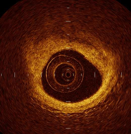
2 Comparison among coronary imaging techniques OCT IVUS MRI CAG Angioscopy Resolution Probe Size Contact Ionizing Radiation No No Yes No No No N/A No Yes < No No Other Tissue Character ization N/A N/A Flow Only Surface Only Advantages of OCT are its high resolution and accuracy of tissue characterization.
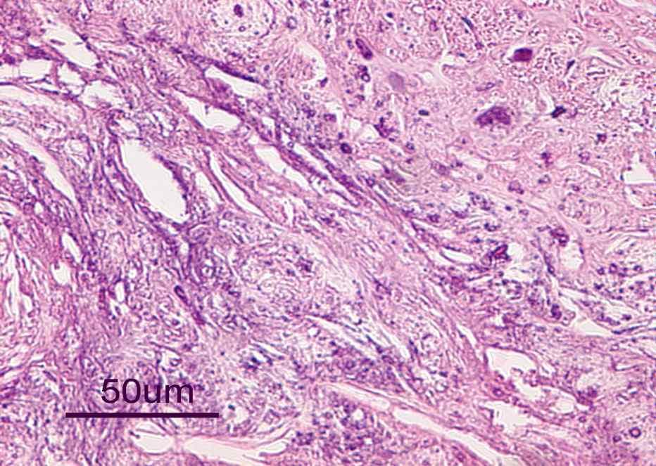
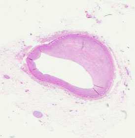
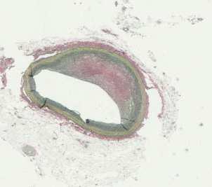
3 Intracoronary Imaging Comparison among OCT, IVUS, VH & Angioscopy Tissue characterization: comparison with histology Vulnerable plaque identification Stent follow-up

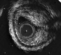
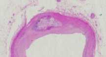
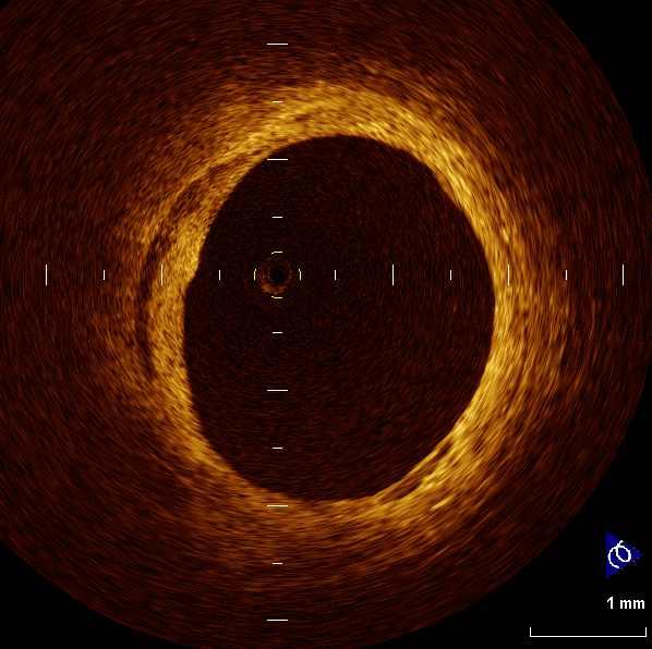
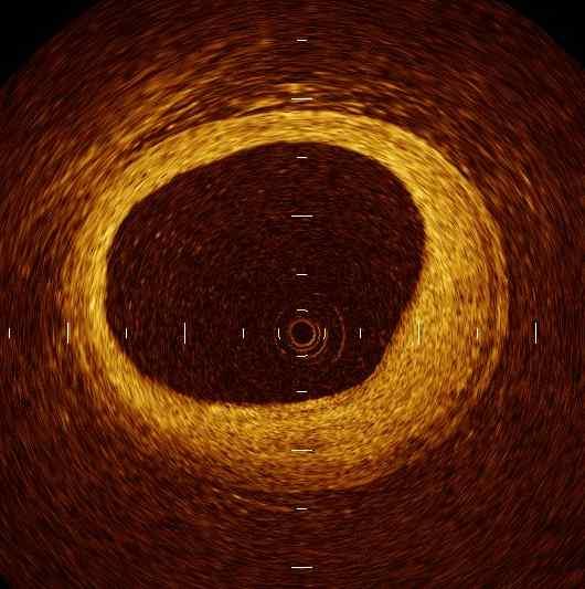
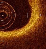
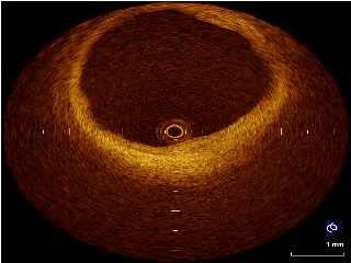
4 Fibrous plaque Attenuation 1mm 100μm Signal rich Diffuse border

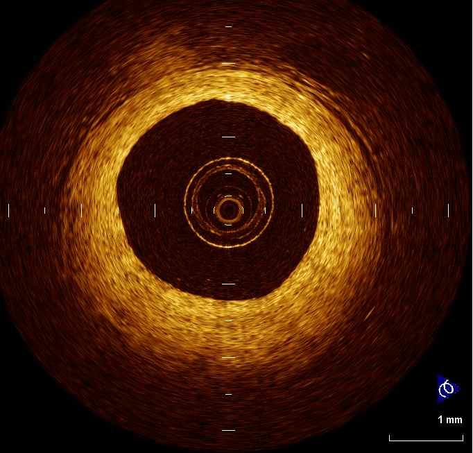
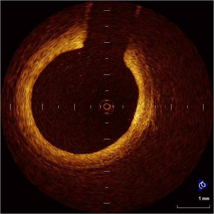
5 Fibrous plaque
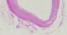
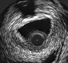
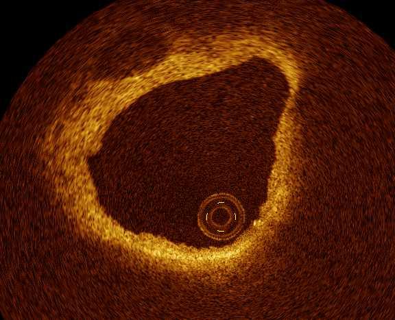
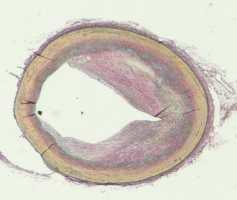
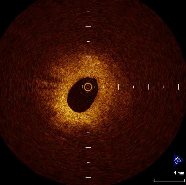
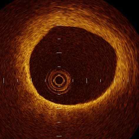
6 Fibrocalcific plaque 1mm 100μm Signal poor Sharp border

7 Calcified plaque Superficial calcified nodule


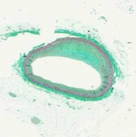
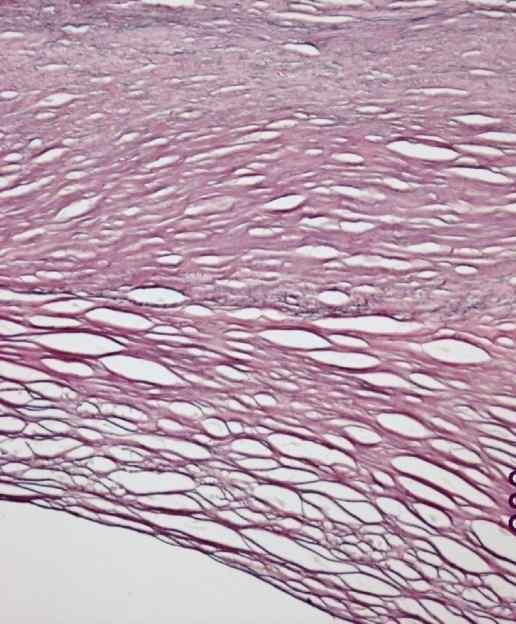
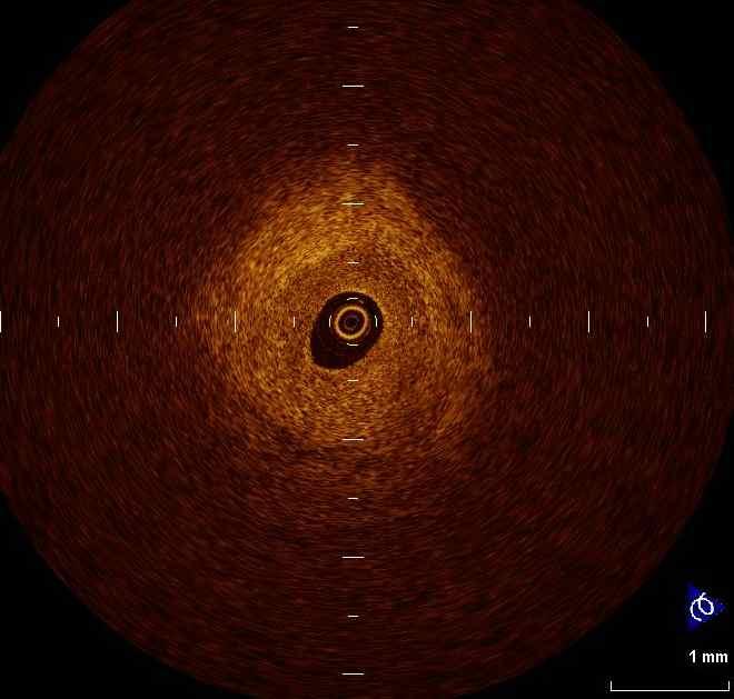
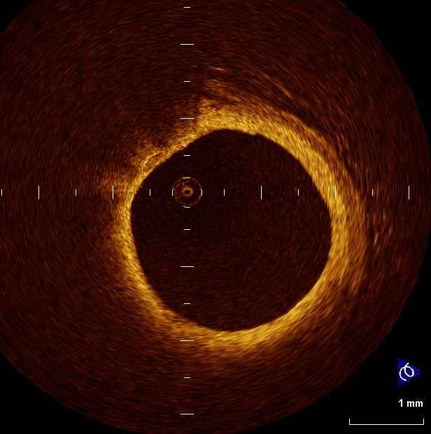
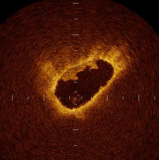
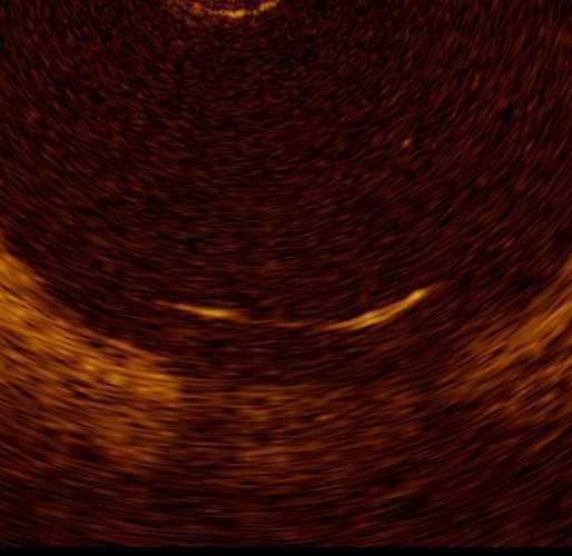
8 Signal poor Diffuse border Fibro-lipidic plaque
9 Fibrofatty plaque
 Kubo T, Akasaka T, et al.")
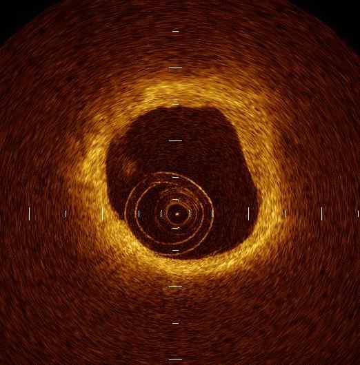
10 Red & white thrombus Red thrombus White thrombus Mixed thrombus Protrusion mass with shadow Protrusion mass without shadow Protrusion mass with & without shadow Kume T, Akasaka T, et al ( Am J Cardiol 97: , 2006 ) Kubo T, Akasaka T, et al. ( J Am Coll Cardiol 50: ,2007)


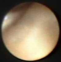
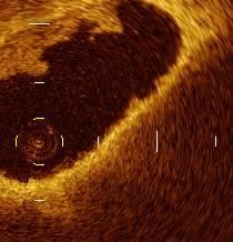
11 Thrombus WT WT WT RT 1mm RT 1mm OCT CAS IVUS
12 Accuracy of intra-coronary OCT for differentiation between red and white thrombus Angioscopy Red thrombus White thrombus OCT Intensity half distance <250μm Intensity half distance 250μm Sensitivity = 95% Specificity = 88% Positive predictive value = 86% Negative predictive value =95%
13 Intracoronary Imaging Comparison among OCT, IVUS & Angioscopy Tissue characterization: comparison with histology Vulnerable plaque identification Stent follow-up
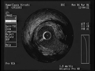
14 Study Design Oral aspirin (162 mg) and intravenous heparin (100 U/kg) were administered before PCI. Cardiac catheterization was performed by the femoral approach, using a 7F sheath and catheters. Thrombectomy (Export catheter Medtronic Japan) TIMI grade III IVUS (Atlantis SR Pro 2.5F, 40-MHz; Boston Scientific, Natick, MA, USA) CAS (Angioscope MC-800E and the optic fiber AS-003, Nihon Kohden) OCT (ImageWire ; LightLab Imaging, Westford, MA, USA)

15 Inferior AMI (71y.o. Male)


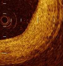
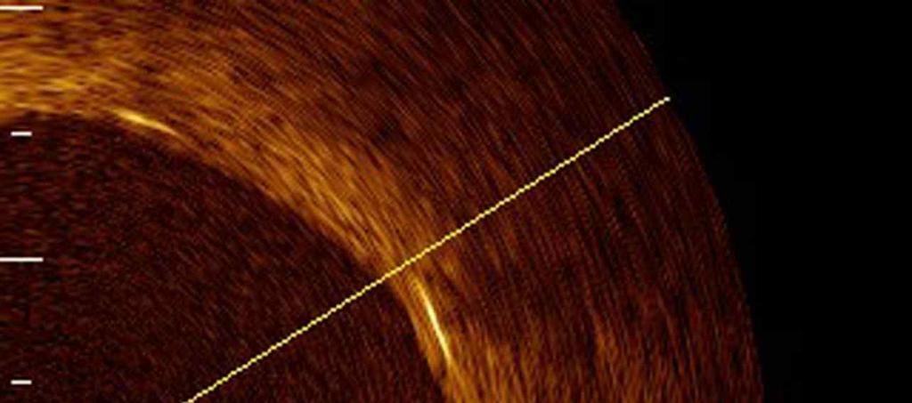
16 Inferior-AMI(71y.o., M) Plaque Rupture Ruptured Fibrous Cap Fibrous Cap Thickness = 40μm TL TL : True Lumen UL : Ulceration UL

")
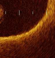
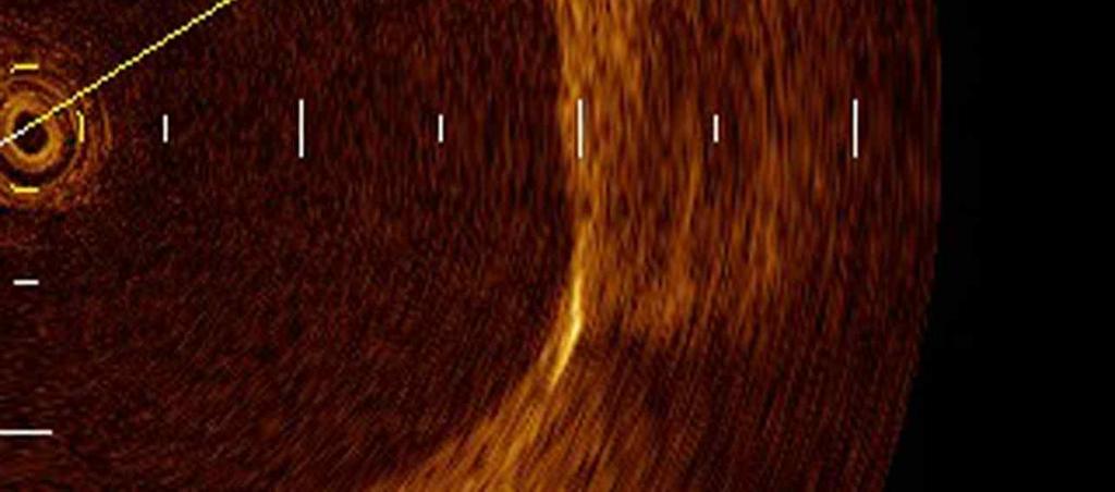
17 Anteroseptal AMI (80y.o., M) Erosion (Ulceration) Thrombus
18 Comparison of plaque Images in AMI (OCT vs. CAS vs. IVUS) n=30 (Kubo T, Akasaka T, et al. J Am Coll Cardiol in press) OCT *CAS **IVUS *p **p Plaque Rupture (%) Ulceration (erosion) (%) Thrombus (%) <0.001 Red thrombus (%) White thrombus (%) TCFA( 65μm) (%) Fibrous cap thickness (μm) 49± LRP (Lipid Arch>180 ) (%) NS TCFA; Thin Cap Fibro-Atheroma, LRP; Lipid Rich Plaque


19 Thin-cap fibroatheroma (TCFA) Possibility to identify TCFA has been demonstrated by several pilot studies.
20 Corresponding Images of OCT and Angioscopy A-1 B-1 C-1 D-1 A-2 B-2 * * * * C-2 * * D-2 * * * * A-3 B-3 C-3 D-3 (Kubo T, et al. J Am Coll Cardiol Intv 1:74-80,2008) Figure. 1
21 Angioscopy vs OCT Plaque color vs lipid size Plaque color vs fibrous cap thickness (Kubo T, et al. J Am Coll Cardiol Intv 1:74-80,2008)
 Percent atheroma volume = (EEM area Lumen area)/eem area x100 40%")
22 IVUS-derived TCFA ( Rodriguez-Granillo GA, et al. J Am Coll Cardiol 46: , 2005 ) Percent atheroma volume = (EEM area Lumen area)/eem area x100 40% Nectrotic core 10% Without evident overlying fibrous tissue


23 Figure 2 Concordant VH-IVUS vs OCT Discordant Without evident overlying fibrous tissue Without evident overlying fibrous tissue With evident overlying fibrous tissue
24 Concordance & discordance between VH-IVUS and OCT Table 4 in the assessment of TCFA IVUS-VH Diagnosis OCT Diagnosis TCFA (n=11) Not TCFA (n=36) VH-TCFA (n=31) 9 22 Not VH-TCFA (n=16) 2 14 Discordance between VH-IVUS & OCT has been described by Sawada T, et al. (Eur Heart J 29: , 2008)


25 OCT assessment of non-culprit lesion (47y.o. male) Baseline 9 month later T.Chol. 200 mg/dl TG 79 mg/dl HDL-C 47 mg/dl LDL-C 128 mg/dl T.Chol. 187 mg/dl TG 133 mg/dl HDL-C 49 mg/dl LDL-C 98 mg/dl Lumen Area 6.1 mm 2 EEL Area 12.3 mm 2 Plaque Area 6.2 mm 2 %plaque burden 50% FI Green Area 2.1 mm 2 68% FF Light green Area 0.4 mm 2 12% DC White Area 0.1 mm 2 2% NC Red area 0.6 mm 2 18% 8.0 mm mm mm 2 44% 2.5 mm 2 84% 0.3 mm 2 9% 0.1 mm 2 3% 0.1 mm 2 4% (Takarada S, et al. Atherosclerosis 202: , 2009)
26 Changes of plaque characteristics by statin (Takarada S, et al. Atherosclerosis 202: , 2009 ) Baseline Follow-up p Statin group Fibrous cap thickness (μm) 114±83 162±75 <0.01 Lipid arc (degrees) 132±37 116±23 <0.01 Non-statin group Fibrous cap thickness (μm) 117±78 129±54 ns Lipid arc (degrees) 129±37 128±28 ns

27 The correlation between the lipid profile and the % change of fibrous-cap thickness (FCT) and total atheroma volume (TAV). %TAV %LDL/HDL r=0.42 p<0.01 %TAV %CRP p= r=-0.44 p<0.01 %FCT 20 %FCT %LDL/HDL p= %TAV and %LDL/HDL were positively correlated (p<0.01, r = 0.42). %FCT and %CRP were inversely correlated (p<0.01, r = -0.44). (Takarada S, et al. JACC Interv. 2010, in press ) -40 %CRP


28 Changes of plaque, media & lumen area PIT VH-TCFA ThCFA Fibrotic Fibrocalcific ( Kubo T, et al. J Am Coll Cardiol 55; , 2010 )


29 Coronary lesion morphology by VH-IVUS Figure 2 ( Kubo T, et al. J Am Coll Cardiol 55; , 2010 ) TCFA ThCFA TCFA Fibrous TCFA TCFA PIT TCFA ThCFA TCFA
30 Intracoronary Imaging Comparison among OCT, IVUS & Angioscopy Tissue characterization: comparison with histology Vulnerable plaque identification Stent follow-up


31 ACS; 69 y.o. M #6 Cypher 3.5 x 18 mm Post PCI


32 Stent malapposition Tissue protrusion 1mm Incomplete stent apposition 1mm Stent edge dissection 1mm 1mm
33 Vascular response after stent implantation between unstable and stable AP 24 unstable and 31 stable AP patients were examined by OCT to evaluate lesion morphologies after stent implantation. 100 (%) * * * * * p<0.05 Unstable AP Stable AP PCI induced Plaque rupture Inadequate stent apposition Tissue protrusion Intracoronary Thrombus Stent edge dissection Conclusion: The inadequate lesion morphologies after stenting were observed more frequently in unstable AP patients. Kubo T, et al, JACC Img :


34 OCT and IVUS images of stented lesions Malappostion Tissue protrusion Thrombi Dissection A-1 B-1 C-1 D-1 A-2 B-2 C-2 D-2 Kubo T, et al, JACC Img :
35 Comparison of the ability for monitoring stent deployment between OCT and IVUS 55 patients were examined by OCT and IVUS to evaluate lesion morphologies after stent implantation. (%) * * * * * p<0.005 OCT IVUS 20 0 Inadequate stent apposition Tissue protrusion Intracoronary Thrombus Stent edge dissection Conclusion: OCT can provide more detailed morphological information after stenting than IVUS. Kubo T, et al, JACC Img :


36 Classification of strut condition Wellapposed with neointima Wellapposed without neointima Malapposed with neointima Side branch orfice without neointimal coverage Malapposed without neointima Side branch orfice with neointimal coverage
37 Post-stent follow up
38 Distribution of the neointima thickness on SES strut (6 months f/u) % pts, 6840 stent strut cross sections 30 64% beyond IVUS resolution >100 Neointimal thickhess (μm) Matsumoto, D. et al. Eur Heart J : Neointima thickness is under IVUS resolution in more than 70% pts.
 implanted proximally in the LAD Stent struts bulged into the lumen and, although")
39 An SES (Cypher, 3.5x23 mm) implanted proximally in the LAD Stent struts bulged into the lumen and, although covered, were transparently visible Awata, M. et al. Circulation 2007;116:
40 Changes in neointimal coverage grades from the first to the third follow-up in 28 stents Stent struts condition Fully embedded and not visible Embedded by the neointima, but still visible translucently Bulged into the lumen, although covered, transparently visible Fully visible similar to soon after implantation Awata, M. et al. Circulation 2007;116:
41 Asymptomiatic instent thombus by CAS SES : 33% BMS : 8% SES : 19% PES : 43% Takano et al. Eur Heart J 2006; 27: Awata et al. J Am Coll Cardiol Intv 2009; 2:


42 Instent thrombus DES BMS Distal to DES
43 Plevalence of in-stent thrombus (%) Instent thrombus by OCT 27% SES P=0.63 P= % PES P= % BMS
44 Conclusions OCT can identify lipid-rich plaques & differentiate the plaque types more sensitively compared with IVUS. OCT can demonstrate rupture or ulceration of fibrous cap with higher detection rate than that of IVUS or CAS. OCT could detect intracoronary thrombus almost exclusively which was confirmed by CAS. OCT may demonstrate the results of PCIs precisely, including mal-appositions, tissue (or thrombus) protrusion, and edge dissection immediately after the procedure and thin neo-intima formation and small thrombus within stents late after DES.


 3 Fibrous-cap")
45 Representative case of plaque stabilization : 66yo, male primary PCI Total atheroma volume=63mm 9-months follow-up 3 Fibrous-cap thickness=90μm Total atheroma volume=61mm (Takarada S, et al. JACC Interv. 2010, in press ) 3 Fibrous-cap thickness=310μm
Assessment of vulnerable plaque by OCT
 Assessment of vulnerable plaque by OCT Comparison with histology and possible clinical applications Takashi Akasaka, M.D. Department of Cardiovascular Medicine Wakayama, Japan Identification of vulnerable
Assessment of plaque morphology by OCT in patients with ACS
 Assessment of plaque morphology by OCT in patients with ACS Takashi Akasaka, M.D. Department of Cardiovascular Medicine Wakayama, Japan Unstable plaque Intima Lipid core Plaque rupture and coronary events
Usefulness of OCT during coronary intervention
 Usefulness of OCT during coronary intervention Takashi Akasaka, M.D. Department of Cardiovascular Medicine Wakayama, Japan Predictors at 12 Months of Stent Thrombosis and Target Lesion Revascularization
Can We Identify Vulnerable Patients & Vulnerable Plaque?
 Can We Identify Vulnerable Patients & Vulnerable Plaque? We Know Enough to Treat High-Risk Lesions? Takashi Akasaka, MD, PhD Department of Cardiovascular Medicine, Japan Disclosure Statement of Financial
Imaging Atheroma The quest for the Vulnerable Plaque
 Imaging Atheroma The quest for the Vulnerable Plaque P.J. de Feijter 1. Department of Cardiology 2. Department of Radiology Coronary Heart Disease Remains the Leading Cause of Death in the U.S, Causing
OCT Findings: Lesson from Stable vs Unstable Plaques
 ANGIOPLASTY SUMMIT TCTAP 2010 Imaging Workshop OCT Findings: Lesson from Stable vs Unstable Plaques Giulio Guagliumi MD Ospedali Riuniti di Bergamo, Italy DISCLOSURE OF FINANCIAL INTERESTS Consultant Boston
Assessment of Vulnerable Plaque by IVUS and VH-IVUS
 Assessment of Vulnerable Plaque by IVUS and VH-IVUS Akiko Maehara, MD Director of Intravascular Imaging & Physiology Core Laboratories Associate Director of MRI/MDCT Core Laboratory Cardiovascular Research
CLINICAL APPLICATIONS OF OPTICAL COHERENCE TOMOGRAPHY. Konstantina P. Bouki, FESC 2 nd Department of Cardiology General Hospital Of Nikea, Pireaus
 CLINICAL APPLICATIONS OF OPTICAL COHERENCE TOMOGRAPHY Konstantina P. Bouki, FESC 2 nd Department of Cardiology General Hospital Of Nikea, Pireaus OPTICAL COHERENCE TOMOGRAPHY (OCT) IVUS and OCT IVUS OCT
Optical Coherence Tomography for Intracoronary Imaging
 Optical Coherence Tomography for Intracoronary Imaging Lorenz Räber Stephan Windecker Department of Cardiology Swiss Cardiovascular Center and Clinical Trials Unit Bern Bern University Hospital, Switzerland
Added Value of Invasive Coronary Imaging for Plaque Rupture and Erosion
 Assessment of Coronary Plaque Rupture and Erosion Added Value of Invasive Coronary Imaging for Plaque Rupture and Erosion Yukio Ozaki, MD, PhD, FACC, FESC Cardiology Dept., Fujita Health Univ. Toyoake,
Can IVUS Define Plaque Features that Impact Patient Care?
 Can IVUS Define Plaque Features that Impact Patient Care? A Pichard L Satler, K Kent, R Waksman, W Suddath, N Bernardo, N Weissman, M Angelo, D Harrington, J Lindsay, J Panza. Washington Hospital Center
Cardiovascular Research Foundation and Columbia University Medical Center, New York.
 Virtual Histology Intravascular Ultrasound Analysis of Non-culprit Attenuated Plaques Detected by Grayscale Intravascular Ultrasound in Patients with Acute Coronary Syndromes Xiaofan Wu, Akiko Maehara,
Imaging Overview for Vulnerable Plaque: Data from IVUS Trial and An Introduction to VH-IVUS Imgaging
 Imaging Overview for Vulnerable Plaque: Data from IVUS Trial and An Introduction to VH-IVUS Imgaging Gary S. Mintz,, MD Cardiovascular Research Foundation New York, NY Today, in reality, almost everything
2yrs 2-6yrs >6yrs BMS 0% 22% 42% DES 29% 41% Nakazawa et al. J Am Coll Cardiol 2011;57:
 Pathology of In-stent Neoatherosclerosis in BMS and DES 197 BMS, 103 SES, and 106 PES with implant duration >30 days The incidence of neoatherosclerosis was significantly greater in DES (31%) than BMS
Invasive Coronary Imaging Modalities for Vulnerable Plaque Detection
 Invasive Coronary Imaging Modalities for Vulnerable Plaque Detection Gary S. Mintz, MD Cardiovascular Research Foundation New York, NY Greyscale IVUS studies have shown Plaque ruptures do not occur randomly
Analysis of macrophage accumulation using optical coherence tomography one year after sirolimus, paclitaxel and zotarolimus-eluting stent
 Analysis of macrophage accumulation using optical coherence tomography one year after sirolimus, paclitaxel and zotarolimus-eluting stent implantation. Department of Cardiology, Ehime Prefectural Imabari
Clinical Value of OCT. Guidance for Coronary Stenting. Giulio Guagliumi, MD
 Clinical Value of OCT Guidance for Coronary Stenting Giulio Guagliumi, MD 100 % Endovascular Imaging Indications of use 87.5 % 75 % 57.5 % 50 % 45 % 25 % 15 % 0 Lesion morphology Stent optimization Lesion
IVUS Analysis. Myeong-Ki. Hong, MD, PhD. Cardiac Center, Asan Medical Center University of Ulsan College of Medicine, Seoul, Korea
 IVUS Analysis Myeong-Ki Hong, MD, PhD Cardiac Center, Asan Medical Center University of Ulsan College of Medicine, Seoul, Korea Intimal disease (plaque) is dense and will appear white Media is made of
1st Department of Cardiology, University of Athens, Hippokration Hospital, Athens, Greece
 Konstantinos Toutouzas, Maria Riga, Antonios Karanasos, Eleftherios Tsiamis, Andreas Synetos, Maria Drakopoulou, Chrysoula Patsa, Georgia Triantafyllou, Aris Androulakis, Christodoulos Stefanadis 1st Department
Review Article Optical Coherence Tomography Imaging in Acute Coronary Syndromes
 SAGE-Hindawi Access to Research Cardiology Research and Practice Volume 2011, Article ID 312978, 7 pages doi:10.4061/2011/312978 Review Article Optical Coherence Tomography Imaging in Acute Coronary Syndromes
The PROSPECT Trial. A Natural History Study of Atherosclerosis Using Multimodality Intracoronary Imaging to Prospectively Identify Vulnerable Plaque
 The PROSPECT Trial Providing Regional Observations to Study Predictors of Events in the Coronary Tree A Natural History Study of Atherosclerosis Using Multimodality Intracoronary Imaging to Prospectively
as a Mechanism of Stent Failure
 In-Stent t Neoatherosclerosis e osc e os s as a Mechanism of Stent Failure Soo-Jin Kang MD., PhD. University of Ulsan College of Medicine, Heart Institute Asan Medical Center, Seoul, Korea Disclosure I
Left main coronary artery (LMCA): The proximal segment
: The proximal segment") Anatomy and Pathology of Left main coronary artery G Nakazawa Tokai Univ. Kanagawa, Japan 1 Anatomy Difinition Left main coronary artery (LMCA): The proximal segment RCA AV LAD LM LCX of the left coronary
Anatomy and Pathology of Left main coronary artery G Nakazawa Tokai Univ. Kanagawa, Japan 1 Anatomy Difinition Left main coronary artery (LMCA): The proximal segment RCA AV LAD LM LCX of the left coronary
Plaque Characteristics in Coronary Artery Disease. Chourmouzios Arampatzis MD, PhD, FESC
 Plaque Characteristics in Coronary Artery Disease Chourmouzios Arampatzis MD, PhD, FESC Disclosure Statement of Financial Interest Regarding this Presentation NONE Atherosclerosis Model proposed by Stary
Optical Coherence Tomography
 Optical Coherence Tomography Disclosure Information Demetrius Lopes MD The following relationships exist related to this presentation: University Grant/Research Support: Rush University Industry Grant
Pathology of Vulnerable Plaque Angioplasty Summit 2005 TCT Asia Pacific, Seoul, April 28-30, 2005
 Pathology of Vulnerable Plaque Angioplasty Summit 25 TCT Asia Pacific, Seoul, April 28-3, 25 Renu Virmani, MD CVPath, A Research Service of the International Registry of Pathology Gaithersburg, MD Plaque
Assessment of Culprit Lesion Morphology in Acute Myocardial Infarction
 Journal of the American College of Cardiology Vol. 50, No. 10, 2007 2007 by the American College of Cardiology Foundation ISSN 0735-1097/07/$32.00 Published by Elsevier Inc. doi:10.1016/j.jacc.2007.04.082
Evaluation of stent placement and outcomes with optical coherence tomography
 REVIEW Evaluation of stent placement and outcomes with optical coherence tomography Optical coherence tomography (OCT) is an imaging modality based on fiberoptic technology. OCT imaging systems use optical
Culprit Lesion Remodeling and Long-term (> 5years) Prognosis in Patients with Acute Coronary Syndrome
 Prognosis in Patients with Acute Coronary Syndrome") Culprit Lesion Remodeling and Long-term (> 5years) Prognosis in Patients with Acute Coronary Syndrome Hiroyuki Okura*, MD; Nobuya Matsushita**,MD Kenji Shimeno**, MD; Hiroyuki Yamaghishi**, MD Iku Toda**,
Culprit Lesion Remodeling and Long-term (> 5years) Prognosis in Patients with Acute Coronary Syndrome Hiroyuki Okura*, MD; Nobuya Matsushita**,MD Kenji Shimeno**, MD; Hiroyuki Yamaghishi**, MD Iku Toda**,
Clinical Application of OCT in Stent Evaluation
 Imaging & Physiology Summit 2010 in Soul #1. Basics of Image Interpretation: IVUS/VH/OCT Clinical Application of OCT in Stent Evaluation Mitsuyasu Terashima, MD, PhD, FACC Stent implantation Stent Apposition
Insights in Thrombosis and In-Stent Restenosis
 Clinical Value of OCT Insights in Thrombosis and In-Stent Restenosis Fernando Alfonso MD, PhD, FESC Interventional Cardiology. Cardiovascular Institute. Clinico San Carlos University Hospital. Madrid.
Invasive Imaging (IVUS, VH-IVUS, and OCT): How I Implement into My
: How I Implement into My") Invasive Imaging (IVUS, VH-IVUS, and OCT): How I Implement into My Practice Gary S. Mintz, MD Cardiovascular Research Foundation Modalities FFR IVUS (with or without VH, imap, or IB-IVUS) OCT NIRS (with
Invasive Imaging (IVUS, VH-IVUS, and OCT): How I Implement into My Practice Gary S. Mintz, MD Cardiovascular Research Foundation Modalities FFR IVUS (with or without VH, imap, or IB-IVUS) OCT NIRS (with
Catch-up Phenomenon: Insights from Pathology
 Catch-up Phenomenon: Insights from Pathology Michael Joner, MD CVPath Institute Inc. Gaithersburg, MD USA Path Lessons learned from the BMS and DES (1 st Gen) era Neointimal Thickness [mm] In Stent Re
Optical Coherence Tomography (OCT): A New Imaging Tool During Carotid Artery Stenting
: A New Imaging Tool During Carotid Artery Stenting") Chapter 6 Optical Coherence Tomography (OCT): A New Imaging Tool During Carotid Artery Stenting Shinichi Yoshimura, Masanori Kawasaki, Kiyofumi Yamada, Arihiro Hattori, Kazuhiko Nishigaki, Shinya Minatoguchi
Chapter 6 Optical Coherence Tomography (OCT): A New Imaging Tool During Carotid Artery Stenting Shinichi Yoshimura, Masanori Kawasaki, Kiyofumi Yamada, Arihiro Hattori, Kazuhiko Nishigaki, Shinya Minatoguchi
Cottrell Memorial Lecture. Has Reversing Atherosclerosis Become the New Gold Standard in the Treatment of Cardiovascular Disease?
 Cottrell Memorial Lecture Has Reversing Atherosclerosis Become the New Gold Standard in the Treatment of Cardiovascular Disease? Stephen Nicholls MBBS PhD @SAHMRI_Heart Disclosures Research support: AstraZeneca,
CPIS So-Yeon Choi, MD., PhD. Department of Cardiology Ajou University School of MedicineSuwon, Korea
 So-Yeon Choi, MD., PhD. Department of Cardiology Ajou University School of MedicineSuwon, Korea Coronary Artery Imaging The ideal coronary imaging technology would be capable of identifying not only vessel
Drug eluting stents (DES) have decreased
 have decreased") JACC: CARDIOVASCULAR IMAGING VOL. 5, NO. 11, 1 1 BY THE AMERICAN COLLEGE OF CARDIOLOGY FOUNDATION ISSN 1936-878X/$36. PUBLISHED BY ELSEVIER INC. http://dx.doi.org/1.116/j.jcmg.1.. BRIEF REPORT OCT-Verified
JACC: CARDIOVASCULAR IMAGING VOL. 5, NO. 11, 1 1 BY THE AMERICAN COLLEGE OF CARDIOLOGY FOUNDATION ISSN 1936-878X/$36. PUBLISHED BY ELSEVIER INC. http://dx.doi.org/1.116/j.jcmg.1.. BRIEF REPORT OCT-Verified
malapposition assessed by OCT
 Stent t coverage and malapposition assessed by OCT Myeong-Ki Hong, M.D. Ph D Professor of Medicine Division of Cardiology, Severance Cardiovascular Hospital Yonsei University College of Medicine, Seoul,
Yukio Ozaki, M Okumura, TF Ismail 2, S Motoyama, H. Naruse, K. Hattori, H. Kawai, M. Sarai, J. Ishii, Jagat Narula 3
 Culprit Lesion Characteristics in Acute Coronary Syndrome and Stable Angina Assessed by Optical Coherence Tomography (OCT), Angioscopy, IVUS and Multidetector Computed Tomography (MDCT) Yukio Ozaki, M
Carotid Intravascular Imaging Technique and Indication
 Nurse and Technician Forum Carotid Intravascular Imaging Technique and Indication Gianmarco de Donato Assistant Professor Vascular and Endovascular Surgery University of Siena - Italy Disclosure Speaker
Neointimal coverage of bare-metal and sirolimuseluting stents evaluated with optical coherence tomography
 Neointimal coverage of bare-metal and sirolimuseluting stents evaluated with optical coherence tomography B X Chen, F Y Ma, W Luo, J H Ruan, W L Xie, X Z Zhao, S H Sun, X M Guo, F Wang, T Tian, X W Chu
Gary S. Mintz,, MD. IVUS Observations in Acute (vs Chronic) Coronary Artery Disease: Structure vs Function
 Coronary Artery Disease: Structure vs Function") Gary S. Mintz,, MD IVUS Observations in Acute (vs Chronic) Coronary Artery Disease: Structure vs Function Important IVUS Observations: Remodeling Originally used (first by Glagov) ) to explain atherosclerosis
Gary S. Mintz,, MD IVUS Observations in Acute (vs Chronic) Coronary Artery Disease: Structure vs Function Important IVUS Observations: Remodeling Originally used (first by Glagov) ) to explain atherosclerosis
The Future of intravascular Is there light or sound at the end of the tunnel? Hybrid Imaging
 The Future of intravascular imaging : Is there light or sound at the end of the tunnel? Hybrid Imaging Jung Ho Heo MD, PhD Kosin University Hospital Busan, Korea From: A Heart With 67 Stents J Am Coll
Optical coherence tomography evaluation of zotarolimus-eluting stents at 9-month follow-up: comparison with sirolimus-eluting stents
 Optical coherence tomography evaluation of zotarolimus-eluting stents at 9-month follow-up: comparison with sirolimus-eluting stents J-S Kim, 1 I-K Jang, 2 J-S Kim, 1 T H Kim, 1 M Takano, 3 T Kume, 4 N
The Role of Optical Coherence Tomography in Coronary Intervention
 review korean j intern med 2012;27:1-12 pissn 1226-3303 eissn 2005-6648 The Role of Optical Coherence Tomography in Coronary Intervention Mitsuyasu Terashima 1, Hideaki Kaneda 2, and Takahiko Suzuki 1
Analysis of neointimal coverage after silolimus-eluting stent implantation using optical coherence tomography.
 Analysis of neointimal coverage after silolimus-eluting stent implantation using optical coherence tomography. Division of Cardiology, Department of Internal Medicine, Fasculty of Medicine, Kinki University,
OCT GUIDED TREATMENT OF CALCIFIED LESIONS RICHARD SHLOFMITZ, MD CHAIRMAN OF DEPT. OF CARDIOLOGY ST. FRANCIS HOSPITAL ROSLYN, NEW YORK
 OCT GUIDED TREATMENT OF CALCIFIED LESIONS RICHARD SHLOFMITZ, MD CHAIRMAN OF DEPT. OF CARDIOLOGY ST. FRANCIS HOSPITAL ROSLYN, NEW YORK Disclosure Statement of Financial Interest Within the past 12 months,
Κλινική Χρήση IVUS και OCT PERIKLIS A. DAVLOUROS ASSOCIATE PROFESSOR OF CARDIOLOGY INVASIVE CARDIOLOGY & CONGENITAL HEART DISEASE
 Κλινική Χρήση IVUS και OCT PERIKLIS A. DAVLOUROS ASSOCIATE PROFESSOR OF CARDIOLOGY INVASIVE CARDIOLOGY & CONGENITAL HEART DISEASE Conflict of interest None to declare While IVUS is the most used intravascular
대한심장학회춘계학술대회 Satellite Symposium
 대한심장학회춘계학술대회 Satellite Symposium Coronary Plaque Regression and Compositional Changes by Lipid-Lowering Therapy: IVUS Substudy in Livalo (Pitavastatin) in Acute Myocardial Infarction Study (LAMIS) Livalo
Optimal assessment observation of intravascular ultrasound
 Optimal assessment observation of intravascular ultrasound Katsutoshi Kawamura and Atsunori Okamura Division of Radiology Cardiovascular Center Sakurabashi Watanabe Hospital SAKURABASHI WATANABE Hospital
Intracoronary Optical Diagnostics
 The MIT Faculty has made this article openly available. Please share how this access benefits you. Your story matters. Citation As Published Publisher Lowe, Harry C., Jagat Narula, James G. Fujimoto, and
Vulnerable Plaque Pathophysiology, Detection, and Intervention. VP: A Local Problem or Systemic Disease. Erling Falk, Denmark
 Vulnerable Plaque Pathophysiology, Detection, and Intervention VP: A Local Problem or Systemic Disease Erling Falk, Denmark Vulnerable Plaque Pathophysiology, Detection, and Intervention VP: A Local Problem
OCT guidance for distal LM lesions
 OCT guidance for distal LM lesions FRANCESCO BURZOTTA INSTITUTE OF CARDIOLOGY CATHOLIC UNIVERSITY OF THE SACRED HEART ROME, ITALY LM suitability for OCT At FU in stented LM Parodi G et al. Eurointervention
Failure of positive. Recanalization and CTO formation. TCFA rupture with (fatal) thrombotic occlusion. TCFA Lipid pool
 thrombotic occlusion. TCFA Lipid pool") Vulnerable Plaque features on coronary CT Jin Ho Choi, MD, PhD Department of Internal Medicine, Emergency Medicine Samsung Medical Center, Sungkyunkwan University School of Medicine, Seoul, Korea IPS /
Vulnerable Plaque features on coronary CT Jin Ho Choi, MD, PhD Department of Internal Medicine, Emergency Medicine Samsung Medical Center, Sungkyunkwan University School of Medicine, Seoul, Korea IPS /
OCT Analysis in Patients With Very Late Stent Thrombosis
 JACC: CARDIOVASCULAR IMAGING VOL. 6, NO. 6, 2013 ª 2013 BY THE AMERICAN COLLEGE OF CARDIOLOGY FOUNDATION ISSN 1936-878X/$36.00 PUBLISHED BY ELSEVIER INC. http://dx.doi.org/10.1016/j.jcmg.2013.02.006 OCT
Coronary Artery Thermography
 Coronary Artery Thermography The 10th Anniversary, Interventional Vascular Therapeutics Angioplasty Summit 2005 TCT Asia Pacific Christodoulos Stefanadis Professor of Cardiology Athens Medical School In
Index. B Bare metal stents (BMS) vs. DES, 172 OCT findings, 170, 172
 vs. DES, 172 OCT findings, 170, 172") Index A Absorbable metal stent (AMS), 189 Absorb BVS, 184 187 Acquired malapposition in DES, stent thrombosis. See also Incomplete stent apposition (ISA) coronary angiography, 155, 156 DAPT therapy, 155
Index A Absorbable metal stent (AMS), 189 Absorb BVS, 184 187 Acquired malapposition in DES, stent thrombosis. See also Incomplete stent apposition (ISA) coronary angiography, 155, 156 DAPT therapy, 155
Characterization of coronary plaques with combined use of intravascular ultrasound, virtual histology and optical coherence tomography
 Heart International 2010; volume 5:e12 Characterization of coronary plaques with combined use of intravascular ultrasound, virtual histology and optical coherence tomography Guillermo Sánchez-Elvira, 1
Cover Page. The handle holds various files of this Leiden University dissertation.
 Cover Page The handle http://hdl.handle.net/1887/64938 holds various files of this Leiden University dissertation. Author: Liu, S. Title: Optical coherence tomography for coronary artery disease : analysis
The Dynamic Nature of Coronary Artery Lesion Morphology Assessed by Serial Virtual Histology Intravascular Ultrasound Tissue Characterization
 Journal of the American College of Cardiology Vol. 55, No. 15, 2010 2010 by the American College of Cardiology Foundation ISSN 0735-1097/10/$36.00 Published by Elsevier Inc. doi:10.1016/j.jacc.2009.07.078
What Does the Yellow Color of Angioscopy Mean? Why Yellow Plaque Is Always Vulnerable?
 Review Angioscopy 2017; 3: 9 18 What Does the Yellow Color of Angioscopy Mean? Why Yellow Plaque Is Always Vulnerable? Kyoichi Mizuno, MD, PhD, *1 and Masamichi Takano, MD, PhD 2 1 Mitsukoshi Health and
Bifurcation Stenting: IVUS and OCT Information
 Bifurcation Stenting: IVUS and OCT Information Yoshinobu Murasato MD, PhD (New Yukuhashi Hospital) On behalf of J-REVERSE investigators October 14-15, 2011, Lisbon Proximal stent deformation induced by
JACC: CARDIOVASCULAR INTERVENTIONS VOL. 2, NO. 5, PUBLISHED BY ELSEVIER INC. DOI: /j.jcin
 JACC: CARDIOVASCULAR INTERVENTIONS VOL. 2, NO. 5, 2009 2009 BY THE AMERICAN COLLEGE OF CARDIOLOGY FOUNDATION ISSN 1936-8798/09/$36.00 PUBLISHED BY ELSEVIER INC. DOI: 10.1016/j.jcin.2009.01.012 Incomplete
High-risk vulnerable plaques. Kostis Raisakis G.Gennimatas General Hospital of Athens
 High-risk vulnerable plaques. Kostis Raisakis G.Gennimatas General Hospital of Athens Overview: 1 Definition-Pathology 2 3 Diagnostic Strategies Invasive Non Invasive Prognostic Value of Detection 4 Treatment
Intravascular Ultrasound
 May 2008 Beth Israel Deaconess Medical Center Harvard Medical School Intravascular Ultrasound Matthew Altman, HMS III Gillian Lieberman, MD BIDMC Department of Radiology Presentation Overview 1. Patient
Dynamic Nature of Nonculprit Coronary Artery Lesion Morphology in STEMI
 JACC: CARDIOVASCULAR IMAGING VOL. 6, NO. 1, 2013 2013 BY THE AMERICAN COLLEGE OF CARDIOLOGY FOUNDATION ISSN 1936-878X/$36.00 PUBLISHED BY ELSEVIER INC. http://dx.doi.org/10.1016/j.jcmg.2012.08.010 Dynamic
Coronary plaque erosion: a clinical case. Dr. Giampaolo Niccoli, MD, PhD, FESC Institute of Cardiology Catholic University, Rome, Italy
 Coronary plaque erosion: a clinical case, MD, PhD, FESC Institute of Cardiology Catholic University, Rome, Italy Coronary plaque erosion: a clinical case B.M. Age: 59 years Sex: female. Cardiological risk
Béla MERKELY MD, PhD, DSc, FESC. Stent thrombosis: patophysiology, predisposing factors, definition, classification, prevention and treatment
 Semmelweis University Heart Center Budapest, Hungary Béla MERKELY MD, PhD, DSc, FESC Stent thrombosis: patophysiology, predisposing factors, definition, classification, prevention and treatment 10th Interventional
Que nos puede aportar el OCT intracoronario
 XXXI Jornadas SOLACI. 10ª Región CONOSUR LIIIº Congreso Chileno de Cardiología y Cirugía Cardiovascular Hotel Patagónico. Puerto Varas. Chile (30 Nov 1 Dic 2016) Que nos puede aportar el OCT intracoronario
IVUS vs FFR Debate: IVUS-Guided PCI
 IVUS vs FFR Debate: IVUS-Guided PCI Gary S. Mintz, MD Cardiovascular Research Foundation New York, NY Disclosure Statement of Financial Interest Within the past 12 months, I have had a financial interest/arrangement
FESC, FACC, MAHA, MSCAI, MEAPSI, ESH
 «Απεικόνιση και φυσιολογία στο αιμοδυναμικό εργαστήριο». Eυάλωτη αθηρωματική πλάκα. Πού βρισκόμαστε? Ηλίας Α. Σανίδας MD, PhD, FESC, FACC, MAHA, MSCAI, MEAPSI, ESH Specialist Επεμβατικός Kαρδιολόγος Επιμελητής,
A Novel Low Pressure Self Expanding Nitinol Coronary Stent (vprotect): Device Design and FIH Experience
: Device Design and FIH Experience") A Novel Low Pressure Self Expanding Nitinol Coronary Stent (vprotect): Device Design and FIH Experience Juan F. Granada, MD Medical Director, Skirball Center for Cardiovascular Research The Cardiovascular
A Novel Low Pressure Self Expanding Nitinol Coronary Stent (vprotect): Device Design and FIH Experience Juan F. Granada, MD Medical Director, Skirball Center for Cardiovascular Research The Cardiovascular
Appearance of Lipid-Laden Intima and Neovascularization After Implantation of Bare-Metal Stents
 Journal of the American College of Cardiology Vol. 55, No. 1, 2010 2010 by the American College of Cardiology Foundation ISSN 0735-1097/10/$36.00 Published by Elsevier Inc. doi:10.1016/j.jacc.2009.08.032
OCT Technology: Differences between Biodegradable and Durable Polymers: Insights from the LEADERS Trial LEADERS OCT
 OCT Technology: Differences between Biodegradable and Durable Polymers: Insights from the LEADERS Trial LEADERS OCT Substudy Carlo Di Mario, MD Peter Barlis, MD Evelyn Regar, MD Peter Juni, MD Patrick
Advance Publication. Atherosclerotic Component of the Yellow Segment After Drug-Eluting Stent Implantation on Coronary Angioscopy
 Circulation Journal doi: 10.1253/circj.CJ-18-0671 ORIGINAL ARTICLE Imaging Atherosclerotic Component of the Yellow Segment After Drug-Eluting Stent Implantation on Coronary Angioscopy An Ex-Vivo Validation
Invasive Evaluation of High Risk or Vulnerable Plaque A Powerful Tool to Address Potential Pharmacological Agents? Jagat Narula, MD PhD MACC
 Invasive Evaluation of High Risk or Vulnerable Plaque A Powerful Tool to Address Potential Pharmacological Agents? Jagat Narula, MD PhD MACC No Conflicts of Interest to Declare Histological Signatures
Sites of Atherosclerosis In order of Frequency
 Pathological Features of Peripheral Atherosclerosis: Implication for Device Development G Nakazawa Tokai Univ. Kanagawa, Japan 1 Sites of Atherosclerosis In order of Frequency carotid (3) (3) Coronary
Cover Page. Author: Wang, Ancong Title: Automatic quantification of intravascular optical coherence tomography Issue Date:
 Cover Page The handle http://hdl.handle.net/1887/29690 holds various files of this Leiden University dissertation Author: Wang, Ancong Title: Automatic quantification of intravascular optical coherence
Multimodality Imaging Atlas of Coronary Atherosclerosis
 JCC: CRDIOVSCUR IMGING VO. 3, NO. 8, 2010 2010 BY THE MERICN COEGE OF CRDIOOGY FOUNDTION ISSN 0735-1097/$36.00 PUBISHED BY ESEVIER INC. DOI:10.1016/j.jcmg.2010.06.006 IMGING VIGNETTE Multimodality Imaging
Clinical Investigations
 Clinical Investigations Serial Angioscopic Evaluation of Neointimal Coverage and Incidence of Thrombus Formation After Paclitaxel-Eluting Stent Implantation: Comparison Between 6- and 18-Month Follow-Up
Noninvasive Coronary Imaging: Plaque Imaging by MDCT
 Coronary Physiology & Imaging Summit 2007 Noninvasive Coronary Imaging: Plaque Imaging by MDCT Byoung Wook Choi Department of Radiology Yonsei University, Seoul, Korea Stary, H. C. et al. Circulation
Spotty Calcification as a Marker of Accelerated Progression of Coronary Atherosclerosis : Insights from Serial Intravascular Ultrasound
 Spotty Calcification as a Marker of Accelerated Progression of Coronary Atherosclerosis : Insights from Serial Intravascular Ultrasound Department of Cardiovascular Medicine Heart and Vascular Institute
The Year in Intracoronary Imaging
 JACC: CARDIOVASCULAR IMAGING VOL. 3, NO. 8, 2010 2010 BY THE AMERICAN COLLEGE OF CARDIOLOGY FOUNDATION ISSN 1936-878X/$36.00 PUBLISHED BY ELSEVIER INC. DOI:10.1016/j.jcmg.2010.05.010 THE YEAR IN IMAGING
Quick guide. Core. precision guided therapy system
 Quick guide Core precision guided therapy system The Philips Volcano imaging system should only be operated by trained personnel. The following information is presented for your convenience and is not
OCT in the Evaluation of Vascular Healing Following DES Implantation: Will It Be a Helpful Tool to Reduce Stent Thrombosis?
 OCT in the Evaluation of Vascular Healing Following DES Implantation: Will It Be a Helpful Tool to Reduce Stent Thrombosis? Juan F. Granada, MD Medical Director, Skirball Center for Cardiovascular Research
Calcified nodule as a cause of myocardial infarction with nonobstructive
 www.edoriumjournals.com CASE REPORT PEER REVIEWED OPEN ACCESS Calcified nodule as a cause of myocardial infarction with nonobstructive coronary artery disease Kaitlyn E. Dugan, Akiko Maehara, Raymond Y.
Very late thrombosis of sirolimus-eluting stent due to late malapposition: Serial observations with optical coherence tomography
 Journal of Cardiology (2008) 52, 290 295 CASE REPORT Very late thrombosis of sirolimus-eluting stent due to late malapposition: Serial observations with optical coherence tomography Takahiro Sawada (MD),
Medical sciences 1 (2017) 1 9
 1 9") Medical sciences 1 (2017) 1 9 TISSUE CHARACTERISTICS OF CULPRIT CORONARY LESIONS IN ACUTE CORONARY SYNDROME AND TARGET CORONARY LESIONS IN STABLE ANGINA PECTORIS: VIRTUAL HISTOLOGY AND INTRAVASCULAR ULTRASOUND
Medical sciences 1 (2017) 1 9 TISSUE CHARACTERISTICS OF CULPRIT CORONARY LESIONS IN ACUTE CORONARY SYNDROME AND TARGET CORONARY LESIONS IN STABLE ANGINA PECTORIS: VIRTUAL HISTOLOGY AND INTRAVASCULAR ULTRASOUND
From the Vulnerable Atherosclerotic Plaque to CAD Management
 33 rd Panhellenic Congress of Cardiology Athens, November 1-3, 2012 From the Vulnerable Atherosclerotic Plaque to CAD Management Filippos Triposkiadis, MD, FESC, FACC Professor of Cardiology Director,
Shockwave Intravascular Lithotripsy System treatment of calcified lesions: Intravascular OCT analysis
 Shockwave Intravascular Lithotripsy System treatment of calcified lesions: Intravascular OCT analysis Andrew Holden, MBChB, FRANZCR, EBIR Director of Interventional Radiology Auckland, New Zealand LINC
Subclinical hypothyroidism is associated with lipid-rich plaques in patients with coronary artery disease as assessed by optical coherence tomography
 Journal of Geriatric Cardiology (2018) 15: 534 539 2018 JGC All rights reserved; www.jgc301.com Research Article Open Access Subclinical hypothyroidism is associated with lipid-rich plaques in patients
Brief History of Development of OCT
 So-Yeon Choi, MD., PhD. Department of Cardiology Ajou University School of Medicine, Suwon, Korea Brief History of Development of OCT 1990-9191 Invention of OCT by Fujimoto (USA), Tanno (Japan) 1996 Exploratory
Defining Plaque Composition by CTA: The Latest Tool to Monitor Therapy?
 Defining Plaque Composition by CTA: The Latest Tool to Monitor Therapy? John McB. Hodgson, M.D., FSCAI Chairman, Department of Cardiology Geisinger Health System Wilkes Barre,, Pa Disclosure Information
Assessment of Coronary Plaque Vulnerability with Optical Coherence Tomography
 Review Article Acta Cardiol Sin 2014;30:1 9 Assessment of Coronary Plaque Vulnerability with Optical Coherence Tomography Shiro Uemura, Tsunenari Soeda, Yu Sugawara, Tomoya Ueda, Makoto Watanabe and Yoshihiko
Intracoronary Optical Coherence Tomography: A Comprehensive Review
 JACC: CARDIOVASCULAR INTERVENTIONS VOL. 2, NO. 11, 2009 2009 BY THE AMERICAN COLLEGE OF CARDIOLOGY FOUNDATION ISSN 1936-8798/09/$36.00 PUBLISHED BY ELSEVIER INC. DOI: 10.1016/j.jcin.2009.06.019 STATE-OF-THE-ART
MR Imaging of Atherosclerotic Plaques
 MR Imaging of Atherosclerotic Plaques Yeon Hyeon Choe, MD Department of Radiology, Samsung Medical Center, Sungkyunkwan University, Seoul MRI for Carotid Atheroma Excellent tissue contrast (fat, fibrous
Journal of Cardiovascular Emergencies 2016;2(4): DOI: /jce
: DOI: /jce") Journal of Cardiovascular Emergencies 2016;2(4):173-184 DOI: 10.1515/jce-2016-0028 JOURNAL OF CARDIOVASCULAR EMERGENCIES ORIGINAL RESEARCH Assessment of Coronary Plaque Vulnerability in Acute Coronary
Journal of Cardiovascular Emergencies 2016;2(4):173-184 DOI: 10.1515/jce-2016-0028 JOURNAL OF CARDIOVASCULAR EMERGENCIES ORIGINAL RESEARCH Assessment of Coronary Plaque Vulnerability in Acute Coronary
Journal of the American College of Cardiology Vol. 38, No. 1, by the American College of Cardiology ISSN /01/$20.
 Journal of the American College of Cardiology Vol. 38, No. 1, 2001 2001 by the American College of Cardiology ISSN 0735-1097/01/$20.00 Published by Elsevier Science Inc. PII S0735-1097(01)01315-8 Coronary
Post PCI functional testing and imaging: case based lessons from FFR React
 Post PCI functional testing and imaging: case based lessons from FFR React Joost Daemen, MD, PhD, FESC Optics in Cardiology 2018 April 21st, 2018 10.15 10.30h Disclosure Statement of Financial Interest