Pericardial Diseases. Smonporn Boonyaratavej, MD. Division of Cardiology, Department of Medicine Chulalongkorn University
|
|
|
- Clyde Ball
- 5 years ago
- Views:
Transcription


1 Pericardial Diseases Smonporn Boonyaratavej, MD Division of Cardiology, Department of Medicine Chulalongkorn University Cardiac Center, King Chulalongkorn Memorial Hospital 21 AUGUST 2016
2
3
4 Pericardial Diseases Etiologic Dx Clinical Dx/ conditions Echo findings Purulent Acute pericarditis Pericardial effusions Tuberculous Recurrent pericarditis Tamponade physiology Viral Cardiac tamponade Constrictive physiology Autoimmune Constrictive pericarditis Pericardial Masses (Tumors,Cysts, and Diverticulum) Post surgery Idiopathic Congenital Malignancy Trauma Effusive constrictive pericarditis Congenital Absence of the Pericardium
5
6 Echo Findings in Pericardial Diseases Pericardial effusion Tamponade physiology Constrictive physiology & Pericardial constriction


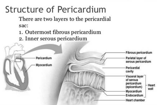
7 Pericardial Disease Pericardial Effusion Normal pericardial fluid = ml Intrapericardial pressure -5 to +5 mmhg Pericardial effusion Separation of visceral and parietal pericardium with echo-free space
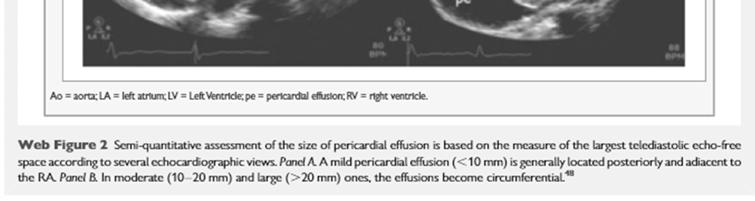
, moderate (10 20")

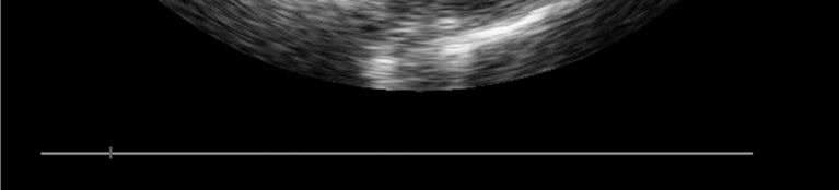
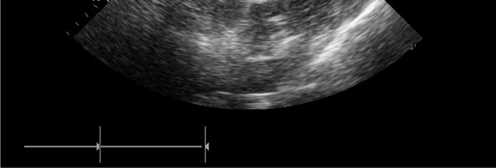
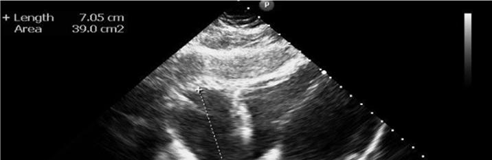
8 Reporting amount of pericardial effusion Trivial, small (<10 mm), moderate (10 20 mm), large (>20 mm) size of the echo-free space seen end-diastole Klein. JASE 2013
9
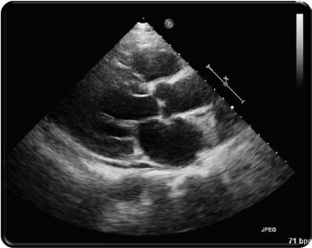
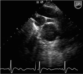
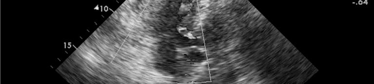
10 Small Pericardial Effusion

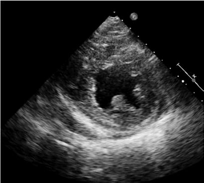
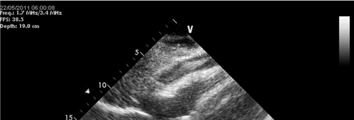
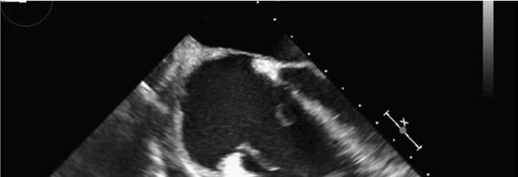
11 Large Pericardial Effusion Pe eff Pl eff * Pl eff Pe eff


12 Pericardial Effusion Pleural vs. Pericardial effusion Pleural vs. Pericardial effusion * A B * *


13 From Netter F. Atlas of Human Anatomy. Ciba-Geigy 1995.
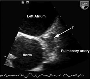
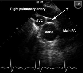
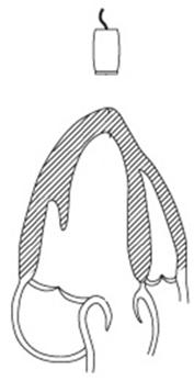
14 Pericardial Attachments SVC Transverse sinus Pulmonary veins Aorta Pulmonary a. Oblique sinus IVC Visceral pericardium Parietal pericardium From Netter F. Atlas of Human Anatomy. Ciba-Geigy 1995.

15 Pericardial Attachments 1-5 Transverse sinus 1. Anterior portions of superior aortic recess 2. Posterior portions of superior aortic recess 3. Inferior aortic recess 4. Left pulmonic recess 5. Right pulmonic recess 6 Oblique sinus 7. Postcaval recess 8. Left pulmonary venous recess 9. Right pulmonary venous recess From Kodama et al. AJR 2003; 181:1101
16 Pericardial Attachments 1-5 Transverse sinus 1. Anterior portions of superior aortic recess 2. Posterior portions of superior aortic recess 3. Inferior aortic recess 4. Left pulmonic recess 5. Right pulmonic recess 6 Oblique sinus 7. Postcaval recess 8. Left pulmonary venous recess 9. Right pulmonary venous recess From Kodama et al. AJR 2003; 181:1101

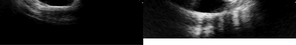
17 Transverse Sinus


18 Anterior view J Am Soc Echocardiogr 2013;26:


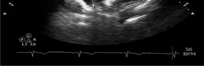
19 Right lateral & Left Lateral views of the pericardium Phrenic n. J Am Soc Echocardiogr 2013;26:


20 Coronal section epicardial fat RV LV epipericardial fat J Am Soc Echocardiogr 2013;26:

21 Epicardial Fat

22 Cardiac Tamponade Abnormal rise in intrapericardial pressure Impairment of diastolic filling

23 Cardiac Tamponade Beck s triad low arterial blood pressure distended neck veins distant, muffled heart sounds
24 Figure 1. Pericardial pressure-volume relations determined in pericardium obtained from a normal experimental animal and from an animal with chronic cardiac dilation produced by volume loading. Little W C, and Freeman G L Circulation 2006;113: Copyright American Heart Association


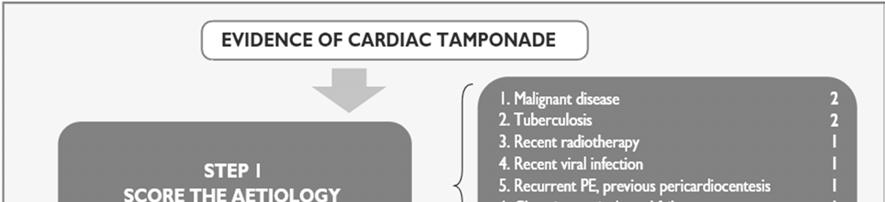
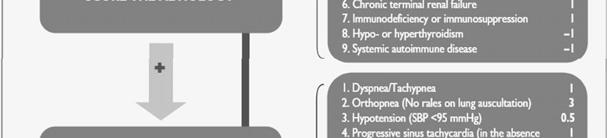
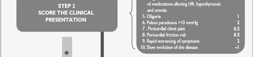
25 Grading of Hemodynamic Compromise Caused by Pericardial Effusion Sagristà-Sauleda. World J Cardiol 2011; 3(5):
26 Role of Echocardiography: in Cardiac Tamponade A Diagnosis of tamponade and evaluation of the size of pericardial effusion B Assessment of the distribution of pericardial effusion C Detection of intrapericardial adhesions D Diagnosis of intrapericardial clot E Assessment of the suitability for pericardiocentesis F Assist in monitoring pericardiocentesis G Diagnosis of effusive constrictive pericarditis Chandraratna, Echocardiography 2014
27 Cardiac Tamponade M mode/2d echocardiogram Diastolic collapse of the anterior RV free wall, RA collapse, LA and very rarely LV collapse increased LV diastolic wall thickness pseudohypertrophy IVC dilatation Swinging heart Doppler Exaggerated respiratory variation of LV and RV inflow
28 Cardiac Tamponade 2-D Echocardiographic diagnosis RV diastolic collapse persistent inward motion of RV free wall after mitral valve opening Most common (supine) anterior free wall and proximal infundibulum

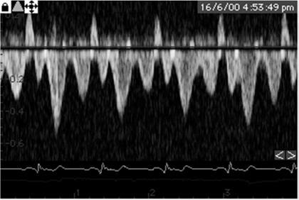
29 RV diastolic collapse


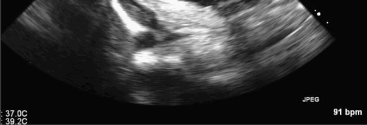
30 RV diastolic collapse M-mode

31 Tamponade 2-D Echocardiographic RA inversion Normally rounded throughout cardiac cycle Sensitivity 90 + % Specificity 82% Predictive value 50% RA inversion lasting > 1/3 cardiac cycle Sensitivity 94 % Specificity 100 %

32 Swinging Heart
33 Cardiac Tamponade 2-D Echocardiographic diagnosis RA inversion & RV collapse False negative Pace rhythm (RA inversion) Adhesion Loculated effusion Increase chamber stiffness
34 Cardiac Tamponade Echocardiographic features suggesting cardiac tamponade Early diastolic collapse of the right ventricle Late diastolic right atrial inversion Plethora of the inferior vena cava with blunted respiratory change Suggestive but indirect Doppler echocardiography more sensitive


35


36 LV inflow PW Doppler

37 Cardiac Tamponade Transmitral Doppler
38 Pericardial effusion?
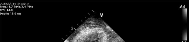
39 Pericardial effusion?
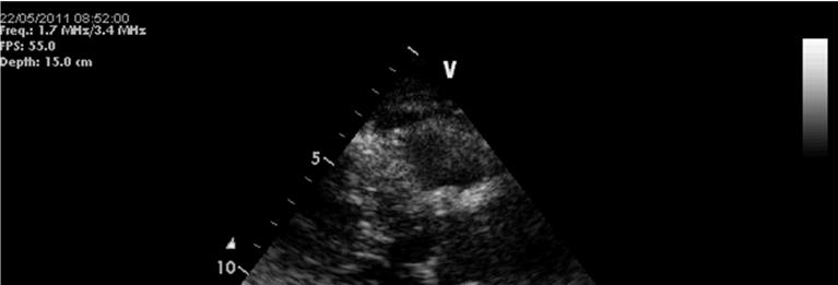
40 Pericardial effusion? Subcostal View

41 LV Free Wall Rupture Hemopericardium


42 BMJ Case Rep Published online: doi: /bcr
43 Role of Echocardiography: in Cardiac Tamponade A Diagnosis of tamponade and evaluation of the size of pericardial effusion B Assessment of the distribution of pericardial effusion C Detection of intrapericardial adhesions D Diagnosis of intrapericardial clot E Assessment of the suitability for pericardiocentesis F Assist in monitoring pericardiocentesis G Diagnosis of effusive constrictive pericarditis

44 Echo-guided pericardiocentesis Jung HK. Korean Circ J November; 42(11):
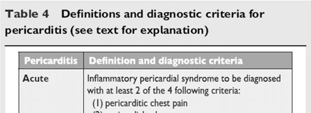
45 2015ESCGuidelines for the diagnosis and management of pericardial diseases


46 2015ESCGuidelines for the Dx and management of pericardial diseases(2)


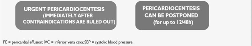
47 2015ESCGuidelines for the Dx and management of pericardial diseases(3)


48 Loculated cardiac hematoma Rev Port Cardiol 2015;34:561.e1-3


49 Loculated cardiac hematoma Rev Port Cardiol 2015;34:561.e1-3
50 Pericardial Cyst
51 Constrictive Pericarditis

52 Constrictive pericarditis Physiology Dissociation of intrathoracic and intracardiac pressures Enhanced ventricular interaction (exaggerated ventricular interdependence)
53 Constrictive pericarditis 2-D Echocardiography Increase pericardial thickness Myocardial tethering Inspiratory septal shift (septal bounce) Abnormal septal motion (septal shudder) IVC plethora (max diameter 21 mm and degree of inspiratory collapse <50%)
54 Constrictive pericarditis 2-D Echocardiography Increase pericardial thickness Myocardial tethering Inspiratory septal shift (septal bounce) Abnormal septal motion (septal shudder) IVC plethora

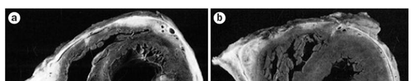
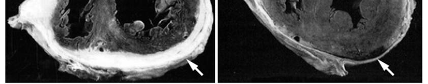
55 Constrictive pericarditis Pericardial Thickness

56 Constrictive pericarditis Pericardial Thickness
57 Constrictive pericarditis Pericardial Thickness


58 Constrictive pericarditis Pericardial Thickness Pericardial thickness from TTE usually NOT reliable Thickness from TEE correlates better with EBCT

59 Pericardial thickness CT scan Normal Increase pericardial thickness
60 Pericardial Thickness MRI Normal Increase pericardial thickness 60
61 Constrictive pericarditis Pericardial Thickness Pericardial thickness does NOT mean Constriction
62 Constrictive pericarditis Pericardial Thickness
63 Constrictive pericarditis 2-D Echocardiography Increase pericardial thickness Myocardial tethering Inspiratory septal shift (septal bounce) Abnormal septal motion (septal shudder) IVC plethora
64 Constrictive pericarditis Myocardial Tethering RA RA No tethering
65 Constrictive pericarditis Myocardial Tethering
66 Constrictive pericarditis 2-D Echocardiography Increase pericardial thickness Myocardial tethering Inspiratory septal shift (septal bounce) Abnormal septal motion (septal shudder) IVC plethora


67 Constrictive pericarditis Inspiratory Septal Shift LV RV


68 Constrictive pericarditis Inspiratory Septal Shift
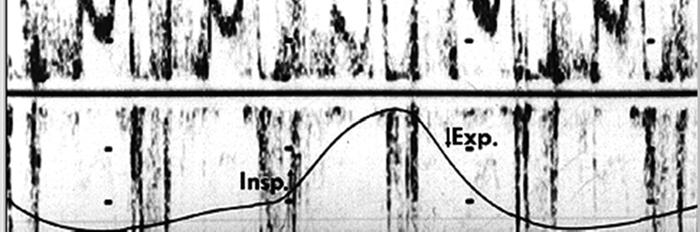
69 Constrictive pericarditis 2-D Echocardiography Increase pericardial thickness Myocardial tethering Inspiratory septal shift (septal bounce) Abnormal septal motion (septal shudder) IVC plethora

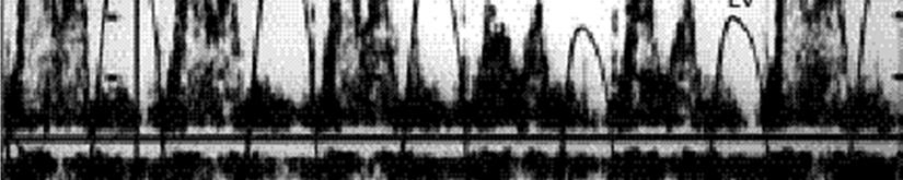
70 Constrictive pericarditis Abnormal Septal motion (Septal Shudder)
71 Interventricular Septal Diastolic Motion (IVSDM) J Candell-Riera. Circulation 1978
72 Constrictive pericarditis 2-D Echocardiography Increase pericardial thickness Myocardial tethering Inspiratory septal shift (septal bounce) Abnormal septal motion (septal shudder) IVC plethora

73 Constrictive pericarditis IVC Plethora

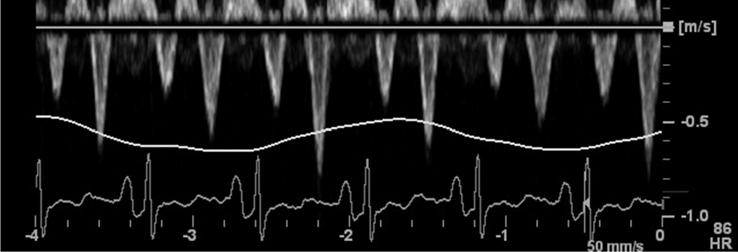
74 Pericardial thickness CT scan Normal Increase pericardial thickness


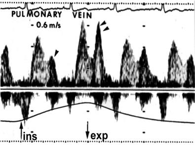
75 Pericardial Thickness MRI Normal Increase pericardial thickness
76 Constrictive pericarditis 2D echo Pericardial thickness Inspiratory septal shift Abnormal septal motion IVC plethora

77 Inspiratory Septal Shift Expiration Inspiration
78 Constrictive Pericarditis Doppler echo with respirometer
79 Constrictive Pericarditis Doppler echo
80 Constrictive Pericarditis Doppler echo


81

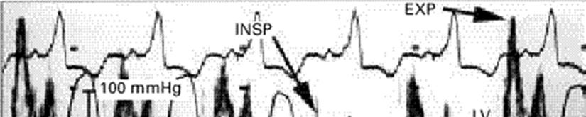
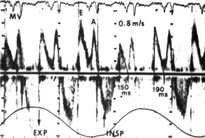
82 Mitral Doppler

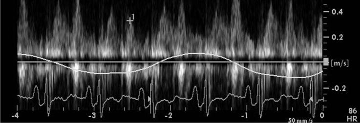
83 Pulmonary vein Doppler

84 Hepatic Vein Doppler


85 Hepatic Vein Doppler Diastolic reversal ratio = reversal velocity forward velocity 0.79

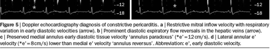
86 Mitral PW Doppler


87 Pulmonary vein Doppler LV LA S D AR TTE TEE


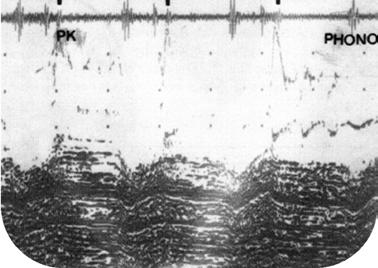
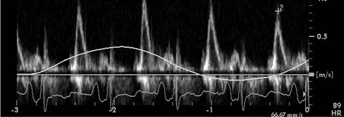
88 Hepatic Vein Doppler Subcostal window Color-Flow Doppler AR S D

89 Superior Vena Cava Dopper Right supraclavicular window Color-Flow Doppler AR S 1 D S 2
90 Constrictive Pericarditis Doppler echocardiography Mitral = 100 * (Expiration Inpiration) / Inpiration > 25% Tricuspid = 100 * (Inspiration Expiration) / Expiration > 30%
91 Constriction and Restriction Normal 1. Mitral inflow E vel.< 10% respiratory variation DT > Hepatic vein flow Systolic forward > Diastolic forward Diastolic reversal < 20% forward flow From: Oh JK et al. The Echo Manual. Little, Brown
92 Constriction and Restriction Constrictive Physiology 1. Mitral inflow > 25% increase E vel. with expiration DT usually < Hepatic vein flow Diastolic reversal > 25% diastolic forward flow From: Oh JK et al. The Echo Manual. Little, Brown

93 Constriction and Restriction Restrictive physiology 1. Mitral inflow E/A usually > 1.5 E vel.< 10% respiratory variation DT < Hepatic vein flow Diastolic forward > Systolic forward Increase reversals with inspiration From: Oh JK et al. The Echo Manual. Little, Brown


94 Constriction vs Restriction Annulus Paradoxus E/E < 15 E/E > 15


95 Constrictive pericarditis TDI Medial Mitral annulus


96 Constrictive Pericarditis Doppler echocardiography (expiration inspiration) x 100% inspiration
97 Constrictive Pericarditis Mitral Pulmonary V

98 Doppler Mitral Inflow

99 Constrictive pericarditis Hepatic vein SVC
100 Restriction vs. Constriction SVC ins exp HV Restrictive Cardiomyopathy Constrictive pericarditis Appleton et al. JACC 1988;11:

101


102 Echocardiographic Diagnosis of Constrictive Pericarditis: Mayo Clinic Criteria Welch et al. Circ Cardiovasc Imaging. 2014;7:


103 Echocardiography diagnostic criteria algorithm for constrictive pericarditis Syed, F. F. et al. Nat. Rev. Cardiol. 11, (2014);
104 Constrictive pericarditis Differential Diagnosis Respiratory variation of mitral E velocity Acute dilatation of the heart Pulmonary embolism RV infarct Pleural effusion Chronic obstructive lung disease
105 Constrictive pericarditis Differential Diagnosis Respiratory variation of mitral inflow velocities Acute dilatation of the heart Pulmonary embolism RV infarct Pleural effusion Clinical 2-D echo Chronic obstructive lung disease


106 COPD vs. Constriction Boonyaratavej S et al. JACC 1998:2043-8


107 COPD vs. Constriction Boonyaratavej S et al. JACC 1998:2043-8
108 Pericardial Diseases Etiologic Dx Clinical Dx/ conditions Echo findings Purulent Acute pericarditis Pericardial effusions Tuberculous Recurrent pericarditis Tamponade physiology Viral Cardiac tamponade Constrictive physiology Autoimmune Constrictive pericarditis Pericardial Masses (Tumors,Cysts, and Diverticulum) Post surgery Idiopathic Congenital Malignancy Effusive constrictive pericarditis Congenital Absence of the Pericardium

109 Thank You
Adel Hasanin Ahmed 1
 Adel Hasanin Ahmed 1 PERICARDIAL DISEASE The pericardial effusion ends anteriorly to the descending aorta and is best visualised in the PLAX. PSAX is actually very useful sometimes for looking at posterior
Pericardial Disease: Case Examples. Echo Fiesta 2017
 Pericardial Disease: Case Examples Echo Fiesta 2017 2014 2014 MFMER MFMER 3346252-1 slide-1 Objectives Have a systematic approach to evaluation of constriction 2014 MFMER 3346252-2 CASE 1 2013 MFMER 3248567-3
Outline. Echocardiographic Assessment of Pericardial Effusion/Tamponade: The Essentials
 Echocardiographic Assessment of Pericardial Effusion/Tamponade: The Essentials John R Schairer DO FACC Henry Ford Heart and Vascular Institute No Disclosures Outline Normal Anatomy and Physiology Pathophysiology
ΚΑΡΔΙΟΛΟΓΟΣ EUROPEAN ACCREDITATION IN TRANSTHORACIC AND TRANSESOPHAGEAL ECHOCARDIOGRAPHY
 1 ΚΑΡΔΙΟΛΟΓΟΣ EUROPEAN ACCREDITATION IN TRANSTHORACIC AND TRANSESOPHAGEAL ECHOCARDIOGRAPHY 2 Constrictive pericarditis (CP) is characterized by impaired ventricular filling due to a stiffened or noncompliant
Constrictive Pericarditis
 Constrictive Pericarditis Never Confused with Anything Else Jae K. Oh, MD 2018 MFMER 3712003-1 ARS #1 CP Which of following patients has constrictive pericarditis? 1 2 3 Medial e 13 cm/s Medial e 3 cm/s
THE PERICARDIUM: LOOKING OUTSIDE THE HEART
 THE PERICARDIUM: LOOKING OUTSIDE THE HEART DISCLOSURE Relevant relationships with commercial entities none Potential for conflicts of interest within this presentation none Steps taken to review and mitigate
10/1/2016. Constrictive Pericarditis Unique Hemodynamics. What s New in Pericardial Disease? Case-based Discussion
 Mayo Clinic Department of Cardiovascular Diseases Mayo Clinic Echocardiography Review Course for Boards and Recertification What s New in Pericardial Disease? Case-based Discussion Jae K. Oh, MD Samsung
Choose the grading of diastolic function in 82 yo woman
 Question #1 Choose the grading of diastolic function in 82 yo woman E= 80 cm/s A= 70 cm/s LAVI < 34 ml/m 2 1= Grade 1 2= Grade 2 3= Grade 3 4= Normal 5= Indeterminate 2018 MFMER 3712003-1 Choose the grading
Constriction vs Restriction Case-based Discussion
 Mayo Clinic Department of Cardiovascular Diseases Mayo Clinic Echocardiography Review Course for Boards and Recertification Constriction vs Restriction Case-based Discussion Jae K. Oh, MD Samsung Professor
Echocardiography as a diagnostic and management tool in medical emergencies
 Echocardiography as a diagnostic and management tool in medical emergencies Frank van der Heusen MD Department of Anesthesia and perioperative Care UCSF Medical Center Objective of this presentation Indications
PERICARDIAL DIAESE. Kaijun Cui Associated professor Sichuan University
 PERICARDIAL DIAESE Kaijun Cui Associated professor Sichuan University CLASSIFICATION acute pericarditis pericardial effusion cardiac tamponade constrictive pericarditis congenitally absent pericardium
Adult Echocardiography Examination Content Outline
 Adult Echocardiography Examination Content Outline (Outline Summary) # Domain Subdomain Percentage 1 2 3 4 5 Anatomy and Physiology Pathology Clinical Care and Safety Measurement Techniques, Maneuvers,
TAMPONADE CARDIAQUE. Dr Cédrick Zaouter TUSAR 15 décembre 2015
 TAMPONADE CARDIAQUE Dr Cédrick Zaouter TUSAR 15 décembre 2015 OUTLINE History Incidence Definition Pathophysiology Aetiologies Investigations - Echocardiography Treatment of cardiac tamponade Pericardial
UPDATE ON CONSTRICTIVE PERICARDITIS ECHOCARDIOGRAPHY AND CARDIAC CATHETERISATION
 Arsen D. Ristić, MD, PhD, FESC (no conflicts of interest to disclose regarding this presentation) UPDATE ON CONSTRICTIVE PERICARDITIS ECHOCARDIOGRAPHY AND CARDIAC CATHETERISATION Department of Cardiology,
Doppler Basic & Hemodynamic Calculations
 Doppler Basic & Hemodynamic Calculations August 19, 2017 Smonporn Boonyaratavej MD Division of Cardiology, Department of Medicine Chulalongkorn University Cardiac Center, King Chulalongkorn Memorial Hospital
Pericardial disease. Se-Jung Yoon Cardiology division NHIS Ilsan hospital
 Pericardial disease Se-Jung Yoon Cardiology division NHIS Ilsan hospital Normal pericardial effusion Normal pericardium Normal pericardium Pericardial Layers: Visceral layer Parietal layer Fibrous pericardium
Transient Constrictive Pericarditis: Causes and Natural History
 Journal of the American College of Cardiology Vol. 43, No. 2, 2004 2004 by the American College of Cardiology Foundation ISSN 0735-1097/04/$30.00 Published by Elsevier Inc. doi:10.1016/j.jacc.2003.08.032
Structural Heart Disease. Echocardiographic Diagnosis of Constrictive Pericarditis Mayo Clinic Criteria
 Structural Heart Disease Echocardiographic Diagnosis of Constrictive Pericarditis Mayo Clinic Criteria Terrence D. Welch, MD; Lieng H. Ling, MBBS, MD; Raul E. Espinosa, MD; Nandan S. Anavekar, MBBCh; Heather
Rotation: Echocardiography: Transthoracic Echocardiography (TTE)
") Rotation: Echocardiography: Transthoracic Echocardiography (TTE) Rotation Format and Responsibilities: Fellows rotate in the echocardiography laboratory in each clinical year. Rotations during the first
Rotation: Echocardiography: Transthoracic Echocardiography (TTE) Rotation Format and Responsibilities: Fellows rotate in the echocardiography laboratory in each clinical year. Rotations during the first
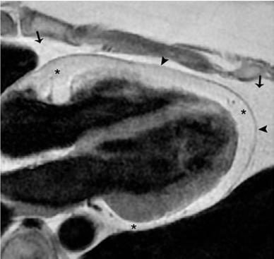
Constrictive Pericarditis Pitfalls in MR Diagnosis Cylen Javidan-Nejad Associate Professor Mallinckrodt Institute of Radiology Washington University
 Constrictive Pericarditis Pitfalls in MR Diagnosis Cylen Javidan-Nejad Associate Professor Mallinckrodt Institute of Radiology Washington University in St. Louis Goal o To review the imaging criteria of
Department of Cardiac, Thoracic and Vascular Sciences University of Padua Cardiac Tamponade. Echocardiography in Diagnosis and Management
 Department of Cardiac, Thoracic and Vascular Sciences University of Padua Cardiac Tamponade. Echocardiography in Diagnosis and Management Luigi P. Badano, MD, FESC, FACC Declaration of interest **Dr. Badano
ECHOCARDIOGRAPHY. Patient Care. Goals and Objectives PF EF MF LF Aspirational
 Patient Care Be able to: Perform and interpret basic TTE and X cardiac Doppler examinations Perform and interpret a comprehensive X TTE and cardiac Doppler examination Perform and interpret a comprehensive
We are now going to review the diagnosis and management of pericardial collections and tamponade
 We are now going to review the diagnosis and management of pericardial collections and tamponade FEEL COURSE PAGE 1 Paying particular attention to the difference between a collection and cardiac tamponade
Pericardial Diseases/Tamponade Illustrative Cases
 Pericardial Diseases/Tamponade Illustrative Cases Jae K. Oh, MD Echo Hawaii 2017 2012 MFMER 3200268v3(2010)-1 Case #1 47 year old man Chest pain Not exertional Normal Examination 2016 MFMER slide-2 1 47
The role of bedside ultrasound in the diagnosis of pericardial effusion and cardiac tamponade
 Symposium The role of bedside ultrasound in the diagnosis of pericardial effusion and cardiac tamponade Adam Goodman, Phillips Perera, Thomas Mailhot, Diku Mandavia Department of Emergency Medicine, Los
A Case of Impending Cardiac Tamponade Caused by Effusive Constrictive Pericarditis
 Archives of Clinical and Medical Case Reports doi: 10.26502/acmcr.96550038 Volume 2, Issue 5 Case Report A Case of Impending Cardiac Tamponade Caused by Effusive Constrictive Pericarditis Catalina Sanchez-Alvarez
Index. K Knobology, TTE artifact, image resolution, ultrasound, 14
 A Acute aortic regurgitation (AR), 124 128 Acute aortic syndrome (AAS) classic aortic dissection diagnosis, 251 263 evolutive patterns, 253 255 pathology, 250 251 classifications, 247 248 incomplete aortic
Imaging in Heart Failure: A Multimodality Approach. Thomas Ryan, MD
 Imaging in Heart Failure: A Multimodality Approach Thomas Ryan, MD Heart Failure HFrEF HFpEF EF50% Lifetime risk 20% Prevalence 6M Americans Societal costs - $30B 50% 5-year survival 1 Systolic
Certificate in Clinician Performed Ultrasound (CCPU) Syllabus. Rapid Cardiac Echo (RCE)
 Syllabus. Rapid Cardiac Echo (RCE)") Certificate in Clinician Performed Ultrasound (CCPU) Syllabus Rapid Cardiac Echo (RCE) Purpose: Rapid Cardiac Echocardiography (RCE) This unit is designed to cover the theoretical and practical curriculum
Certificate in Clinician Performed Ultrasound (CCPU) Syllabus Rapid Cardiac Echo (RCE) Purpose: Rapid Cardiac Echocardiography (RCE) This unit is designed to cover the theoretical and practical curriculum
Constrictive Pericarditis Curable Paradoxical Diastolic Heart Failure ASE EBRC 2018
 Constrictive Pericarditis Curable Paradoxical Diastolic Heart Failure ASE EBRC 2018 May 8 th, 2018 Jae K. Oh, MD Samsung Professor of CV Diseases Director, Pericardial Disease Clinic Co-Director, Integrated
Evalua&on)of)Le-)Ventricular)Diastolic) Dysfunc&on)by)Echocardiography:) Role)of)Ejec&on)Frac&on)
of)Le-)Ventricular)Diastolic) Dysfunc&on)by)Echocardiography:) Role)of)Ejec&on)Frac&on)") Evalua&on)of)Le-)Ventricular)Diastolic) Dysfunc&on)by)Echocardiography:) Role)of)Ejec&on)Frac&on) N.Koutsogiannis) Department)of)Cardiology) University)Hospital)of)Patras)! I have no conflicts of interest
Evalua&on)of)Le-)Ventricular)Diastolic) Dysfunc&on)by)Echocardiography:) Role)of)Ejec&on)Frac&on) N.Koutsogiannis) Department)of)Cardiology) University)Hospital)of)Patras)! I have no conflicts of interest
Diastology State of The Art Assessment
 Diastology State of The Art Assessment Dr. Mohammad AlGhamdi Assistant professor, KSAU-HS Consultant Cardiologist King AbdulAziz Cardiac Center Ministry of National Guard Health Affairs Diagnostic Clinical
2/4/2011. Nathan Kerner, M.D.
 Nathan Kerner, M.D. Definition Elevated pressures - cut off usually >40 mmhg pulmonary artery systolic pressure (PASP) Usually associated with elevated pulmonary vascular resistance (PVR) measured in dynessec/cm
Echocardiography Conference
 Echocardiography Conference David Stultz, MD Cardiology Fellow, PGY-6 September 20, 2005 Atrial Septal Aneurysm Bulging of Fossa Ovalis Associated commonly with Atrial septal defect or small perforations
Disclosures. Cardiac Ultrasound. Introductory Case. 80 y/o male Syncope at home Emesis x 3 in ambulance Looks sick. No pain.
 Disclosures Cardiac Ultrasound Justin A Davis, MD MPH RDMS Subchief for Emergency Ultrasound Kaiser Permanente East Bay Medical Center I have nothing to disclose. Introductory Case HR 118 BP 65/43 RR 27
Normal Pericardial Physiology
 Normal Pericardial Physiology Normal pericardium contains 20-30 ml of lymphoid fluid lubricating function that facilitates normal myocardial rotation and translation during each cardiac cycle in that the
Cardiac tamponade and Pericardiocentesis Made Easy
 Cardiac tamponade and Pericardiocentesis Made Easy www.cardiconcept.com Etiology of pericardial diseases. Non Infectious cause Infectious cause European Heart Journal (2015) 36, 2921 2964 Recommendations
COMPREHENSIVE EVALUATION OF FETAL HEART R. GOWDAMARAJAN MD
 COMPREHENSIVE EVALUATION OF FETAL HEART R. GOWDAMARAJAN MD Disclosure No Relevant Financial Relationships with Commercial Interests Fetal Echo: How to do it? Timing of Study -optimally between 22-24 weeks
Diastolic Function: What the Sonographer Needs to Know. Echocardiographic Assessment of Diastolic Function: Basic Concepts 2/8/2012
 Diastolic Function: What the Sonographer Needs to Know Pat Bailey, RDCS, FASE Technical Director Beaumont Health System Echocardiographic Assessment of Diastolic Function: Basic Concepts Practical Hints
Atrial Septal Defects
 Supplementary ACHD Echo Acquisition Protocol for Atrial Septal Defects The following protocol for echo in adult patients with atrial septal defects (ASDs) is a guide for performing a comprehensive assessment
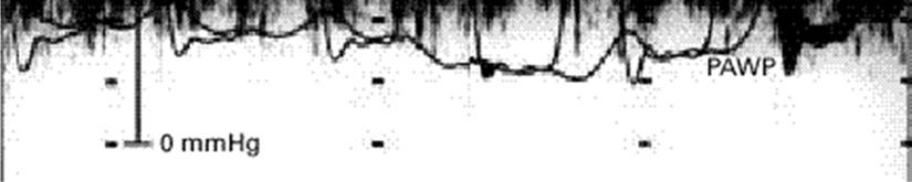
Comprehensive Hemodynamics By Doppler Echocardiography. The Echocardiographic Swan-Ganz Catheter.
 Comprehensive Hemodynamics By Doppler Echocardiography. The Echocardiographic Swan-Ganz Catheter. Itzhak Kronzon, MD, FASE, FACC, FESC, FAHA, FACP, FCCP North Shore HS, LIJ/Lenox Hill Hospital, New York
Echocardiographic Evaluation of Pericardial Disease
 Echocardiographic Evaluation of Pericardial Disease Edwin G. Avery, IV, M.D. Chief, Division of Cardiac Anesthesia University Hospitals Case Medical Center Associate Professor of Anesthesiology Case Western
Right Heart Hemodynamics: Echo-Cath Discrepancies
 Department of cardiac, thoracic and vascular sciences University of Padua, School of Medicine Padua, Italy Right Heart Hemodynamics: Echo-Cath Discrepancies Luigi P. Badano, MD, PhD, FESC, FACC **Dr. Badano
PART II ECHOCARDIOGRAPHY LABORATORY OPERATIONS ADULT TRANSTHORACIC ECHOCARDIOGRAPHY TESTING
 PART II ECHOCARDIOGRAPHY LABORATORY OPERATIONS ADULT TRANSTHORACIC ECHOCARDIOGRAPHY TESTING STANDARD - Primary Instrumentation 1.1 Cardiac Ultrasound Systems SECTION 1 Instrumentation Ultrasound instruments
Hemodynamic Assessment. Assessment of Systolic Function Doppler Hemodynamics
 Hemodynamic Assessment Matt M. Umland, RDCS, FASE Aurora Medical Group Milwaukee, WI Assessment of Systolic Function Doppler Hemodynamics Stroke Volume Cardiac Output Cardiac Index Tei Index/Index of myocardial
Echo Emergencies. Outline. Michael H. Picard, MD Massachusetts General Hospital Harvard Medical School No disclosures
 Echo Emergencies Michael H. Picard, MD Massachusetts General Hospital Harvard Medical School No disclosures Outline Common emergency / on call scenarios Tamponade Pulmonary embolism/rv strain Cardiogenic
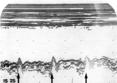
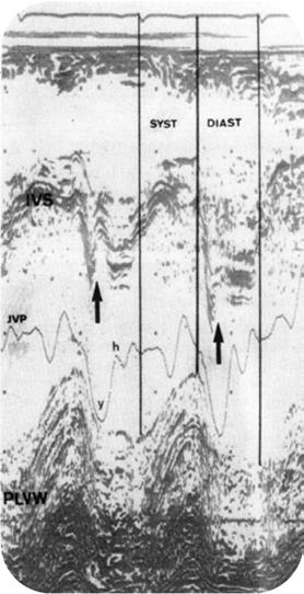
Changes in the Venous Pulse Waveform in Pericardial Effusion Revealed by Doppler. Benoy N Shah MD(Res) MRCP FESC & Dhrubo J Rakhit PhD FRCP FACC
 MRCP FESC & Dhrubo J Rakhit PhD FRCP FACC") Page 1 of 5 Title of image and video article Changes in the Venous Pulse Waveform in Pericardial Effusion Revealed by Doppler Echocardiography of the Superior Vena Cava Authors Benoy N Shah MD(Res) MRCP
Page 1 of 5 Title of image and video article Changes in the Venous Pulse Waveform in Pericardial Effusion Revealed by Doppler Echocardiography of the Superior Vena Cava Authors Benoy N Shah MD(Res) MRCP
Pericardial effusion, Cardiac Tamponade, and echo guided pericardiocentesis
 KSC 2017 Echo5- Myocardial and Pericardial disease Pericardial effusion, Cardiac Tamponade, and echo guided pericardiocentesis Ji-Hyun Jung Division of Cardiology Sejong Hospital KSC 2017 The 61 th Annual
Acute Viral Myopericarditis Presenting as a Transient Effusive-Constrictive Pericarditis Caused by Coinfection with Coxsackieviruses A4 and B3
 case report korean j intern med 2012;27:216-220 ORIGINL RTICLE pissn 1226-3303 eissn 2005-6648 cute Viral Myopericarditis Presenting as a Transient Effusive-Constrictive Pericarditis Caused y Coinfection
M-Mode Echocardiography Is it still Alive? Itzhak Kronzon, MD,FASE. Sampling Rate M-Mode: 1800 / sec 2D: 30 / sec
 M-Mode Echocardiography Is it still Alive? Itzhak Kronzon, MD,FASE Honoraria: Philips Classical M-mode Echocardiography M-Mode offers better time and image resolution. Sampling Rate M-Mode: 1800 / sec
Restrictive Cardiomyopathy
 ESC Congress 2011, Paris Imaging Unusual Causes of Cardiomyopathy Restrictive Cardiomyopathy Kazuaki Tanabe, MD, PhD Professor of Medicine Chair, Division of Cardiology Izumo, Japan I Have No Disclosures
The Cardiovascular System
 The Cardiovascular System The Manila Times College of Subic Prepared by: Stevens B. Badar, RN, MANc THE HEART Anatomy of the Heart Location and Size approx. the size of a person s fist, hollow and cone-shaped,
CMS Limitations Guide - Radiology Services
 CMS Limitations Guide - Radiology Services Starting October 1, 2015, CMS will update their existing medical necessity limitations on tests and procedures to correspond to ICD-10 codes. This limitations
Constrictive pericarditis: Morphological, functional and haemodynamic evaluation
 Constrictive pericarditis: Morphological, functional and haemodynamic evaluation Poster No.: C-0743 Congress: ECR 2010 Type: Educational Exhibit Topic: Cardiac Authors: B. Graca, P. Donato, M. Ferreira,
British Society of Echocardiography
 British Society of Echocardiography Affiliated to the British Cardiac Society A Minimum Dataset for a Standard Adult Transthoracic Echocardiogram From the British Society of Echocardiography Education
Appendix II: ECHOCARDIOGRAPHY ANALYSIS
 Appendix II: ECHOCARDIOGRAPHY ANALYSIS Two-Dimensional (2D) imaging was performed using the Vivid 7 Advantage cardiovascular ultrasound system (GE Medical Systems, Milwaukee) with a frame rate of 400 frames
Evaluation of the Right Ventricle in Candidates for Right Ventricular Assist Device Implantation.
 Evaluation of the Right Ventricle in Candidates for Right Ventricular Assist Device Implantation. Evaluation of RVAD Function. Ioannis A Paraskevaidis Attikon University Hospital Historical Perspective
JFICMI Basic Critical Care Echocardiography (BCCE)
") JFICMI Basic Critical Care Echocardiography (BCCE) 2017 Introduction The International expert statement on training standards for critical care ultrasonography position paper published in Intensive Care
JFICMI Basic Critical Care Echocardiography (BCCE) 2017 Introduction The International expert statement on training standards for critical care ultrasonography position paper published in Intensive Care
Echo Doppler Assessment of Right and Left Ventricular Hemodynamics.
 Echo Doppler Assessment of Right and Left Ventricular Hemodynamics. Itzhak Kronzon, MD, FASE, FACC, FESC, FAHA, FACP, FCCP Northwell, Lenox Hill Hospital, New York Professor of Cardiology Hofstra University
Breakout Session: Transesophageal Echocardiography
 Breakout Session: Transesophageal Echocardiography Doris Ockert, MD Andrew Schroeder, MD University of Wisconsin School of Medicine and Public Health Jutta Novalija, MD, PhD Medical College of Wisconsin
ASCeXAM / ReASCE. Practice Board Exam Questions Monday Morning
 ASCeXAM / ReASCE Practice Board Exam Questions Monday Morning Ultrasound Physics Artifacts Doppler Physics Imaging, Knobology, and Artifacts Echocardiographic Evaluation of the RV Tricuspid and Pulmonary
Pediatric Echocardiography Examination Content Outline
 Pediatric Echocardiography Examination Content Outline (Outline Summary) # Domain Subdomain Percentage 1 Anatomy and Physiology Normal Anatomy and Physiology 10% 2 Abnormal Pathology and Pathophysiology
Essentials of Pericardial Diseases
 Essentials of Pericardial Diseases 1 Nikolaos Skubas MD, 2 Manuel Fontes MD The pericardial diseases result in cardiovascular perturbations ranging from asymptomatic electrocardiographic findings (in pericarditis
P = 4V 2. IVC Dimensions 10/20/2014. Comprehensive Hemodynamic Evaluation by Doppler Echocardiography. The Simplified Bernoulli Equation
 Comprehensive Hemodynamic Evaluation by Doppler Echocardiography Itzhak Kronzon, MD North Shore LIJ/ Lenox Hill Hospital New York, NY Disclosure: Philips Healthcare St. Jude Medical The Simplified Bernoulli
The right ventricle in chronic heart failure
 The right ventricle in chronic heart failure ESC 2012 Christian Opitz, Berlin There are no conflicts of interest relevant to this presentation Percent of Population Prevalence of Heart Failure by Age and
The pericardial sac is composed of the outer fibrous pericardium
 Pericardiectomy for Constrictive or Recurrent Inflammatory Pericarditis Mauricio A. Villavicencio, MD, Joseph A. Dearani, MD, and Thoralf M. Sundt, III, MD Anatomy and Preoperative Considerations The pericardial
DOPPLER HEMODYNAMICS (1) QUANTIFICATION OF PRESSURE GRADIENTS and INTRACARDIAC PRESSURES
 QUANTIFICATION OF PRESSURE GRADIENTS and INTRACARDIAC PRESSURES") THORAXCENTRE DOPPLER HEMODYNAMICS (1) QUANTIFICATION OF PRESSURE GRADIENTS and INTRACARDIAC PRESSURES J. Roelandt DOPPLER HEMODYNAMICS Intracardiac pressures and pressure gradients Volumetric measurement
THORAXCENTRE DOPPLER HEMODYNAMICS (1) QUANTIFICATION OF PRESSURE GRADIENTS and INTRACARDIAC PRESSURES J. Roelandt DOPPLER HEMODYNAMICS Intracardiac pressures and pressure gradients Volumetric measurement
Looking Outside the Box: Incidental Extracardiac Finding in Echo
 Looking Outside the Box: Incidental Extracardiac Finding in Echo Dr. Aijaz Shah Head of Division, Adult Echocardiography Laboratory Prince Sultan Cardiac Centre Riyadh Case 1 17 year old boy presented
LV FUNCTION ASSESSMENT: WHAT IS BEYOND EJECTION FRACTION
 LV FUNCTION ASSESSMENT: WHAT IS BEYOND EJECTION FRACTION Jamilah S AlRahimi Assistant Professor, KSU-HS Consultant Noninvasive Cardiology KFCC, MNGHA-WR Introduction LV function assessment in Heart Failure:
JOINT MEETING 2 Tricuspid club Chairpersons: G. Athanassopoulos, A. Avgeropoulou, M. Khoury, G. Stavridis
 JOINT MEETING 2 Tricuspid club Chairpersons: G. Athanassopoulos, A. Avgeropoulou, M. Khoury, G. Stavridis Similarities and differences in Tricuspid vs. Mitral Valve Anatomy and Imaging. Echo evaluation
Data Collected: June 17, Reported: June 30, Survey Dates 05/24/ /07/2010
 Job Task Analysis for ARDMS Pediatric Echocardiography Data Collected: June 17, 2010 Reported: Analysis Summary For: Pediatric Echocardiography Exam Survey Dates 05/24/2010-06/07/2010 Invited Respondents
The Patient with Atrial Fibrilation
 Assessment of Diastolic Function The Patient with Atrial Fibrilation Assoc. Prof. Adriana Ilieşiu, FESC University of Medicine Carol Davila Bucharest, Romania Associated Conditions with Atrial Fibrillation
The Doppler Examination. Katie Twomley, MD Wake Forest Baptist Health - Lexington
 The Doppler Examination Katie Twomley, MD Wake Forest Baptist Health - Lexington OUTLINE Principles/Physics Use in valvular assessment Aortic stenosis (continuity equation) Aortic regurgitation (pressure
Right Heart Evaluation ASE Guidelines Review. Chris Mann RDCS, RCS, FASE Faculty, Echocardiography Pitt Community College Greenville, NC
 Right Heart Evaluation ASE Guidelines Review Chris Mann RDCS, RCS, FASE Faculty, Echocardiography Pitt Community College Greenville, NC Objectives Briefly review right atrial and right ventricular anatomy
Πνευμονική υπέρταση και περικαρδιακή συλλογή. Τρόποι αντιμετώπισης
 Πνευμονική υπέρταση και περικαρδιακή συλλογή. Τρόποι αντιμετώπισης Γεώργιος Λάζαρος Καρδιολόγος, Διευθυντής ΕΣΥ Α Πανεπιστημιακή Καρδιολογική Κλινική Ιπποκράτειο Γ.Ν. Αθηνών Pericardial syndromes o Acute
Advanced imaging of the left atrium - strain, CT, 3D, MRI -
 Advanced imaging of the left atrium - strain, CT, 3D, MRI - Monica Rosca, MD Carol Davila University of Medicine and Pharmacy, Bucharest, Romania Declaration of interest: I have nothing to declare Case
Normal TTE Examination, Doppler Echocardiography and Normal Antegrade Flow Patterns
 Normal TTE Examination, Doppler Echocardiography and Normal Antegrade Flow Patterns Pravin Patil, MD FACC FASE Associate Professor of Medicine Director, Cardiovascular Disease Training Program Lewis Katz
Copyright 2017 American College of Emergency Physicians. All rights reserved.
 POLICY Approved April 2017 Guidelines for the Use of Transesophageal Echocardiography (TEE) in the ED for Cardiac Arrest Approved by the ACEP Board of Directors April 2017 1. Introduction The American
Case # 1. Page: 8. DUKE: Adams
 Case # 1 Page: 8 1. The cardiac output in this patient is reduced because of: O a) tamponade physiology O b) restrictive physiology O c) coronary artery disease O d) left bundle branch block Page: 8 1.
An Uncommon Cardiac Etiology of Liver Cirrhosis, Recurrent Ascites, Atrial Fibrillation and Congestive Heart Failure
 Cronicon OPEN ACCESS EC CARDIOLOGY Case Report An Uncommon Cardiac Etiology of Liver Cirrhosis, Recurrent Ascites, Atrial Fibrillation and Congestive Heart Failure Montaser Y Ismail 1 *, Mohammed I Nassar
Ιπποκράτειες μέρες καρδιολογίας Θεσσαλονίκη, 9-10 Μαρτίου Φωτεινή Α. Λαζαρίδου Επιμελήτρια Α Γενικό Νοσοκομείο Αγιος Παύλος, Θεσσαλονίκη
 Ιπποκράτειες μέρες καρδιολογίας Θεσσαλονίκη, 9-10 Μαρτίου 2018 Φωτεινή Α. Λαζαρίδου Επιμελήτρια Α Γενικό Νοσοκομείο Αγιος Παύλος, Θεσσαλονίκη RV shape Triangular shape in frontal plane crescent shape in
Echo in Pulmonary HTN
 Echo in Pulmonary HTN Steven A. Goldstein MD FACC FASE Professor of Medicine Georgetown University Medical Center MedStar Heart Institute Washington Hospital Center Monday, October 10, 2017 Pulmonary Artery
PRACTICAL ECHOCARDIOGRAPHY IN THE ADULT with Doppler and color-doppler flow imaging
 PRACTICAL ECHOCARDIOGRAPHY IN THE ADULT with Doppler and color-doppler flow imaging PRACTICAL ECHOCARDIOGRAPHY IN THE ADULT with Doppler and color-doppler flow imaging by J.P.M. HAMER Thoraxcentre, Department
Echocardiographic Cardiovascular Risk Stratification: Beyond Ejection Fraction
 Echocardiographic Cardiovascular Risk Stratification: Beyond Ejection Fraction October 4, 2014 James S. Lee, M.D., F.A.C.C. Associates in Cardiology, P.A. Silver Spring, M.D. Disclosures Financial none
Mitral Valve Disease, When to Intervene
 Mitral Valve Disease, When to Intervene Swedish Heart and Vascular Institute Ming Zhang MD PhD Interventional Cardiology Structure Heart Disease Conflict of Interest None Current ACC/AHA guideline Stages
Ultrasound. FAST Focused Assessment with Sonography in Trauma
 Ultrasound FAST Focused Assessment with Sonography in Trauma Rohit Patel, MD University of Florida Health Director, Critical Care Ultrasound Surgical ICU Center for Intensive Care Gainesville, Florida
Cardiac Mass and Mass-like Structures
 KSE 2017 Basic Echo Review Course (4) Nov 26, 2017 Cardiac Mass and Mass-like Structures Sun Hwa Lee, MD, PhD Chonbuk National University Hospital & Medical School Introduction Although cardiac tumors
Cardiac ultrasound protocols
 Cardiac ultrasound protocols IDEXX Telemedicine Consultants Two-dimensional and M-mode imaging planes Right parasternal long axis four chamber Obtained from the right side Displays the relative proportions
Constrictive Pericarditis in the Modern Era
 Journal of the American College of Cardiology Vol. 51, No. 3, 2008 2008 by the American College of Cardiology Foundation ISSN 0735-1097/08/$34.00 Published by Elsevier Inc. doi:10.1016/j.jacc.2007.09.039
Redistribution of pericardial effusion during respiration simulating the echocardiographic features of cardiac tamponade
 www.edoriumjournals.com CASE REPORT PEER REVIEWED OPEN ACCESS Redistribution of pericardial effusion during respiration simulating the echocardiographic features of cardiac tamponade Raymond Maung M. Khin
Integrative Clinical Hemodyamics
 Integrative Clinical Hemodyamics James A. Goldstein, MD Director, Research & Education Division of Cardiology William Beaumont Hospital Disclosure Information Integrative Clinical Hemodyamics James A.
Management Options and Risks
 Session: Update on Constrictive Pericarditis Management Options and Risks Prof. Dr. Bernhard Maisch Director of Internal Medicine-Cardiology the UKGM GmbH and Philipps University Marburg Marburg, Germany
ADVANCED CARDIOVASCULAR IMAGING. Medical Knowledge. Goals and Objectives PF EF MF LF Aspirational
 Medical Knowledge Goals and Objectives PF EF MF LF Aspirational Know the basic principles of magnetic resonance imaging (MRI) including the role of the magnetic fields and gradient coil systems, generation
ΔΙΑΧΕΙΡΙΣΗ ΑΣΘΕΝΩΝ ΜΕ ΜΕΣΟΚΟΛΠΙΚΗ ΕΠΙΚΟΙΝΩΝΙΑ ΖΑΧΑΡΑΚΗ ΑΓΓΕΛΙΚΗ ΚΑΡΔΙΟΛΟΓΟΣ ΗΡΑΚΛΕΙΟ - ΚΡΗΤΗ
 ΔΙΑΧΕΙΡΙΣΗ ΑΣΘΕΝΩΝ ΜΕ ΜΕΣΟΚΟΛΠΙΚΗ ΕΠΙΚΟΙΝΩΝΙΑ ΖΑΧΑΡΑΚΗ ΑΓΓΕΛΙΚΗ ΚΑΡΔΙΟΛΟΓΟΣ ΗΡΑΚΛΕΙΟ - ΚΡΗΤΗ European Accreditation in TTE, TEE and CHD Echocardiography NOTHING TO DECLARE ATRIAL SEPTAL DEFECT TYPES SECUNDUM
Quantitation of right ventricular dimensions and function
 SCCS Basics of cardiac assessment Quantitation of right ventricular dimensions and function Tomasz Kukulski, MD PhD Dept of Cardiology, Congenital Heart Disease and Electrotherapy Silesian Medical University
MITRAL STENOSIS. Joanne Cusack
 MITRAL STENOSIS Joanne Cusack BSE Breakdown Recognition of rheumatic mitral stenosis Qualitative description of valve and sub-valve calcification and fibrosis Measurement of orifice area by planimetry
A classic case of amyloid cardiomyopathy
 Images in... A classic case of amyloid cardiomyopathy Hayan Jouni, 1 William G Morice, 2 S Vincent Rajkumar, 3 Joerg Herrmann 4 1 Department of Internal Medicine, Mayo Clinic, Rochester, Minnesota, USA
Constrictive/Restrictive Cardiomyopathies: Diagnosis and Management Update; Radiation Induced Heart Disease. Alexander (Sandy) Dick, MD
 Dick, MD") Constrictive/Restrictive Cardiomyopathies: Diagnosis and Management Update; Radiation Induced Heart Disease Alexander (Sandy) Dick, MD Outline Pericardial Constriction Diagnosis: Imaging, Hemodynamics
Constrictive/Restrictive Cardiomyopathies: Diagnosis and Management Update; Radiation Induced Heart Disease Alexander (Sandy) Dick, MD Outline Pericardial Constriction Diagnosis: Imaging, Hemodynamics
Evaluation of Left Ventricular Diastolic Dysfunction by Doppler and 2D Speckle-tracking Imaging in Patients with Primary Pulmonary Hypertension
 ESC Congress 2011.No 85975 Evaluation of Left Ventricular Diastolic Dysfunction by Doppler and 2D Speckle-tracking Imaging in Patients with Primary Pulmonary Hypertension Second Department of Internal
Review of Cardiac Imaging Modalities in the Renal Patient. George Youssef
 Review of Cardiac Imaging Modalities in the Renal Patient George Youssef ECHO Left ventricular hypertrophy (LVH) assessment Diastolic dysfunction Stress ECHO Cardiac CT angiography Echocardiography - positives