Section 3 and 4. Objectives. Bundle Branches 10/9/2018. LBBB, RBBB Bifascicular, Trifascicular Block
|
|
|
- Tracy Russell
- 5 years ago
- Views:
Transcription
1 Section 3 and 4 LBBB, RBBB Bifascicular, Trifascicular Block Objectives At the conclusion of this presentation the participant will be able to Outline a systematic approach to 12 lead ECG interpretation Demonstrate the process for determining axis List criteria for LVH, RVH, RAE, LAE LBBB, RBBB, Bifasicular and trifasicular block, acute and chronic MI changes Define QTc significance Contrast features of VT vs SVT with aberrancy 2 Bundle of His divides into right and left bundle branches Left bundle branch divides into septal, anterior and posterior fascicles Bundle Branches I 3 1

2 Conduction Abnormalities BBB Causes of BBB Arterial occlusion total Arterial occlusion partial Structural changes Helpful hints r/t BBB ST segment and the T wave are opposite deflection of QRS If T waves same deflection, may mean ischemia 4 Narrow < 0.12 seconds in duration Electrical axis between 0 and +90 Normal QRS Complex I 5 QRS Interval/Bundle Branch Block Assess QRS Duration 1. QRS duration can be measured from any of the 12 leads 2. All that matters is whether the QRS is normal or wide 3. Judge QRS prolongation from the lead where the QRS appears longest 6 2
 5.")

3 QRS Interval/Bundle Branch Block Assess QRS Duration cont. 4. If the QRS is: < 0.12 seconds than the QRS is normal > 0.12 seconds than the QRS is wide (greater than half a large box) 5. The limits given do not hold for children 7 Bundle Branch Block Leads to one or both bundle branches failing to conduct impulses Produces delay in depolarization of the ventricle it supplies I 8 Widened QRS complex RR configuration in chest leads Bundle Branch Block I 9 3
4 QRS Interval/Bundle Branch Block QRS Widening Typical RBBB Typical LBBB Neither typical RBBB or LBBB IVCD 10 Key Leads 11 ECG Findings for BBB s 12 4

5 Conduction Abnormalities RBBB RBBB Thin fiber, runs along intraventricular septum to the base of the papillary muscle of the right ventricle. No sub divisions. Septal perforator of LAD 13 Right Bundle Branch Block Look for RR in leads V 1 or V 2 14 Conduction Abnormalities LBBB LBBB Divides two primary fascicles anterior and posterior branches, rare median branch Blood supply: LAFB ; septal perforator of LAD, LPFB ; PDA, or septal perforator 15 5


6 Left Bundle Branch Block Look for RR in leads V 5 or V 6 16 Criteria for RBBB ECG changes with RBBB QRS > 0.12 sec Rabbit ear rsr in V1 Wide S wave in V1 Slurred S wave in Lead I

7 19 ECG Findings RBBB 20 Criteria for LBBB ECG changes with LBBB QRS > 0.12 sec Absence of Q wave and presence of R wave usually notched in leads V1 & V6 rs or QS in V1 21 7

8 ECG Findings for LBBB 22 LBBB 23 LBBB 24 8
9 ECG Findings for BBB s r R r R S waves S waves RBBB 27 9


10 10/9/ Lead I and V6 look similar T wave opposite of the QRS Lead I and V6 look similar T wave opposite of the QRS LBBB pattern 30 10


11 10/9/ Section IV Continued Hemiblock Blocks 11
12 A Word about Hemiblocks LAHB M ore common than LPHB If net deflection of Lead II is Negative and more than -30degrees LPHB Distinctly uncommon Rarely isolated finding Often associated with accompanying RBBB Dramatically deepened S wave in Lead I More dangerous 34 Hemiblocks Occur when one of fascicles of LBB blocked Key to detecting is a change in the QRS axis I 35 Identifying Hemiblocks What to look for in what leads Fasicular Blocks LAFB LPFB Leads I and avl qr rs II, III, avf rs qr 36 12


13 10/9/2018 Hemiblock Anatomy 37 Left Anterior Hemiblock I


14 10/9/2018 Small q wave Lead I Tall R wave avl Left Posterior Hemiblock I 42 14

15 10/9/ r S R r q R q S R q

16 Identifying Hemiblocks What to look for in what leads Fasicular Blocks LAFB LPFB Leads I and avl qr rs II, III, avf rs qr
17 RBBB RBBB r, S r, S RBBB with LAFB 49 Trifascicular Block (TFB) TFB refers to presence of conduction disease in all three fascicles RBB LAF LPF 50 Incomplete TFB Fixed block of two fascicles with evidence of delayed conduction in the remaining fascicle (1 st or 2 nd degree AV block) Fixed block of one fascicle (RBBB) with intermittent failure of the other two fascicles (alternating LAFB/LPFB) 51 17
18 Complete TFB Complete TFB produces 3 rd degree AV block with features of bifascicular block This is due to escape rhythms that may arise from either LAF or LPF 52 Main Causes Ischemic Heart disease HTN AS AWMI Primary degenerative disease of the conducting system (Lenergre s disease) Congenital heart disease Hyperkalemia Digoxin Toxicity

19 Incomplete TFB RBBB: look at lead I and V1, V6 LAD: Lead I is +, Lead avf is LAFB: Tall qr in I and avl, rs in II, III, avf 1 st degree AV block: prolonged PR Complete Tirfascicular Block RBBB LAD LAFB 3 rd degree AV block 57 19

20 Section 5 Landscape of an MI Objectives At the conclusion of this presentation the participant will be able to Outline a systematic approach to 12 lead ECG interpretation Demonstrate the process for determining axis List criteria for LVH, RVH, LBBB, RBBB, Bifasicular and trifasicular block, acute and chronic MI changes Define QTc significance Contrast features of VT vs SVT with aberrancy OAAPN SWM First part of QRS complex First downward deflection from baseline Q Wave I OAAPN SWM


21 Flat line that follows the QRS complex and connects it to T wave ST Segment I OAAPN SWM Slightly asymmetrical and oriented in same direction as preceding QRS complex T Wave OAAPN SWM Ischemia, Injury, and Infarction Occurs with interruption of coronary artery blood flow Often a progressive process OAAPN SWM I A 2

22 Landscape of an MI Changes in the 12 lead that may indicate : Ischemia Injury Infarct Must have changes in two or more contiguous leads OAAPN SWM OAAPN SWM ECG Indicators I OAAPN SWM


23 Myocardial Ischemia Characteristic signs: OAAPN SWM Occurs because ischemic tissue does not repolarize normally T Wave Inversion I OAAPN SWM T Wave Inversion I OAAPN SWM
 T")


24 May be seen in early stages of acute myocardial infarction Within a short time (two hours) T waves invert Peaked T Waves OAAPN SWM ST Segment Depression May or may not include T wave inversion I OAAPN SWM Flat ST Segment Depression Results from Non STEMI OAAPN SWM
 OAAPN SWM 2018 16 OAAPN SWM 2018 17 Inferior and Lateral Ischemia OAAPN SWM 2018")
25 Landscape of an MI Ischemia: T wave inversion ST segment depression Other causes of T wave inversion Cardiac: BBB Ventricular hypertrophy Pericarditis Non cardiac: Electrolyte disorders Shock Positional changes CNS disorders(subarachnoid hemorrhage) OAAPN SWM OAAPN SWM Inferior and Lateral Ischemia OAAPN SWM

26 ST Segment Elevation Earliest reliable sign that myocardial infarction has occurred I OAAPN SWM Landscape of an MI cont. Injury: ST elevation Indicates acute injury: 1mm or > in limb leads 2mm or> in precordial leads Other causes: Pericarditis Ventricular aneurysm OAAPN SWM OAAPN SWM
 and 25% of the entire QRS complex.")
27 Landscape of an MI cont. Necrosis (infarction): Q wave Q wave: indicates dead tissue, results in a negative deflection. Significant or pathologic Q waves are wide and deep. A Q wave is at least 0.04 in duration(1mm) and 25% of the entire QRS complex. Other causes: Ventricular hypertrophy Diffuse myocardial disease Fascicular blocks Small Q waves may be present in presence of Non STEMI OAAPN SWM Pathologic Q Waves Indicate presence of irreversible myocardial damage or myocardial infarction I OAAPN SWM Landscape of an MI cont. Myocardial ischemia Results from temporary interruption of blood flow Least acute phase Electrically irritable, prone to dysrhythmias Alters repolarization of ischemic cells Appears on ECG as ST segment or T wave changes Reversible with prompt treatment OAAPN SWM
28 Landscape of an MI cont. Myocardial Injury Results from prolonged interruption of oxygen and nutrients Causes tissue damage Appears on ECG as ST elevation > 1mm with or without loss of R wave Reversible with prompt treatment OAAPN SWM Landscape of an MI cont. Myocardial Infarction Results from cell destruction Causes electrically inert tissue, non-conducted electrical impulses Prevents depolarization/repolarazation of myocardial cells ECG is abnormal with evidence of abnormal Q waves, ST or T wave abnormalities Irreversible due to scar tissue OAAPN SWM Landscape of an MI cont. Diagnosis of infarcts Importance of lead grouping Inferior wall MI: Leads II, III, avf High Lateral wall MI: Leads I, avl Low Lateral wall MI: Leads V5, V6 Anterior wall MI: V1-V4 Septal wall MI: V1, V2 Posterior wall MI: V7- V9, or mirror changes V1-V3 Right ventricular wall MI: V2R, V3R, V4R OAAPN SWM
29 Landscape of an MI cont. Review Coronary Anatomy Right Coronary Artery 55% supply to SA node 90% supply to AV node RA and RV Posterior wall of left ventricle Inferior wall of left ventricle Posterior interventricular septum Left posterior fasicle OAAPN SWM Landscape of an MI cont. Review Coronary Anatomy Left Anterior Descending Anterior wall of left ventricle Apex of heart Anterior interventricular septum RBB LAF LPF Bundle of His OAAPN SWM Landscape of an MI cont. Review Coronary Anatomy Left Circumflex 45% of blood SA node 10% of blood to AV node LA Lateral wall of left ventricle Posterior wall of left ventricle Small percentage of population the CX is dominant and supplies the entire left posterior ventricle and interventricular septum OAAPN SWM
30 Landscape of an MI Most common and complications Inferior MI Leads II, III, avf Characterized first by hypodynamic response (bradycardia and hypotension) Transient AV HB Papillary muscle dysfunction leading to Valvular insufficiency CHF A-Fib/A-Flutter Increase parasympathetic tone OAAPN SWM What is your diagnosis? OAAPN SWM What is your diagnosis? Acute Inferior Wall MI OAAPN SWM

31 What is your diagnosis? OAAPN SWM What is your diagnosis? Acute Inferior lateral MI OAAPN SWM What is your diagnosis? OAAPN SWM
 Decreased LV Function CHF Pulmonary Edema")
32 What is your diagnosis? Acute Inferior MI OAAPN SWM Landscape of an MI Most common and complications Anterior MI Hyperdynamic response (tachycardia and hypertension) Decreased LV Function CHF Pulmonary Edema Cardiogenic shock Multifascicular BBB and AV blocks Ventricular aneurysm Increased sympathetic stimulation Leads V1-V4 OAAPN SWM What is your diagnosis? OAAPN SWM
33 What is your diagnosis? Acute Anterior Lateral MI OAAPN SWM What is your diagnosis? OAAPN SWM What is your diagnosis? Acute Anterior Lateral MI with Tombstone T waves OAAPN SWM
34 What is your diagnosis? OAAPN SWM What is your diagnosis? Q waves Acute Anterior MI with q waves in V1, V2, V3, V4 OAAPN SWM What is your diagnosis? OAAPN SWM
35 What is your diagnosis? ST Elevation ST Elevation ST Elevation ST Elevation Q waves ST Elevation Q waves Acute Anterior Lateral MI with q waves in V1 V4 OAAPN SWM Landscape of an MI Most common and complications Lateral Wall MI 1 st and 2 nd degree blocks CHF Atrial arrhythmias Posterior wall involvement Changes in Leads I, avl, V5, V6 Reciprocal Changes II, III, avf OAAPN SWM What is your diagnosis? OAAPN SWM
36 What is your diagnosis? Acute Lateral MI with reciprocal changes in inferior anterior leads OAAPN SWM What is your diagnosis? OAAPN SWM What is your diagnosis? Acute Lateral MI with reciprocal changes inferiorly OAAPN SWM
37 What is your diagnosis? OAAPN SWM What is your diagnosis? Acute Anterior lateral MI with reciprocal changes in inferior leads OAAPN SWM What is your diagnosis? OAAPN SWM
38 What is your diagnosis? Acute Anterior lateral MI with reciprocal changes in inferior leads OAAPN SWM Other MI s Septal Wall Leads involved: V1 V2 Reciprocal leads: II, III, avf Complications: BBB, hemiblocks OAAPN SWM Other MI s Posterior MI Indicative leads: Posterior leads with ST, T wave changes (mirror changes, increase in R wave in V1 2 Reciprocal changes: V1 2 Complication: same as Inferior MI OAAPN SWM

39 Posterior Myocardial Infarction Involve posterior surface of the heart Look for reciprocal changes in leads V 1 and V 2 OAAPN SWM What is your diagnosis? OAAPN SWM What is your diagnosis? Acute Inferior Posterior MI with lateral involvement Likely the circumflex region and dominant left circ OAAPN SWM


40 What is your diagnosis? OAAPN SWM What is your diagnosis? Posterior leads Acute Inferior Posterior MI OAAPN SWM Additional Leads OAAPN SWM

41 Other MI s Right Ventricular Infarct Indicative leads: V 3-6R (II, III, avf) Reciprocal leads: I avl Complications: Right ventricular failure, same as inferior wall MI OAAPN SWM Right sided leads OAAPN SWM What is your diagnosis? OAAPN SWM
42 What is your diagnosis? Acute Inferior, RV infarct OAAPN SWM What is your diagnosis? OAAPN SWM What is your diagnosis? Acute Inferior MI with RV involvement OAAPN SWM
43 ECG Sensible Approach Rate Rhythm Axis Hypertrophy 4 I s Intervals, Ischemia, Injury, Infarction If possible, always have an old ECG for comparison OAAPN SWM
44 Section 6 and 7 QT Abnormalities Other Cardiac Conditions and EKG Abnormalities Objectives At the conclusion of this presentation the participant will be able to Outline a systematic approach to 12 lead ECG interpretation Demonstrate the process for determining axis List criteria for LVH, RVH, LBBB, RBBB, Bifasicular and trifasicular block, acute and chronic MI changes Define QTc significance Contrast features of VT vs SVT with aberrancy OAAPN SWM Causes of Regular, Wide Complex Tachycardia Ventricular Tachycardia SVT with preexisting BBB SVT with aberrant conduction OAAPN SWM
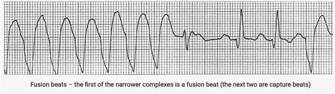
 Very broad complexes (> 160 ms) Capture beats Fusion beats Positive or negative")
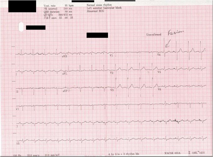
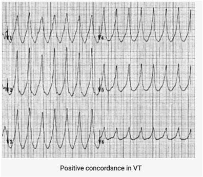
45 HIS DEBS H ypoxia I schemia S ympathomimetic disturbances D rugs E lectrolytes B rady S tretch OAAPN SWM VT vs. SVT with aberrancy IT is more likely VT if: Absence of typical RBBB or LBBB Extreme axis deviation (northwest axis) Very broad complexes (> 160 ms) Capture beats Fusion beats Positive or negative concordance throughout chest leads RSR complexes with a taller left rabbit ear. This is the most specific finding in favor of VT OAAPN SWM Capture Beats OAAPN SWM
46 OAAPN SWM OAAPN SWM OAAPN SWM

47 OAAPN SWM OAAPN SWM Northwest Axis OAAPN SWM

48 Indeterminate Axis or Northwest Axis OAAPN SWM SVT or AV nodal re entry tachycardia (AVNRT) Classified based on site of origin (atria or AV node) or regularity (regular or irregular) QRS width not helpful and influenced by preexisting BBB, Rate related aberrant conduction or accessary pathways OAAPN SWM Classification of SVT by site of Origin Atrial Atrioventricular Regular ST Atrial Tach Atrial Flutter Inappropriate ST SN re entrant tach AV re entry tach (AVRT) AV nodal re entry Tach (AVNRT) Automatic Junctional tachycardia Irregular Atrial Fibrillation Atrial Flutter with variable block Multifocal atrial Tach OAAPN SWM

49 AVNRT Most common cause of palpitations in pts with structurally normal hearts Occurs spontaneously or upon provocation (caffeine, ETOH, Beta agonists, sympathomimetics (amphetamines) More common in women and may occur in young healthy patients Sudden onset of rapid, regular palpitations SOB Pts with CAD may c/o angina Tachy rate bpm Generally well tolerated May cease spontaneously and abruptly OAAPN SWM Typical ECG findings Regular tachy bpm QRS complexes usually narrow (< 120 msec) unless pre existing BBB ST segment depression may be seen without CAD QRS alternans P waves if visible exhibit retrograde conduction with P wave inversion in leads II, III, avf P waves may be buried in the QRS OAAPN SWM Slow Fast AVNRT OAAPN SWM


50 Typical AVNRT Narrow complex Tachycardia No visible P waves There are pseudo R waves in V1 2 OAAPN SWM OAAPN SWM Fast Slow AVNRT Narrow complex Tachycardia Retrograde P waves are visible after each QRS complex OAAPN SWM
51 Pre Excitation & Accessory Pathways Activation of the ventricles due to impulse bypassing the AV node via an accessary pathway Abnormal conduction pathways Impulses conduct either antergrade towards the ventricle or retrograde, away or in both directions Majority conduct in both directions Reentry circuit involving accessary pathways termed Atrioventricular reentry tachycardias (AVRT) OAAPN SWM Wolf Parkinson White (WPW) PR interval < 120 ms Delta wave: slurring slow rise of initial portion of the QRS QRS prolongation > 110 ms ST segment and T wave discordant changes Pseudo infraction pattern can be seen in up to 70% of patients (pseudo q waves, or prominent R wave in V1 V3 mimicking posterior infarction) OAAPN SWM OAAPN SWM

52 Other Pre Excitation Syndromes Lown Ganong Levine (LGL) syndrome Accessary pathway composed of James Fibres ECG PR interval < 120 ms Normal QRS morphology The term should not be used in the absence of paroxysmal tachycardia Existence is disputed and may not exist OAAPN SWM Delta wave Sinus rhythm with a very short PR interval Broad QRS with slurred upstroke (delta wave) Dominant R wave V1 Tall R wave and inverted T wave in V1 3 mimicking RVH OAAPN SWM 2018 Negative Delta wave in avl (pseudo infarction pattern) 26 Common causes of QT Prolongation Drugs Type IA, III Antiarrythmic Tricyclic antidepressants Phenothiazines Electrolyte disturbances Hypokalelmia Hypomagnesemia Hypocalcaemia CNS disturbances Stroke ICB or Brainstem bleed Coma OAAPN SWM


53 OAAPN SWM OAAPN SWM Common causes of ST Depression Ischemia Strain Digitalis effect Hypokalemia/hypomagnesemia Rate related changes Any combination of the above OAAPN SWM
54 Common causes of Tall R wave in V1 WPW RBBB RVH Posterior MI Normal variant OAAPN SWM Common causes of Nonspecific ST-T wave Abnormalities Ischemia LVH Cardiomyopathy MVP Drug effect Lyte abnormalities CNS disorder Hyperventilation Severe medical illness Severe emotional stress Exercise Hypoxemia Acidosis Temp extremes Other causes OAAPN SWM Other Cardiac Conditions Many conditions cause changes to the ECG Electrolyte abnormality Ischemia Infarction Inflammation Medications OAAPN SWM


55 ECG Changes in Pericarditis T wave initially upright and elevated but then during recovery phase it inverts ST segment elevated and usually flat or concave OAAPN SWM Pericardial Effusion Can occur with pericarditis Can cause lowvoltage QRS complexes in all leads and electrical alternans OAAPN SWM Electrical Alternans QRS complexes change in height with each successive beat OAAPN SWM
56 Pulmonary Embolism Acute blockage of one of the pulmonary arteries Leads to obstruction of blood flow to the lung segment supplied by the artery Produces large S wave in lead I, deep Q wave in lead III, inverted T wave in lead III Called the S1 Q3 T3 pattern OAAPN SWM A OAAPN SWM S1 Q3 T3 Sinus Tach RBBB T wave inversions in right precordial leads OAAPN (V1 3) SWM as 2018 well Lead III

57 Pulmonary Embolism OAAPN SWM Electrolyte Imbalances Increases or decreases in potassium and calcium serum levels can have a profound effect on the ECG OAAPN SWM Hyperkalemia Key characteristics include: T wave peaking Flattened P waves 1 st -degree AV heart block Widened QRS complexes Deepened S waves Merging of S and T waves OAAPN SWM

58 OAAPN SWM Hyperkalemia OAAPN SWM Suspect Hyperkalemia New bradycardia New AV block especially with CKD or ESRD taking ACE I or potassium sparing meds OAAPN SWM


59 Hypokalemia Key ECG characteristics include: ST segment depression Flattening of the T wave Appearance of U waves OAAPN SWM OAAPN SWM OAAPN SWM


60 OAAPN SWM Hypocalcemia QT interval slightly prolonged OAAPN SWM OAAPN SWM
61 OAAPN SWM Systematic approach Compare with old ECG Look at Rate Look at Rhythm Look at Axis Look at Hypertrophy Look at I s Intervals, ischemia, injury, infarct OAAPN SWM
Section V. Objectives
 Section V Landscape of an MI Objectives At the conclusion of this presentation the participant will be able to Outline a systematic approach to 12 lead ECG interpretation Demonstrate the process for determining
12 Lead EKG Interpretation. Disclosures
 12 Lead EKG Interpretation Louann B. Bailey, DNP, APRN, FAANP ACNP BC I have no disclosures Disclosures 2 1 Objectives At the conclusion of this presentation the participant will be able to Outline a systematic
Conduction Problems / Arrhythmias. Conduction
 Conduction Problems / Arrhythmias Conduction Wolf-Parkinson White Syndrome (WPW) and Lown-Ganong-Levine (LGL): Atrial impulses bypass the AV node through an accessory pathway or bypass tract (bundle of
12-Lead ECG Interpretation. Kathy Kuznar, RN, ANP
 12-Lead ECG Interpretation Kathy Kuznar, RN, ANP The 12-Lead ECG Objectives Identify the normal morphology and features of the 12- lead ECG. Perform systematic analysis of the 12-lead ECG. Recognize abnormalities
12 Lead ECG. Presented by Rebecca Sevigny BSN, RN Professional Practice & Development Dept.
 12 Lead ECG Presented by Rebecca Sevigny BSN, RN Professional Practice & Development Dept. Two Main Coronary Arteries RCA LCA which branches into Left Anterior Descending Circumflex Artery Two Main Coronary
REtrive. REpeat. RElearn Design by. Test-Enhanced Learning based ECG practice E-book
 Test-Enhanced Learning Test-Enhanced Learning Test-Enhanced Learning Test-Enhanced Learning based ECG practice E-book REtrive REpeat RElearn Design by S I T T I N U N T H A N G J U I P E E R I Y A W A
2017 EKG Workshop Advanced. Family Medicine Review Course Lou Mancano, MD, FAAFP Reading Health System Family and Community Medicine Reading, PA
 2017 EKG Workshop Advanced Family Medicine Review Course Lou Mancano, MD, FAAFP Reading Health System Family and Community Medicine Reading, PA Part II - Objective Describe a useful approach to interpreting
Pennsylvania Academy of Family Physicians Foundation & UPMC 43rd Refresher Course in Family Medicine CME Conference March 10-13, 2016
 Pennsylvania Academy of Family Physicians Foundation & UPMC 43rd Refresher Course in Family Medicine CME Conference March 10-13, 2016 Disclosures: EKG Workshop Louis Mancano, MD Speaker has no disclosures
Huseng Vefali MD St. Luke s University Health Network Department of Cardiology
 Huseng Vefali MD St. Luke s University Health Network Department of Cardiology Learning Objectives Establish Consistent Approach to Interpreting ECGs Review Essential Cases for Paramedics and first responders
ECG Basics Sonia Samtani 7/2017 UCI Resident Lecture Series
 ECG Basics Sonia Samtani 7/2017 UCI Resident Lecture Series Agenda I. Introduction II.The Conduction System III.ECG Basics IV.Cardiac Emergencies V.Summary The Conduction System Lead Placement avf Precordial
ECG Interpretation Made Easy
 ECG Interpretation Made Easy Dr. A Tageldien Abdellah, MSc MD EBSC Lecturer of Cardiology- Hull University Hull York Medical School 2007-2008 ECG Interpretation Made Easy Synopsis Benefits Objectives Process
Myocardial Infarction. Reading Assignment (p66-78 in Outline )
") Myocardial Infarction Reading Assignment (p66-78 in Outline ) Objectives 1. Why do ST segments go up or down in ischemia? 2. STEMI locations and culprit vessels 3. Why 15-lead ECGs? 4. What s up with avr?
Myocardial Infarction Reading Assignment (p66-78 in Outline ) Objectives 1. Why do ST segments go up or down in ischemia? 2. STEMI locations and culprit vessels 3. Why 15-lead ECGs? 4. What s up with avr?
Please check your answers with correct statements in answer pages after the ECG cases.
 ECG Cases ECG Case 1 Springer International Publishing AG, part of Springer Nature 2018 S. Okutucu, A. Oto, Interpreting ECGs in Clinical Practice, In Clinical Practice, https://doi.org/10.1007/978-3-319-90557-0
ECG CONVENTIONS AND INTERVALS
 1 ECG Waveforms and Intervals ECG waveforms labeled alphabetically P wave== represents atrial depolarization QRS complex=ventricular depolarization ST-T-U complex (ST segment, T wave, and U wave)== V repolarization.
, David Stultz, MD.
 http://www.dilbert.com EKG Rounds Handouts available at http://www.drstultz.com January 5, 2004 David Stultz, MD Cardiology Fellow, PGY 4 Overview of Topics How to read an EKG Normal EKG Determination
Electrical System Overview Electrocardiograms Action Potentials 12-Lead Positioning Values To Memorize Calculating Rates
 Electrocardiograms Electrical System Overview James Lamberg 2/ 74 Action Potentials 12-Lead Positioning 3/ 74 4/ 74 Values To Memorize Inherent Rates SA: 60 to 100 AV: 40 to 60 Ventricles: 20 to 40 Normal
Acute Coronary Syndromes Unstable Angina Non ST segment Elevation MI (NSTEMI) ST segment Elevation MI (STEMI)
 ST segment Elevation MI (STEMI)") Leanna R. Miller, RN, MN, CCRN-CSC, PCCN-CMC, CEN, CNRN, CMSRN, NP Education Specialist LRM Consulting Nashville, TN Objectives Evaluate common abnormalities that mimic myocardial infarction. Identify
Leanna R. Miller, RN, MN, CCRN-CSC, PCCN-CMC, CEN, CNRN, CMSRN, NP Education Specialist LRM Consulting Nashville, TN Objectives Evaluate common abnormalities that mimic myocardial infarction. Identify
Chapter 2 Practical Approach
 Chapter 2 Practical Approach There are beginners in electrocardiogram (ECG) analysis who are fascinated by a special pattern (e.g., a bundle-branch block or a striking Q wave) and thereby overlook other
The Electrocardiogram part II. Dr. Adelina Vlad, MD PhD
 The Electrocardiogram part II Dr. Adelina Vlad, MD PhD Basic Interpretation of the ECG 1) Evaluate calibration 2) Calculate rate 3) Determine rhythm 4) Determine QRS axis 5) Measure intervals 6) Analyze
ECG ABNORMALITIES D R. T AM A R A AL Q U D AH
 ECG ABNORMALITIES D R. T AM A R A AL Q U D AH When we interpret an ECG we compare it instantaneously with the normal ECG and normal variants stored in our memory; these memories are stored visually in
Bundle Branch & Fascicular Blocks. Reading Assignment (p53-58 in Outline )
") Bundle Branch & Fascicular Blocks Reading Assignment (p53-58 in Outline ) Objectives 1. QRS analysis of Right and Left BBB 2. Uncomplicated vs complicated BBB 3. Diagnosis of RBBB with LAFB and LPFB 4.
Bundle Branch & Fascicular Blocks Reading Assignment (p53-58 in Outline ) Objectives 1. QRS analysis of Right and Left BBB 2. Uncomplicated vs complicated BBB 3. Diagnosis of RBBB with LAFB and LPFB 4.
Supraventricular Arrhythmias. Reading Assignment. Chapter 5 (p17-30)
") Supraventricular Arrhythmias Reading Assignment Chapter 5 (p17-30) The Supraventricular Rhythms In Our Lives Site of Origin Single Events Slow Rates Intermediate Rates Fast Rates (>100 bpm) Sinus Sinus
Supraventricular Arrhythmias Reading Assignment Chapter 5 (p17-30) The Supraventricular Rhythms In Our Lives Site of Origin Single Events Slow Rates Intermediate Rates Fast Rates (>100 bpm) Sinus Sinus
General Introduction to ECG. Reading Assignment (p2-16 in PDF Outline )
") General Introduction to ECG Reading Assignment (p2-16 in PDF Outline ) Objectives 1. Practice the 5-step Method 2. Differential Diagnosis: R & L axis deviation 3. Differential Diagnosis: Poor R-wave progression
General Introduction to ECG Reading Assignment (p2-16 in PDF Outline ) Objectives 1. Practice the 5-step Method 2. Differential Diagnosis: R & L axis deviation 3. Differential Diagnosis: Poor R-wave progression
Basic electrocardiography reading. R3 lee wei-chieh
 Basic electrocardiography reading R3 lee wei-chieh The Normal Conduction System Lead Placement avf Limb Leads Precordial Leads Interpretation Rate Rhythm Interval Axis Chamber abnormality QRST change What
ECG Practice Strips Discussion part 1:
 ECG Practice Strips Discussion part 1: The first 20 strips are for teaching various abnormalities of the morphology of the waves of the ECG. Strips 21 and following are for teaching some abnormalities
ECG Cases and Questions. Ashish Sadhu, MD, FHRS, FACC Electrophysiology/Cardiology
 ECG Cases and Questions Ashish Sadhu, MD, FHRS, FACC Electrophysiology/Cardiology 32 yo female Life Insurance Physical 56 yo male with chest pain Terminology Injury ST elevation Ischemia T wave inversion
ECGs: Everything a finalist needs to know. Dr Amy Coulden As part of the Simply Finals series
 ECGs: Everything a finalist needs to know Dr Amy Coulden As part of the Simply Finals series Aims and objectives To be able to interpret basic ECG abnormalities To be able to recognise commonly tested
Blocks & Dissociations. Reading Assignment (p47-52 in Outline )
") Blocks & Dissociations Reading Assignment (p47-52 in Outline ) Objectives Who are Wenckebach and Mobitz? Review SA and AV Blocks AV Dissociations: learning who s the boss and why 2 nd degree SA Block:
Blocks & Dissociations Reading Assignment (p47-52 in Outline ) Objectives Who are Wenckebach and Mobitz? Review SA and AV Blocks AV Dissociations: learning who s the boss and why 2 nd degree SA Block:
Making Sense of Those Little Lines Advanced ECG Interpretation
 Making Sense of Those Little Lines Advanced ECG Interpretation Cara Solodky-Hardy, ND, ANP-BC, AACC MD24 House Call Cardiology My patients www.ecglibrary.com Image Sources The Alan E. Lindsay Ecg Learning
Ekg pra pr c a tice D.HAMMOUDI.MD
 Ekg practice D.HAMMOUDI.MD Anatomy Revisited RCA (Right Coronary Artery) Right ventricle Inferior wall of LV Posterior wall of LV (75%) SA Node (60%) AV Node (>80%) LCA (Left Coronary Artery) Septal wall
402 Index. B β-blockers, 4, 5 Bradyarrhythmias, 76 77
 Index A Acquired immunodeficiency syndrome (AIDS), 126, 163 Action potentials, 1, 5, 27 Acute coronary syndromes, 123t, 129 Adenosine, intravenous, 277 Alcohol abuse, as T wave inversion cause, 199 Aneurysm,
Electrocardiography for Healthcare Professionals. Chapter 14 Basic 12-Lead ECG Interpretation
 Electrocardiography for Healthcare Professionals Chapter 14 Basic 12-Lead ECG Interpretation 2012 The Companies, Inc. All rights reserved. Learning Outcomes 14.1 Discuss the anatomic views seen on a 12-lead
ECGs and Arrhythmias: Family Medicine Board Review 2009
 Rate Rhythm Intervals Hypertrophy ECGs and Arrhythmias: Family Medicine Board Review 2009 Axis Jess (Fogler) Waldura, MD University of California, San Francisco walduraj@nccc.ucsf.edu Ischemia Overview
10 ECGs No Practitioner Can Afford to Miss. Objectives
 10 ECGs No Practitioner Can Afford to Miss Mary L. Dohrmann, MD Professor of Clinical Medicine Division of Cardiovascular Medicine University of Missouri School of Medicine No disclosures Objectives 1.
Family Medicine for English language students of Medical University of Lodz ECG. Jakub Dorożyński
 Family Medicine for English language students of Medical University of Lodz ECG Jakub Dorożyński Parts of an ECG The standard ECG has 12 leads: six of them are considered limb leads because they are placed
UNDERSTANDING YOUR ECG: A REVIEW
 UNDERSTANDING YOUR ECG: A REVIEW Health professionals use the electrocardiograph (ECG) rhythm strip to systematically analyse the cardiac rhythm. Before the systematic process of ECG analysis is described
Relax and Learn at the FARM 2012: Session 8: 12 Lead ECG 401: ECG Variants
 Relax and Learn at the FARM 2012: Session 8: 12 Lead ECG 401: ECG Variants A Ship in the Harbor is Safe But that is not what ships are built for. Karen Marzlin DNP, RN, CCNS, CCRN-CMC, CHFN Cardiovascular
12 Lead EKG. The Basics
 12 Lead EKG The Basics Objectives Demonstrate proper 12 EKG lead placement Determine electrical axis Identify ST and T wave changes as they relate to myocardial ischemia Describe possible complications
Acute Coronary Syndromes. Disclosures
 Acute Coronary Syndromes Disclosures I work for Virginia Garcia Memorial Health Center, Beaverton, OR. Jon Tardiff, BS, PA-C OHSU Clinical Assistant Professor And I am a medical editor for Jones & Bartlett
SIMPLY ECGs. Dr William Dooley
 SIMPLY ECGs Dr William Dooley Content Basic ECG interpretation pattern Some common (examined) abnormalities Presenting ECGs in context Setting up an ECG Setting up an ECG 1 V1-4 th Right intercostal space
Electrocardiography Abnormalities (Arrhythmias) 7. Faisal I. Mohammed, MD, PhD
 7. Faisal I. Mohammed, MD, PhD") Electrocardiography Abnormalities (Arrhythmias) 7 Faisal I. Mohammed, MD, PhD 1 Causes of Cardiac Arrythmias Abnormal rhythmicity of the pacemaker Shift of pacemaker from sinus node Blocks at different
Electrocardiography Abnormalities (Arrhythmias) 7 Faisal I. Mohammed, MD, PhD 1 Causes of Cardiac Arrythmias Abnormal rhythmicity of the pacemaker Shift of pacemaker from sinus node Blocks at different
Cardiology Flash Cards
 Cardiology Flash Cards EKG in a nut shell www.brain101.info Conduction System www.brain101.info 2 Analyzing EKG Step by step Steps in Analyzing ECG'S 1. Rhythm: - Regular _ Sinus, Junctional or Ventricular.
Office ECG Interpretation
 Office ECG Interpretation Jason Evanchan, DO Assistant Professor of Medicine Division of Cardiovascular Medicine The Ohio State University Wexner Medical Center Outline of topics High risk ischemia T wave
ΔΙΑΤΑΡΑΧΕΣ ΕΝΔΟΚΟΙΛΙΑΚΗΣ ΑΓΩΓΙΜΟΤΗΤΑΣ ΔΗΜΗΤΡΙΟΣ Δ. ΜΑΝΩΛΑΤΟΣ Β ΚΑΡΔΙΟΛΟΓΙΚΗ ΚΛΙΝΙΚΗ ΕΡΓΑΣΤΗΡΙΟ ΗΛΕΚΤΡΟΦΥΣΙΟΛΟΓΙΑΣ Γ.Ν.Α.
 ΔΙΑΤΑΡΑΧΕΣ ΕΝΔΟΚΟΙΛΙΑΚΗΣ ΑΓΩΓΙΜΟΤΗΤΑΣ ΔΗΜΗΤΡΙΟΣ Δ. ΜΑΝΩΛΑΤΟΣ Β ΚΑΡΔΙΟΛΟΓΙΚΗ ΚΛΙΝΙΚΗ ΕΡΓΑΣΤΗΡΙΟ ΗΛΕΚΤΡΟΦΥΣΙΟΛΟΓΙΑΣ Γ.Ν.Α. «ΕΥΑΓΓΕΛΙΣΜΟΣ» Intraventricular conduction delay and Blocks Right Bundle Branch
Return to Basics. ECG Rate and Rhythm. Management of the Hospitalized Patient September 25, 2009
 Management of the Hospitalized Patient September 25, 2009 ECG Refresher and Update 2009 Return to Basics Determine rate and rhythm Determine intervals and axes Define morphology of P-QRS-T-U Compare with
ECGs and Arrhythmias: Family Medicine Board Review 2012
 Overview ECGs and Arrhythmias: Family Medicine Board Review 2012 Jess Waldura, MD University of California, San Francisco walduraj@nccc.ucsf.edu Bundle branch blocks Quick review of ischemia Arrhythmias
ELECTROCARDIOGRAPH. General. Heart Rate. Starship Children s Health Clinical Guideline
 General Heart Rate QRS Axis T Wave Axis PR Interval according to Heart Rate & Age P Wave Duration and Amplitude QRS Duration according to Age QT Interval R & S voltages according to Lead & Age R/S ratio
Appendix D Output Code and Interpretation of Analysis
 Appendix D Output Code and Interpretation of Analysis 8 Arrhythmia Code No. Description 8002 Marked rhythm irregularity 8110 Sinus rhythm 8102 Sinus arrhythmia 8108 Marked sinus arrhythmia 8120 Sinus tachycardia
2017 EKG Workshop Basic. Family Medicine Review Course Lou Mancano, MD, FAAFP Reading Health System Family and Community Medicine Reading, PA
 2017 EKG Workshop Basic Family Medicine Review Course Lou Mancano, MD, FAAFP Reading Health System Family and Community Medicine Reading, PA Part I - Objectives Discuss a systematic approach to EKG interpretation
3/4/2018. March Martina Frost, PA C Desert Cardiology. Electricity moving towards/away from electrode create downward/upward directions of waves
 March 2018 Martina Frost, PA C Desert Cardiology Electricity moving towards/away from electrode create downward/upward directions of waves Frontal view Limb leads: I, II, III, avl, avf, (avr) Horizontal
SIMPLY ECGs. Dr William Dooley
 SIMPLY ECGs Dr William Dooley 1 No anatomy just interpretation 2 Setting up an ECG 3 Setting up an ECG 1 V1-4 th Right intercostal space at sternal border 2 V2-4 th Left intercostal space at sternal border
ECG Interpretation Cat Williams, DVM DACVIM (Cardiology)
") ECG Interpretation Cat Williams, DVM DACVIM (Cardiology) Providing the best quality care and service for the patient, the client, and the referring veterinarian. GOAL: Reduce Anxiety about ECGs Back to
ECG Interpretation Cat Williams, DVM DACVIM (Cardiology) Providing the best quality care and service for the patient, the client, and the referring veterinarian. GOAL: Reduce Anxiety about ECGs Back to
12 Lead ECG Skills: Building Confidence for Clinical Practice. Presented By: Cynthia Webner, BSN, RN, CCRN-CMC. Karen Marzlin, BSN, RN,CCRN-CMC
 12 Lead ECG Skills: Building Confidence for Clinical Practice NTI 2009 Preconference Session 803 Presented By: Karen Marzlin, BSN, RN,CCRN-CMC 1 12 Lead ECG Fundamentals: The Starting Place for Linking
Return to Basics. Normal Intervals & Axes. ECG Rate and Rhythm
 Return to Basics Management of the Hospitalized Patient October 15, 2010 ECG Refresher and Update 2010 Determine rate and rhythm Determine intervals and axes Define morphology of P-QRS-T-U Compare with
Arrhythmic Complications of MI. Teferi Mitiku, MD Assistant Clinical Professor of Medicine University of California Irvine
 Arrhythmic Complications of MI Teferi Mitiku, MD Assistant Clinical Professor of Medicine University of California Irvine Objectives Brief overview -Pathophysiology of Arrhythmia ECG review of typical
ECG Interpretation. Best to have a system to methodically evaluate ECG (from Dubin) * Rate * Rhythm * Axis * Intervals * Hypertrophy * Infarction
 * Rate * Rhythm * Axis * Intervals * Hypertrophy * Infarction") ECG to save Babies ECG Interpretation Best to have a system to methodically evaluate ECG (from Dubin) * Rate * Rhythm * Axis * Intervals * Hypertrophy * Infarction Electrical Activity in the heart 5 events
ECG to save Babies ECG Interpretation Best to have a system to methodically evaluate ECG (from Dubin) * Rate * Rhythm * Axis * Intervals * Hypertrophy * Infarction Electrical Activity in the heart 5 events
Step by step approach to EKG rhythm interpretation:
 Sinus Rhythms Normal sinus arrhythmia Small, slow variation of the R-R interval i.e. variation of the normal sinus heart rate with respiration, etc. Sinus Tachycardia Defined as sinus rhythm with a rate
Paediatric ECG Interpretation
 Paediatric ECG Interpretation Dr Sanj Fernando (thanks to http://lifeinthefastlane.com/ecg-library/paediatric-ecginterpretation/) 3 yo boy complaining of abdominal pain and chest pain Child ECG vs Adult
If the P wave > 0.12 sec( 3 mm) usually in any lead. Notched P wave usually in lead I,aVl may be lead II Negative terminal portion of P wave in V1, 1
 usually in any lead. Notched P wave usually in lead I,aVl may be lead II Negative terminal portion of P wave in V1, 1") If the P wave > 0.12 sec( 3 mm) usually in any lead. Notched P wave usually in lead I,aVl may be lead II Negative terminal portion of P wave in V1, 1 mm depth and 3 mm width( most specific) Since Mitral
If the P wave > 0.12 sec( 3 mm) usually in any lead. Notched P wave usually in lead I,aVl may be lead II Negative terminal portion of P wave in V1, 1 mm depth and 3 mm width( most specific) Since Mitral
Study methodology for screening candidates to athletes risk
 1. Periodical Evaluations: each 2 years. Study methodology for screening candidates to athletes risk 2. Personal history: Personal history of murmur in childhood; dizziness, syncope, palpitations, intolerance
Return to Basics. ECG Rate and Rhythm. Management of the Hospitalized Patient October 4, 2007
 Management of the Hospitalized Patient October 4, 2007 ECG Refresher for the Hospitalists Return to Basics Determine rate and rhythm Determine intervals and axes Define morphology of P-QRS-T-U Compare
All About STEMIs. Presented By: Brittney Urvand, RN, BSN, CCCC. Essentia Health Fargo Cardiovascular Program Manager.
 All About STEMIs Presented By: Brittney Urvand, RN, BSN, CCCC Essentia Health Fargo Cardiovascular Program Manager Updated 10/2/2018 None Disclosures Objectives Identify signs and symptoms of a heart attack
Cardiac Arrhythmia How to approach นพ.พ น จ แกวส วรรณะ หน วยโรคห วใจและหลอดเล อด
 Cardiac Arrhythmia How to approach นพ.พ น จ แกวส วรรณะ หน วยโรคห วใจและหลอดเล อด EKG paper is a grid where time is measured along the horizontal axis. Each small square is 1 mm in length and represents
The Efficient and Smart Methods for Diagnosis of SVT 대구파티마병원순환기내과정병천
 The Efficient and Smart Methods for Diagnosis of SVT 대구파티마병원순환기내과정병천 Differentiation Supraventricular Origin from Ventricular Origin on ECG. QRS-Complex Width. 1. Narrow QRS-Complex Tachycardia (
How To Think About Rhythms and Conduction
 How To Think About Rhythms and Conduction Frank Yanowitz, MD Professor of Medicine (Cardiology) University of Utah School of Medicine Medical Director, IHC ECG Services Intermountain Healthcare http://ecg.utah.edu
2) Heart Arrhythmias 2 - Dr. Abdullah Sharif
 Heart Arrhythmias 2 - Dr. Abdullah Sharif") 2) Heart Arrhythmias 2 - Dr. Abdullah Sharif Rhythms from the Sinus Node Sinus Tachycardia: HR > 100 b/m Causes: o Withdrawal of vagal tone & Sympathetic stimulation (exercise, fight or flight) o Fever
2) Heart Arrhythmias 2 - Dr. Abdullah Sharif Rhythms from the Sinus Node Sinus Tachycardia: HR > 100 b/m Causes: o Withdrawal of vagal tone & Sympathetic stimulation (exercise, fight or flight) o Fever
By the end of this lecture, you will be able to: Understand the 12 lead ECG in relation to the coronary circulation and myocardium Perform an ECG
 By the end of this lecture, you will be able to: Understand the 12 lead ECG in relation to the coronary circulation and myocardium Perform an ECG recording Identify the ECG changes that occur in the presence
Reading Assignment (p1-91 in Outline ) Objectives What s in an ECG?
 Objectives What s in an ECG?") Reading Assignment (p1-91 in Outline ) Objectives What s in an ECG? The 5-Step Method ECG #: Mearurements: Rhythm (s): Conduction: Waveform: Interpretation: A= V= PR= QRS= QT= Axis= 1. Compute the 5 basic
Reading Assignment (p1-91 in Outline ) Objectives What s in an ECG? The 5-Step Method ECG #: Mearurements: Rhythm (s): Conduction: Waveform: Interpretation: A= V= PR= QRS= QT= Axis= 1. Compute the 5 basic
also aid the clinician in recognizing both the obvious and subtle abnormalities that may help guide therapy.
 Karen Lieberman, MS, CRNP f the many diagnostic tools used to screen for and evaluate cardiac abnormalities, the 12-lead electrocardiogram (ECG) is among the most basic. This inexpensive and noninvasive
THE ELECTROCARDIOGRAM A UBIQUITOUS AND COST-EFFECTIVE DIAGNOSTIC TOOL FOR THE FAMILY MEDICINE REFRESHER COURSE MARCH 8, 2019
 THE ELECTROCARDIOGRAM A UBIQUITOUS AND COST-EFFECTIVE DIAGNOSTIC TOOL FOR THE FAMILY MEDICINE REFRESHER COURSE MARCH 8, 2019 Major Clinical Disorders Pulmonary Embolism 69 y/o woman with dyspnea and an
Ben Taylor, PhD, PA-C
 Ben Taylor, PhD, PA-C The patient is a 23-year-old white male with a history of polysubstance abuse who was found unresponsive, last seen the day before. Classic signs of systemic hypothermia with prominent
Relax and Learn At the Farm 2012
 Relax and Learn At the Farm 2012 Session 2: 12 Lead ECG Fundamentals 101 Cynthia Webner DNP, RN, CCNS, CCRN-CMC, CHFN Though for Today Mastery is not something that strikes in an instant, like a thunderbolt,
PATIENT S NAME, DATE/TIME,
 ECG and Arrhythmias Dec, 1 st 2014 Doctor Mohammad Jarrah References: - Lecture and Slides - ECG Made Easy - Davidson Principles of Medicine - First Aid Cases for the USMLE Step 1 - Mini-OSCE Archive ECG
Left posterior hemiblock (LPH)/
/") ECG OF THE MONTH Left Postero-inferior Depolarization Delay Keywords Electrocardiography Intraventricular conduction delay, Inferoposterior hemiblock, Left posterior fascicular block, Left posterior hemiblock
ECG OF THE MONTH Left Postero-inferior Depolarization Delay Keywords Electrocardiography Intraventricular conduction delay, Inferoposterior hemiblock, Left posterior fascicular block, Left posterior hemiblock
Understanding the 12-lead ECG, part II
 Bundle-branch blocks Understanding the 12-lead ECG, part II Most common electrocardiogram (ECG) abnormality Appears as a wider than normal S complex Occurs when one of the two bundle branches can t conduct
FLB s What Are Those Funny-Looking Beats?
 FLB s What Are Those Funny-Looking Beats? Reading Assignment (pages 27-45 in Outline ) The 5-Step Method ECG #: Mearurements: Rhythm (s): Conduction: Waveform: Interpretation: A= V= PR= QRS= QT= Axis=
PAEDIATRIC ECG Dimosthenis Avramidis, MD.
 PAEDIATRIC ECG Dimosthenis Avramidis, MD. Consultant Mitera Children s Hospital Athens Greece S. Associate 1st Cardiology Dpt Evangelismos Hospital Athens Greece 5 y/o with sinus tach Background ECG changes
HR: 50 bpm (Sinus) PR: 280 ms QRS: 120 ms QT: 490 ms Axis: -70. Sinus bradycardia with one ventricular escape (*)
 PR: 280 ms QRS: 120 ms QT: 490 ms Axis: -70. Sinus bradycardia with one ventricular escape (*)") 1? HR: 50 bpm (Sinus) PR: 280 ms QRS: 120 ms QT: 490 ms Axis: -70 1 Sinus P waves? 2 sinus cycles The pause (2 sinus cycles) suggests that the sinus fired (?) but did not conduct to the atria (i.e., missing
1? HR: 50 bpm (Sinus) PR: 280 ms QRS: 120 ms QT: 490 ms Axis: -70 1 Sinus P waves? 2 sinus cycles The pause (2 sinus cycles) suggests that the sinus fired (?) but did not conduct to the atria (i.e., missing
Miscellaneous Stuff Keep reading the Outline
 Miscellaneous Stuff Keep reading the Outline Welcome to the 5-Step Method ECG #: Mearurements: Rhythm (s): Conduction: Waveform: Interpretation: A= V= PR= QRS= QT= Axis= 1. Compute the 5 basic measurements:
Abnormalities Caused by Left Bundle Branch Block
 Marquette University e-publications@marquette Physician Assistant Studies Faculty Research and Publications Physician Assistant Studies, Department 12-17-2010 Abnormalities Caused by Left Bundle Branch
12 Lead ECG Interpretation
 12 Lead ECG Interpretation Julie Zimmerman, MSN, RN, CNS, CCRN Significant increase in mortality for every 15 minutes of delay! N Engl J Med 2007;357:1631-1638 Who should get a 12-lead ECG? Also include
Introduction to ECG Gary Martin, M.D.
 Brief review of basic concepts Introduction to ECG Gary Martin, M.D. The electrical activity of the heart is caused by a sequence of rapid ionic movements across cell membranes resulting first in depolarization
MICS OF MYOCARDIAL ISCHEMIA AND INFARCTION REVISED FOR LAS VEGAS
 ECG MIMICS OF MYOCARDIAL ISCHEMIA AND INFARCTION 102.06.05 Tzong-Luen Wang MD, PhD, JM, FESC, FACC Professor. Medical School, Fu-Jen Catholic University Chief, Emergency Department, Shin-Kong Wu Ho-Su
The Fundamentals of 12 Lead EKG. ECG Recording. J Point. Reviewing the Cardiac Conductive System. Dr. E. Joe Sasin, MD Rusty Powers, NRP
 The Fundamentals of 12 Lead EKG Dr. E. Joe Sasin, MD Rusty Powers, NRP SA Node Intranodal Pathways AV Junction AV Fibers Bundle of His Septum Bundle Branches Purkinje System Reviewing the Cardiac Conductive
Introduction to Electrocardiography
 Introduction to Electrocardiography Class Objectives: Introduction to ECG monitoring Discuss principles of interpretation Identify the components and measurements of the ECG ECG analysis ECG Monitoring
Appendix. Table 1: Causes for abnormal axis deviation Left axis deviation
 Appendix Table 1: Causes for abnormal axis deviation Left axis deviation Normal variant (2 5%) Left anterior fascicular block Left ventricular hypertrophy Inferior wall myocardial infarction Primum atrial
Rhythm ECG Characteristics Example. Normal Sinus Rhythm (NSR)
") Normal Sinus Rhythm (NSR) Rate: 60-100 per minute Rhythm: R- R = P waves: Upright, similar P-R: 0.12-0.20 second & consistent P:qRs: 1P:1qRs Sinus Tachycardia Exercise Hypovolemia Medications Fever Substances
Normal Sinus Rhythm (NSR) Rate: 60-100 per minute Rhythm: R- R = P waves: Upright, similar P-R: 0.12-0.20 second & consistent P:qRs: 1P:1qRs Sinus Tachycardia Exercise Hypovolemia Medications Fever Substances
ECG Interpretation. Introduction to Cardiac Telemetry. Michael Peters, RN, CCRN, CFRN CALSTAR Air Medical Services
 ECG Interpretation Introduction to Cardiac Telemetry Michael Peters, RN, CCRN, CFRN CALSTAR Air Medical Services Disclosures Nothing to disclose Objectives Describe the electrical conduction pathway in
Dr. Schroeder has no financial relationships to disclose
 Valerie A Schroeder MD MS Assistant Professor University of Kansas Medical Center READING THE WAVES- THE HEART S ELECTRICAL MESSAGE FINANCIAL DISCLOSURE Dr. Schroeder has no financial relationships to
Preface: Wang s Viewpoints
 AHA/ACCF/HRS Recommendations for the Standardization and Interpretation of the Electrocardiogram: Part IV, Ischemia and Infarction Presented by: WANG, TZONG LUEN, MD, PhD, JM, FACC, FESC, FCAPSC Professor,
12 LEAD EKG BASICS. By: Steven Jones, NREMT P CLEMC
 12 LEAD EKG BASICS By: Steven Jones, NREMT P CLEMC ECG Review Waves and Intervals P wave: the sequential activation (depolarization) of the right and left atria QRS complex: right and left ventricular
You Don t Want to Miss This One! Focus on can t miss EKG tracings
 You Don t Want to Miss This One! Focus on can t miss EKG tracings Renaissance St. Louis Grand Hotel Convention Center October 23, 2014 David K. Tan, M.D., EMT-T, FAAEM EMS Section Chief, Division of Emergency
December 2018 Tracings
 Tracings Tracing 1 Tracing 4 Tracing 1 Answer Tracing 4 Answer Tracing 2 Tracing 5 Tracing 2 Answer Tracing 5 Answer Tracing 3 Tracing 6 Tracing 3 Answer Tracing 6 Answer Questions? Contact Dr. Nelson
ECG Interactive Session
 SA HEART 2018 PRE-CONGRESS WORKSHOP 4 OCTOBER 2018 ECG Interactive Session Ashley Chin Cardiologist/Electrophysiologist University of Cape Town Groote Schuur Hospital ECG 1 Which ONE of the following is
12 Lead ECG Interpretation: The Basics and Beyond
 12 Lead ECG Interpretation: The Basics and Beyond Cindy Weston, DNP, RN, CCRN, CNS-CC, FNP-BC Assistant Professor Texas A&M University College of Nursing cweston@tamhsc.edu Objectives Review the basics
Masqueraders of STEMI
 Masqueraders of STEMI Steven M. Costa, M.D. Assistant Professor Department of Medicine Division of Cardiology Scott & White Memorial Hospital and Clinic Texas A&M University Health Science Center Disclosures
ECG pre-reading manual. Created for the North West Regional EMET training program
 ECG pre-reading manual Created for the North West Regional EMET training program Author:- Dr Juan Carlos Ascencio-Lane juan.ascencio-lane@ths.tas.gov.au 1 Disclaimer This handbook has been created for
A Review of Cardiac Pathophysiology and EKG. Jamie Dyson PT, DPT Kathy Swanick PT, DPT, OCS
 A Review of Cardiac Pathophysiology and EKG Jamie Dyson PT, DPT Kathy Swanick PT, DPT, OCS Cardiac Pathophysiology Coronary Artery Disease Congestive Heart Failure Valvular Heart Disease Athletic Heart
Electrocardiogram ECG. Hilal Al Saffar FRCP FACC College of medicine,baghdad University
 Electrocardiogram ECG Hilal Al Saffar FRCP FACC College of medicine,baghdad University Tuesday 29 October 2013 ECG introduction Wednesday 30 October 2013 Abnormal ECG ( ischemia, chamber hypertrophy, heart
Case 1. Case 2. Case 3
 Case 1 The correct answer is D. Occasionally, the Brugada syndrome can present similar morphologies to A and also change depending on the lead position but in the Brugada pattern the r is wider and ST