NCVH. Ultrasongraphy: State of the Art Vein Forum 2015 A Multidisciplinary Approach to Otptimizing Venous Circulation From Wounds to WOW
|
|
|
- Howard Lewis
- 5 years ago
- Views:
Transcription

1 Ultrasongraphy: State of the Art 2015 NCVH New Cardiovascular Horizons Vein Forum 2015 A Multidisciplinary Approach to Otptimizing Venous Circulation From Wounds to WOW Anil K. Chagarlamudi, M.D. Cardiovascular Institute of the South Houma, LA


2 Disclosures None
3
4 US - Introduction Duplex US has become the test of choice for evaluation of venous insufficiency US is : Safe Non-invasive Cost-effective Reliable
5 US Probe Selection 4-7 MHz transducer is ideal for most veins Superficial veins Higher frequency probe ( MHx) -provide better resolution Deep veins Curvilinear probe (3MHz) better depth of penetration Variable frequency probes Change frequency up or down
6 US - Settings During imaging low flow settings are used Pulse repetition frequency (PRF) set at 1500 Hz or lower Focus is set with the posterior wall to allow better resolution in the field of imaging Time Gain Compensation (TGC) set according to the echogenicity and depth of the relevant tissues Gain set to have a dark background when obtaining velocity waveforms to avoid overestimation Angle of insonation often set at 0 degrees
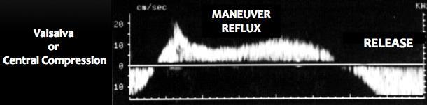
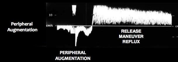
7 US - Maneuvers Distal augmentation Valsalva Active dorsiflexion and plantar flexion Parana maneuver Automatic cuffs Proximal augmentation REFLUX IS NOT STATIC

8 Distal Augmentation Manual compressions over calf or the varicose capacitance bed Squeeze and hold for 0.25 secs and release This allows interpretation of both phases systole and diastole Allows the spectral curve to display change in direction of flow

9 Valsalva Produces higher intra-abdominal pressure Done with forceful contraction of the abdomen Excellent for evaluation of reflux of the terminal and subterminal valves at the SFJ
10 Active dorsiflexion & plantar flexion Technically difficult for the examiner due to patient movement and pulsed doppler aiming Very effective in eliminating false negatives Helpful in patients with severe leg edema or lymphatically compromised lower extremities due to the extrinsic pressure on the vein from the edema

11 Parana maneuver Variation to the active muscular contraction Patient standing Rock forwards or backwards Causes isometric contraction of the calf muscles Also done by shifting weight from one leg to other Mimics muscular changes similar to walking

12 Automatic cuff devices Easy to use Facilitate compression augmentation Pressure from mmhg Rapid, large caliber deflation port mimicks diastole
13 Proximal augmentation Similar to valsalva Apply proximal pressure to force blood down the vein Reverse velocities > 30 cm/sec can result in valve closure Cannot lead to reliable results Generally avoided as a testing method for reflux

14 Spontaneous flow Present in all normal veins Absence of spontaneous flow means obstruction central or peripheral to the point of augmentation

15 Phasic flow Phasicity flow in response to respiration Normal venous flow is phasic Changes in response to quiet respiration Non-phasic flow = Continuous flow

16 Continuous flow Usually low Usually due to obstruction central or peripheral to the site of interrogation

17 Pulsatile flow A-V malformations
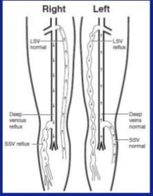
18 Competent valve - PW
19 Incompetent valve - PW
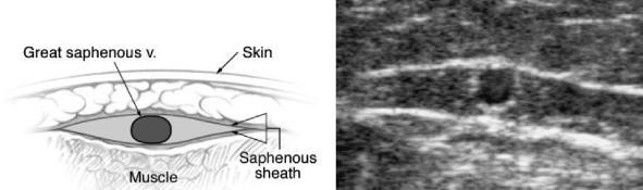
20 Incompetent valve - PW

21 Competent valve - CDI

22 Incompetent valve - CDI
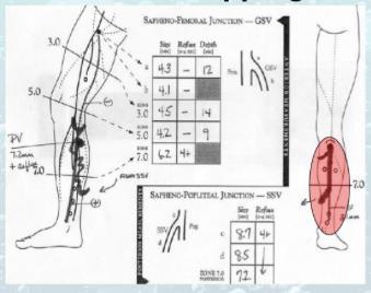
23 Ultrasound 3 Stages Diagnostic US Procedural US Follow-up US
24 DIAGNOSTIC ULTRASOUND


25 Diagnostic US
26 US Patient position 1. Standing 2. Standing 3. Standing

27 Patient position for scanning GSV STANDING on floor or platform Facing the examiner Open stance External rotation of the hip Knee slightly bent with heel flat Weight on the contralateral limb

28 Patient position for scanning - SSV STANDING on floor or platform Turned around facing away from examiner Open stance Step forward, knee slightly bent and heel flat Weight on contralateral limb
29 Imaging - Tip Observe in SAX and doppler in LAX
30 IF THERE IS REFLUX Measure true diameter Supine Measure largest diameter Standing Measure the diameter and depth of vein in SAX from SFJ / SPJ to the intended access point Evaluate access site in SAX and LAX

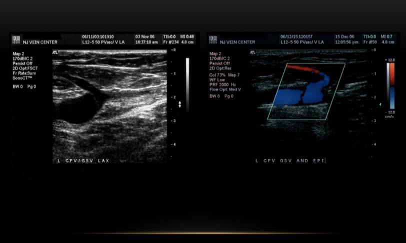
31 US - Report

32 US - Report
33 PROCEDURAL ULTRASOUND


34 Mapping From SFJ / SPJ to access point or vice versa Mark every 1 2 inches Identify and mark any aneurysms, tortuosities, large tributaries, access point, size, depth

35 Mapping Why? To determine size To determine depth Course of the vessel Location of large tributaries or perforators


36 Patient position for procedure GSV Supine Rev. Trendelenberg Leg slightly abducted and externally rotated SSV Prone Rev. Trendelenberg Pillow under foot to flex knee

37 Procedure - Access


38 Procedure - Access SAX short axis or transverse view Divides the vessel in cross section LAX long axis Divides the vessel longitudinally LAX long axis or longitudinal view Divides the vessel lengthwise

39 Procedure - Access
40 POST-PROCEDURE ULTRASOUND
41 Post-ablation scans - Why To confirm occlusion of the treated vein Insure patency of the deep system CFV, PV Insure patency of the epigastric vein
42 Immediate post-ablation scan Modified rule-out DVT protocol involving the ablated vein and the associated deep veins SAX - B-mode with and without compression SAX - Confirm the lumen is echo-free and demonstrate full coaptation of walls of deep vein LAX - with and without color demonstrating a patent SFJ, SPJ or other deep vein if perforators involved LAX with color to demonstrate patency of EV
43 Post-ablation scan
44 Post-ablation scans - Frequency Immediate post-ablation 3-7 days 6 weeks 3 months 6 months 1 year Yearly there after up to 5 years
45 Surveillance US Done to minimize the chance of recurrence Observe progressive nature of disease Arrest recruitment of new vessels
46 US Tips and Tricks GSV start at mid-thigh and then move to groin SSV start at mid-calf and move to popliteal fossa Do not concern yourself with every tributary; concentrate on tributaries that are same size or larger than the truncal vein it is connecting with If a vein is < 2mm no need for spectral doppler Do not waste time on perforators < 3mm Document normal and abnormal findings in main truncal veins Diameter measured in SAX ( ant. to post. walls) Spectral doppler waveforms done in LAX
47 US - Conclusions Reflux is not STATIC Look for the source does it match the clinical picture? Exam is very operator dependent Failure to identify and treat all sources may lead to early recurrence


48 Legs are staple articles and will never go out of fashion while the world lasts. - Jarrett and Palmer
49 Thank You
Venous Reflux Duplex Exam
 Venous Reflux Duplex Exam GWENDOLYN CARMEL, RVT PHYSIOLOGIST, DEPARTMENT OF VASCULAR SURGERY NEW JERSEY VETERANS HEALTHCARE CENTER EAST ORANGE, NJ PURPOSE: To identify patterns of incompetence and which
Protocols for the evaluation of lower extremity venous reflux: supine, sitting, or standing?
 Protocols for the evaluation of lower extremity venous reflux: supine, sitting, or standing? Susan Whitelaw RVT, RDMS PURPOSE Duplex imaging of the lower extremity veins is performed to assess the deep
Step by step ultrasound examination of varicose veins. Dr. Özgün Sensebat Vascular Surgeon Private Vascular Clinic Dorsten & Borken, Germany
 Step by step ultrasound examination of varicose Dr. Özgün Sensebat Vascular Surgeon Private Vascular Clinic Dorsten & Borken, Germany Required technical setup: B-mode vessel imaging combined with color
Bedside Ultrasound for DVT. Linear Probe. Leg Veins
 Bedside Ultrasound for DVT J. Christian Fox, MD, RDMS, FAAEM, FAIUM Director of Emergency Ultrasound Fellowship University of California, Irvine Jchrsitianfox@gmail.com Linear Probe High frequency transducer
Let s Take a Look Venous Insufficiency Ultrasound Techniques
 Let s Take a Look Venous Insufficiency Ultrasound Techniques Brent Wilkinson RVT, RDMS Steve Schomaker RVT, RDCS, RDMS Let s take a look Differentiate between normal venous flow and venous insufficiency
LOWER EXTREMITY VENOUS COMPRESSION ULTRASOUND. CPT Stacey Good, DO Emergency Medicine Ultrasound Fellow Madigan Army Medical Center
 LOWER EXTREMITY VENOUS COMPRESSION ULTRASOUND CPT Stacey Good, DO Emergency Medicine Ultrasound Fellow Madigan Army Medical Center Learning Objectives Setup and patient positioning for optimizing success
Introduction. Background Evidence System of examination Diagnoses & Variants Final actions Limitation of the examination
 Rule in DVT Introduction Background Evidence System of examination Diagnoses & Variants Final actions Limitation of the examination BACKGROUND Common presentation Influence initial management NICE Guidelines
Lower Extremity Venous Insufficiency Evaluation
 VASCULAR TECHNOLOGY PROFESSIONAL PERFORMANCE GUIDELINES Lower Extremity Venous Insufficiency Evaluation This Protocol was prepared by members of the Society for Vascular Ultrasound (SVU) as a template
The role of ultrasound duplex in endovenous procedures
 The role of ultrasound duplex in endovenous procedures Neophytos A. Zambas MD, PhD Vascular Surgeon Polyclinic Ygia, Limassol, Cyprus ΚΕΑΕΧ ΚΥΠΡΙΑΚΗ ΕΤΑΙΡΕΙΑ ΑΓΓΕΙΑΚΗΣ ΚΑΙ ΕΝΔΑΓΓΕΙΑΚΗΣ ΧΕΙΡΟΥΡΓΙΚΗΣ Pre
BEDSIDE ULTRASOUND BEDSIDE ULTRASOUND. Deep Vein Thrombosis. Probe used
 BEDSIDE ULTRASOUND Part 2 Diagnosis of deep vein thrombosis Kishore Kumar Pichamuthu, Professor, Department of Critical Care, CMC, Vellore Summary: Deep vein thrombosis (DVT) is a problem encountered in
Anatomy. Patterns of reflux. Technique. Testing Reflux time Patient position. Difficult! Learning. NOT system optimisation. Clinical Assesment
 Anatomy Patterns of reflux Awareness Technique Testing Reflux time Patient position Difficult! Learning NOT system optimisation Enlarged Clinical Assesment Twisted Where are the symptoms? Why they are
Clinical/Duplex Evaluation of Varicose Veins: Who to Treat?
 Clinical/Duplex Evaluation of Varicose Veins: Who to Treat? Sanjoy Kundu MD, FASA, FCIRSE, FSIR The Vein Institute of Toronto Scarborough Vascular Group Scarborough Vascular Ultrasound Scarborough Vascular
Segmental GSV reflux
 Segmental GSV reflux History of presentation A 43 year old female presented with right lower extremity varicose veins and swelling. She had symptoms of aching, heaviness and tiredness in the right leg.
Duplex ultrasound is first-line imaging for all
 Our Protocol for Transabdominal Pelvic Vein Duplex Ultrasound A summary of s protocol for pelvic vein duplex ultrasonography, including equipment, patient positioning, ultrasound settings, and technique.
Guidelines, Policies and Statements D20 Statement on Peripheral Venous Ultrasound
 Guidelines, Policies and Statements D20 Statement on Peripheral Venous Ultrasound Disclaimer and Copyright The ASUM Standards of Practice Board have made every effort to ensure that this Guideline/Policy/Statement
Chronic Venous Insufficiency Compression and Beyond
 Disclosure of Conflict of Interest Chronic Venous Insufficiency Compression and Beyond Shawn Amyot, MD, CCFP Fellow of the Canadian Society of Phlebology Ottawa Vein Centre I do not have relevant financial
Bedside Ultrasound for Detection of Deep Vein Thrombosis: the Two-Point Compression Method
 Bedside Ultrasound for Detection of Deep Vein Thrombosis: the Two-Point Compression Method Tom Ashar MD RDMS a, Krishnaraj Jayarama DO, Raymond Yun MD Department of Emergency Medicine, Newark Beth Israel
Endo-Thermal Heat Induced Thrombosis (E-HIT)
") Endo-Thermal Heat Induced Thrombosis (E-HIT) Michael Ombrellino MD FACS The Cardiovascular Care Group Clinical Associate Professor of Surgery Rutgers School of Medicine Objectives: What is E-HIT? How do
Endo-Thermal Heat Induced Thrombosis (E-HIT) Michael Ombrellino MD FACS The Cardiovascular Care Group Clinical Associate Professor of Surgery Rutgers School of Medicine Objectives: What is E-HIT? How do
TREATMENT OPTIONS FOR CHRONIC VENOUS INSUFFICIENCY
 TREATMENT OPTIONS FOR CHRONIC VENOUS INSUFFICIENCY TL LUK Consultant Vascular Surgeon Sarawak General Hospital HKL Vascular Conference 19/06/2013 PREVALENCE OF LOWER LIMB VENOUS DISEASE Affects half of
Carotid Abnormalities Coils, Kinks and Tortuosity David Lorelli M.D., RVT, FACS Michigan Vascular Association Conference Saturday, October 20, 2012
 Carotid Abnormalities Coils, Kinks and Tortuosity David Lorelli M.D., RVT, FACS Michigan Vascular Association Conference Saturday, October 20, 2012 Page 1 Table of Contents Carotid Anatomy Carotid Duplex
For exam: VL DUPLEX EXTREMITY VEINS UNILAT LT
 For exam: VL DUPLEX EXTREMITY VEINS UNILAT LT - 8870390 METHOD/TECHNIQUE: The veins of the left upper extremity were studied at multiple For exam: VL DUPLEX EXTREMITY VEINS UNILAT RT - 8870400 METHOD/TECHNIQUE:
DOPPLER ULTRASOUND OF DEEP VENOUS THROMBOSIS
 TOKUDA HOSPITAL SOFIA DOPPLER ULTRASOUND OF DEEP VENOUS THROMBOSIS MILENA STANEVA, MD, PhD Department of vascular surgery and angiology Venous thromboembolic disease continues to cause significant morbidity
Non-invasive examination
 Non-invasive examination Segmental pressure and Ankle-Brachial Index (ABI) The segmental blood pressure (SBP) examination is a simple, noninvasive method for diagnosing and localizing arterial disease.
LOWER LIMB DOPPLER ULTRASOUND FOR THE STUDY OF VENOUS INSUFFICIENCY
 Revista Chilena de Radiología. 2009; 15(4): -. 1 LOWER LIMB DOPPLER ULTRASOUND FOR THE STUDY OF VENOUS INSUFFICIENCY Dr. Paola Paolinelli G. Diagnostic Imaging Service, Clinica Las Condes, Santiago, Chile.
Lower Limb Venous Ultrasound. Colin P. Griffin MSc, BSc (Hons)
") Lower Limb Venous Ultrasound Colin P. Griffin MSc, BSc (Hons) Peripheral Vessels Lower Limb Peripheral Vessels Lower Limb Venous Deep System Common Iliac External/Internal Iliac Common Femoral Femoral
Lower Limb Venous Ultrasound Colin P. Griffin MSc, BSc (Hons) Peripheral Vessels Lower Limb Peripheral Vessels Lower Limb Venous Deep System Common Iliac External/Internal Iliac Common Femoral Femoral
Certificate in Clinician Performed Ultrasound (CCPU) Syllabus
 Syllabus") Certificate in Clinician Performed Ultrasound (CCPU) Syllabus Proximal Deep Vein Thrombosis (DVT) Page 1 of 6 03/17 Deep Vein Thrombosis (DVT) Syllabus Purpose: This unit is designed to cover the theoretical
Certificate in Clinician Performed Ultrasound (CCPU) Syllabus Proximal Deep Vein Thrombosis (DVT) Page 1 of 6 03/17 Deep Vein Thrombosis (DVT) Syllabus Purpose: This unit is designed to cover the theoretical
Guide to Small Animal Vascular Imaging using the Vevo 770 Micro-Ultrasound System
 Guide to Small Animal Vascular Imaging using the Vevo 770 Micro-Ultrasound System January 2007 Objectives: After completion of this module, the participant will be able to accomplish the following: Understand
Recurrent Varicose Veins We All See Them
 We All See Them November 4, 2017 Austin, TX Arlington Heights, IL No conflicts Terminology REVAS REcurrent Varices After Surgery PREVAIT PREsence of Varices After Interventional Treatment Recurrent varices
Saphenous Vein Wall Thickness in Age and Venous Reflux-Associated Remodeling in Adults
 Saphenous Vein Wall Thickness in Age and Venous Reflux-Associated Remodeling in Adults Nicos Labropoulos Professor of Surgery Director, Vascular Laboratory Division of Vascular Surgery Stony Brook Medicine
Terminology Tissue Appearance
 By Marc Nielsen, MD Advantages/Disadvantages Generation of Image Ultrasound Machine/Transducer selection Modes of Ultrasound Terminology Tissue Appearance Scanning Technique Real-time Portable No ionizing
Bedside Emergency Ultrasound For Deep Venous Thrombosis
 Bedside Emergency Ultrasound For Deep Venous Thrombosis Michael Blaivas, MD, MBA(candidate) FACEP, FAIUM Professor of Medicine University of South Carolina School of Medicine AIUM Third Vice President
Doppler ultrasound evaluation of pattern of venous incompetance and relation with skin changes in varicose vein patients
 Doppler ultrasound evaluation of pattern of venous incompetance and relation with skin changes in varicose vein patients Pant HP 1, Sharma S 2, Bhattarai S 1, Pandit SP 3, Maharjan D 2 1 Radiology resident,
Background & Indications Probe Selection
 Teresa S. Wu, MD, FACEP Director, EM Ultrasound Program & Fellowship Co-Director, Simulation Based Training Program & Fellowship Associate Program Director, EM Residency Program Maricopa Medical Center
Certificate in Clinician Performed Ultrasound (CCPU) Syllabus. Above Knee Deep Vein Thrombosis (DVT)
 Syllabus. Above Knee Deep Vein Thrombosis (DVT)") Certificate in Clinician Performed Ultrasound (CCPU) Syllabus Above Knee Deep Vein Thrombosis (DVT) Deep Vein Thrombosis (DVT) Purpose: Prerequisites: Training: Assessments: This unit is designed to cover
Certificate in Clinician Performed Ultrasound (CCPU) Syllabus Above Knee Deep Vein Thrombosis (DVT) Deep Vein Thrombosis (DVT) Purpose: Prerequisites: Training: Assessments: This unit is designed to cover
validation study Original article Clinical examination of varicose veins - a Jong Kim, Simon Richards, Patrick J Kent
 The Royal College of Surgeons of England : 171175 Original article Clinical examination of varicose veins a validation study Jong Kim, Simon Richards, Patrick J Kent Department of Vascular and Endovascular
Optimising your Doppler settings for an accurate PI. Alison McGuinness Mid Yorks Hospitals
 Optimising your Doppler settings for an accurate PI Alison McGuinness Mid Yorks Hospitals Applications Both maternal uterine and fetal circulations can be studied with doppler sonography Uterine arteries
RECOGNITION AND ENDOVASCULAR TREATMENT OF CHRONIC VENOUS INSUFFICIENCY
 RECOGNITION AND ENDOVASCULAR TREATMENT OF CHRONIC VENOUS INSUFFICIENCY Paul Kramer, MD, FACC, FSCAI Liberty Cardiovascular Specialists Liberty Regional Heart and Vascular Center DISCLOSURES NONE Venous
Physician s Vascular Interpretation Examination Content Outline
 Physician s Vascular Interpretation Examination Content Outline (Outline Summary) # Domain Subdomain Percentage 1 2 3 4 5 6 Cerebrovascular Abdominal Peripheral Arterial - Duplex Imaging Peripheral Arterial
Where should you palpate the pulse of different arteries in the lower limb?
 Where should you palpate the pulse of different arteries in the lower limb? The femoral artery In the femoral triangle, its pulse is easily felt just inferior to the inguinal ligament midway between the
POINT OF CARE ULTRASOUND - Venous US for DVT
 POINT OF CARE ULTRASOUND - Venous US for DVT The diagnosis of deep venous thrombosis (DVT) using ultrasound in the emergency department. DVT US is easy to perform and can be usually be completed in less
Vascular Sonography Examination
 Vascular Sonography Examination The purpose of The American Registry of Radiologic Technologists (ARRT ) Vascular Sonography Examination is to assess the knowledge and cognitive skills underlying the intelligent
Goals. Access flow and renal artery stenosis evaluation by Doppler ultrasound. Reimbursement. WHY use of Doppler Ultrasound
 Access flow and renal artery stenosis evaluation by Doppler ultrasound Adina Voiculescu, MD Interventional Nephrology Brigham and Women s Hospital Boston Instructor at Harvard Medical School Understand
High Level Overview: Venous Anatomy of Lower Extremities. Anatomy of a Vein 5/11/2015. Barbara Deusterman, RN
 High Level Overview: Venous Anatomy of Lower Extremities Barbara Deusterman, RN What does this anatomy lecture have to do with visually guided sclerotherapy (VGS)? May 11, 2015 2 Anatomy of a Vein Almeida,
Chronic Venous Insufficiency
 Chronic Venous Insufficiency None Disclosures Lesley Enfinger, MSN,NP-C Chronic Venous Insufficiency Over 24 Million Americans affected by Chronic Venous Insufficiency (CVI) 10 x More Americans suffer
Additional Information S-55
 Additional Information S-55 Network providers are encouraged, but not required to participate in the on-line American Venous Forum Registry (AVR) - The First National Registry for the Treatment of Varicose
Patient assessment and strategy making for endovenous treatment
 Patient assessment and strategy making for endovenous treatment Raghu Kolluri, MD Director Vascular Medicine OhioHealth Riverside Methodist Hospital Columbus, OH Disclosures Current Medtronic Consultant/
Routine For: Total Hip Arthroplasty - Standard Precautions
 Standard Hip Precautions TOTAL HIP - 4 Ankle Pump not bend your operative hip beyond a 90 degree angle. not sit with your legs crossed or lie on your back with your operative leg crossed over the other
2017 Florida Vascular Society
 Current Management of Venous Leg Ulcers: How to Identify Patients with Correctable Venous Disease and Interventional Procedures to Heal and Prevent Recurrence 2017 Florida Vascular Society Bill Marston
: A guide to Doppler US evaluation of chronic lower limb venous insufficiency
 : A guide to Doppler US evaluation of chronic lower limb venous insufficiency Poster No.: C-1781 Congress: ECR 2011 Type: Educational Exhibit Authors: T. M. O. Couto, H. Patricio, Â. Moreira, A. Estevao
How varicose veins occur
 Varicose veins are a very common problem, generally appearing as twisting, bulging rope-like cords on the legs, anywhere from groin to ankle. Spider veins are smaller, flatter, red or purple veins closer
Upper Extremity Venous Duplex Evaluation
 VASCULARTECHNOLOGY PROFESSIONAL PERFORMANCE GUIDELINES Upper Extremity Venous Duplex Evaluation This Guideline was prepared by the Professional Guidelines Subcommittee of the Society for Vascular Ultrasound
Clinical case. Symptomatic anterior accessory great saphenous vein (AAGSV) reflux
 reflux") Clinical case Symptomatic anterior accessory great saphenous vein (AAGSV) reflux A 70 year-old female presents with symptomatic varicose veins on left leg for more than 10 years. She complains of heaviness,
Clinical case Symptomatic anterior accessory great saphenous vein (AAGSV) reflux A 70 year-old female presents with symptomatic varicose veins on left leg for more than 10 years. She complains of heaviness,
Peripheral Vascular Examination. Dr. Gary Mumaugh Western Physical Assessment
 Peripheral Vascular Examination Dr. Gary Mumaugh Western Physical Assessment Competencies 1. Inspection of upper extremity for: size symmetry swelling venous pattern color Texture nail beds Competencies
Exercise Therapy for Patients with Knee OA Knee Exercise Protocol Knee Home Exercise Programme
 Chapter FOUR Exercise Therapy for Patients with Knee OA Knee Exercise Protocol Knee Home Exercise Programme Chris Higgs Cathy Chapple Daniel Pinto J. Haxby Abbott 99 n n 100 General Guidelines Knee Exercise
Clinico-Anatomical and Radiological Correlation of Varicose Veins of Lower Limb A Cross-sectional Study
 ORIGINAL RESEARCH www.ijcmr.com Clinico-Anatomical and Radiological Correlation of Varicose Veins of Lower Limb A Cross-sectional Study Lalatendu Swain 1, Mamata Singh 2, Prabhat Nalini Rautray 3 ABSTRACT
Preventative Exercises for the Achilles
 Preventative Exercises for the Achilles Outline 1. Toe walk x 15 each foot 2. Feet out walk x 15 each foot 3. Feet in walk x 15 each foot 4. Ankle in walk x 10 each foot 5. Ankle out walk x 10 each foot
STRUCTURED EDUCATION REQUIREMENTS IMPLEMENTATION DATE: JULY 1, 2016
 STRUCTURED EDUCATION REQUIREMENTS Vascular Sonography The purpose of structured education is to provide the opportunity for individuals to develop mastery of discipline-specific knowledge that, when coupled
Vein Disease Treatment
 MP9241 Covered Service: Yes when meets criteria below Prior Authorization Required: Yes as indicated in 2.0, 3.0, 4.0 and 5.0 Additional Information: None Prevea360 Health Plan Medical Policy: Vein disease
Selection and work up for the right patients suspected of deep venous disease
 Selection and work up for the right patients suspected of deep venous disease R A G H U K O L L U R I, M S, M D, R V T S Y S T E M M E D I C A L D I R E C T O R V A S C U L A R M E D I C I N E / V A S
Endothermal Ablation for Venous Insufficiency. Dr. S. Kundu Medical Director The Vein Institute of Toronto
 Endothermal Ablation for Venous Insufficiency Dr. S. Kundu Medical Director The Vein Institute of Toronto Objective: remove the GSV from the circulation 1. Surgical - HL & stripping 2. Chemical sclerotherapy
GENERAL EXERCISES KNEE BMW MANUFACTURING CO. PZ-AM-G-US I July 2017
 GENERAL EXERCISES KNEE BMW MANUFACTURING CO. PZ-AM-G-US I July 2017 Disclosure: The exercises, stretches, and mobilizations provided in this presentation are for educational purposes only are not to be
Principles of Ultrasound. Cara C. Prideaux, M.D. University of Utah PM&R Sports Medicine Fellow March 14, 2012
 Principles of Ultrasound Cara C. Prideaux, M.D. University of Utah PM&R Sports Medicine Fellow March 14, 2012 None Disclosures Outline Introduction Benefits and Limitations of US Ultrasound (US) Physics
How to choose which treatment method(s) to use for a particular varicose veins patient ESTABLISHING A TREATMENT PLAN.
 to use for a particular varicose veins patient ESTABLISHING A TREATMENT PLAN.") How to choose which treatment method(s) to use for a particular varicose veins patient ESTABLISHING A TREATMENT PLAN Surgeon Dr G Mark Malouf Sydney Australia Following History and Physical examination
How to choose which treatment method(s) to use for a particular varicose veins patient ESTABLISHING A TREATMENT PLAN Surgeon Dr G Mark Malouf Sydney Australia Following History and Physical examination
Developed by: Physiotherapy Department Surrey Memorial Hospital. Printshop #
 Developed by: Physiotherapy Department Surrey Memorial Hospital Printshop # 255171 The following exercises are intended for you to continue at home. Your physiotherapist will teach and mark the exercises
Clinical Examination of VASCULAR PATIENTS. Stephanie Hirst & Alexander Sunde
 Clinical Examination of VASCULAR PATIENTS Stephanie Hirst & Alexander Sunde Goals of Medical History To record the patient s symptoms at time of presentation. To organize the events which have lead to
Image Formation (10) 2 Evaluation and Selection of Representative Images (10)
 2 Evaluation and Selection of Representative Images (10)") STRUCTURED SELF ASSESSMENT CONTENT SPECIFICATIONS SSA LAUNCH DATE: JANUARY 1, 2018 Vascular Sonography The purpose of continuing qualifications requirements (CQR) is to assist registered technologists
STRUCTURED SELF ASSESSMENT CONTENT SPECIFICATIONS SSA LAUNCH DATE: JANUARY 1, 2018 Vascular Sonography The purpose of continuing qualifications requirements (CQR) is to assist registered technologists
Fig MHz cm/s. Table 1 Fig. 2. Fig. 3, 4. Fig. 5
 GE Fig. 1 3. 5 MHz 7 10 MHz 3. 5 5. 0 MHz B 10 20 cm/s Table 1 Fig. 2 Fig. 1 1 2 3 3 3 : 1 2 3 Fig. 3, 4 Fig. 5 Table 1 a b c Fig. 2 a B b B c Fig. 6 Table 1 Fig. 7 a b c Fig. 3 a AV b A VV c 1 cm 2 1
Surgical Options for revascularisation P E T E R S U B R A M A N I A M
 Surgical Options for revascularisation P E T E R S U B R A M A N I A M The goal Treat pain Heal ulcer Preserve limb Preserve life The options Conservative Endovascular Surgical bypass Primary amputation
VFI Technology to Change the Way You Work
 Analogic Ultrasound VFI Technology to Change the Way You Work Vascular Ultrasound Made Easier Vector Flow Imaging VFI VFI is a ground-breaking technology that can revolutionize the workflow for many Doppler
Flexibility. STRETCH: Kneeling gastrocnemius. STRETCH: Standing gastrocnemius. STRETCH: Standing soleus. Adopt a press up position
 STRETCH: Kneeling gastrocnemius Adopt a press up position Rest one knee on mat with the opposite leg straight Maintain a neutral spine position Push through arms to lever ankle into increased dorsiflexion
Lesson 07: Ultrasound Transducers. This lesson contains 73 slides plus 16 multiple-choice questions.
 Lesson 07: Ultrasound Transducers This lesson contains 73 slides plus 16 multiple-choice questions. This lesson was derived from pages 33 through 42 in the textbook: Ultrasound Transducers Ultrasound Transducers
Doppler ultrasound in the evaluation of chronic venous insufficiency: A step-by-step morphological and hemodynamic review
 Doppler ultrasound in the evaluation of chronic venous insufficiency: A step-by-step morphological and hemodynamic review Poster No.: C-3206 Congress: ECR 2010 Type: Educational Exhibit Topic: Vascular
Doppler Basic & Hemodynamic Calculations
 Doppler Basic & Hemodynamic Calculations August 19, 2017 Smonporn Boonyaratavej MD Division of Cardiology, Department of Medicine Chulalongkorn University Cardiac Center, King Chulalongkorn Memorial Hospital
Page 1. Ruling out deep venous obstruction prior to superficial vein treatment. Disclosures. Indications for saphenous vein ablation (SVA)
") 1 Ruling out deep venous obstruction prior to superficial vein treatment Deepak Sudheendra, MD, RPVI Assistant Professor of Clinical Radiology & Surgery Disclosures No financial disclosures Indications
1 Ruling out deep venous obstruction prior to superficial vein treatment Deepak Sudheendra, MD, RPVI Assistant Professor of Clinical Radiology & Surgery Disclosures No financial disclosures Indications
Venous drainage of the lower limb
 Venous drainage of the lower limb INTRODUCTION It is of immense clinical and surgical importance. The venous blood against gravity. FACTORS HELPING THE VENOUS DRAINAGE OF THE LOWER LIMB The contraction
Exercise Report For: Augusta James
 Exercise Report For: Optimizing Sport Performance Provided By: Greg Redman BScPT, BScKin, Wave Physiotherapy Phone: 250-763-9283 Fax:, www.wavephysio.ca Page: 1 Stretch hip flexor kneel w/ball Stretch
Ex Fix Rehab Phase II Strengthening
 Perform repetitions of each exercise, twice daily. Increase to repetitions. Ankle Pumps: With leg resting on bed and knee straight, slowly pump ankle up and down as far as possible. Quad sets: Tighten
2017 COS ANNUAL MEETING AND EXHIBITION HOME EXERCISES
 UPPER BODY Push Up From a push up position. Lower whole body down to floor. Press up to return to start position. Maintain abdominal hollow and neutral spinal alignment throughout movement. Note: Perform
Overview Functional Training
 Overview Functional Training Exercises with Therapist 1. Sitting 2. Standing up vs. Sitting down 3. Standing 4. Stance phase ( Static and dynamic ) 5. Swing phase 6. Gait Evaluation 7. Walking level ground
Post-Thrombotic Syndrome(PTS) Conservative Treatment Options
 Conservative Treatment Options") Post-Thrombotic Syndrome(PTS) Conservative Treatment Options Dr. S. Kundu Scarborough Hospital-General Division Scarborough Vascular Group Toronto Endovascular Centre The Vein Institute of Toronto Scarborough
Post-Thrombotic Syndrome(PTS) Conservative Treatment Options Dr. S. Kundu Scarborough Hospital-General Division Scarborough Vascular Group Toronto Endovascular Centre The Vein Institute of Toronto Scarborough
GOLFERS TEN PROGRAM 1. SELF STRETCHING OF THE SHOULDER CAPSULE
 GOLFERS TEN PROGRAM 1. SELF STRETCHING OF THE SHOULDER CAPSULE POSTERIOR CAPSULAR STRETCH Bring your arm across your chest toward the opposite shoulder. With the opposite arm grasp your arm at your elbow.
The Golfers Ten Program. 1. Self Stretching of the Shoulder Capsule
 The Golfers Ten Program 1. Self Stretching of the Shoulder Capsule A. Posterior capsular stretch Bring your arm across your chest toward the opposite shoulder. With the opposite arm grasp your arm at your
Controversies & updates in Vascular Surgery
 Controversies & updates in Vascular Surgery Paris - january 24 2018 Venous session VENOUS ODDITIES DUPLEX IMAGE Philippe LEMASLE Le Chesnay - France I have no financial relationship to disclose Case n
REHABILITATION PROTOCOL Criteria-Based Postoperative ACL Reconstruction Rehabilitation Protocol
 REHABILITATION PROTOCOL Criteria-Based Postoperative ACL Reconstruction Rehabilitation Protocol Phase I (Days 1 7) WEIGHTBEARING STATUS 1- Two crutches, weightbearing as tolerated. Exercises 1- Heel slides/wall
Randomized trial comparing cyanoacrylate embolization and radiofrequency ablation for incompetent great saphenous vein
 Randomized trial comparing cyanoacrylate embolization and radiofrequency ablation for incompetent great saphenous vein Raghu Kolluri, MD, Director Vascular Medicine OhioHealth Riverside Methodist Hospital
Lumbar. Physician. Technique: Continue this. back pain is. bent. under the contralatera. Copyright
 Lumbar myofascial releasee Lumbar spine Brief description: Low back pain is a common problem and lumbar myofascial releasee can be useful as part of a comprehensiv ve treatment of low back pain. By usingg
HD Scanning: Velocities and Volume Flow
 HD Scanning: Velocities and Volume Flow Non-Invasive Lab Symposium West Orange, NJ April 27, 2018 Volume Flow Cindy Sturt, MD, FACS, RVT 500,000 Americans on dialysis 20-25% annual mortality 65% 5 year
KNEE AND LEG EXERCISE PROGRAM
 KNEE AND LEG EXERCISE PROGRAM These exercises are specifically designed to rehabilitate the muscles of the hip and knee by increasing the strength and flexibility of the involved leg. This exercise program
ULTRASOUND. OB/Gyn (Core) Ultrasound PIEZOELECTRIC EFFECT. Principles of Ultrasound Physics and Instrumentation. Nathan Pinkney, BS, CDOS
 Ultrasound PIEZOELECTRIC EFFECT. Principles of Ultrasound Physics and Instrumentation. Nathan Pinkney, BS, CDOS") 1 OB/Gyn (Core) Ultrasound Principles of Ultrasound Physics and Instrumentation Nathan Pinkney, BS, CDOS Philadelphia College of Osteopathic Medicine 2016 ULTRASOUND CATEGORIES OF SOUND INFRASOUND = below
1 OB/Gyn (Core) Ultrasound Principles of Ultrasound Physics and Instrumentation Nathan Pinkney, BS, CDOS Philadelphia College of Osteopathic Medicine 2016 ULTRASOUND CATEGORIES OF SOUND INFRASOUND = below
The Peripheral Vascular System
 The Peripheral Vascular System Anatomy and Physiology Arteries Arteries contain 3 concentric layers of tissue: - the intima - the media - the adventitia The intima The endothelium of the intima has metabolic
VASCULAR DISEASE: THREE THINGS YOU SHOULD KNOW JAMES A.M. SMITH, D.O. KANSAS VASCULAR MEDICINE, P.A. WICHITA, KANSAS
 VASCULAR DISEASE: THREE THINGS YOU SHOULD KNOW JAMES A.M. SMITH, D.O. KANSAS VASCULAR MEDICINE, P.A. WICHITA, KANSAS KANSAS ASSOCIATION OF OSTEOPATHIC MEDICINE ANNUAL CME CONVENTION APRIL 13, 2018 THREE
Complex Iliocaval Reconstruction PNEC. Seattle WA. Bill Marston MD Professor, Div of Vascular Surgery University of N.
 Complex Iliocaval Reconstruction 2017 PNEC. Seattle WA Bill Marston MD Professor, Div of Vascular Surgery University of N. Carolina DISCLOSURES William Marston, MD Consultant/Advisory Board: Veniti, Cardinal
Post Operative Total Hip Replacement Protocol Brian J. White, MD
 Post Operative Total Hip Replacement Protocol Brian J. White, MD www.western-ortho.com The intent of this protocol is to provide guidelines for progression of rehabilitation. It is not intended to serve
Nicky Schmidt PT, C/NDT 1
 Preparing the foot for third rocker and initial contact Nicky Schmidt PT, C/NDT copyright 2012 References Laboratory Strategies developed and taught by Nicky Schmidt, P.T. in the NDTA Approved Advanced
Terms of Movements by Prof. Dr. Muhammad Imran Qureshi
 Terms of Movements by Prof. Dr. Muhammad Imran Qureshi Three systems of the body work in coordination to perform various movements of the body. These are: A System of Bones (Osteology), A System of Muscles
Body Organizations Flashcards
 1. What are the two main regions of the body? 2. What three structures are in the Axial Region? 1. Axial Region (Goes down midline of the body) 2. Appendicular Region (limbs) 3. Axial Region (Goes down
Hip Arthroscopy Protocol
 The intent of this protocol is to provide guidelines for progression of rehabilitation, it is not intended to serve as a substitute for clinical decision making. Progression through each phase of rehabilitation
Prof. Nabil CHAKFE et coll.
 Prof. Nabil CHAKFE et coll. For the Department of Vascular Surgery and Kidney Transplantation University Hospital of Strasbourg, FRANCE Popliteal artery entrapment: misdiagnosed Epidemiology Prevalence:
Ultrasound Guided Lower Extremity Blocks
 Ultrasound Guided Lower Extremity Blocks CONTENTS: 1. Femoral Nerve Block 2. Popliteal Nerve Block Updated December 2017 1 1. Femoral Nerve Block Indications Surgery involving the knee, anterior thigh,