Can We Identify Vulnerable Patients & Vulnerable Plaque?
|
|
|
- Aubrey Dean
- 5 years ago
- Views:
Transcription
1 Can We Identify Vulnerable Patients & Vulnerable Plaque? We Know Enough to Treat High-Risk Lesions? Takashi Akasaka, MD, PhD Department of Cardiovascular Medicine, Japan
2 Disclosure Statement of Financial Interest Within the past 12 months, I or my spouse/partner have had a financial interest/arrangement or affiliation with the organization(s) listed below. Affiliation/Financial Relationship Grant/Research Support Consulting Fees/Honoraria : Abbott Vascular Japan Boston Scientific Japan Goodman Inc. Sent Jude Medical Japan Terumo Inc. :Goodman Inc. GE Medical Healthcare Sent Jude Medical Japan Terumo Inc.
3 Cardiovascular event-free survival probability according to high or low hs-crp & LDL cholesterol Ridker PM et al. N Engl J Med 2002;347:
4 動脈硬化プラークの進展 Libby, P. Circulation 2001;104: Progression of atherosclerotic plaque ( Naghavi M, et al. Circulation 2003;108: ) Positive remodering is an adaption for atherosclerotic change. ACS may occur even in insignificant stenosis.
5 Coronary lesion assessment Anatomical assessment CAG IVUS MSCT MRI Echo OCT Molecular Imaging Physiological assessment Stress ECG Stress scintigraphy Stress Echo PET MRI (Perfusion image) Contrast Echo Doppler Echo TTE TEE Transcatheter(Doppler GW) Coronary pressure Transcatheter(Pressure GW)


6 Comparison among coronary imaging techniques OCT IVUS MRI CAG MDCT Angioscopy Resolution <200 Probe Size N/A N/A 800 Contact Ionizing Radiation Imaging Target Other No No Layer Tissue Character ization Yes No Layer N/A No No Density N/A No Yes Blood Flow Flow Only No Yes Density CT number No No Surface Surface Only Each modality may have advantages and disadvantages.


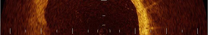
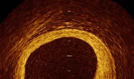
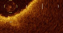
7 Progression of atherosclerosis & corresponding OCT Images A B C D E F Normal Intimal thickening Early yplaque formation with neovascularization Fibrous cap atheroma Thin-cap fiboratheroma Plaque rupture lipid arc = 206 degree 400µm 260µm 40µm lipid lipid Intimal thickening Neovascularization lipid arc = 126 degree Extracelluler lipid Machrophage form cells Smooth muscle cells Neovascular vessel Necrotic core Calcified plaque Thrombus Collagen


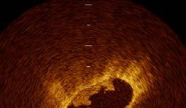
8 Plaque rupture (Plaque disruption) Plaque rupture could be identified by the findings of discontinuity of the fibrous cap and ulcer (cavity) formation at the site of the discontinuing fibrous cap.


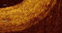
9 Red & white thrombus Red thrombus White thrombus Mixed thrombus Protrusion mass with shadow Protrusion mass without shadow Protrusion mass with & without shadow Kume T, Akasaka T, et al(am J Cardiol 97: , 2006 ) Kubo T, Akasaka T, et al. ( J Am Coll Cardiol 50: ,2007)


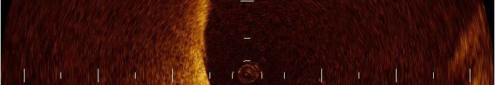
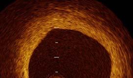
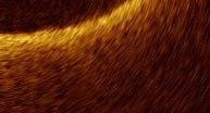
10 Thin-capped Fibroatheroma (TCFA) The TCFA was defined as a plaque with lipid content in more than 2 quadrants and the thinnest part of a fibrous cap measuring less than 65 μm by histology. Lipid id The cap thickness is measured from the surface of the lumen to the portion just starting the attenuation Thickness of fibrous cap


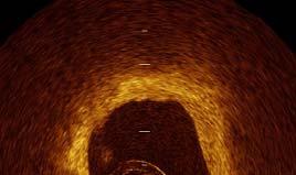
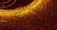
11 Erosion Plaque ulceration Plaque ulceration could be identified a hollow at the culprit site, especially if there is no rupture. Plaque erosion could be identified in a broad band spectrum from denudation of several endothelium to ulcer formation without rupture in the culprit site.


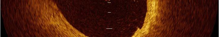
12 Calcified nodule Virmani R et al. Am J Cardiol Calcified nodule is identified as a protrusion of well-delineated, heterogeneous region with attenuation of OCT signal. Kubo T, Akasaka T, et al. Cardiol Res Pract 2011.


")

13 OCT findings of macrophages Low Mφ High Mφ 250 µm OCT CD68 (macrophage) Tearney GJ et al. Circulation, 107: , 2003


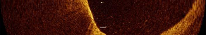


14 Identification of macrophage Extremely high signal with rapid attenuation on the surface of the vessel wall or within fibrous tissue might demonstrate macrophage accumuration.
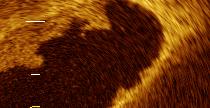
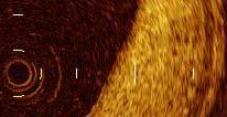
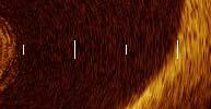
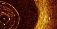
15 Corresponding Images of OCT and Angioscopy A-1 B-1 C-1 D-1 A-2 B-2 C-2 D-2 * * * A-3 B-3 C-3 D-3 * * * * * * * (Kubo T, et al. J Am Coll Cardiol Intv 1:74-80,2008)
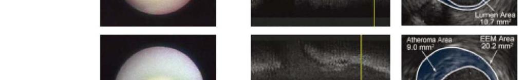
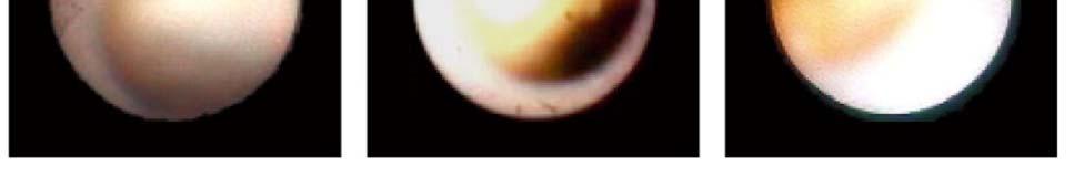
16 Angioscopy vs OCT Plaque color vs lipid size Plaque color vs fibrous cap thickness (Kubo T, et al. J Am Coll Cardiol Intv 1:74-80,2008)
17 Criteria for defining vulnerable plaque Major criteria ( Naghavi M, et al. Circulation 2003;108: ) Active inflammation (monocyte/macrophage and sometimes T-cell infiltration) Thin cap (< 65 μm) with large lipid core Endotherial denudation with superficial platelet aggregation Fissued plaque Stenosis > 90% Minor criteria Superficial calcified nodule Glistening yellow Intraplaque hemorrhage Endotherial dysfunction Outward (positive) remodering
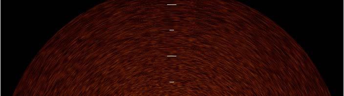
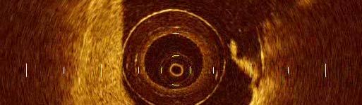
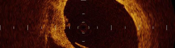
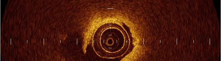
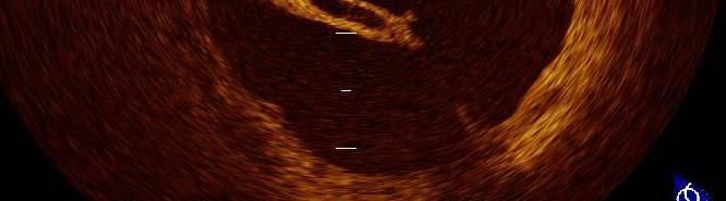
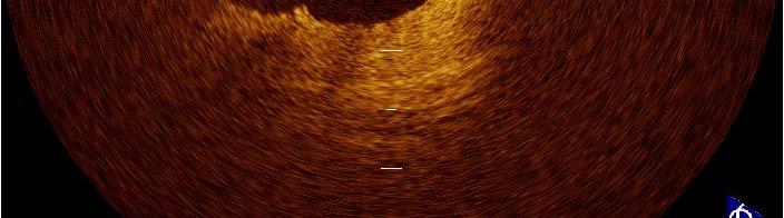
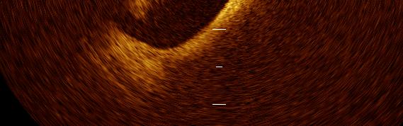
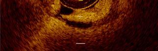
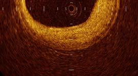
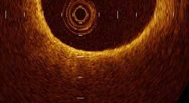
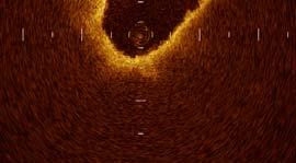
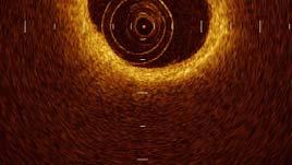
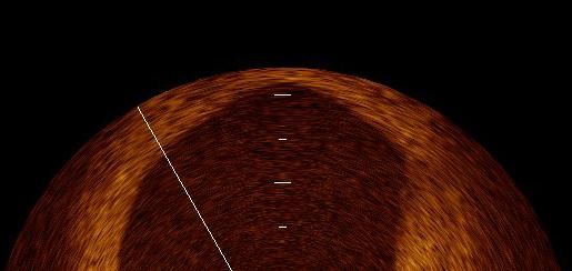
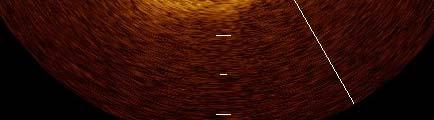
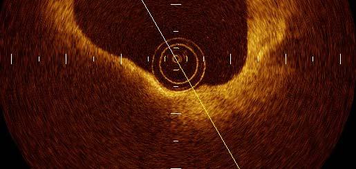
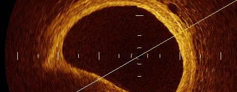
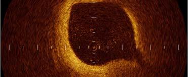
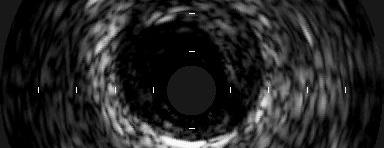
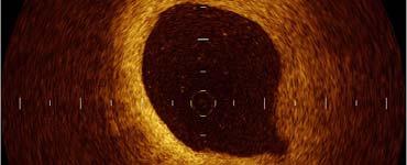
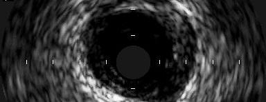
18 Lipid pool Vulnerable plaque Fibrous cap 1 Positive remodering 2 Eccentric plaque 3 Low echoic area (lipid pool) 4 Thin fibrous cap Eccentric plaque IVUS allow us to identify plaque characteristics, but it is not sufficient enough in resolution & tissue characterization.
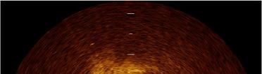
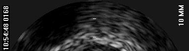
19 Tissue characterization by IB ( Kawasaki M, et al. Circulation 105: , 2002 )
20 Tissue characterization by IB CL; calcification ML; mixed lesion FI ; fibrosis LC; lipid core IH ; intimal hyperplasia p TH; thrombus ( Kawasaki M, et al. Circulation 105: , 2002 )
21 Prediction of ACS by IB Baseline IVUS Characteristics ( Kawasaki M, et al. J Am Coll Cardiol 47: , 2006 )

22 VH IVUS Tissue characterization is performed by several indexes obtained from RF signal including frequency, IB, power, spectral gradient, etc. Fibrous Tissue Fibro-fatty Necrotic Core Dense Calcium
23 IVUS-derived TCFA ( Rodriguez-Granillo GA, et al. J Am Coll Cardiol 46: , ) Percent atheroma volume = (EEM area Lumen area)/eem area x100 40% Nectrotic core 10% Without evident overlying fibrous tissue
24 PROSPECT trial ( Stone GW, et al. N Engl J Med 364: , ) Predictive value of IVUS tissue characterization is not so high compared with gray-scale IVUS information such as MLA & PB.


25 VH-IVUS vs OCT ( Sawada T, et al Eur Heart J 29: , 1146, 2008 ) Without evident overlying fibrous tissue Without evident overlying fibrous tissue With evident overlying fibrous tissue
26 Concordance & discordance between VH-IVUS and OCT Table 4 in the assessment of TCFA IVUS-VH Diagnosis OCT Diagnosis TCFA (n=11) Not TCFA (n=36) VH-TCFA (n=31) 9 22 Not VH-TCFA (n=16) 2 14 Discordance between VH-IVUS & OCT has been described by Sawada T, et al. (Eur Heart J 29: , 1146, 2008)
27 Changes of plaque, media & lumen area PIT VH-TCFA ThCFA Fibrotic Fibrocalcific ( Kubo T, et al. J Am Coll Cardiol 55; , 2010 )Wakayama Medical University


")

28 Coronary lesion morphology by VH-IVUS ( Kubo T, et al. J Am Coll Cardiol 55; , ) Figure 2 TCFA ThCFA TCFA Fibrous TCFA TCFA PIT TCFA ThCFA TCFA
29 Controversy in plaque characterization by VH-IVUS ( Thim T, et al. Cir Cardiovasc Imaging. 2010;3: ) Figure 2
30 Intravascular imaging modalities IVUS Gray scale IB Virtual histology Elastography Palpography available not available Angioscopy OCT NIR system available


31 Assessment by MDCT Outer vessel area Outer vessel area Lumen area % Plaque area = vessel area lumen area Positive remodeling (Remodeling Index 1.05) CT attenuation value Ring-like sign Lumen CT attenuation value Lumen area
 Remodeling (B) Distal")


32 Remodeling Index Assessed by MDCT Proximal (A) Remodeling (B) Distal (C) A B C Remodeling Index = 2 Culprit area Prox. + Distal reference area Positive remodeling: Remodeling Index 1.05 The arterial remodeling index was defined as the ratio between the outer vessel area at the site of maximal luminal l narrowing and the mean of the proximal and distal reference sites. Positive Remodeling was defined as remodeling index


")

33 The Assessment of CT Attenuation Value Lumen Plaque The CT values of plaques were measured in multiple l (at least 3 sections) cross- sectional images along the plaque and averaged.
34 Patient population & event 1059 patients PR & LAP 45 PR or LAP 27 Neither PR nor LAP 820 No plaque 167 ACS No ACS ACS No ACS ACS No ACS ACS No ACS PR: positive remodeling LAP: low-attenuation plaque Motoyama S, et al. J Am Coll Cardiol Cardiol 54: 49-57, 2009
35 Treatment of vulnerable plaque Local approach Plaque sealing by stent Plaque stabilization by local drug delivery Systemic approach Change lifestyle Reduction of risk factor Medication
36 LDL cholesterol & coronary event %) event (% ronary e y of cor equency Fre AFCAPS lovastatin Diabetes Mellitus Secondary prevention Primary yprevention 4S DM LIPID DM simvastatin placebo LIPID DM CARE DM pravastatin placebo CARE DM 4S LIPID pravastatin simvastatin LIPID placebo pravastatin CARE CARE pravastatinastatin placebo WOSCOPS pravastatin WOSCOPS placebo AFCAPS placebo 4S placebo Mean LDL cholesterol (mmol/l)
37 LDL vs Atheroma volume CAMELOT placebo REVERSAL pravastatin REVERSAL atorvastatin ACTIVATE placebo ILLUSTRATE atorvastatin ASTEROID rosuvastatin


")
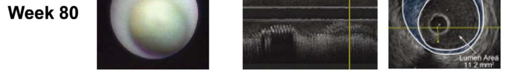
38 Changes in plaque color & volume by statin (Hirayama A, et al: Circ J 73; ; , 725, 2009)
39 Changes in plaque color & volume by statin (Hirayama A, et al: Circ J 73; , 725, 2009)


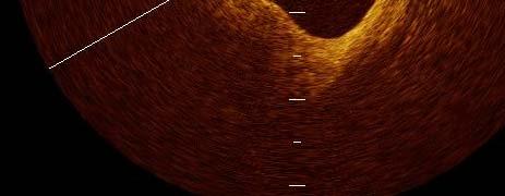
40 OCT assessment of non-culprit lesion (47y.o. male) Baseline 9 month later T.Chol. 200 mg/dl TG 79 mg/dl HDL-C 47 mg/dl LDL-C 128 mg/dl T.Chol. 187 mg/dl TG 133 mg/dl HDL-C 49 mg/dl LDL-C 98 mg/dl (Takarada S, et al. Atherosclerosis 202: , 2009 )
41 The correlation between the lipid profile and the % change of fibrous-cap thickness (FCT) and total atheroma volume (TAV). %TAV %TA AV r= p< %LDL/HDL %CRP p= r=-0.44 p<0.01 %FCT 20 %FCT %LDL/HDL p= %TAV and %LDL/HDL were positively correlated (p<0.01, r = 0.42). %FCT and %CRP were inversely correlated (p<0.01, r = -0.44). (Takarada S, et al. JACC Interv. 2010;3: ) %CRP
42 Univariable and multivariable logistic regression analyses as predictors of plaque stabilization univariable analysis multivariable analysis p-value : OR(95% CI) :OR(95%CI) p-value age,y g,y 0.52 ( ) p=0.60 gender 1.38 ( ) p=0.86 HLP 0.91( ) p=0.86 HT 0.53 ( ) p= ( ) p=0.73 DM 0.56 ( ) p= ( ) p=0.84 statin tti 357( ( ) 6) p= ( ( ) p= Plaques stabilization was defined by decreasing TAV and increasing FCT. In the present study, 31 plaques (39%) stabilized. (Takarada S, et al. JACC Interv. 2010;3: )
43 JUPITER trial N Engl J Med 2008;359: % % reduction HR 0.56 P<
44 Conclusions Newly developed invasive and non-invasive imaging modalities may improve the assessment of tissue characterization and coronary pathophysiology for the identification of vulnerable plaques (VPs). OCT may have a potential to demonstrate the pathophysiology of the coronary artery disease in vivo in detail compared with other imaging modalities. Future development of molecular imaging and chemical mediators may allow us to identify VPs more precisely. VPs are the manifestation of systemic atherosclerosis, and not local but systemic approach should be ideal for their treatment.
OCT; Comparative Imaging Results with IVUS, VH and Angioscopy
 OCT; Comparative Imaging Results with IVUS, VH and Angioscopy Takashi Akasaka, M.D. Department of Cardiovascular Medicine Wakayama, Japan Comparison among coronary imaging techniques OCT IVUS MRI CAG Angioscopy
Added Value of Invasive Coronary Imaging for Plaque Rupture and Erosion
 Assessment of Coronary Plaque Rupture and Erosion Added Value of Invasive Coronary Imaging for Plaque Rupture and Erosion Yukio Ozaki, MD, PhD, FACC, FESC Cardiology Dept., Fujita Health Univ. Toyoake,
Imaging Atheroma The quest for the Vulnerable Plaque
 Imaging Atheroma The quest for the Vulnerable Plaque P.J. de Feijter 1. Department of Cardiology 2. Department of Radiology Coronary Heart Disease Remains the Leading Cause of Death in the U.S, Causing
Imaging Overview for Vulnerable Plaque: Data from IVUS Trial and An Introduction to VH-IVUS Imgaging
 Imaging Overview for Vulnerable Plaque: Data from IVUS Trial and An Introduction to VH-IVUS Imgaging Gary S. Mintz,, MD Cardiovascular Research Foundation New York, NY Today, in reality, almost everything
Invasive Coronary Imaging Modalities for Vulnerable Plaque Detection
 Invasive Coronary Imaging Modalities for Vulnerable Plaque Detection Gary S. Mintz, MD Cardiovascular Research Foundation New York, NY Greyscale IVUS studies have shown Plaque ruptures do not occur randomly
Can IVUS Define Plaque Features that Impact Patient Care?
 Can IVUS Define Plaque Features that Impact Patient Care? A Pichard L Satler, K Kent, R Waksman, W Suddath, N Bernardo, N Weissman, M Angelo, D Harrington, J Lindsay, J Panza. Washington Hospital Center
Cardiovascular Research Foundation and Columbia University Medical Center, New York.
 Virtual Histology Intravascular Ultrasound Analysis of Non-culprit Attenuated Plaques Detected by Grayscale Intravascular Ultrasound in Patients with Acute Coronary Syndromes Xiaofan Wu, Akiko Maehara,
The PROSPECT Trial. A Natural History Study of Atherosclerosis Using Multimodality Intracoronary Imaging to Prospectively Identify Vulnerable Plaque
 The PROSPECT Trial Providing Regional Observations to Study Predictors of Events in the Coronary Tree A Natural History Study of Atherosclerosis Using Multimodality Intracoronary Imaging to Prospectively
Cottrell Memorial Lecture. Has Reversing Atherosclerosis Become the New Gold Standard in the Treatment of Cardiovascular Disease?
 Cottrell Memorial Lecture Has Reversing Atherosclerosis Become the New Gold Standard in the Treatment of Cardiovascular Disease? Stephen Nicholls MBBS PhD @SAHMRI_Heart Disclosures Research support: AstraZeneca,
High-risk vulnerable plaques. Kostis Raisakis G.Gennimatas General Hospital of Athens
 High-risk vulnerable plaques. Kostis Raisakis G.Gennimatas General Hospital of Athens Overview: 1 Definition-Pathology 2 3 Diagnostic Strategies Invasive Non Invasive Prognostic Value of Detection 4 Treatment
Usefulness of OCT during coronary intervention
 Usefulness of OCT during coronary intervention Takashi Akasaka, M.D. Department of Cardiovascular Medicine Wakayama, Japan Predictors at 12 Months of Stent Thrombosis and Target Lesion Revascularization
Assessment of plaque morphology by OCT in patients with ACS
 Assessment of plaque morphology by OCT in patients with ACS Takashi Akasaka, M.D. Department of Cardiovascular Medicine Wakayama, Japan Unstable plaque Intima Lipid core Plaque rupture and coronary events
Assessment of vulnerable plaque by OCT
 Assessment of vulnerable plaque by OCT Comparison with histology and possible clinical applications Takashi Akasaka, M.D. Department of Cardiovascular Medicine Wakayama, Japan Identification of vulnerable
Assessment of Vulnerable Plaque by IVUS and VH-IVUS
 Assessment of Vulnerable Plaque by IVUS and VH-IVUS Akiko Maehara, MD Director of Intravascular Imaging & Physiology Core Laboratories Associate Director of MRI/MDCT Core Laboratory Cardiovascular Research
The PROSPECT Trial. A Natural History Study of Atherosclerosis Using Multimodality Intracoronary Imaging to Prospectively Identify Vulnerable Plaque
 The PROSPECT Trial Providing Regional Observations to Study Predictors of Events in the Coronary Tree A Natural History Study of Atherosclerosis Using Multimodality Intracoronary Imaging to Prospectively
Plaque Characteristics in Coronary Artery Disease. Chourmouzios Arampatzis MD, PhD, FESC
 Plaque Characteristics in Coronary Artery Disease Chourmouzios Arampatzis MD, PhD, FESC Disclosure Statement of Financial Interest Regarding this Presentation NONE Atherosclerosis Model proposed by Stary
Pathology of Vulnerable Plaque Angioplasty Summit 2005 TCT Asia Pacific, Seoul, April 28-30, 2005
 Pathology of Vulnerable Plaque Angioplasty Summit 25 TCT Asia Pacific, Seoul, April 28-3, 25 Renu Virmani, MD CVPath, A Research Service of the International Registry of Pathology Gaithersburg, MD Plaque
Pathology of Coronary Artery Disease
 Pathology of Coronary Artery Disease Seth J. Kligerman, MD Pathology of Coronary Artery Disease Seth Kligerman, MD Assistant Professor Medical Director of MRI University of Maryland Department of Radiology
2yrs 2-6yrs >6yrs BMS 0% 22% 42% DES 29% 41% Nakazawa et al. J Am Coll Cardiol 2011;57:
 Pathology of In-stent Neoatherosclerosis in BMS and DES 197 BMS, 103 SES, and 106 PES with implant duration >30 days The incidence of neoatherosclerosis was significantly greater in DES (31%) than BMS
1st Department of Cardiology, University of Athens, Hippokration Hospital, Athens, Greece
 Konstantinos Toutouzas, Maria Riga, Antonios Karanasos, Eleftherios Tsiamis, Andreas Synetos, Maria Drakopoulou, Chrysoula Patsa, Georgia Triantafyllou, Aris Androulakis, Christodoulos Stefanadis 1st Department
Review Article Optical Coherence Tomography Imaging in Acute Coronary Syndromes
 SAGE-Hindawi Access to Research Cardiology Research and Practice Volume 2011, Article ID 312978, 7 pages doi:10.4061/2011/312978 Review Article Optical Coherence Tomography Imaging in Acute Coronary Syndromes
대한심장학회춘계학술대회 Satellite Symposium
 대한심장학회춘계학술대회 Satellite Symposium Coronary Plaque Regression and Compositional Changes by Lipid-Lowering Therapy: IVUS Substudy in Livalo (Pitavastatin) in Acute Myocardial Infarction Study (LAMIS) Livalo
Coronary Artery Thermography
 Coronary Artery Thermography The 10th Anniversary, Interventional Vascular Therapeutics Angioplasty Summit 2005 TCT Asia Pacific Christodoulos Stefanadis Professor of Cardiology Athens Medical School In
OCT Findings: Lesson from Stable vs Unstable Plaques
 ANGIOPLASTY SUMMIT TCTAP 2010 Imaging Workshop OCT Findings: Lesson from Stable vs Unstable Plaques Giulio Guagliumi MD Ospedali Riuniti di Bergamo, Italy DISCLOSURE OF FINANCIAL INTERESTS Consultant Boston
Vulnerable Plaque Pathophysiology, Detection, and Intervention. VP: A Local Problem or Systemic Disease. Erling Falk, Denmark
 Vulnerable Plaque Pathophysiology, Detection, and Intervention VP: A Local Problem or Systemic Disease Erling Falk, Denmark Vulnerable Plaque Pathophysiology, Detection, and Intervention VP: A Local Problem
Gary S. Mintz,, MD. IVUS Observations in Acute (vs Chronic) Coronary Artery Disease: Structure vs Function
 Coronary Artery Disease: Structure vs Function") Gary S. Mintz,, MD IVUS Observations in Acute (vs Chronic) Coronary Artery Disease: Structure vs Function Important IVUS Observations: Remodeling Originally used (first by Glagov) ) to explain atherosclerosis
Gary S. Mintz,, MD IVUS Observations in Acute (vs Chronic) Coronary Artery Disease: Structure vs Function Important IVUS Observations: Remodeling Originally used (first by Glagov) ) to explain atherosclerosis
Left main coronary artery (LMCA): The proximal segment
: The proximal segment") Anatomy and Pathology of Left main coronary artery G Nakazawa Tokai Univ. Kanagawa, Japan 1 Anatomy Difinition Left main coronary artery (LMCA): The proximal segment RCA AV LAD LM LCX of the left coronary
Anatomy and Pathology of Left main coronary artery G Nakazawa Tokai Univ. Kanagawa, Japan 1 Anatomy Difinition Left main coronary artery (LMCA): The proximal segment RCA AV LAD LM LCX of the left coronary
Chapter 43 Noninvasive Coronary Plaque Imaging
 hapter 43 Noninvasive oronary Plaque Imaging NIRUDH KOHLI The goal of coronary imaging is to define the extent of luminal narrowing as well as composition of an atherosclerotic plaque to facilitate appropriate
IVUS Analysis. Myeong-Ki. Hong, MD, PhD. Cardiac Center, Asan Medical Center University of Ulsan College of Medicine, Seoul, Korea
 IVUS Analysis Myeong-Ki Hong, MD, PhD Cardiac Center, Asan Medical Center University of Ulsan College of Medicine, Seoul, Korea Intimal disease (plaque) is dense and will appear white Media is made of
as a Mechanism of Stent Failure
 In-Stent t Neoatherosclerosis e osc e os s as a Mechanism of Stent Failure Soo-Jin Kang MD., PhD. University of Ulsan College of Medicine, Heart Institute Asan Medical Center, Seoul, Korea Disclosure I
State of the Art. Advances in Cardiovascular Imaging. ESC Congres Stockholm September 1, 2010 Frank E. Rademakers, MD, PhD, FESC
 State of the Art Advances in Cardiovascular Imaging ESC Congres Stockholm September 1, 2010 Frank E. Rademakers, MD, PhD, FESC Coronary Artery Disease Content Patho Physiology Imaging requirements Economical
Failure of positive. Recanalization and CTO formation. TCFA rupture with (fatal) thrombotic occlusion. TCFA Lipid pool
 thrombotic occlusion. TCFA Lipid pool") Vulnerable Plaque features on coronary CT Jin Ho Choi, MD, PhD Department of Internal Medicine, Emergency Medicine Samsung Medical Center, Sungkyunkwan University School of Medicine, Seoul, Korea IPS /
Vulnerable Plaque features on coronary CT Jin Ho Choi, MD, PhD Department of Internal Medicine, Emergency Medicine Samsung Medical Center, Sungkyunkwan University School of Medicine, Seoul, Korea IPS /
OCT GUIDED TREATMENT OF CALCIFIED LESIONS RICHARD SHLOFMITZ, MD CHAIRMAN OF DEPT. OF CARDIOLOGY ST. FRANCIS HOSPITAL ROSLYN, NEW YORK
 OCT GUIDED TREATMENT OF CALCIFIED LESIONS RICHARD SHLOFMITZ, MD CHAIRMAN OF DEPT. OF CARDIOLOGY ST. FRANCIS HOSPITAL ROSLYN, NEW YORK Disclosure Statement of Financial Interest Within the past 12 months,
Defining Plaque Composition by CTA: The Latest Tool to Monitor Therapy?
 Defining Plaque Composition by CTA: The Latest Tool to Monitor Therapy? John McB. Hodgson, M.D., FSCAI Chairman, Department of Cardiology Geisinger Health System Wilkes Barre,, Pa Disclosure Information
Optical Coherence Tomography for Intracoronary Imaging
 Optical Coherence Tomography for Intracoronary Imaging Lorenz Räber Stephan Windecker Department of Cardiology Swiss Cardiovascular Center and Clinical Trials Unit Bern Bern University Hospital, Switzerland
Le espressioni della placca che preoccupano il clinico: progressione o vulnerabilità? CardioLUCCA Marzo 2017
 Le espressioni della placca che preoccupano il clinico: progressione o vulnerabilità? CardioLUCCA Marzo 2017 F Prati San Giovanni H. and CLI Foundation, Rome Euro Image Research Che cosa preoccupa il cardiologo
CT Imaging of Atherosclerotic Plaque. William Stanford MD Professor-Emeritus Radiology University of Iowa College of Medicine Iowa City, IA
 CT Imaging of Atherosclerotic Plaque William Stanford MD Professor-Emeritus Radiology University of Iowa College of Medicine Iowa City, IA PREVALENCE OF CARDIOVASCULAR DISEASE In 2006 there were 80 million
CLINICAL APPLICATIONS OF OPTICAL COHERENCE TOMOGRAPHY. Konstantina P. Bouki, FESC 2 nd Department of Cardiology General Hospital Of Nikea, Pireaus
 CLINICAL APPLICATIONS OF OPTICAL COHERENCE TOMOGRAPHY Konstantina P. Bouki, FESC 2 nd Department of Cardiology General Hospital Of Nikea, Pireaus OPTICAL COHERENCE TOMOGRAPHY (OCT) IVUS and OCT IVUS OCT
Optical Coherence Tomography (OCT): A New Imaging Tool During Carotid Artery Stenting
: A New Imaging Tool During Carotid Artery Stenting") Chapter 6 Optical Coherence Tomography (OCT): A New Imaging Tool During Carotid Artery Stenting Shinichi Yoshimura, Masanori Kawasaki, Kiyofumi Yamada, Arihiro Hattori, Kazuhiko Nishigaki, Shinya Minatoguchi
Chapter 6 Optical Coherence Tomography (OCT): A New Imaging Tool During Carotid Artery Stenting Shinichi Yoshimura, Masanori Kawasaki, Kiyofumi Yamada, Arihiro Hattori, Kazuhiko Nishigaki, Shinya Minatoguchi
Invasive Evaluation of High Risk or Vulnerable Plaque A Powerful Tool to Address Potential Pharmacological Agents? Jagat Narula, MD PhD MACC
 Invasive Evaluation of High Risk or Vulnerable Plaque A Powerful Tool to Address Potential Pharmacological Agents? Jagat Narula, MD PhD MACC No Conflicts of Interest to Declare Histological Signatures
Noninvasive Coronary Imaging: Plaque Imaging by MDCT
 Coronary Physiology & Imaging Summit 2007 Noninvasive Coronary Imaging: Plaque Imaging by MDCT Byoung Wook Choi Department of Radiology Yonsei University, Seoul, Korea Stary, H. C. et al. Circulation
FESC, FACC, MAHA, MSCAI, MEAPSI, ESH
 «Απεικόνιση και φυσιολογία στο αιμοδυναμικό εργαστήριο». Eυάλωτη αθηρωματική πλάκα. Πού βρισκόμαστε? Ηλίας Α. Σανίδας MD, PhD, FESC, FACC, MAHA, MSCAI, MEAPSI, ESH Specialist Επεμβατικός Kαρδιολόγος Επιμελητής,
CPIS So-Yeon Choi, MD., PhD. Department of Cardiology Ajou University School of MedicineSuwon, Korea
 So-Yeon Choi, MD., PhD. Department of Cardiology Ajou University School of MedicineSuwon, Korea Coronary Artery Imaging The ideal coronary imaging technology would be capable of identifying not only vessel
Optical Coherence Tomography
 Optical Coherence Tomography Disclosure Information Demetrius Lopes MD The following relationships exist related to this presentation: University Grant/Research Support: Rush University Industry Grant
actually rupture! Challenges to the vulnerable plaque concept
 An Update on the Pathogenesis of the Acute Coronary Syndromes Peter Libby Brigham & Women s Hospital Harvard Medical School ADVANCES IN HEART DISEASE University of California San Francisco December 20,
Arterial Wall Remodeling in Response to Atheroma Regression with Very Intensive Lipid Lowering
 Arterial Wall Remodeling in Response to Atheroma Regression with Very Intensive Lipid Lowering Matthew I. Worthley MB BS, PhD, FRACP, FCSANZ, FACC Senior Lecturer/ Interventional Cardiologist University
Ambiguity in Detection of Necrosis in IVUS Plaque Characterization Algorithms and SDH as Alternative Solution
 Ambiguity in Detection of Necrosis in IVUS Plaque Characterization Algorithms and SDH as Alternative Solution Amin Katouzian, Ph.D., Debdoot Sheet, M.S., Abouzar Eslami, Ph.D., Athanasios Karamalis, M.Sc.,
Multimodality Imaging Atlas of Coronary Atherosclerosis
 JCC: CRDIOVSCUR IMGING VO. 3, NO. 8, 2010 2010 BY THE MERICN COEGE OF CRDIOOGY FOUNDTION ISSN 0735-1097/$36.00 PUBISHED BY ESEVIER INC. DOI:10.1016/j.jcmg.2010.06.006 IMGING VIGNETTE Multimodality Imaging
Assessment of Culprit Lesion Morphology in Acute Myocardial Infarction
 Journal of the American College of Cardiology Vol. 50, No. 10, 2007 2007 by the American College of Cardiology Foundation ISSN 0735-1097/07/$32.00 Published by Elsevier Inc. doi:10.1016/j.jacc.2007.04.082
Culprit Lesion Remodeling and Long-term (> 5years) Prognosis in Patients with Acute Coronary Syndrome
 Prognosis in Patients with Acute Coronary Syndrome") Culprit Lesion Remodeling and Long-term (> 5years) Prognosis in Patients with Acute Coronary Syndrome Hiroyuki Okura*, MD; Nobuya Matsushita**,MD Kenji Shimeno**, MD; Hiroyuki Yamaghishi**, MD Iku Toda**,
Culprit Lesion Remodeling and Long-term (> 5years) Prognosis in Patients with Acute Coronary Syndrome Hiroyuki Okura*, MD; Nobuya Matsushita**,MD Kenji Shimeno**, MD; Hiroyuki Yamaghishi**, MD Iku Toda**,
Impact of Body Mass Index and Metabolic Syndrome on the Characteristics of Coronary Plaques Using Computed Tomography Angiography
 Impact of Body Mass Index and Metabolic Syndrome on the Characteristics of Coronary Plaques Using Computed Tomography Angiography Cardiovascular Division, Faculty of Medicine, University of Tsukuba Akira
Imaging the Vulnerable Plaque. David A. Dowe, MD Atlantic Medical Imaging
 Imaging the Vulnerable Plaque David A. Dowe, MD Atlantic Medical Imaging Why is this so important? The Acute Situation Coronary disease-important Diagnosis of cardiovascular disease cost $148 billion in
ZEUS Trial ezetimibe Ultrasound Study
 Trial The lower, The better Is it True for Plaque Regression? Statin alone versus Combination of Ezetimibe and Statin Juntendo University, Department of Cardiology, Tokyo, Japan Katsumi Miyauchi, Naohisa
Κλινική Χρήση IVUS και OCT PERIKLIS A. DAVLOUROS ASSOCIATE PROFESSOR OF CARDIOLOGY INVASIVE CARDIOLOGY & CONGENITAL HEART DISEASE
 Κλινική Χρήση IVUS και OCT PERIKLIS A. DAVLOUROS ASSOCIATE PROFESSOR OF CARDIOLOGY INVASIVE CARDIOLOGY & CONGENITAL HEART DISEASE Conflict of interest None to declare While IVUS is the most used intravascular
Non-invasive Plaque Imaging
 Characterization of Coronary Atherosclerotic Plaques Using CTA Koen Nieman, MD Erasmus University Medical Center Thoraxcenter, dept. of Cardiology Dept. of Radiology Rotterdam, The Netherlands Massachusetts
IVUS Virtual Histology. Listening through Walls D. Geoffrey Vince, PhD The Cleveland Clinic Foundation
 IVUS Virtual Histology Listening through Walls D. Geoffrey Vince, PhD Disclosure VH is licenced to Volcano Therapeutics Grant funding from Pfizer, Inc. Grant funding from Boston-Scientific Most Myocardial
Best Lipid Treatments
 Best Lipid Treatments Pam R. Taub MD, FACC Director of Step Family Cardiac Rehabilitation and Wellness Center Associate Professor of Medicine UC San Diego Health System Overview of Talk Review of pathogenesis
Pathology of the Vulnerable Plaque
 Journal of the American College of Cardiology Vol. 47, No. 8 Suppl C 2006 by the American College of Cardiology Foundation ISSN 0735-1097/06/$32.00 Published by Elsevier Inc. doi:10.1016/j.jacc.2005.10.065
Atherosclerosis Regression An Overview of Recent Findings & Issues
 Atherosclerosis Regression An Overview of Recent Findings & Issues 13th Angioplasty Summit 2008 Cheol Whan Lee, MD University of Ulsan, Asan Medical Center, Seoul, Korea CardioVascular Research Foundation
The Dynamic Nature of Coronary Artery Lesion Morphology Assessed by Serial Virtual Histology Intravascular Ultrasound Tissue Characterization
 Journal of the American College of Cardiology Vol. 55, No. 15, 2010 2010 by the American College of Cardiology Foundation ISSN 0735-1097/10/$36.00 Published by Elsevier Inc. doi:10.1016/j.jacc.2009.07.078
OCT. molecular imaging J Jpn Coll Angiol, 2008, 48: molecular imaging MRI positron-emission tomography PET IMT
 48 6 CT MRI PET OCT molecular imaging J Jpn Coll Angiol, 2008, 48: 456 461 atherosclerosis, imaging gold standard computed tomography CT magnetic resonance imaging MRI CT B intima media thickness IMT B
From the Vulnerable Atherosclerotic Plaque to CAD Management
 33 rd Panhellenic Congress of Cardiology Athens, November 1-3, 2012 From the Vulnerable Atherosclerotic Plaque to CAD Management Filippos Triposkiadis, MD, FESC, FACC Professor of Cardiology Director,
MR Imaging of Atherosclerotic Plaques
 MR Imaging of Atherosclerotic Plaques Yeon Hyeon Choe, MD Department of Radiology, Samsung Medical Center, Sungkyunkwan University, Seoul MRI for Carotid Atheroma Excellent tissue contrast (fat, fibrous
Intravascular Ultrasound
 May 2008 Beth Israel Deaconess Medical Center Harvard Medical School Intravascular Ultrasound Matthew Altman, HMS III Gillian Lieberman, MD BIDMC Department of Radiology Presentation Overview 1. Patient
Imaging Biomarkers: utilisation for the purposes of registration. EMEA-EFPIA Workshop on Biomarkers 15 December 2006
 Imaging Biomarkers: utilisation for the purposes of registration EMEA-EFPIA Workshop on Biomarkers 15 December 2006 Vascular Imaging Technologies Carotid Ultrasound-IMT IVUS-PAV QCA-% stenosis 2 ICH E
Inflammation, plaque progression and vulnerability: evidence from intravascular ultrasound imaging
 Review Article Inflammation, plaque progression and vulnerability: evidence from intravascular ultrasound imaging Yu Kataoka, Rishi Puri, Stephen J. Nicholls South Australian Health & Medical Research
Characterization of coronary plaques with combined use of intravascular ultrasound, virtual histology and optical coherence tomography
 Heart International 2010; volume 5:e12 Characterization of coronary plaques with combined use of intravascular ultrasound, virtual histology and optical coherence tomography Guillermo Sánchez-Elvira, 1
Optimal assessment observation of intravascular ultrasound
 Optimal assessment observation of intravascular ultrasound Katsutoshi Kawamura and Atsunori Okamura Division of Radiology Cardiovascular Center Sakurabashi Watanabe Hospital SAKURABASHI WATANABE Hospital
Spotty Calcification as a Marker of Accelerated Progression of Coronary Atherosclerosis : Insights from Serial Intravascular Ultrasound
 Spotty Calcification as a Marker of Accelerated Progression of Coronary Atherosclerosis : Insights from Serial Intravascular Ultrasound Department of Cardiovascular Medicine Heart and Vascular Institute
Non-invasive Imaging of Carotid Artery Atherosclerosis
 Non-invasive Imaging of Carotid Artery Atherosclerosis 최연현 성균관의대삼성서울병원영상의학과 Noninvasive Techniques US with Doppler CT MRI Ultrasonography Techniques of Carotid US US Anatomy (ICA vs ECA) Gray scale and
Medical sciences 1 (2017) 1 9
 1 9") Medical sciences 1 (2017) 1 9 TISSUE CHARACTERISTICS OF CULPRIT CORONARY LESIONS IN ACUTE CORONARY SYNDROME AND TARGET CORONARY LESIONS IN STABLE ANGINA PECTORIS: VIRTUAL HISTOLOGY AND INTRAVASCULAR ULTRASOUND
Medical sciences 1 (2017) 1 9 TISSUE CHARACTERISTICS OF CULPRIT CORONARY LESIONS IN ACUTE CORONARY SYNDROME AND TARGET CORONARY LESIONS IN STABLE ANGINA PECTORIS: VIRTUAL HISTOLOGY AND INTRAVASCULAR ULTRASOUND
Jose Mª de la Torre Hernandez, MD, PhD, FESC. Cardiologia Valdecilla Hospital Universitario Marques de Valdecilla Santander. SPAIN
 Validation and application of IVUS-MLA in LMCA disease Jose Mª de la Torre Hernandez, MD, PhD, FESC Interventional Cardiology Dpt Cardiologia Valdecilla Hospital Universitario Marques de Valdecilla Santander.
Malaysian Healthy Ageing Society
 Organised by: Co-Sponsored: Malaysian Healthy Ageing Society CAD INVESTIGATIONS PHYSIOLOGICAL / FUNCTIONAL VS ANATOMICAL / STRUCTURAL Disclosure iheal medical centre is a one stop cardiac centre with
CARDIAC IMAGING FOR SUBCLINICAL CAD
 CARDIAC IMAGING FOR SUBCLINICAL CAD WHY DON'T YOU ADOPT MORE SMART TECHNIQUE? Whal Lee, M.D. Seoul National University Hospital Department of Radiology We are talking about Coronary artery Calcium scoring,
TVA_C02.qxd 8/8/06 10:27 AM Page 19 PART 2. Pathology
 TVA_C2.qxd 8/8/6 :27 AM Page 19 2 PART 2 Pathology TVA_C2.qxd 8/8/6 :27 AM Page TVA_C2.qxd 8/8/6 :27 AM Page 21 2 CHAPTER 2 The pathology of vulnerable plaque Renu Virmani, Allen P Burke, James T Willerson,
Vascular disease. Structural evaluation of vascular disease. Goo-Yeong Cho, MD, PhD Seoul National University Bundang Hospital
 Vascular disease. Structural evaluation of vascular disease Goo-Yeong Cho, MD, PhD Seoul National University Bundang Hospital resistance vessels : arteries
Cardiac CT Angiography
 Cardiac CT Angiography Dr James Chafey, Radiologist Why do we need a better test for C.A.D? 1. CAD is the leading cause of death in the US CAD 31% Cancer 23% Stroke 7% 2. The prevalence of atherosclerosis
Analysis of macrophage accumulation using optical coherence tomography one year after sirolimus, paclitaxel and zotarolimus-eluting stent
 Analysis of macrophage accumulation using optical coherence tomography one year after sirolimus, paclitaxel and zotarolimus-eluting stent implantation. Department of Cardiology, Ehime Prefectural Imabari
Plaque Imaging: What It Can Tell Us. Kenneth Snyder, MD, PhD L Nelson Hopkins MD FACS Elad Levy MD MBA FAHA FACS Adnan Siddiqui MD PhD
 Plaque Imaging: What It Can Tell Us Kenneth Snyder, MD, PhD L Nelson Hopkins MD FACS Elad Levy MD MBA FAHA FACS Adnan Siddiqui MD PhD Buffalo Disclosure Information FINANCIAL DISCLOSURE: Research and consultant
Yukio Ozaki, M Okumura, TF Ismail 2, S Motoyama, H. Naruse, K. Hattori, H. Kawai, M. Sarai, J. Ishii, Jagat Narula 3
 Culprit Lesion Characteristics in Acute Coronary Syndrome and Stable Angina Assessed by Optical Coherence Tomography (OCT), Angioscopy, IVUS and Multidetector Computed Tomography (MDCT) Yukio Ozaki, M
Appearance of Lipid-Laden Intima and Neovascularization After Implantation of Bare-Metal Stents
 Journal of the American College of Cardiology Vol. 55, No. 1, 2010 2010 by the American College of Cardiology Foundation ISSN 0735-1097/10/$36.00 Published by Elsevier Inc. doi:10.1016/j.jacc.2009.08.032
Fielder XT: Initial and. Department of Cardiology, Asan Medical Center, Ulsan University of college of medicine
 Fielder XT: Initial and Professional Use for CTO Seung-Whan Lee, MD, PhD D t t f C di l A M di l C t Department of Cardiology, Asan Medical Center, Ulsan University of college of medicine Plastic-Jacket
The Atherogenic Dyslipidemia of Diabetes Mellitus- Not just a question of LDL-C
 The Atherogenic Dyslipidemia of Diabetes Mellitus- Not just a question of LDL-C Eun-Jung Rhee Department of Endocrinology and Metabolism Kangbuk Samsung Hospital, Sungkyunkwan University School of Medicine
What Does the Yellow Color of Angioscopy Mean? Why Yellow Plaque Is Always Vulnerable?
 Review Angioscopy 2017; 3: 9 18 What Does the Yellow Color of Angioscopy Mean? Why Yellow Plaque Is Always Vulnerable? Kyoichi Mizuno, MD, PhD, *1 and Masamichi Takano, MD, PhD 2 1 Mitsukoshi Health and
Assessment of Coronary Plaque Vulnerability with Optical Coherence Tomography
 Review Article Acta Cardiol Sin 2014;30:1 9 Assessment of Coronary Plaque Vulnerability with Optical Coherence Tomography Shiro Uemura, Tsunenari Soeda, Yu Sugawara, Tomoya Ueda, Makoto Watanabe and Yoshihiko
Evaluation of Intermediate Coronary lesions: Can You Handle the Pressure? Jeffrey A Southard, MD May 4, 2013
 Evaluation of Intermediate Coronary lesions: Can You Handle the Pressure? Jeffrey A Southard, MD May 4, 2013 Disclosures Consultant- St Jude Medical Boston Scientific Speaker- Volcano Corporation Heart
pissn: , eissn: Yonsei Med J 54(2): , 2013
: , 2013") Original Article http://dx.doi.org/10.3349/ymj.2013.54.2.336 pissn: 0513-5796, eissn: 1976-2437 Yonsei Med J 54(2):336-344, 2013 Early Differential Changes in Coronary Plaque Composition According to Plaque
Original Article http://dx.doi.org/10.3349/ymj.2013.54.2.336 pissn: 0513-5796, eissn: 1976-2437 Yonsei Med J 54(2):336-344, 2013 Early Differential Changes in Coronary Plaque Composition According to Plaque
The 10 th International & 15 th National Congress on Quality Improvement in Clinical Laboratories
 The 10 th International & 15 th National Congress on Quality Improvement in Clinical Laboratories Cardiac biomarkers in atherosclerosis Najma Asadi MD-APCP Ross and Colleagues in 1973: Response to Injury
Innate Immunity in Atherosclerosis
 Innate Immunity in Atherosclerosis Peter Libby Brigham & Women s Hospital Harvard Medical School IAS Amsterdam May 26, 2015 ACS Stable demand angina Characteristics of Atherosclerotic Plaques Associated
Dyslipedemia New Guidelines
 Dyslipedemia New Guidelines New ACC/AHA Prevention Guidelines on Blood Cholesterol November 12, 2013 Mohammed M Abd El Ghany Professor of Cardiology Cairo Universlty 1 1 0 Cholesterol Management Pharmacotherapy
THE EFFECT OF CALCIFIED PLAQUE ON STRESS WITHIN A FIBROUS THIN CAP ATHEROMA IN AN ATHEROSCLEROTIC CORONARY ARTERY USING FINITE ELEMENT ANALYSIS (FEA)
") THE EFFECT OF CALCIFIED PLAQUE ON STRESS WITHIN A FIBROUS THIN CAP ATHEROMA IN AN ATHEROSCLEROTIC CORONARY ARTERY USING FINITE ELEMENT ANALYSIS (FEA) A Thesis Presented to the Faculty of California Polytechnic
THE EFFECT OF CALCIFIED PLAQUE ON STRESS WITHIN A FIBROUS THIN CAP ATHEROMA IN AN ATHEROSCLEROTIC CORONARY ARTERY USING FINITE ELEMENT ANALYSIS (FEA) A Thesis Presented to the Faculty of California Polytechnic
Review of guidelines for management of dyslipidemia in diabetic patients
 2012 international Conference on Diabetes and metabolism (ICDM) Review of guidelines for management of dyslipidemia in diabetic patients Nan Hee Kim, MD, PhD Department of Internal Medicine, Korea University
CHAPTER (2) THE VULNERABLE PLAQUE
 THE VULNERABLE PLAQUE") CHAPTER (2) THE VULNERABLE PLAQUE UNSTABLE OR HIGH RISK ATHEROSCLEROTIC PLAQUE - Definition and Composition - Plaque Destabilization and Disruption - Fate of Disrupted Plaque - Clinical Presentation -
CHAPTER (2) THE VULNERABLE PLAQUE UNSTABLE OR HIGH RISK ATHEROSCLEROTIC PLAQUE - Definition and Composition - Plaque Destabilization and Disruption - Fate of Disrupted Plaque - Clinical Presentation -
Tissue Characterization of Coronary Plaques Using Intravascular Ultrasound/Virtual Histology
 REVIEW Korean Circulation J 2006;36:553-558 ISSN 1738-5520 c 2006, The Korean Society of Circulation Tissue Characterization of Coronary Plaques Using Intravascular Ultrasound/Virtual Histology Jang-Ho
The Future of intravascular Is there light or sound at the end of the tunnel? Hybrid Imaging
 The Future of intravascular imaging : Is there light or sound at the end of the tunnel? Hybrid Imaging Jung Ho Heo MD, PhD Kosin University Hospital Busan, Korea From: A Heart With 67 Stents J Am Coll
Subclinical hypothyroidism is associated with lipid-rich plaques in patients with coronary artery disease as assessed by optical coherence tomography
 Journal of Geriatric Cardiology (2018) 15: 534 539 2018 JGC All rights reserved; www.jgc301.com Research Article Open Access Subclinical hypothyroidism is associated with lipid-rich plaques in patients
21st Annual Contemporary Therapeutic Issues in Cardiovascular Disease
 21st Annual Contemporary Therapeutic Issues in Cardiovascular Disease Noninvasive Evaluation of Coronary Artery Disease: Anatomical, Functional, Clinical May 5, 2018 Mark Hansen MD FRCPC Cardiologist,
Statin Use and Cardiovascular Disease in HIV
 Statin Use and Cardiovascular Disease in HIV Steven K. Grinspoon, MD Professor of Medicine Harvard Medical School Boston, Massachusetts FLOWED: 04/18/16 Los Angeles, California: April 25, 2016 Statin Use
Animesh Rathore, MD 4/21/17. Penetrating atherosclerotic ulcers of aorta
 Animesh Rathore, MD 4/21/17 Penetrating atherosclerotic ulcers of aorta Disclosures No financial disclosures Thank You Dr. Panneton for giving this lecture for me. I am stuck at Norfolk with an emergency
Title for Paragraph Format Slide
 Title for Paragraph Format Slide Presentation Title: Month Date, Year Atherosclerosis A Spectrum of Disease: February 12, 2015 Richard Cameron Padgett, MD Executive Medical Director, OHVI Pt RB Age 38