HUMAN ANATOMY AND PHYSIOLOGY
|
|
|
- Colin Henry
- 6 years ago
- Views:
Transcription

1 HUMAN ANATOMY AND PHYSIOLOGY NAME Detection of heart sounds. Clean the ear pieces of the stethoscope before using. The ear pieces should be pointing slightly forward when inserted into the ears because they fit more comfortably this way and hearing is facilitated. Listen to heart sounds at several positions on the chest wall. You may listen to your own heart and /or your partner's. You should be able to distinguish between the first and second heart sounds. The sounds are produced by the parts of the valves coming together and the turbulence this produces. a. Describe any change in loudness in either of the sounds when the stethoscope is moved to the each of the four locations indicated on the diagram. b. Where on the chest is the first sound the loudest? c. Where on the chest is the second sound the loudest? closing of the atrioventricular valves. Why would this create sound? d 1. The first sound involves the d 2. The second sound is caused by the closing of the semilunar valves. Why would this create a sound louder than the first one? (Hint: higher pressure pushes harder on the valves as they close) Perform one minute of moderate exercise and check the sounds again. e. An increase in exercise will increase preload. How would this increase affect the force closing ( and thus the sound) the atrio-ventricular valves? 1
2 f. Why is an increase in arterial pressure associated with increased afterload? How would this increased pressure affect the force of the closing of the semilunar valves. What effect would this have on the second heart sound? Determination of arterial pressure The blood pressure cuff or sphygmomanometer is placed around the biceps and inflated using a hand pump. The pressure on the arm is indicated by a gage attached to the cuff. DO NOT leave the cuff inflated on the arm for more than two minutes. When the cuff is inflated to about 140 mm of mercury pressure, the cuff will be applying enough pressure to close the brachial artery. While watching the pressure gage, slowly release the pressure in the cuff. When the pressure in the cuff is equal to the pressure in the artery, the systolic pressure has been reached and the pressure on the gage should be noted. When the systolic pressure has been reached, blood flow will resume. However, the compressed artery wall will cause turbulence as the blood squeezes through. These sounds can be detected by listening to the brachial artery with a stethoscope. Continue the slow release of pressure from the cuff and note the pressure at which the sound of blood squirting through the constricted artery first disappears. At this point the artery has returned to its normal diameter, and there is no more turbulence as the blood flows through it. This indicates the diastolic blood pressure. Average systolic and diastolic pressures for a 15 year old are 113 mm and 75 mm of mercury respectively. The average pressures for a 60 year old are 135 mm and 89 mm of mercury. Blood Pressure with Stethoscope = Blood Pressure with Electronic Device = a. How do your pressures compare to the average for 15 year-olds? b 1. Why were there no sounds when you first inflated the cuff? b 2. Why did you hear sounds as you let air out of the cuff? b 3. Why did the sounds disappear as you let out air from the cuff? 2
3 Determination of Venous pressure (demo) Stand against a whiteboard, estimate the level of the right atrium. Mark this point on the board. Hold your hand at your side and allow blood to accumulate in the veins in your hand. Slowly raise your hand until the veins in your hand collapse. Mark this point on the board. Measure the difference between the level of the right atrium the point at which the veins in your hand collapsed. The difference represents the venous pressure in mm of water, and the density of water is 1gm/ cm 3. Convert this pressure to pressure in mm of mercury (the density of mercury is about 13.5 g/cm 3 ). Complete your calculations here and enter your results below. Difference between atrium and vein collapse height = cm water, which would be equal to a column of mercury cm in height. Converting to mm, this would be mm Hg. The Electrocardiogram (ECG or EKG) An electrocardiogram (ECG or EKG) is a graphical recording of the electrical events occurring within the heart. In a healthy heart there is a natural pacemaker in the right atrium (the sinoatrial node) which initiates an electrical sequence. This impulse then passes down natural conduction pathways between the atria to the atrioventricular node and from there to both ventricles. The natural conduction pathways facilitate orderly spread of the impulse and coordinated contraction of first the atria and then the ventricles. The electrical journey creates unique deflections in the EKG that tell a story about heart function and health (Figure 1). Even more information is obtained by looking at the story from different angles, which is accomplished by placing electrodes in various positions on the chest and extremities. A positive deflection in an EKG tracing represents electrical activity moving toward the active lead (the green lead in this experiment). Figure 1 Doctors and other trained personnel can look at an EKG tracing and see evidence for disorders of the heart such as abnormal slowing, speeding, irregular rhythms, injury to muscle tissue (angina), and death of muscle tissue (myocardial infarction). The length of an interval indicates whether an impulse is following its normal pathway. A long interval reveals that an impulse has been slowed or has taken a longer route. A short interval reflects an impulse which followed a shorter route. If a complex is absent, the electrical impulse did not rise normally, or was blocked at that part of the heart. Lack of normal depolarization of the atria leads to an absent P wave. An absent QRS complex after a normal P wave indicates the electrical impulse was blocked before it reached the ventricles. Abnormally shaped complexes result from abnormal spread of the impulse through the muscle tissue, such as in myocardial infarction where the impulse cannot follow its normal pathway because of tissue death or injury. Electrical patterns may also be changed by metabolic abnormalities and by various medicines. 3

4 In this section you will use the EKG sensor to make a five second graphical recording of your heart s electrical activity. Be sure to record your EKG with both procedures! Procedure A. Standard limb lead EKG 1. Connect the EKG Sensor to the computer interface. Open Logger Pro. It should automatically open the file Analyzing Heart EKG from the Human Physiology with Vernier folder. In Open, go to the templates file and open HAP EKG Template. It should be set for 2 seconds. If not, select the icon to the right of the green collect button and select 2 seconds. 2. Attach three electrode tabs to your arms, as shown in Figure 2. Place a single patch on the inside of the right wrist, on the inside of the right upper forearm (distal to the elbow), and on the inside of the left upper forearm (distal to elbow). *** Remember, if you move during the recording of the ECG, electromyograms from other muscles will also be recorded. 3. Connect the EKG clips to the electrode tabs as shown in Figure 2. Sit in a relaxed position in a chair, with your forearms resting on your legs or on the arms of the chair. The EKG template is set to record for two seconds, which should be enough for one complete cycle. Use this EKG to show the stages and to measure the time intervals indicated in the table in results. BE SURE TO INDICATE PQTST and the time interval between them. When you are properly positioned, and relaxed, have someone click the green to begin data collection. You may need to do this several times before you get smooth, repetitive curves. Figure 2 4 Go to Print, in the print menu be sure to enter your name and make sure all black grids in NOT selected. Use the MGcart01-LJ1 printer 5. Now, go to the tab just to the right of the green button and change the time interval to 5 seconds. Delete your 2 second recording by going to the experiment menu and clicking on remove previous experiment. Set the program to record for 5 seconds. When you are properly positioned, and relaxed, have someone click the green to begin data collection. Once data collection is finished, click and drag to highlight four or five complete cycles (use the R peaks) and use this number to determine heart rate (beats per minute). The x value (shown at the bottom left) will indicate the time interval for the selected region. Follow the printing directions as in #4. 6. Tape your two charts on the data and analysis page. 4
5 5
6 CUT OUT AND ATTACH YOUR SHORTER, 2 SECOND, LABELED ECG HERE Use your electrocardiogram and the information on the previous page complete the following. a. On the shorter graph label the parts (pqrst) of one cardiac cycle. b. Determine how much time is spent in the atrial phase (p-r interval) and ventricular phase(q-t interval) of your cycle. Be sure to show these on your ECG. Compare to the information on the table. Interval Your time Standard times (secs) P-R 0.12 to 0.20 QRS less than 0.12 Q-T 0.30 to 0.40 R-R b. Describe the source of each wave in the ECG wave pattern. p: qrs: t: 6
7 CUT OUT AND ATTACH YOUR LONGER TIME INTERVAL LABELED ECG HERE d. Calculate the duration of your cardiac cycle in seconds. To do this, measure the time interval between 4 or 5 R peaks. Be sure to show this on your ECG! # of cycles Total time seconds e. Using these numbers, calculate your heart rate in beats per minute. # of cycles/ Total seconds : X cycles/60 seconds 7


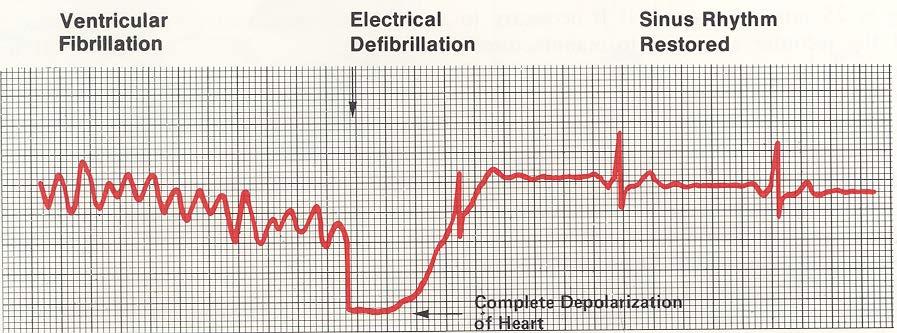
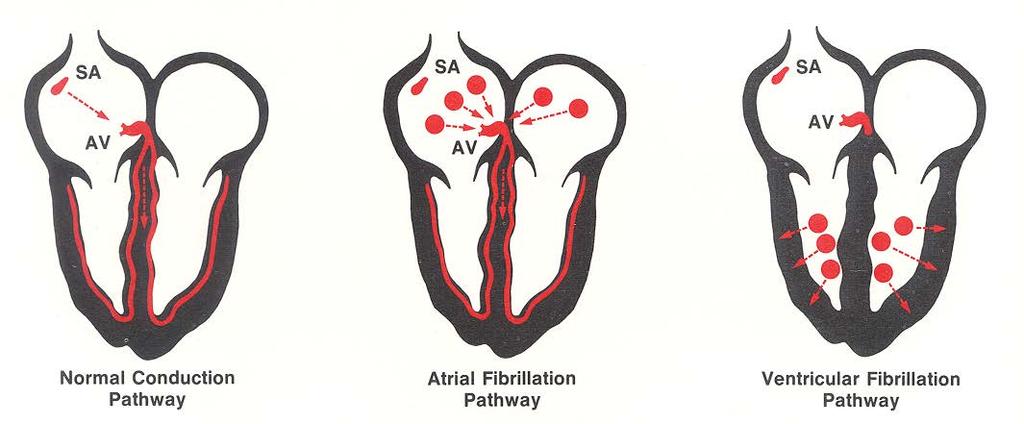
8 When things go awry. Atrial Fibrillation Ventricular Fibrillation 8
Analyzing the Heart with EKG
 Analyzing the Heart with EKG LabQuest An electrocardiogram (ECG or EKG) is a graphical recording of the electrical events occurring within the heart. In a healthy heart there is a natural pacemaker in
BIO 360: Vertebrate Physiology Performing and analyzing an EKG Lab 11: Performing and analyzing an EKG Lab report due April 17 th
 BIO 60: Vertebrate Physiology Lab : Lab report due April 7 th All muscles produce an electrical current when they contract. The heart is no exception. An electrocardiogram (ECG or EKG) is a graphical recording
Figure 1 muscle tissue to its resting state. By looking at several beats you can also calculate the rate for each component.
 ANALYZING THE HEART WITH EKG WITH LABQUEST LAB From Human Physiology with Vernier Westminster College INTRODUCTION An electrocardiogram (ECG or EKG) is a graphical recording of the electrical events occurring
Sample. Analyzing the Heart with EKG. Computer
 Analyzing the Heart with EKG Computer An electrocardiogram (ECG or EKG) is a graphical recording of the electrical events occurring within the heart. In a healthy heart there is a natural pacemaker in
Lab #3: Electrocardiogram (ECG / EKG)
") Lab #3: Electrocardiogram (ECG / EKG) An introduction to the recording and analysis of cardiac activity Introduction The beating of the heart is triggered by an electrical signal from the pacemaker. The
Lab #3: Electrocardiogram (ECG / EKG) An introduction to the recording and analysis of cardiac activity Introduction The beating of the heart is triggered by an electrical signal from the pacemaker. The
Cardiac Cycle. Each heartbeat is called a cardiac cycle. First the two atria contract at the same time.
 The Heartbeat Cardiac Cycle Each heartbeat is called a cardiac cycle. First the two atria contract at the same time. Next the two ventricles contract at the same time. Then all the chambers relax. http://www.youtube.com/watch?v=frd3k6lkhws
CAMOSUN COLLEGE BIOLOGY 144 (2010) LABS
 LABS") LAB 8: CARDIOVASCULAR PHYSIOLOGY PART 1. HEART SOUNDS AND PULSE DETERMINATIONS Introduction Two distinct sounds can be heard during each cardiac cycle. These sounds are commonly described as lub and dup
LAB 8: CARDIOVASCULAR PHYSIOLOGY PART 1. HEART SOUNDS AND PULSE DETERMINATIONS Introduction Two distinct sounds can be heard during each cardiac cycle. These sounds are commonly described as lub and dup
Cardiovascular Physiology
 Cardiovascular Physiology The mammalian heart is a pump that pushes blood around the body and is made of four chambers: right and left atria and right and left ventricles. The two atria act as collecting
Electrical Conduction
 Sinoatrial (SA) node Electrical Conduction Sets the pace of the heartbeat at 70 bpm AV node (50 bpm) and Purkinje fibers (25 40 bpm) can act as pacemakers under some conditions Internodal pathway from
Objectives of the Heart
 Objectives of the Heart Electrical activity of the heart Action potential EKG Cardiac cycle Heart sounds Heart Rate The heart s beat separated into 2 phases Relaxed phase diastole (filling of the chambers)
Lab #3: Electrocardiogram (ECG / EKG)
") Lab #3: Electrocardiogram (ECG / EKG) An introduction to the recording and analysis of cardiac activity Introduction The beating of the heart is triggered by an electrical signal from the pacemaker. The
Lab #3: Electrocardiogram (ECG / EKG) An introduction to the recording and analysis of cardiac activity Introduction The beating of the heart is triggered by an electrical signal from the pacemaker. The
LABORATORY INVESTIGATION
 LABORATORY INVESTIGATION Recording Electrocardiograms The taking of an electrocardiogram is an almost universal part of any complete physical examination. From the ECG record of the electrical activity
Lab 4: Introduction to Physiological Measurements - Cardiovascular
 Lab 4: Introduction to Physiological Measurements - Cardiovascular INTRODUCTION: This lab will demonstrate cardiovascular measurements by creating an ECG with instruments used in previous labs. Students
PART I: HEART ANATOMY
 Lab 7: Heart Sounds and Blood Pressure PART I: HEART ANATOMY a) You should be able to identify the following structures on an adult human heart diagram. the 4 chambers the bicuspid (mitral) and tricuspid
12.2 Monitoring the Human Circulatory System
 12.2 Monitoring the Human Circulatory System Video 1: 3D Animation of Heart Pumping Blood blood flow through the heart... Video 2: Hank Reviews Everything on the Heart https://www.youtube.com/watch?v=x9zz6tcxari
#6 - Cardiovascular III Heart Sounds, Pulse Rate, Hemoglobin Saturation, and Blood Pressure
 #6 - Cardiovascular III Heart Sounds, Pulse Rate, Hemoglobin Saturation, and Blood Pressure Objectives: Observe slide of artery and vein cross-section Auscultate heart sounds using a stethoscope Measure
Biology 13A Lab #10: Cardiovascular System II ECG & Heart Disease
 Biology 13A Lab #10: Cardiovascular System II ECG & Heart Disease Lab #10 Table of Contents: Expected Learning Outcomes...... 83 Introduction....... 84 Activity 1: Collecting ECG Data..... 85 Activity
Science in Sport. 204a ECG demonstration (Graph) Read. The Electrocardiogram. ECG Any 12 bit EASYSENSE. Sensors: Loggers: Logging time: 10 seconds
 Read. The Electrocardiogram. ECG Any 12 bit EASYSENSE. Sensors: Loggers: Logging time: 10 seconds") Sensors: Loggers: ECG Any 12 bit EASYSENSE Science in Sport Logging time: 10 seconds 204a ECG demonstration (Graph) Read Regular medical check ups are essential part of the life of a professional sports
Sensors: Loggers: ECG Any 12 bit EASYSENSE Science in Sport Logging time: 10 seconds 204a ECG demonstration (Graph) Read Regular medical check ups are essential part of the life of a professional sports
The Circulatory System. Lesson Overview. Lesson Overview The Circulatory System
 33.1 THINK ABOUT IT More than one-third of the 1.2 million Americans who suffer a heart attack each year die. This grim evidence shows that the heart and the circulatory system it powers are vital to life.
Blood Pressure Laboratory
 Introduction The blood that circulates throughout the body maintains a flow and pressure. The nervous system can change the flow and pressure based on the particular needs at a given time. For example,
IB TOPIC 6.2 THE BLOOD SYSTEM
 IB TOPIC 6.2 THE BLOOD SYSTEM TERMS TO KNOW circulation ventricle artery vein THE BLOOD SYSTEM 6.2.U1 - Arteries convey blood at high pressure from the ventricles to the tissues of the body Circulation
Biology 212: Anatomy and Physiology II. Lab #5: Physiology of the Cardiovascular System For Labs Associated With Dr. Thompson s Lectures
 Biology 212: Anatomy and Physiology II Lab #5: Physiology of the Cardiovascular System For Labs Associated With Dr. Thompson s Lectures References: Saladin, KS: Anatomy and Physiology, The Unity of Form
Large Arteries of Heart
 Cardiovascular System (Part A-2) Module 5 -Chapter 8 Overview Arteries Capillaries Veins Heart Anatomy Conduction System Blood pressure Fetal circulation Susie Turner, M.D. 1/5/13 Large Arteries of Heart
IB TOPIC 6.2 THE BLOOD SYSTEM
 IB TOPIC 6.2 THE BLOOD SYSTEM THE BLOOD SYSTEM TERMS TO KNOW circulation ventricle artery vein 6.2.U1 - Arteries convey blood at high pressure from the ventricles to the tissues of the body Circulation
Cardiac Conduction System
 Cardiac Conduction System What causes the Heart to Beat? Heart contracts by electrical signals! Cardiac muscle tissue contracts on its own an electrical signal is sent out by the heart so that all cells
Warm Up- Monday -AND- Setup Cornell Notes.
 Warm Up- Monday Brainstorm in your notebook: If the heart sends blood to all organs, how and where does the heart get blood to provide oxygen for its muscles? -AND- Setup Cornell Notes. Announcements Unit
Collin County Community College
 Collin County Community College BIOL. 2402 Anatomy & Physiology WEEK 5 The Heart 1 The Heart Beat and the EKG 2 1 The Heart Beat and the EKG P-wave = Atrial depolarization QRS-wave = Ventricular depolarization
37 1 The Circulatory System
 H T H E E A R T 37 1 The Circulatory System The circulatory system and respiratory system work together to supply cells with the nutrients and oxygen they need to stay alive. a) The respiratory system:
CRITICAL THINKING QUESTIONS AND ANSWERS AND CYCLE 2 LAB EXAM TEMPLATE. There are two main mechanisms that work in conjunction to return the blood
 CRITICAL THINKING QUESTIONS AND ANSWERS AND CYCLE 2 LAB EXAM TEMPLATE There are two main mechanisms that work in conjunction to return the blood THE CARDIAC PUMP 1) The forward pull(vis a fronte) This
12 Lead EKG. Brigham and Women's Hospital
 12 Lead EKG What is the heart? The heart is the organ that is responsible for pumping blood rich in oxygen to all parts of the body It is located in the center of the chest and is approximately the size
The Circulatory System
 The Circulatory System Key Questions What are the functions of the circulatory system? How does the heart pump blood through the body? What are three types of blood vessels? Vocabulary myocardium atrium
BIO 360: Vertebrate Physiology Lab 8b: Electrical activity of muscular contractions
 Lab report DUE 3/19 Muscle tissues maintain electrical imbalances, or potentials, across cell membranes by concentrating positive or negative charges on opposite sides of those membranes. These potentials
CARDIAC CYCLE CONTENTS. Divisions of cardiac cycle 11/13/13. Definition. Badri Paudel GMC
 CARDIAC CYCLE Badri Paudel GMC CONTENTS Ø DEFINATION Ø DIVISION OF CARDIAC CYCLE Ø SUB DIVISION AND DURATION OF CARDIAC CYCLE Ø SYSTOLE Ø DIASTOLE Ø DESCRIPTION OF EVENTS OF CARDIAC CYCLE Ø SUMMARY Ø ELECTROCARDIOGRAPHY
Heart Rate, Blood Pressure, and Exercise. Evaluation copy
 Heart Rate, Blood Pressure, and Exercise Computer 11 The adaptability of the heart can be observed during exercise, when the metabolic activity of skeletal muscles increases. The cardiovascular system,
Identify and describe the circulation system that is missing from the organizer above.
 Lesson 15.1 NOTES: The Circulatory System (Unlock) Essential Question: -What are the structures and functions of the circulatory system? Learning Target(s): -I can identify structures and explain functions
Physiology of the Circulatory System modified from
 Physiology of the Circulatory System modified from http://www.ekcsk12.org/science/aplabreview/aplab10.htm Introduction The circulatory system functions to deliver oxygen and nutrients to tissues for growth
Lab #10 Physiology of the Circulatory System
 Lab #10 Physiology of the Circulatory System Introduction The circulatory system functions to deliver oxygen an nutrients to tissues for growth and metabolism, and to remove metabolic wastes. The heart
Circulatory system of mammals
 Circulatory system of mammals Explain the cardiac cycle and its initiation Discuss the internal factors that control heart action Blood flows through the heart as a result of pressure differences Blood
Unit 1: Human Systems. The Circulatory System
 Unit 1: Human Systems The Circulatory System nourish all cells with oxygen, glucose, amino acids and other nutrients and carry away carbon dioxide, urea and other wastes Purposes Transport chemical messengers
Human Cardiovascular Physiology: Blood Pressure and Pulse Determinations
 ighapmlre33apg269_274 5/12/04 3:10 PM Page 269 impos03 302:bjighapmL:ighapmLrevshts:layouts: NAME Human Cardiovascular Physiology: Blood Pressure and Pulse Determinations LAB TIME/DATE REVIEW SHEET exercise
Human Anatomy and Physiology II Laboratory Cardiovascular Physiology
 Human Anatomy and Physiology II Laboratory Cardiovascular Physiology 1 This lab involves two exercises: 1) Conduction System of the Heart and Electrocardiography and 2) Human Cardiovascular Physiology:
Cardiovascular System Notes: Heart Disease & Disorders
 Cardiovascular System Notes: Heart Disease & Disorders Interesting Heart Facts The Electrocardiograph (ECG) was invented in 1902 by Willem Einthoven Dutch Physiologist. This test is still used to evaluate
Interpreting Electrocardiograms (ECG) Physiology Name: Per:
 Physiology Name: Per:") Interpreting Electrocardiograms (ECG) Physiology Name: Per: Introduction The heart has its own system in place to create nerve impulses and does not actually require the brain to make it beat. This electrical
Interpreting Electrocardiograms (ECG) Physiology Name: Per: Introduction The heart has its own system in place to create nerve impulses and does not actually require the brain to make it beat. This electrical
Electrocardiogram sensor (ECG/EKG sensor)
") [KDS-1040] Electrocardiogram sensor (ECG/EKG sensor) User's Manual Note This product is designed for educational use only. Not recommended for industrial, medical, commercial use. What is Electrocardiogram?
[KDS-1040] Electrocardiogram sensor (ECG/EKG sensor) User's Manual Note This product is designed for educational use only. Not recommended for industrial, medical, commercial use. What is Electrocardiogram?
current, and acting like
 Heart 10 IV. HEART PHYSIOLOGY - How the heart beats. How the heart depolarizes the myocardium, which leads to a contraction. A) INTRINSIC CONTROL - Heart controls its own rhythm. HOW? The presence of gap
The cardiovascular system is composed of the heart and blood vessels that carry blood to and from the body s organs. There are 2 major circuits:
 1 The cardiovascular system is composed of the heart and blood vessels that carry blood to and from the body s organs. There are 2 major circuits: pulmonary and systemic. The pulmonary goes out to the
Heart. Heart 2-Tunica media: middle layer (media ='middle') muscle fibers (smooth or cardiac).
 muscle fibers (smooth or cardiac).") t. innermost lumenal General Circulatory system heart and blood vessels walls have 3 layers (inside to outside) 1-Tunica interna: aka tunica intima layer--lumenal layer epithelium--endothelium simple squamous
t. innermost lumenal General Circulatory system heart and blood vessels walls have 3 layers (inside to outside) 1-Tunica interna: aka tunica intima layer--lumenal layer epithelium--endothelium simple squamous
Unit 6: Circulatory System. 6.2 Heart
 Unit 6: Circulatory System 6.2 Heart Functions of Circulatory System 1. The heart is the pump necessary to circulate blood to all parts of the body 2. Arteries, veins and capillaries are the structures
Introduction to Lesson 2 - Heartbeat
 Introduction to Lesson 2 - Heartbeat Activity: Locate your pulse at rest. Count how many times it beats in 15 seconds (look at a clock), then multiply this number by 4. This is your pulse rate Approximately
The Heart. Happy Friday! #takeoutyournotes #testnotgradedyet
 The Heart Happy Friday! #takeoutyournotes #testnotgradedyet Introduction Cardiovascular system distributes blood Pump (heart) Distribution areas (capillaries) Heart has 4 compartments 2 receive blood (atria)
4. The two inferior chambers of the heart are known as the atria. the superior and inferior vena cava, which empty into the left atrium.
 Answer each statement true or false. If the statement is false, change the underlined word to make it true. 1. The heart is located approximately between the second and fifth ribs and posterior to the
WHAT S THAT RHYTHM I AM HEARING? GUIDE TO AUSCULTATION OF ARRHYTHMIAS IN HORSES
 WHAT S THAT RHYTHM I AM HEARING? GUIDE TO AUSCULTATION OF ARRHYTHMIAS IN HORSES Michelle Henry Barton DVM, PhD, DACVIM University of Georgia, Athens, GA INTRODUCTION The purpose of this talk is to review
The Mammalian Circulatory System
 The Mammalian Heart The Mammalian Circulatory System Recall: What are the 3 cycles of the mammalian circulatory system? What are their functions? What are the three main vessel types in the mammalian circulatory
The Circulatory System (p )
") The Circulatory System (p. 268-281) How Does Gravity Affect Blood Circulation? As with all land animals, the giraffe and the corn snake are constantly subject to the force of gravity The circulatory system
The Circulatory System (p. 268-281) How Does Gravity Affect Blood Circulation? As with all land animals, the giraffe and the corn snake are constantly subject to the force of gravity The circulatory system
INTRODUCTION TO ECG. Dr. Tamara Alqudah
 INTRODUCTION TO ECG Dr. Tamara Alqudah Excitatory & conductive system of the heart + - The ECG The electrocardiogram, or ECG, is a simple & noninvasive diagnostic test which records the electrical
EKG Sensor Product Number: ENEKG189
 imagine explore learn EKG Sensor Product Number: ENEKG189 Overview An electrocardiogram abbreviated as EKG or ECG is a test that measures the electrical activity of the heartbeat. With each beat, an electrical
LAB 9: Metabolic Rates
 LAB 9: Metabolic Rates Introduction: The cardiovascular (circulatory) system functions to deliver oxygen and nutrients to tissues for growth and metabolism, and to remove metabolic wastes. The heart pumps
Heart Rate and Blood Pressure as Vital Signs
 Heart Rate and Blood Pressure as Vital Signs Computer 10 Since the earliest days of medicine heart rate has been recognized as a vital sign an indicator of health, disease, excitement, and stress. Medical
Lab 16. The Cardiovascular System Heart and Blood Vessels. Laboratory Objectives
 Lab 16 The Cardiovascular System Heart and Blood Vessels Laboratory Objectives Describe the anatomical structures of the heart to include the pericardium, chambers, valves, and major vessels. Describe
Cardiovascular System Notes: Physiology of the Heart
 Cardiovascular System Notes: Physiology of the Heart Interesting Heart Fact Capillaries are so small it takes ten of them to equal the thickness of a human hair. Review What are the 3 parts of the cardiovascular
Evaluation copy. EMG and Muscle Fatigue. Computer
 EMG and Muscle Fatigue Computer 18 Voluntary muscle contraction is the result of communication between the brain and individual muscle fibers of the musculoskeletal system. A thought is transformed into
Chapter 13 The Cardiovascular System: Cardiac Function
 Chapter 13 The Cardiovascular System: Cardiac Function Overview of the Cardiovascular System The Path of Blood Flow through the Heart and Vasculature Anatomy of the Heart Electrical Activity of the Heart
THE HEART THE CIRCULATORY SYSTEM
 THE HEART THE CIRCULATORY SYSTEM There are three primary closed cycles: 1) Cardiac circulation pathway of blood within the heart 2) Pulmonary circulation blood from the heart to lungs and back 3) Systemic
Name Class Date. Bell Diaphragm. Chest piece. Stethoscope
 Name Class Date Sensing Circulation Exploring the Effects of Exercise on Heart Rate Investigative Lab 30 8 Questions How do the sounds you hear through a stethoscope relate to the stages of a heartbeat?
Lab 7. Physiology of Electrocardiography
 7.1 Lab 7. Physiology of Electrocardiography The heart is a muscular pump that circulates blood throughout the body. To efficiently pump the blood, cardiac contractions must be coordinated and are regulated
Outline. Electrical Activity of the Human Heart. What is the Heart? The Heart as a Pump. Anatomy of the Heart. The Hard Work
 Electrical Activity of the Human Heart Oguz Poroy, PhD Assistant Professor Department of Biomedical Engineering The University of Iowa Outline Basic Facts about the Heart Heart Chambers and Heart s The
The Cardiovascular and Lymphatic Systems
 BIOLOGY OF HUMANS Concepts, Applications, and Issues Fifth Edition Judith Goodenough Betty McGuire 12 The Cardiovascular and Lymphatic Systems Lecture Presentation Anne Gasc Hawaii Pacific University and
Principles of Biomedical Systems & Devices. Lecture 8: Cardiovascular Dynamics Dr. Maria Tahamont
 Principles of Biomedical Systems & Devices Lecture 8: Cardiovascular Dynamics Dr. Maria Tahamont Review of Cardiac Anatomy Four chambers Two atria-receive blood from the vena cave and pulmonary veins Two
CRC 431 ECG Basics. Bill Pruitt, MBA, RRT, CPFT, AE-C
 CRC 431 ECG Basics Bill Pruitt, MBA, RRT, CPFT, AE-C Resources White s 5 th ed. Ch 6 Electrocardiography Einthoven s Triangle Chest leads and limb leads Egan s 10 th ed. Ch 17 Interpreting the Electrocardiogram
Topic 6: Human Physiology
 Topic 6: Human Physiology 6.2 The Blood System D.4 The Heart Essential Questions: 6.2 The blood system continuously transports substances to cells and simultaneously collects waste products. D.3 The chemical
Introduction to EMG. Figure 1
 Experiment 13 An electromyogram, or EMG, is a graphical recording of electrical activity within muscles. Activation of muscles by nerves results in changes in ion flow across cell membranes, which generates
A. Incorrect! The left ventricle receives oxygenated blood from the lungs via the left atrium.
 Anatomy and Physiology - Problem Drill 16: The Cardiovascular System No. 1 of 10 Instruction: (1) Read the problem statement and answer choices carefully (2) Work the problems on paper as needed (3) Pick
PLANK 1 Direct Care Staff Trained in Accurate BP Measurement
 Direct Care Staff Trained in Accurate BP Measurement 1. 2. 3. 5. 22 to 26 cm Small adult (12X22 cm) 27 to 34 cm Adult (16X30 cm) 35 to 44 cm Large adult (16X36 cm) 45 to 52 cm Adult thigh (16X42 cm) widths
HASPI Medical Anatomy & Physiology 13a Station Lab Activity
 HASPI Medical Anatomy & Physiology 13a Station Lab Activity Name(s): Period: Date: The Cardiovascular System The cardiovascular system is made up of the heart, blood, and blood vessels. It functions as
Biology Unit 3 The Human Heart P
 Biology 2201 Unit 3 The Human Heart P 314-321 Structure and Function of the Human Heart Structure of the Human Heart Has four Chambers (2 Atria and 2 Ventricles) Made of Cardiac Muscle Found in Chest Cavity
The Function of an ECG in Diagnosing Heart Conditions. A useful guide to the function of the heart s electrical system for patients receiving an ECG
 The Function of an ECG in Diagnosing Heart Conditions A useful guide to the function of the heart s electrical system for patients receiving an ECG Written by Erhan Selvi July 28, 2014 Audience and Scope
Control of Heart Rate
 Control of Heart Rate Control of Heart Rate The beating of your heart is an involuntary movement one that is beyond your direct control. The nerve impulse that causes the heart to beat originates within
following: the readout..
 Read Me Vital Signs In-Lab Guide We will be studying 5 concepts in lab 1. Study the anatomy of the Intrinsic Conduction System using heart models. 2. Study the ECG tracings, including the following: -
LAB: Blood Pressure Measurable Indicator of the Health of the Circulatory System!
 LAB: Blood Measurable Indicator of the Health of the Circulatory System! Lab Objectives. At the completion of the lab, you should be able to: measure pulse or heart rate (HR) and respiratory rate (RR);
THE CARDIOVASCULAR SYSTEM : (circulatory system) Lab-4
 Lab-4") THE CARDIOVASCULAR SYSTEM : (circulatory system) Lab-4 The Circulatory System: The circulatory system (cardiovascular system) has 3 basic components:- *A muscular pump: heart *Interconnecting tubes: blood
THE CARDIOVASCULAR SYSTEM : (circulatory system) Lab-4 The Circulatory System: The circulatory system (cardiovascular system) has 3 basic components:- *A muscular pump: heart *Interconnecting tubes: blood
The Electrocardiogram
 The Electrocardiogram Chapters 11 and 13 AUTUMN WEDAN AND NATASHA MCDOUGAL The Normal Electrocardiogram P-wave Generated when the atria depolarizes QRS-Complex Ventricles depolarizing before a contraction
Cardiac Telemetry Self Study: Part One Cardiovascular Review 2017 THINGS TO REMEMBER
 Please review the above anatomy of the heart. THINGS TO REMEMBER There are 3 electrolytes that affect cardiac function o Sodium, Potassium, and Calcium When any of these electrolytes are out of the normal
Pearson's Comprehensive Medical Assisting Administrative and Clinical Competencies
 Pearson's Comprehensive Medical Assisting Administrative and Clinical Competencies THIRD EDITION CHAPTER 27 The Cardiovascular System Lesson 1: Overview of the Cardiovascular System Lesson Objectives Upon
The Heart and Cardiovascular System
 The Heart and Cardiovascular System What you will learn The location of the heart 3 layers and covering of the heart Explain the function of the heart as 2 separate pumps Identify the 4 chambers of the
THE CARDIOVASCULAR SYSTEM. Heart 2
 THE CARDIOVASCULAR SYSTEM Heart 2 PROPERTIES OF CARDIAC MUSCLE Cardiac muscle Striated Short Wide Branched Interconnected Skeletal muscle Striated Long Narrow Cylindrical PROPERTIES OF CARDIAC MUSCLE Intercalated
How Do We Sense, Think, and Move? -- Lab #11 Bioelectronics Measuring Electrical Properties of the Body
 How Do We Sense, Think, and Move? -- Lab #11 Bioelectronics Measuring Electrical Properties of the Body Experiment #1 Your Body's Resistance Equipment: Digital multimeter, Banana leads Important Equipment
Do Now. Get out work from last class to be checked
 Do Now Get out work from last class to be checked Heart Actions Cardiac Cycle: One complete heartbeat. The contraction of a heart chamber is called systole and the relaxation of a chamber is called diastole.
Circulation. Circulation = is a process used for the transport of oxygen, carbon! dioxide, nutrients and wastes through-out the body
 Circulation Circulation = is a process used for the transport of oxygen, carbon! dioxide, nutrients and wastes through-out the body Heart = muscular organ about the size of your fist which pumps blood.
ELECTROCARDIOGRAPHY (ECG)
") ELECTROCARDIOGRAPHY (ECG) The heart is a muscular organ, which pumps blood through the blood vessels of the circulatory system. Blood provides the body with oxygen and nutrients, as well as assists in
ELECTROCARDIOGRAPHY (ECG) The heart is a muscular organ, which pumps blood through the blood vessels of the circulatory system. Blood provides the body with oxygen and nutrients, as well as assists in
The Heart A-Chapter Identify the layers of the heart wall and state the type of tissue in each layer.
 Objectives: The Heart AChapter 12 1. Identify the layers of the heart wall and state the type of tissue in each layer. Label a diagram of the heart, identifying the chambers, valves, and associated vessels.
Protocol 4: Measuring Blood Pressure
 Zool 430L Protocols Page 8 of 12 Protocol 4: Measuring Blood Pressure 4.1 Via Auscultation (listening through stethoscope) The estimate of blood pressure is obtained by cutting off blood flow with a known
The Cardiovascular and Lymphatic Systems Cardiovascular System Blood Vessels Blood Vessels Arteries Arteries Arteries
 CH 12 The Cardiovascular and s The Cardiovascular and s OUTLINE: Cardiovascular System Blood Vessels Blood Pressure Cardiovascular System The cardiovascular system is composed of Blood vessels This system
BUSINESS. Articles? Grades Midterm Review session
 BUSINESS Articles? Grades Midterm Review session REVIEW Cardiac cells Myogenic cells Properties of contractile cells CONDUCTION SYSTEM OF THE HEART Conduction pathway SA node (pacemaker) atrial depolarization
Practice Exercises for the Cardiovascular System
 Practice Exercises for the Cardiovascular System On the diagram below, color the oxygen-rich blood red and the oxygen-poor blood blue. Label the parts: Continued on the next page... Label the parts on
2The Concept of Periodic Functions
 The Concept of Periodic Functions EKG INTRODUCTION The heart is a fist-sized muscle that acts as an electrical generator lying in a conducting medium made of body tissue and fluids. The heart pumps oxygen-rich
ECE ECE PRINCIPLES OF BIOMEDICAL SYSTEMS & DEVICES LAB 1 - ELECTROCARDIOGRAM
 ECE0909.404.01 ECE 0909.504.03 PRINCIPLES OF BIOMEDICAL SYSTEMS & DEVICES LAB 1 - ELECTROCARDIOGRAM The purpose of this laboratory is to introduce you to electrocardiogram, its acquisition and interpretation.
Birmingham Regional Emergency Medical Services System
 Birmingham Regional Emergency Medical Services System 2018 ALCTE Summer Conference EKG Basics Brian Gober, MAT, ATC, NRP, CSCS Education Services Manager ECC Training Center Coordinator Birmingham Regional
Electrocardiography I Laboratory
 Introduction The body relies on the heart to circulate blood throughout the body. The heart is responsible for pumping oxygenated blood from the lungs out to the body through the arteries and also circulating
d) Cardiovascular System Higher Human Biology
 Cardiovascular System Higher Human Biology") d) Cardiovascular System Higher Human Biology What can your remember about the heart and blood vessels? What is the Cardiovascular System? The cardiovascular system, also known as the circulatory system,
d) Cardiovascular System Higher Human Biology What can your remember about the heart and blood vessels? What is the Cardiovascular System? The cardiovascular system, also known as the circulatory system,