The Fontan circulation. Folkert Meijboom
|
|
|
- Ethelbert Farmer
- 6 years ago
- Views:
Transcription
1 The Fontan circulation Folkert Meijboom
2 What to expect? Why a Fontan-circulation Indications How does it work Types of Fontan circulation Historical overview Role of echocardiography
3 What to expect? Why a Fontan-circulation Indications How does it work Types of Fontan circulation Historical overview Role of echocardiography
4 Why a Fontan circulation? No functional RV Volume overload LV systemic venous return plus pulmonary venous return Desaturation systemic circulation - cyanosis How van this be solved?

5 Why a Fontan circulation? No functional RV Volume overload LV systemic venous return plus pulmonary venous return Desaturation systemic circulation - cyanosis Not by a biventricular repair Univentricular repair
6 Indication for a Fontan situations for which a biventricular is not possible
7 Indication for a Fontan situations for which a biventricular is not possible HLHS TA MA DILV Unbalanced AVSD Ebstein PA intact IVS etc
8 The essence of the Fontan circulation only 1 functional ventricle systemic ventricle
9 The essence of the Fontan circulation only 1 functional ventricle systemic ventricle no subpulmonary ventricle
10 The essence of the Fontan circulation only 1 functional ventricle systemic ventricle no subpulmonary ventricle Systemic venous return flows, without a pumping ventricle, into the lungs
11 The essence of the Fontan circulation only 1 functional ventricle systemic ventricle no subpulmonary ventricle Systemic venous return flows, without a pumping ventricle, into the lungs no desaturated blood in the systemic circulation
12 The essence of the Fontan circulation only 1 functional ventricle systemic ventricle no subpulmonary ventricle Systemic venous return flows, without a pumping ventricle, into the lungs no desaturated blood in the systemic circulation No longer a volume-overloaded LV
13 How does it work? How can an adequate pulmonary blood flow be achieved? 1. elevated systemic venous pressure 2. low pulmonary vascular resistance 3. Low LA pressures


14 How does it work? Flow depends on pressure difference between systemic venous pressure and LA pressure and pulmonary vascular resistance
15 Pressure difference VCS - LA Push & pull circulation: Elevated systemic venous pressure = push Periodic lowering of LA pressure During systole During rapid filling of ventricle pull
16 a c v y x LA pressure curve = the pull systole H.Hamer, AZG, Groningen

17 Anatomy (history) of the Fontan-operation H.Hamer, AZG, Groningen
18 the patient with a Fontan circulation Does not exist
19 the Fontan circulation Mixed bag many different types of Intracardiac anatomy Type of surgical repair
20 Echocardiography & Fontan Know what the underlying cardiac defect is Know what type of Fontan repair is done Understand the Fontan circulation
21 Echocardiography & Fontan Know what the underlying cardiac defect is Know what type of Fontan repair is done Understand the Fontan circulation Read the surgical report!!
22 Echocardiography & Fontan Know what the underlying cardiac defect is Know what type of Fontan repair is done Understand the Fontan circulation If you do not know where you are looking for, you may not find it
23 Echocardiography & Fontan in univentricular hearts before surgery echo is important for: Intracardiac anatomy Ventricular function AV valve function Biventricular - univentricular repair
24 Echocardiography & Fontan Univentricular hearts after Fontan (Intracardiac anatomy) Ventricular function AV valve function
25 Echocardiography & Fontan Univentricular hearts after Fontan (Intracardiac anatomy) Ventricular function AV valve function Anatomy & function of the Fontan circulation
26 Echocardiography & Fontan Univentricular hearts after Fontan (Intracardiac anatomy) Ventricular function AV valve function Anatomy & function of the Fontan circulation
27 ventricular function & Fontan

28 ventricular & AV valve function

29 ventricular & AV valve function

30 ventricular & AV valve function
31 AV valve function Assessment of AV valve regurgitation = very important Moderate to severe regurgitation elevated LA pressure decrease in transpulmonary gradient decrease in transpulmonary flow decrease in cardiac output compensatory mechanism = fluid retention elevated central venous pressure transpulmonary gradient restored cardiac output restored
32 AV valve function Assessment of AV valve regurgitation = very important Moderate to severe regurgitation elevated LA pressure decrease in transpulmonary gradient decrease in transpulmonary flow decrease in cardiac output compensatory mechanism = fluid retention elevated central venous pressure transpulmonary gradient restored cardiac output restored To a certain limit decompensation
33 Fontan & AV valve function Assessment of AV valve regurgitation = very important If one sees development of AV valve regurgitation Alarm signs Worsening ventricular function How to assess AV valve regurgitation: beyond the scope of this talk
34 Fontan & ventricular function How do we assess & measure function of the systemic ventricle?
35 Fontan & ventricular function EAE & ASE recommendations on chamber quantification LV = systemic ventricle Standardized cross sections 4Ch view Parasternal long axis Normal values for sex and body size
36 Fontan & ventricular function EAE & ASE recommendations on chamber quantification Standardized cross sections??? 4Ch view Parasternal long axis Normal values for sex and body size But embrace the idea behind it: Use reproducible cross section Anatomic landmarks DILV HLFS unbalanced AVSD
37 Fontan &ventricular function Eyeballing Quantify what you can quantify, in a reproducible way Essential for longitudinal follow-up M-mode 2D 3D Doppler (incl TDI) not for comparsion with normal values; patient as his/her own control

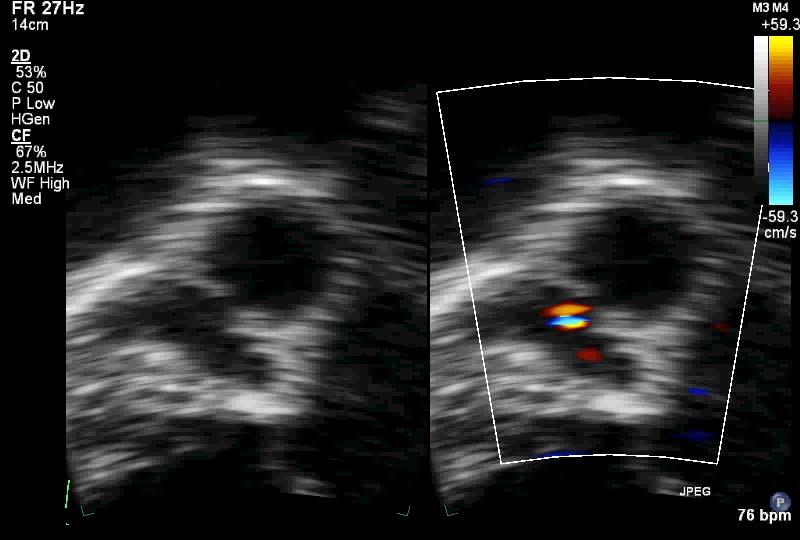
38 2D & color Doppler
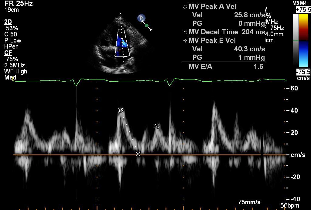
39 Inflow

40 Tissue Doppler

41 Ejection time, Tei-index

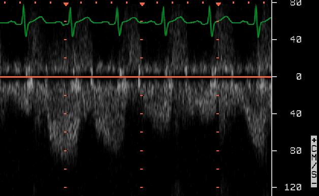
42 Relative duration systole in cardiac cycle (good functioning adult Fontan)

43 duration systole, incl IVCT & IVRT (poorly functioning adult Fontan)

44 duration systole very short diastole (3 yr old with HLHS)
45 SD ratio in the RV Friedberg, J Am Soc Echocardiogr 2007;20:749

46 duration systole short diastole (21 yr old criss-cross, TCPC with PLE)
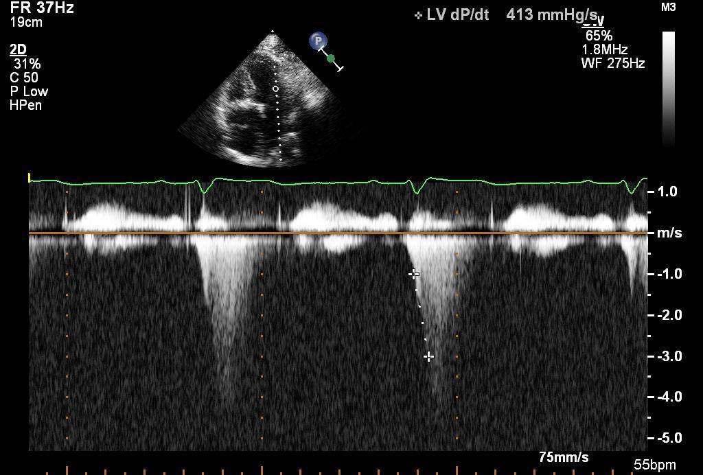
47 CW dp/dt
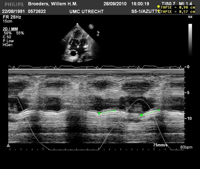
48 TAPSE
49 PV flow pattern: very low S in absence MR
50 a c v y x LA pressure curve systole
51 Echo & Doppler measurements Absolute values difficult to interpret Change over time/ in different conditions may guide us: in clinical decision making When to send a patient to the cathlab
52 Echo & Doppler measurements Absolute values difficult to interpret Change over time/ in different conditions may guide us: in clinical decision making When to send a patient to the cathlab The essence of the Fontan circulation: V = P/R we cannot measure this with echo
53 Visualisation of Fontan anatomy
54 Visualisation of Fontan anatomy Segmental approach Hepatic veins VCI Tunnel Intracardiac Extracardiac RA Connection with PA VCS Connection with PA Pulmonary artery branches Pulmonary veins
55 Visualisation of Fontan anatomy & function Segmental approach Hepatic veins VCI Color Doppler patterns Tunnel Intracardiac Pulsed Doppler flow patterns Extracardiac RA Shunts/leaks in the tunnel Connection with PA VCS thrombi Connection with PA Pulmonary artery branches Pulmonary veins

56 VCI & hepatic veins
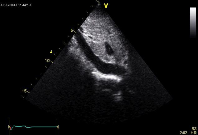
57

58 VCI pulsed Doppler: Scale! Low velocity filter! Respiration!

59 VCI pulsed Doppler: Scale! Low velocity filter! Respiration!

60 TCPC driving force = inspiration no influence of cardiac cycle

61 Connection VCI extracardiac lateral tunnel

62 Lateral, extracardiac tunnel look for thrombi

63 Apical 4 Ch view intracardiac tunnel

64 Intra-atrial tunnel Intra-atrial tunnel initially a small diameter No PV obstruction Apical scan; upside down

65 Intra-atrial tunnel; adult age can dilate in time, become very wide can give PV obstruction Looks like RA PA (different flow patterns)

66 VCI Doppler in RA PA conduit effect of atrial contraction
67 Tricuspid atresia; RA AP connectie

68 RA PA conduit

69 Atrio ventricular connection
70 Tricuspid atresia & RA RV connectie

71 RA RV conduit with stenosis


72 PA flow in RA RV conduit: Pulsatile systolic flow in PA!
73 Doppler patterns & type of Fontan Doppler patterns in different types of connection are different Doppler patterns in different patients with the same type of connections are different registration of ECG p-waves and respiration curve Off-line studying on tracings: What explains flow patterns? What explains phasic changes?

74 intracardiac tunnel VCI - PA
75 Look for shunts/abnormal flows

76 Baffle leakage with R L shunt

77 Baffle leakage with R L shunt Courtesy Jan Marek
78 What to do with a baffle leak? Beyond the scope of this talk
79 Bidirectional Glenn anastomosis

80 VCS Glenn from R supraclavicular
81 VCS Glenn from R supraclavicular
82 Glenn anastomose VCS

83 APL from SSN; almost always obtainable

84 APL bij TCPC

85 From SSN: LPA

86 From SSN: LPA

87 APL in LA AP connection
88
89 R upper pulmonary vein

90 PV Doppler
 PW AP4K Always")

91 RUPV obstruction (RA RV connection) PW AP4K Always look for the R pulmonary veins, especially if the IAS is bulging

92 Dilated RA in RA-AP connection beware of R PV obstruction

93 RUPV obstruction Pulsed wave Doppler ( pull-back )
94 Conclusion
95 Conclusion Why a Fontan-circulation Indications How does it work Types of Fontan circulation Historical overview essential for the understanding of echo& Doppler images Role of echocardiography in the follow-up Measuring function in a reproducible way for longitudinal follow up
96 Conclusion Fontan & echo = difficult & fascinating You need to understand Fontan physiology, especially when you consider intervention (elective redo of old RA- PA to TCPC) Echo is very helpful, but do not hesitate to send a patient to the cathlab when in doubt V= P/R
Atrial Septal Defects
 Supplementary ACHD Echo Acquisition Protocol for Atrial Septal Defects The following protocol for echo in adult patients with atrial septal defects (ASDs) is a guide for performing a comprehensive assessment
Adult Echocardiography Examination Content Outline
 Adult Echocardiography Examination Content Outline (Outline Summary) # Domain Subdomain Percentage 1 2 3 4 5 Anatomy and Physiology Pathology Clinical Care and Safety Measurement Techniques, Maneuvers,
Appendix A.1: Tier 1 Surgical Procedure Terms and Definitions
 Appendix A.1: Tier 1 Surgical Procedure Terms and Definitions Tier 1 surgeries AV Canal Atrioventricular Septal Repair, Complete Repair of complete AV canal (AVSD) using one- or two-patch or other technique,
ADULT CONGENITAL HEART DISEASE. Stuart Lilley
 ADULT CONGENITAL HEART DISEASE Stuart Lilley More adults than children have congenital heart disease Huge variety of congenital lesions from minor to major Heart failure, re-operation and arrhythmia are
Hemodynamic Assessment. Assessment of Systolic Function Doppler Hemodynamics
 Hemodynamic Assessment Matt M. Umland, RDCS, FASE Aurora Medical Group Milwaukee, WI Assessment of Systolic Function Doppler Hemodynamics Stroke Volume Cardiac Output Cardiac Index Tei Index/Index of myocardial
The Fontan operation. Choussat s 10 commandments revisited. Early palliative approach: systemic to pulmonary shunt or banding
 Echographic assessment in Fontan patients: sequential segmental approach Functionnaly Single Ventricle Glenn and Fontan echocardiographic assessment Xavier Iriart 1. Cavopulmonary connection 2. Branch
TGA atrial vs arterial switch what do we need to look for and how to react
 TGA atrial vs arterial switch what do we need to look for and how to react Folkert Meijboom, MD, PhD, FES Dept ardiology University Medical entre Utrecht The Netherlands TGA + atrial switch: Follow-up
Echocardiography in Adult Congenital Heart Disease
 Echocardiography in Adult Congenital Heart Disease Michael Vogel Kinderherz-Praxis München CHD missed in childhood Subsequent lesions after repaired CHD Follow-up of cyanotic heart disease CHD missed in
CMR for Congenital Heart Disease
 CMR for Congenital Heart Disease * Second-line tool after TTE * Strengths of CMR : tissue characterisation, comprehensive access and coverage, relatively accurate measurements of biventricular function/
Echocardiographic assessment of the right ventricle in paediatric pulmonary hypertension.
 Echocardiographic assessment of the right ventricle in paediatric pulmonary hypertension. Mark K. Friedberg, MD No disclosures Outline RV response to increased afterload Echo assessment of RV function
Cardiac MRI in ACHD What We. ACHD Patients
 Cardiac MRI in ACHD What We Have Learned to Apply to ACHD Patients Faris Al Mousily, MBChB, FAAC, FACC Consultant, Pediatric Cardiology, KFSH&RC/Jeddah Adjunct Faculty, Division of Pediatric Cardiology
가천의대길병원소아심장과최덕영 PA C IVS THE EVALUATION AND PRINCIPLES OF TREATMENT STRATEGY
 가천의대길병원소아심장과최덕영 PA C IVS THE EVALUATION AND PRINCIPLES OF TREATMENT STRATEGY PA c IVS (not only pulmonary valve disease) Edwards JE. Pathologic Alteration of the right heart. In: Konstam MA, Isner M, eds.
COMPLEX CONGENITAL HEART DISEASE: WHEN IS IT TOO LATE TO INTERVENE?
 COMPLEX CONGENITAL HEART DISEASE: WHEN IS IT TOO LATE TO INTERVENE? Aurora S. Gamponia, MD, FPPS, FPCC, FPSE OBJECTIVES Identify complex congenital heart disease at high risk or too late for intervention
Echocardiography as a diagnostic and management tool in medical emergencies
 Echocardiography as a diagnostic and management tool in medical emergencies Frank van der Heusen MD Department of Anesthesia and perioperative Care UCSF Medical Center Objective of this presentation Indications
FUNCTIONALLY SINGLE VENTRICLE
 MORPHOLOGICAL DETERMINANTS VI TRAN EuroEcho, Budapest, 7 th December 2011 DECLARATION OF CONFLICT OF INTEREST: I have nothing to declare What is the functionally single ventricle? The heart that is incapable
Echocardiography: Guidelines for Valve Quantification
 Echocardiography: Guidelines for Echocardiography: Guidelines for Chamber Quantification British Society of Echocardiography Education Committee Richard Steeds (Chair), Gill Wharton (Lead Author), Jane
DOPPLER HEMODYNAMICS (1) QUANTIFICATION OF PRESSURE GRADIENTS and INTRACARDIAC PRESSURES
 QUANTIFICATION OF PRESSURE GRADIENTS and INTRACARDIAC PRESSURES") THORAXCENTRE DOPPLER HEMODYNAMICS (1) QUANTIFICATION OF PRESSURE GRADIENTS and INTRACARDIAC PRESSURES J. Roelandt DOPPLER HEMODYNAMICS Intracardiac pressures and pressure gradients Volumetric measurement
THORAXCENTRE DOPPLER HEMODYNAMICS (1) QUANTIFICATION OF PRESSURE GRADIENTS and INTRACARDIAC PRESSURES J. Roelandt DOPPLER HEMODYNAMICS Intracardiac pressures and pressure gradients Volumetric measurement
Transposition of the great arteries
 EuroEcho 2010 - Teaching course on CHD Transposition of the great arteries - Follow-up after the arterial switch Gertjan Tj. Sieswerda, MD PhD Nothing to disclose Interuniversitary Institute for Congenital
Adult Congenital Heart Disease: What All Echocardiographers Should Know Sharon L. Roble, MD, FACC Echo Hawaii 2016
 1 Adult Congenital Heart Disease: What All Echocardiographers Should Know Sharon L. Roble, MD, FACC Echo Hawaii 2016 DISCLOSURES I have no disclosures relevant to today s talk 2 Why should all echocardiographers
Appendix II: ECHOCARDIOGRAPHY ANALYSIS
 Appendix II: ECHOCARDIOGRAPHY ANALYSIS Two-Dimensional (2D) imaging was performed using the Vivid 7 Advantage cardiovascular ultrasound system (GE Medical Systems, Milwaukee) with a frame rate of 400 frames
Right Ventricle Steven J. Lester MD, FACC, FRCP(C), FASE Mayo Clinic, Arizona
, FASE Mayo Clinic, Arizona") Right Ventricle Steven J. Lester MD, FACC, FRCP(C), FASE Mayo Clinic, Arizona 1. In which scenario will applying the simplified Bernoulli equation to the peak tricuspid regurgitation velocity and adding
Right Ventricle Steven J. Lester MD, FACC, FRCP(C), FASE Mayo Clinic, Arizona 1. In which scenario will applying the simplified Bernoulli equation to the peak tricuspid regurgitation velocity and adding
PART II ECHOCARDIOGRAPHY LABORATORY OPERATIONS ADULT TRANSTHORACIC ECHOCARDIOGRAPHY TESTING
 PART II ECHOCARDIOGRAPHY LABORATORY OPERATIONS ADULT TRANSTHORACIC ECHOCARDIOGRAPHY TESTING STANDARD - Primary Instrumentation 1.1 Cardiac Ultrasound Systems SECTION 1 Instrumentation Ultrasound instruments
MITRAL STENOSIS. Joanne Cusack
 MITRAL STENOSIS Joanne Cusack BSE Breakdown Recognition of rheumatic mitral stenosis Qualitative description of valve and sub-valve calcification and fibrosis Measurement of orifice area by planimetry
Diastolic Function: What the Sonographer Needs to Know. Echocardiographic Assessment of Diastolic Function: Basic Concepts 2/8/2012
 Diastolic Function: What the Sonographer Needs to Know Pat Bailey, RDCS, FASE Technical Director Beaumont Health System Echocardiographic Assessment of Diastolic Function: Basic Concepts Practical Hints
Quantitation of right ventricular dimensions and function
 SCCS Basics of cardiac assessment Quantitation of right ventricular dimensions and function Tomasz Kukulski, MD PhD Dept of Cardiology, Congenital Heart Disease and Electrotherapy Silesian Medical University
MRI and ce-mra in patients who have undergone a Fontan operation or a Bidirectional Cavo-Pulmonary Connection for Single Ventricle physiology
 MRI and ce-mra in patients who have undergone a Fontan operation or a Bidirectional Cavo-Pulmonary Connection for Single Ventricle physiology R.Crepaz, J.Stuefer*, C.Romeo, C.Pedron **, O.Milanesi ^, G.
Echocardiography. Guidelines for Valve and Chamber Quantification. In partnership with
 Echocardiography Guidelines for Valve and Chamber Quantification In partnership with Explanatory note & references These guidelines have been developed by the Education Committee of the British Society
Pediatric Echocardiography Examination Content Outline
 Pediatric Echocardiography Examination Content Outline (Outline Summary) # Domain Subdomain Percentage 1 Anatomy and Physiology Normal Anatomy and Physiology 10% 2 Abnormal Pathology and Pathophysiology
Common Defects With Expected Adult Survival:
 Common Defects With Expected Adult Survival: Bicuspid aortic valve :Acyanotic Mitral valve prolapse Coarctation of aorta Pulmonary valve stenosis Atrial septal defect Patent ductus arteriosus (V.S.D.)
The background of the Cardiac Sonographer Network News masthead is a diagnostic image:
 Number 5 Welcome Number 5 Welcome to the newsletter created just for you: sonographers who perform pediatric echocardiograms in primarily adult echo labs. Each issue features tips on echocardiography of
Doppler Basic & Hemodynamic Calculations
 Doppler Basic & Hemodynamic Calculations August 19, 2017 Smonporn Boonyaratavej MD Division of Cardiology, Department of Medicine Chulalongkorn University Cardiac Center, King Chulalongkorn Memorial Hospital
HEMODYNAMIC ASSESSMENT
 HEMODYNAMIC ASSESSMENT INTRODUCTION Conventionally hemodynamics were obtained by cardiac catheterization. It is possible to determine the same by echocardiography. Methods M-mode & 2D echo alone can provide
4 th Echocardiography Course on Congenital Heart Disease
 4 th Echocardiography Course on Congenital Heart Disease The Hospital for Sick Children s Daniels Hollywood Theatre April 4 6, 2019 Course Directors: Luc Mertens, Mark Friedberg, Andreea Dragulescu Co-directors:
Imaging Assessment of the Pulmonary Valve in Stenosis/Atresia and Regurgitation
 Imaging Assessment of the Pulmonary Valve in Stenosis/Atresia and Regurgitation Craig E Fleishman, MD FACC FASE The Heart Center at Arnold Palmer Hospital for Children SCAI Fall Fellows Course 2014 Las
Echocardiography Conference
 Echocardiography Conference David Stultz, MD Cardiology Fellow, PGY-6 September 20, 2005 Atrial Septal Aneurysm Bulging of Fossa Ovalis Associated commonly with Atrial septal defect or small perforations
Incorporating the New Echo Guidelines Into Everyday Practice
 Incorporating the New Echo Guidelines Into Everyday Practice Clinical Case RIGHT VENTRICULAR FAILURE Gustavo Restrepo MD President Elect Interamerican Society of Cardiology Director Fellowship Training
Data Collected: June 17, Reported: June 30, Survey Dates 05/24/ /07/2010
 Job Task Analysis for ARDMS Pediatric Echocardiography Data Collected: June 17, 2010 Reported: Analysis Summary For: Pediatric Echocardiography Exam Survey Dates 05/24/2010-06/07/2010 Invited Respondents
Right Ventricular Function
 Right Ventricular Function Jan Marek Professor of Cardiology Great Ormond Street Hospital & Institute of Cardiovascular Sciences, University College London No disclosures Right ventricular function: questions
Pediatric Echocardiographic Normal values. SIEC Firenze Febbraio 2016
 Pediatric Echocardiographic Normal values Massimiliano Cantinotti MD Fondazione Toscana G. Monasterio and Institute of Clinical Physiology (CNR) Massa and Pisa SIEC Firenze 18-20 Febbraio 2016 Background
Double outlet right ventricle: navigation of surgeon to chose best treatment strategy
 Double outlet right ventricle: navigation of surgeon to chose best treatment strategy Jan Marek Great Ormond Street Hospital & Institute of Cardiovascular Sciences, University College London Double outlet
Tricuspid and Pulmonary Valve Disease
 Tricuspid and Pulmonary Valve Disease Lawrence Rudski MD FRCPC FACC FASE Professor of Medicine Director, Division of Cardiology Jewish General Hospital McGill University Right Sided Failure Edema Gut congestion
Cardiac Cycle MCQ. Professor of Cardiovascular Physiology. Cairo University 2007
 Cardiac Cycle MCQ Abdel Moniem Ibrahim Ahmed, MD Professor of Cardiovascular Physiology Cairo University 2007 1- Regarding the length of systole and diastole: a- At heart rate 75 b/min, the duration of
Fig.1 Normal appearance of RV in SAX:
 Tutorial 7 - Assessment of the right heart Assessment of the Right heart The right heart assessment clinically and echocardiographically is not a very important part of mainstream cardiology. In the ICU,
Tissue Doppler Imaging in Congenital Heart Disease
 Tissue Doppler Imaging in Congenital Heart Disease L. Youngmin Eun, M.D. Department of Pediatrics, Division of Pediatric Cardiology, Kwandong University College of Medicine The potential advantage of ultrasound
Doppler-echocardiographic findings in a patient with persisting right ventricular sinusoids
 Zurich Open Repository and Archive University of Zurich Main Library Strickhofstrasse 39 CH-8057 Zurich www.zora.uzh.ch Year: 1990 Doppler-echocardiographic findings in a patient with persisting right
Index. K Knobology, TTE artifact, image resolution, ultrasound, 14
 A Acute aortic regurgitation (AR), 124 128 Acute aortic syndrome (AAS) classic aortic dissection diagnosis, 251 263 evolutive patterns, 253 255 pathology, 250 251 classifications, 247 248 incomplete aortic
AIMI-HF PROCEDURE MANUAL TECHNICAL GUIDE FOR ECHOCARDIOGRAPHY. MHI Core Laboratory E. O Meara - J.C. Tardif J. Vincent, G. Grenier, C.
 AIMI-HF PROCEDURE MANUAL TECHNICAL GUIDE FOR ECHOCARDIOGRAPHY MHI Core Laboratory E. O Meara - J.C. Tardif J. Vincent, G. Grenier, C. Roy February 2016 Montreal Heart Institute HF Research Aude Turgeon,
5.8 Congenital Heart Disease
 5.8 Congenital Heart Disease Congenital heart diseases (CHD) refer to structural or functional heart diseases, which are present at birth. Some of these lesions may be discovered later. prevalence of Chd
Adel Hasanin Ahmed 1
 Adel Hasanin Ahmed 1 PERICARDIAL DISEASE The pericardial effusion ends anteriorly to the descending aorta and is best visualised in the PLAX. PSAX is actually very useful sometimes for looking at posterior
RIGHT VENTRICULAR SIZE AND FUNCTION
 RIGHT VENTRICULAR SIZE AND FUNCTION Edwin S. Tucay, MD, FPCC, FPCC, FPSE Philippine Society of Echocardiography Quezon City, Philippines Echo Mission, BRTTH, Legaspi City, July 1-2, 2016 NO DISCLOSURE
Strain imaging in children: from Tissue Doppler to 3 D
 Strain imaging in children: from Tissue Doppler to 3 D Mark kk. Friedberg Fi Outline Deformation in the fetus and neonate Deformation in pediatric cardiomyopathy y (briefly!) Deformation in Congenital
The Doppler Examination. Katie Twomley, MD Wake Forest Baptist Health - Lexington
 The Doppler Examination Katie Twomley, MD Wake Forest Baptist Health - Lexington OUTLINE Principles/Physics Use in valvular assessment Aortic stenosis (continuity equation) Aortic regurgitation (pressure
Echocardiographic assessment in Adult Patients with Congenital Heart Diseases
 Echocardiographic assessment in Adult Patients with Congenital Heart Diseases Athanasios Koutsakis Cardiologist, Cl. Research Fellow George Giannakoulas Ass. Professor in Cardiology 1st Cardiology Department,
Glenn Shunts Revisited
 Glenn Shunts Revisited What is a Super Glenn Patricia O Brien, MSN, CPNP-AC Nurse Practitioner, Pediatric Cardiology No Disclosures Single Ventricle Anatomy Glenn Shunt Cavopulmonary Anastomosis Anastomosis
Basic Approach to the Echocardiographic Evaluation of Ventricular Diastolic Function
 Basic Approach to the Echocardiographic Evaluation of Ventricular Diastolic Function J A F E R A L I, M D U N I V E R S I T Y H O S P I T A L S C A S E M E D I C A L C E N T E R S T A F F C A R D I O T
What is the Definition of Small Systemic Ventricle. Hong Ryang Kil, MD Department of Pediatrics, College of Medicine, Chungnam National University
 What is the Definition of Small Systemic Ventricle Hong Ryang Kil, MD Department of Pediatrics, College of Medicine, Chungnam National University Contents Introduction Aortic valve stenosis Aortic coarctation
SONOGRAPHER & NURSE LED VALVE CLINICS
 SONOGRAPHER & NURSE LED VALVE CLINICS Frequency of visits and alerts AORTIC STENOSIS V max > 4.0 m/s or EOA < 1.0 cm 2 V max 3.5 4.0 m/s + Ca+ V max 3.0 4.0 m/s or EOA 1.0-1.5 cm 2 V max 2.5 3.0 m/s every
MRI (AND CT) FOR REPAIRED TETRALOGY OF FALLOT
 FOR REPAIRED TETRALOGY OF FALLOT") MRI (AND CT) FOR REPAIRED TETRALOGY OF FALLOT Linda B Haramati MD, MS Departments of Radiology and Medicine Bronx, New York OUTLINE Pathogenesis Variants Initial surgical treatments Basic MR protocols
MRI (AND CT) FOR REPAIRED TETRALOGY OF FALLOT Linda B Haramati MD, MS Departments of Radiology and Medicine Bronx, New York OUTLINE Pathogenesis Variants Initial surgical treatments Basic MR protocols
Giovanni Di Salvo MD, PhD, FESC Second University of Naples Monaldi Hospital
 Giovanni Di Salvo MD, PhD, FESC Second University of Naples Monaldi Hospital VSD is one of the most common congenital cardiac abnormalities in the newborn. It can occur as an isolated finding or in combination
British Society of Echocardiography
 British Society of Echocardiography Affiliated to the British Cardiac Society A Minimum Dataset for a Standard Adult Transthoracic Echocardiogram From the British Society of Echocardiography Education
Congenital Heart Disease
 Congenital Heart Disease Mohammed Alghamdi, MD, FRCPC, FAAP, FACC Associate Professor and Consultant Pediatric Cardiology, Cardiac Science King Fahad Cardiac Centre King Saud University INTRODUCTION CHD
Foetal Cardiology: How to predict perinatal problems. Prof. I.Witters Prof.M.Gewillig UZ Leuven
 Foetal Cardiology: How to predict perinatal problems Prof. I.Witters Prof.M.Gewillig UZ Leuven Cardiopathies Incidence : 8-12 / 1000 births ( 1% ) Most frequent - Ventricle Septum Defect 20% - Atrium Septum
Hypoplastic Left Heart Syndrome: Echocardiographic Assessment
 Hypoplastic Left Heart Syndrome: Echocardiographic Assessment Craig E Fleishman, MD, FACC, FASE Director, Non-invasive Cardiac Imaging The Hear Center at Arnold Palmer Hospital for Children, Orlando SCAI
Left ventricular diastolic function and filling pressure in patients with dilated cardiomyopathy
 Left ventricular diastolic function and filling pressure in patients with dilated cardiomyopathy Bogdan A. Popescu University of Medicine and Pharmacy Bucharest, Romania My conflicts of interest: I have
Echo in Pulmonary HTN
 Echo in Pulmonary HTN Steven A. Goldstein MD FACC FASE Professor of Medicine Georgetown University Medical Center MedStar Heart Institute Washington Hospital Center Monday, October 10, 2017 Pulmonary Artery
COMPREHENSIVE EVALUATION OF FETAL HEART R. GOWDAMARAJAN MD
 COMPREHENSIVE EVALUATION OF FETAL HEART R. GOWDAMARAJAN MD Disclosure No Relevant Financial Relationships with Commercial Interests Fetal Echo: How to do it? Timing of Study -optimally between 22-24 weeks
Diastolic Heart Function: Applying the New Guidelines Case Studies
 Diastolic Heart Function: Applying the New Guidelines Case Studies Mitral Regurgitation The New ASE William Guidelines: A. Zoghbi Role MD, of FASE, 2D/3D MACCand CMR Professor and Chairman, Department
Cardiovascular MRI of Adult Congenital Heart Disease
 Cardiovascular MRI of Adult Congenital Heart Disease Anil K. Attili, MD Cardiovascular Magnetic Resonance imaging of Adult Congenital Heart Disease Anil Attili, M.D. Assistant Professor of Radiology /Cardiology
Cor pulmonale. Dr hamid reza javadi
 1 Cor pulmonale Dr hamid reza javadi 2 Definition Cor pulmonale ;pulmonary heart disease; is defined as dilation and hypertrophy of the right ventricle (RV) in response to diseases of the pulmonary vasculature
Certificate in Clinician Performed Ultrasound (CCPU) Syllabus. Rapid Cardiac Echo (RCE)
 Syllabus. Rapid Cardiac Echo (RCE)") Certificate in Clinician Performed Ultrasound (CCPU) Syllabus Rapid Cardiac Echo (RCE) Purpose: Rapid Cardiac Echocardiography (RCE) This unit is designed to cover the theoretical and practical curriculum
Certificate in Clinician Performed Ultrasound (CCPU) Syllabus Rapid Cardiac Echo (RCE) Purpose: Rapid Cardiac Echocardiography (RCE) This unit is designed to cover the theoretical and practical curriculum
Segmental approach to normal and abnormal situs arrangement - Echocardiography -
 Segmental approach to normal and abnormal situs arrangement - Echocardiography - Jan Marek Great Ormond Street Hospital & Institute of Cardiovascular Sciences, University College London No disclosures
DISCLOSURE OBJECTIVES PULMONARY VEIN STENOSIS DIAGNOSTIC TOOLS. Echo with Doppler Catheterization with angiography CT angiography MRI
 1 2 ND INTERNATIONAL CONFERENCE: NEONATAL AND CHILDHOOD PULMONARY VASCULAR DISEASE, MARCH 13-14, 2009, SAN FRANCISCO, USA PATHOPHYSIOLOGY OF PULMONARY VEIN FLOW: IMAGING NORMAL AND ABNORMAL PULMONARY VEIN
Adel Hasanin Ahmed 1 ASD
 Adel Hasanin Ahmed 1 ASD Atrial septal defect (ASD) is the commonest form of congenital heart disease seen in adults. The commonest form of defect is the secundum ASD, accounting for two thirds of cases,
좌심실수축기능평가 Cardiac Function
 Basic Echo Review Course 좌심실수축기능평가 Cardiac Function Seonghoon Choi Cardiology Hallym university LV systolic function Systolic function 좌심실수축기능 - 심근의수축으로심실에서혈액을대동맥으로박출하는기능 실제임상에서 LV function 의의미 1Diagnosis
LV geometric and functional changes in VHD: How to assess? Mi-Seung Shin M.D., Ph.D. Gachon University Gil Hospital
 LV geometric and functional changes in VHD: How to assess? Mi-Seung Shin M.D., Ph.D. Gachon University Gil Hospital LV inflow across MV LV LV outflow across AV LV LV geometric changes Pressure overload
Echocardiography of Congenital Heart Disease
 Echocardiography of Congenital Heart Disease Sunday, April 15 Tuesday, April 17, 2018 Ruth and Tristram Colket, Jr. Translational Research Building on the Raymond G. Perelman Campus Learn more: chop.cloud-cme.com
Cases in Adult Congenital Heart Disease
 Cases in Adult Congenital Heart Disease Sabrina Phillips, MD FACC FASE Associate Professor of Medicine The University of Oklahoma Health Sciences Center No Disclosures I Have Palpitations 18 Year old Man
ECHOCARDIOGRAPHIC APPROACH TO CONGENITAL HEART DISEASE: THE UNOPERATED ADULT
 ECHOCARDIOGRAPHIC APPROACH TO CONGENITAL HEART DISEASE: THE UNOPERATED ADULT Karen Stout, MD, FACC Divisions of Cardiology University of Washington Medical Center Seattle Children s Hospital NO DISCLOSURES
Normal TTE Examination, Doppler Echocardiography and Normal Antegrade Flow Patterns
 Normal TTE Examination, Doppler Echocardiography and Normal Antegrade Flow Patterns Pravin Patil, MD FACC FASE Associate Professor of Medicine Director, Cardiovascular Disease Training Program Lewis Katz
Assessment of fetal heart function and rhythm
 Assessment of fetal heart function and rhythm The fetal myocardium Early Gestation Myofibrils 30% of myocytes Less sarcoplasmic reticula Late Gestation Myofibrils 60% of myocytes Increased force per unit
Little is known about the degree and time course of
 Differential Changes in Regional Right Ventricular Function Before and After a Bilateral Lung Transplantation: An Ultrasonic Strain and Strain Rate Study Virginija Dambrauskaite, MD, Lieven Herbots, MD,
Lab 16. The Cardiovascular System Heart and Blood Vessels. Laboratory Objectives
 Lab 16 The Cardiovascular System Heart and Blood Vessels Laboratory Objectives Describe the anatomical structures of the heart to include the pericardium, chambers, valves, and major vessels. Describe
LV FUNCTION ASSESSMENT: WHAT IS BEYOND EJECTION FRACTION
 LV FUNCTION ASSESSMENT: WHAT IS BEYOND EJECTION FRACTION Jamilah S AlRahimi Assistant Professor, KSU-HS Consultant Noninvasive Cardiology KFCC, MNGHA-WR Introduction LV function assessment in Heart Failure:
Echo assessment of the failing heart
 Echo assessment of the failing heart Mark K. Friedberg, MD The Labatt Family Heart Center The Hospital for Sick Children Toronto, Ontario, Canada Cardiac function- definitions Cardiovascular function:
Bits and Bobs secondary causes of heart problems. Dr Angela McBrien 9 th September 2017
 Bits and Bobs secondary causes of heart problems Dr Angela McBrien 9 th September 2017 Not the heart Dextroposition Heart in the right chest with the apex to the left Often caused by left sided chest mass
The Normal Echocardiogram
 The Normal Echocardiogram Pravin V. Patil, MD FACC Lewis Katz School of Medicine at Temple University Acknowledgments Dr. Susan Wiegers Dr. Martin Keane Temple Cardiac Sonographers Disclosures No relevant
Surgical Treatment for Double Outlet Right Ventricle. Masakazu Nakao Consultant, Paediatric Cardiothoracic Surgery
 for Double Outlet Right Ventricle Masakazu Nakao Consultant, Paediatric Cardiothoracic Surgery 1 History Intraventricular tunnel (Kawashima) First repair of Taussig-Bing anomaly (Kirklin) Taussig-Bing
HISTORY. Question: What category of heart disease is suggested by the fact that a murmur was heard at birth?
 HISTORY 23-year-old man. CHIEF COMPLAINT: Decreasing exercise tolerance of several years duration. PRESENT ILLNESS: The patient is the product of an uncomplicated term pregnancy. A heart murmur was discovered
Failing right ventricle
 Failing right ventricle U. Herberg 1, U. Gembruch 2 1 Pediatric Cardiology, 2 Prenatal Diagnostics and Fetal Therapy, University of Bonn, Germany Prenatal Physiology Right ventricle dominant ventricle
Complex Congenital Heart Disease in Adults
 Complex Congenital Heart Disease in Adults Linda B. Haramati, MD Disclosures Complex Congenital Heart Disease in Adults Linda B. Haramati MD, MS Jeffrey M. Levsky MD, PhD Meir Scheinfeld MD, PhD Department
Preoperative Echocardiographic Assessment of Uni-ventricular Repair
 Preoperative Echocardiographic Assessment of Uni-ventricular Repair Salem Deraz, MD Pediatric Cardiologist, Aswan Heart Centre Magdi Yacoub Heart Foundation Uni-ventricular repair A single or series of
Children with Single Ventricle Physiology: The Possibilities
 Children with Single Ventricle Physiology: The Possibilities William I. Douglas, M.D. Pediatric Cardiovascular Surgery Children s Memorial Hermann Hospital The University of Texas Health Science Center
Fontan Deterioration in Pediatric Cardiologist s s View. Pusan National University Hospital Hyoung Doo Lee M.D.
 Fontan Deterioration in Pediatric Cardiologist s s View Pusan National University Hospital Hyoung Doo Lee M.D. Outcomes of Fontan operation JTCS 2006:131;172-80 Mitchell ME et al Jan. 1992~Dec. 1999, 332
Advanced imaging of the left atrium - strain, CT, 3D, MRI -
 Advanced imaging of the left atrium - strain, CT, 3D, MRI - Monica Rosca, MD Carol Davila University of Medicine and Pharmacy, Bucharest, Romania Declaration of interest: I have nothing to declare Case
Multimodality Imaging of Septal Defects
 Multimodality Imaging of Septal Defects Ohio-ACC 2018 Annual Meeting October 27, 2018 Kan N. Hor, MD Director, Cardiac Magnetic Resonance Imaging Associate Professor of Pediatrics The Heart Center, Nationwide
Squeeze, Squeeze, Squeeze: The Importance of Right Ventricular Function and PH
 Squeeze, Squeeze, Squeeze: The Importance of Right Ventricular Function and PH Javier Jimenez MD PhD FACC Director, Advanced Heart Failure and Pulmonary Hypertension Miami Cardiac & Vascular Institute
Survival Rates of Children with Congenital Heart Disease continue to improve.
 DOROTHY RADFORD Survival Rates of Children with Congenital Heart Disease continue to improve. 1940-20% 1960-40% 1980-70% 2010->90% Percentage of children with CHD reaching age of 18 years 1938 First Patent
Research Presentation June 23, Nimish Muni Resident Internal Medicine
 Research Presentation June 23, 2009 Nimish Muni Resident Internal Medicine Research Question In adult patients with repaired Tetralogy of Fallot, how does Echocardiography compare to MRI in evaluating
Congenital Heart Disease II: The Repaired Adult
 Congenital Heart Disease II: The Repaired Adult Doreen DeFaria Yeh, MD FACC Assistant Professor, Harvard Medical School MGH Adult Congenital Heart Disease Program Echocardiography Section, no disclosures
Evaluation of Left Ventricular Diastolic Dysfunction by Doppler and 2D Speckle-tracking Imaging in Patients with Primary Pulmonary Hypertension
 ESC Congress 2011.No 85975 Evaluation of Left Ventricular Diastolic Dysfunction by Doppler and 2D Speckle-tracking Imaging in Patients with Primary Pulmonary Hypertension Second Department of Internal