Causes of pleural effusion and its imaging approach in pediatrics. M. Mearadji International Foundation for Pediatric Imaging Aid
|
|
|
- Madeleine Holmes
- 6 years ago
- Views:
Transcription

1
2 Causes of pleural effusion and its imaging approach in pediatrics M. Mearadji International Foundation for Pediatric Imaging Aid
3 Pleural fluid A tiny amount of fluid in the pleural cavity is physiological. Fluid is formed primarily from parietal pleura. Fluid is absorbed through the visceral pleura via an extensive network of subpleural lymphatics. These lymphatics extend into the lung, traveling along pulmonary veins in the interlobular septae. From the interlobulair septae they eventually drain into the hilar and mediastinal lymph nodes. Finally the draining ends in either the right lymphatic or the thoracic duct.
4 Pleural effusion Physiological amount of pleural fluid is unknown. An injection of 5 cc of saline or contrast media could be visualized as fluid in adult cadavers. The term pleural effusion is unspecific. The nature of effusion can be chyle, blood, transudate of exudate.
5 Four main factors account for an increase in pleural fluid (with exception of iatrogenic and traumatic causes): 1. Increased hydrostatic pressure (left heart failure) 2. Decreased osmotic pressure (hypoproteinemia) 3. Increased capillary permeability (inflammation neoplastic) 4. Lymphatic obstruction
6 Causes of pleural effusion Congenital Cardiac Renal Infection Traumatic Iatrogenic Neoplastic Other (ascites, hypoproteinemina)
7 Pleural effusion The cause of pleural effusion can be suspected clinically. Imaging procedures are essential for adequate diagnosis as well as therapy. Laboratory examination should be used to differentiate the characteristics of the fluid.

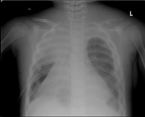
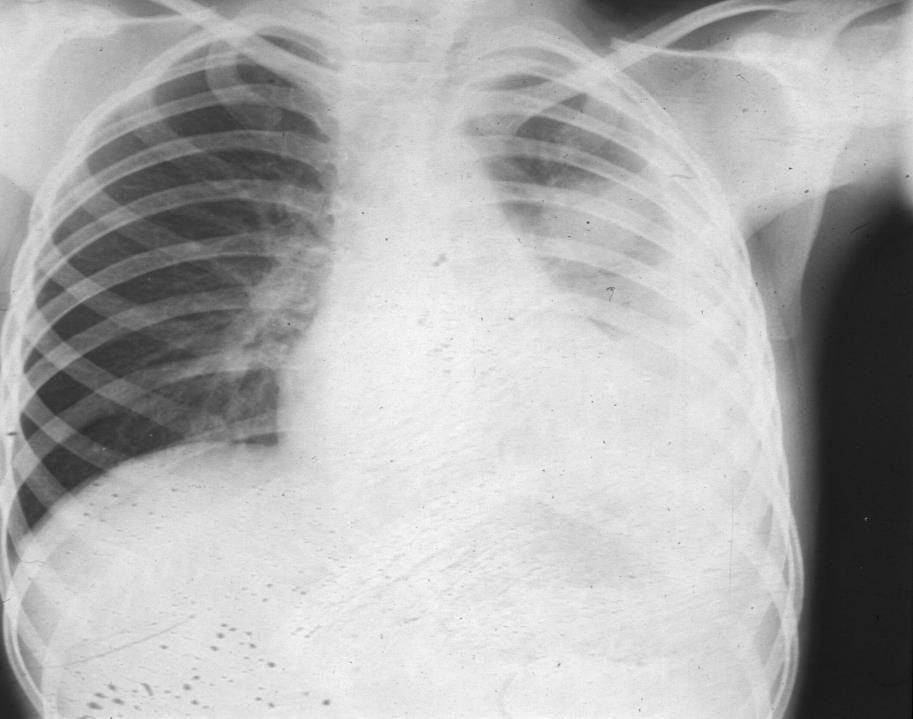
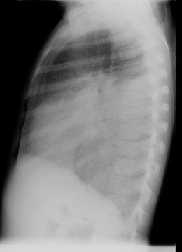
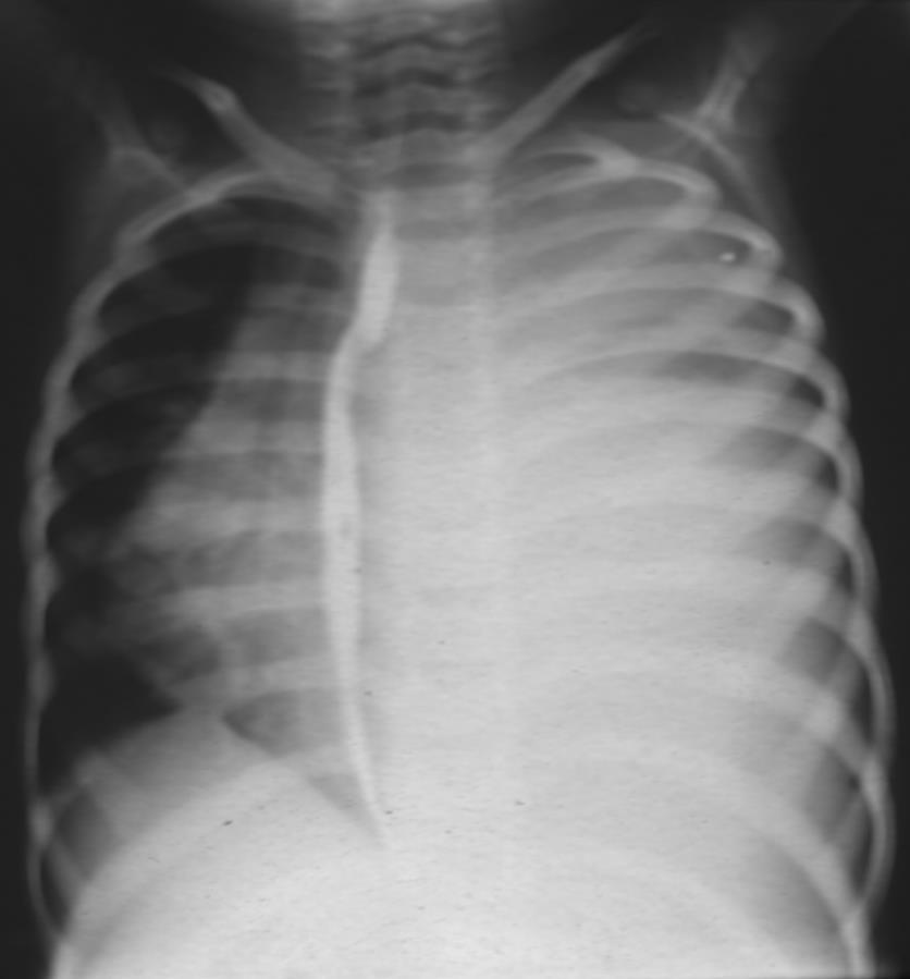
8 9-years-old boy with a normal chestfilm


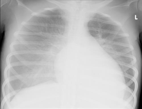
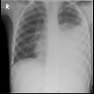
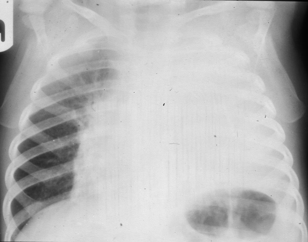
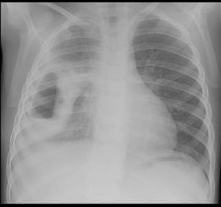
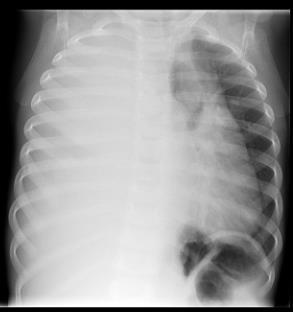
9 Transient dyspnea of newborn Pleural effusion on the right side


10 Congenital causes Congenital chylothorax with unknown etiology Follow-up a few weeks later


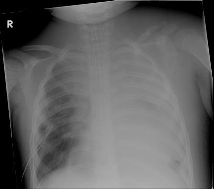
11 Neonatal chylothorax right-sided. Pneumothorax left-sided.
12 Chylothorax and vanishing bone disease by Gorham syndrome.

13 Hematothorax in a neonate. Traumatic? Iatrogenic?

14 Cardial causes Pericarditis purulenta
15 Cardiac decompensation in a case of Duchenne disease


16 Nefrogenic causes (less frequent) Nephrotic syndrome with nephritis
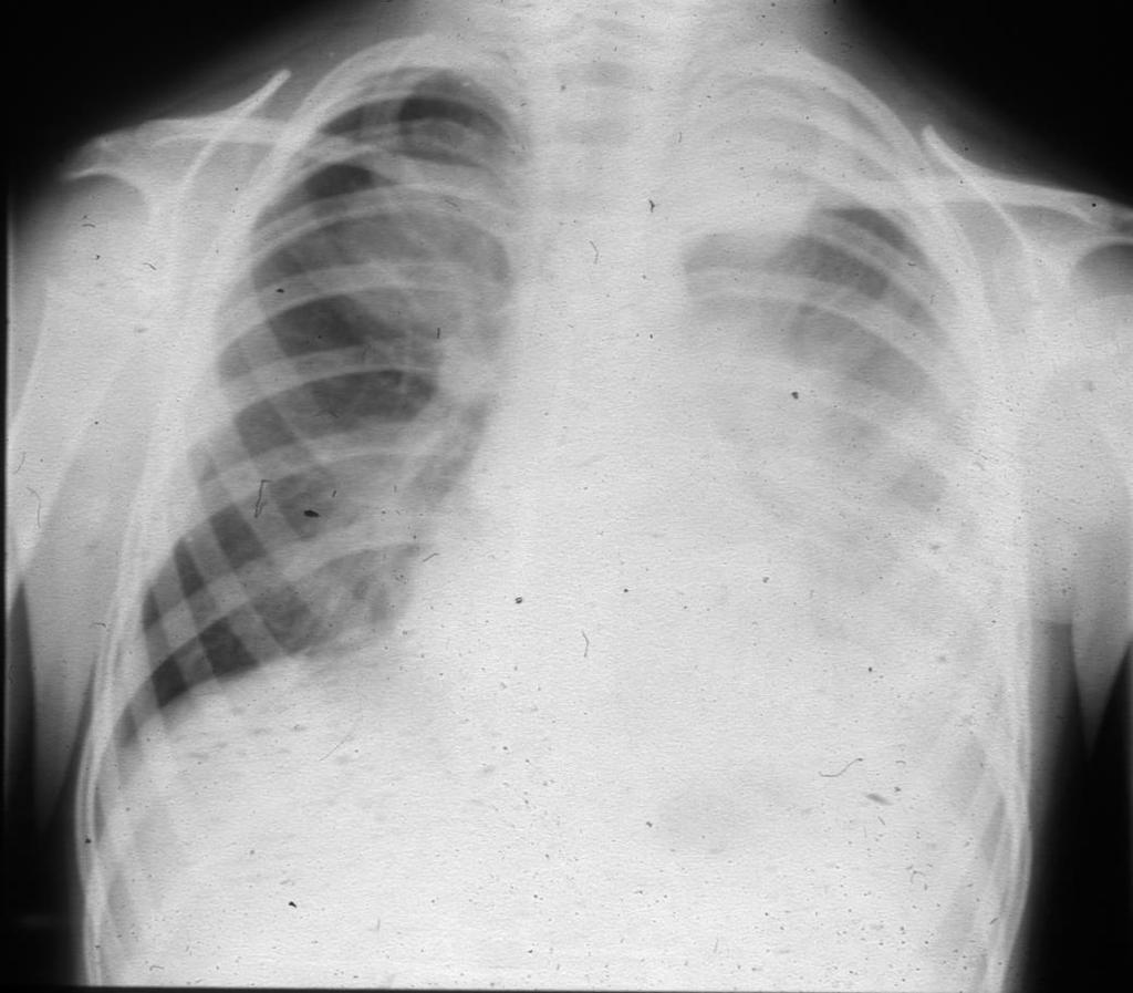
17 Infectious causes Main cause of pleural effusion in childhood: Infection (66%). In adult neoplasm is the most frequent cause of pleural effusion. The most common organisms are pneumococcus and staphylococcus. Viral infections are rarely the cause of pleural effusion. Sterile effusions occur in about 20 to 25% of children with primary pulmonary tuberculosis predominantly in older children.


18 Classification of empyema stages Exudative 2. Fibrinopurulent 3. Organized 1. 2.
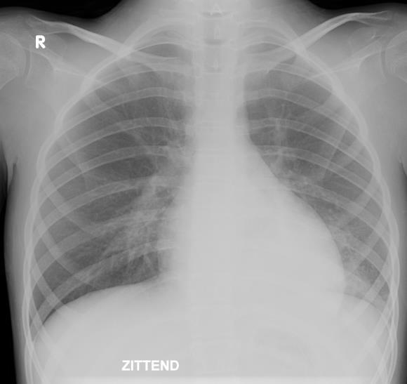
19 Parapneumonic pleural effusion in a case of pneumonia
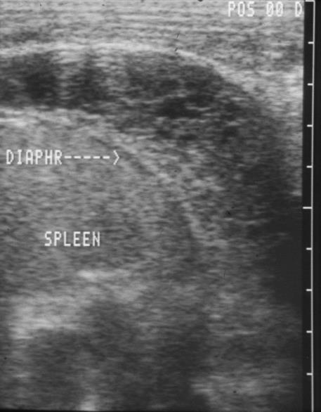
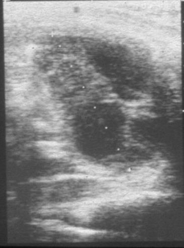
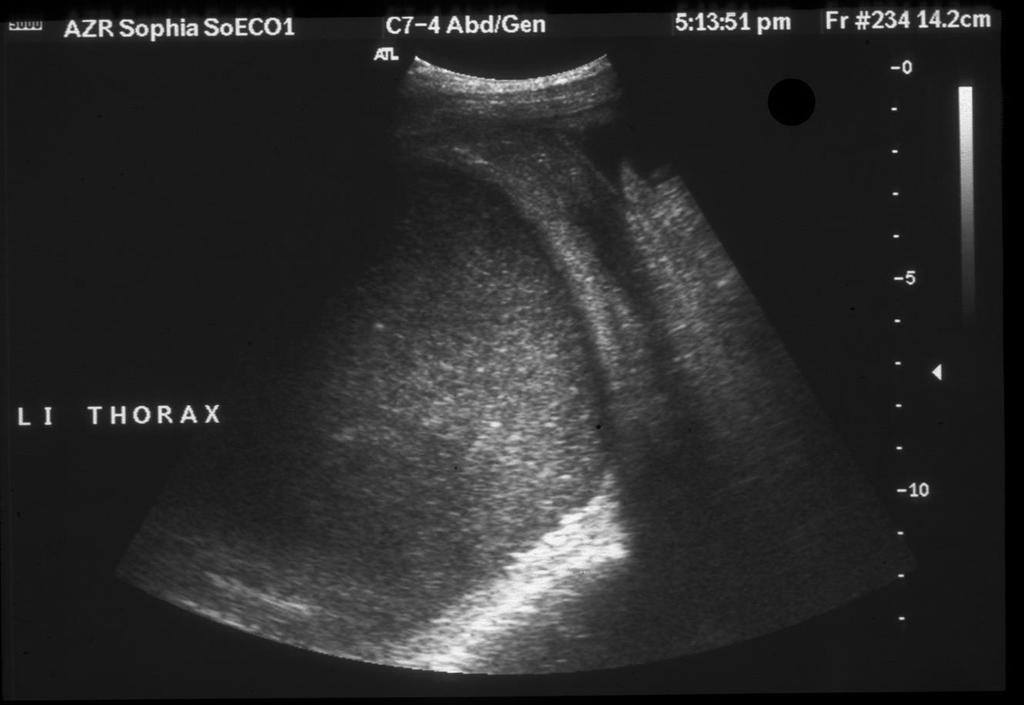
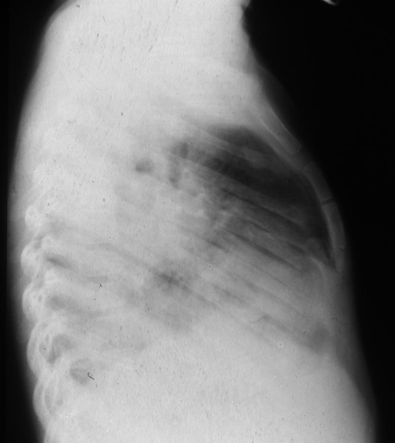
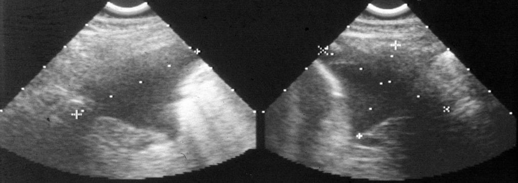
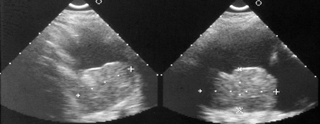
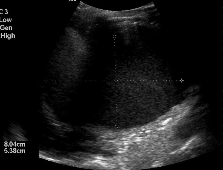
20 Lung tuberculosis with pleuritis tuberculosa

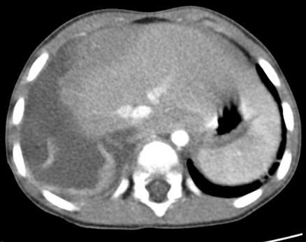
21 Right-sided empyema in a case of pulmonary infection

22 Abscess based on pneumonia

23 Abscess in the lung
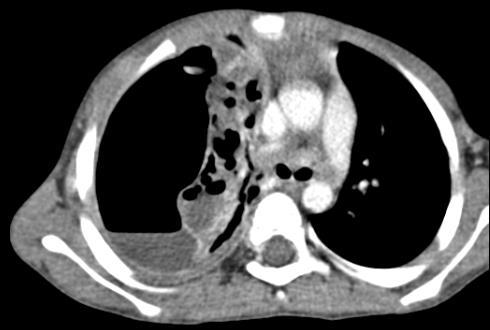
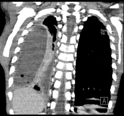
24 13-year-old girl with pleural empyema
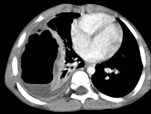
25 Right-sided empyema in a case with pulmonal abscess

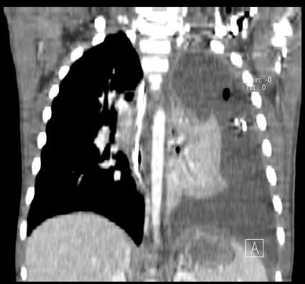
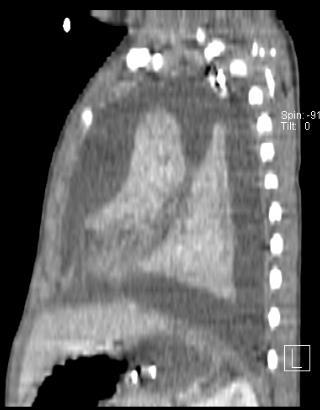
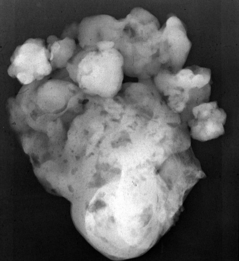
26 Traumatic causes Battered child with multiple rib fractures and left-sided hematothorax.

27 Ultrasound of a post-traumatic hematothorax.


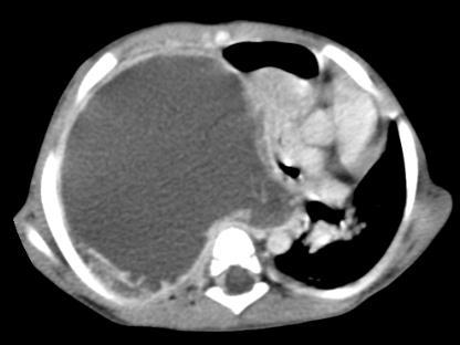
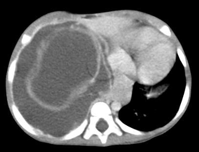
28 Iatrogenic causes Hematothorax due to pseudoaneurysm after an open ductus Botalli operation.


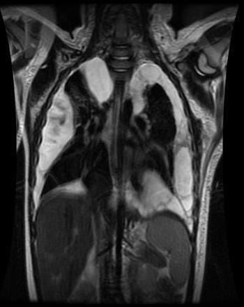
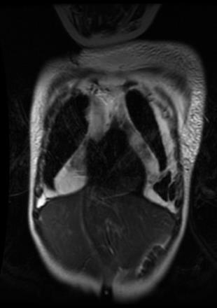
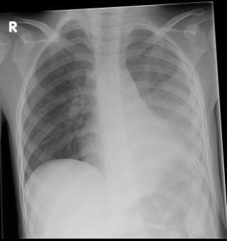
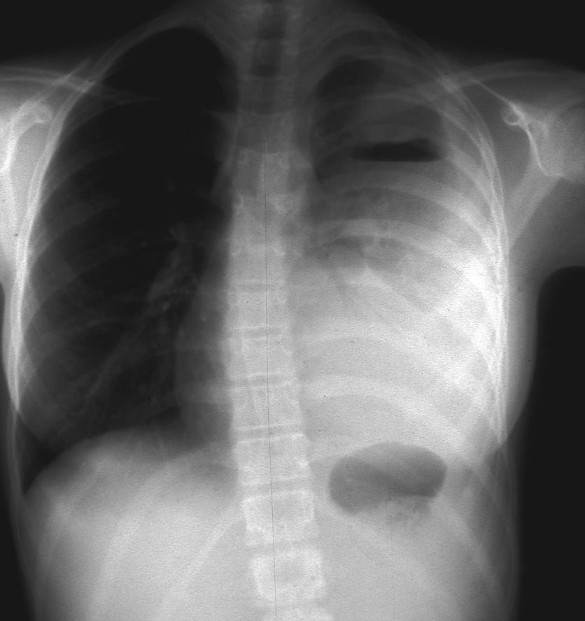
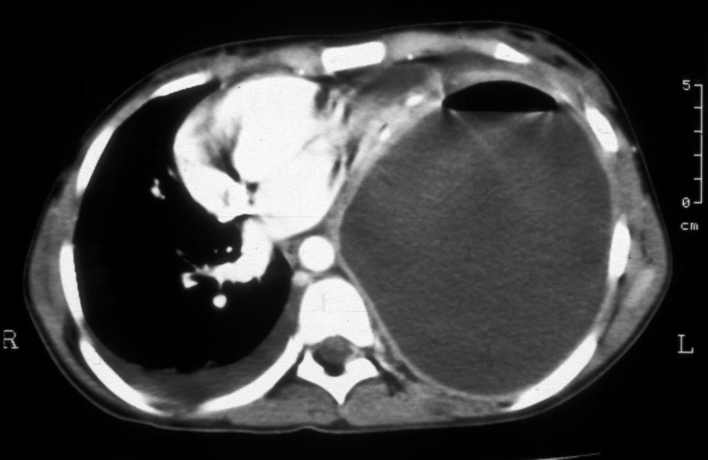
29 1-month old boy with an iatrogenic intrathoracic perforation of the central line


30 Retropleural effusion due to iatrogenic perforated esophagus.


31 21 september oktober 2005 Pleural hemorrhage after resection of a ganglioneurinoma

32 Overloaded circular system by infusion. Note the pleura effusion.
33 Neoplastic causes Malignant teratoma


34 2,5-year-girl with lymphosarcoma with a large lymphoma in left hilus with huge pleural effusion. Note the iatrogenic pneumothorax and collapsed lung after thoracocentesis.
35 Intrathoracic metastases of Wilms tumor with pleuritis carcinomatosa


36 Right-sided Wilms tumor. Tumor infiltration of diaphragm with pleura effusion. Liver and lung metastasis.


37 T-non Hodgkin lymphoma with pleural effusion
38 Pleuropulmonal blastoma
39 Other causes of pleural effusion are: Ascites Collagen-vascular disease Hypoproteinemia of any cause Fluid overload Ventriculopleural shunt


40 Others Pleura effusion in a septic infant with capillary leak
41 Conclusions The chest film in AP en lateral position is the first step in imaging of pleural effusion. Ultrasound is an additional modality in diagnostic imaging of pleural effusion: -amount of pleural effusion -aspect of pleural effusion: echogenicity, septation -localization -intervention
42 Conclusions CT is indicated in: -complicated pulmonary infection -iatrogenic causes -unknown etiology -neoplastic diseases -congenital diseases MRI is indicated in detection of congenital malformation or neoplastic disease of the chest wall.
Malignant Effusions. Anantham Devanand Respiratory and Critical Care Medicine Singapore General Hospital
 Malignant Effusions Anantham Devanand Respiratory and Critical Care Medicine Singapore General Hospital Malignant Effusions Definition: Presence of malignant cells in the pleural space 75% are caused by
Diagnostic Approach to Pleural Effusion
 Diagnostic Approach to Pleural Effusion Objectives Define the leading causes of pleural effusion Classify the type of effusion Identify procedures and tests associated with diagnosis 2 Agenda Basic anatomy
Management of Pleural Effusion
 Management of Pleural Effusion Development of Pleural Effusion pulmonary capillary pressure (CHF) capillary permeability (Pneumonia) intrapleural pressure (atelectasis) plasma oncotic pressure (hypoalbuminemia)
Lung sequestration and Scimitar syndrome
 Lung sequestration and Scimitar syndrome Imaging approaches M. Mearadji International Foundation for Pediatric Imaging Aid Rotterdam, The Netherlands Pulmonary sequestration Pulmonary sequestration (PS)
Serous fluids. Dr. Mohamed Saad Daoud
 Serous fluids 1 Reference Books: Urinanalysis and body fluids (Susan King Strasinger- Marjorie Schaub De Lorenzo) Fifth edition 2 The closed cavities of the body namely, the pleural, pericardial, and peritoneal
Dr. A.Torossian, M.D., Ph. D. Department of Respiratory Diseases
 Pleural effusions Dr. A.Torossian, M.D., Ph. D. Department of Respiratory Diseases A pleural effusion is an abnormal collection of fluid in the pleural space resulting from excess fluid production or decreased
Thoracic Cavity and Tumors of Lung and Pleura
 Tutorial Module 6 Thoracic Cavity and Tumors of Lung and Pleura Alfonso López Atlantic Veterinary College University of Prince Edward Island Canada Sept 28, 2014 Thoracic Cavity There are anatomical differences
Pulmonary Morning Report. Ashley Schmehl D.O. PGY-3 January,
 Pulmonary Morning Report Ashley Schmehl D.O. PGY-3 January, 8 2015 Pleural Effusion Unilateral versus Bilateral Associated symptoms Transudate versus Exudate Light s Criteria: Pleural protein: Serum protein
PLEURAL DISEASES. (Pleural effusion & empyema) Menaldi Rasmin
 Menaldi Rasmin") PLEURAL DISEASES (Pleural effusion & empyema) Menaldi Rasmin Department of Pulmonology & Respiratory Medicine Faculty of Medicine, University of Indonesia Introduction Pleural effusion is the most common
PLEURAL DISEASES (Pleural effusion & empyema) Menaldi Rasmin Department of Pulmonology & Respiratory Medicine Faculty of Medicine, University of Indonesia Introduction Pleural effusion is the most common
Pleural Effusion. Exudative pleural effusion - Involve an increase in capillary permeability and impaired pleural fluid resorption
 Pleural Effusion Definition of pleural effusion Accumulation of fluid between the pleural layers Epidemiology of pleural effusion Estimated prevalence of pleural effusion is 320 cases per 100,000 people
Pleural Diseases. Dr Matthew J Knight Consultant Respiratory Physician
 Pleural Diseases Dr Matthew J Knight Consultant Respiratory Physician What do you need to know? What do you need to know? Pleura- normal anatomy and physiology Pleural effusions Causes and investigations
PLEURAL EFFUSION. Prof. G. Zuliani
 PLEURAL EFFUSION Prof. G. Zuliani Anatomy of pleural membrane and pleural space Pleural membrane consists of parietal pleura and visceral pleura A space situated between parietal and visceral pleura is
like humans, have well-developed mediastinal separation between the left and right hemithorax, thus unilateral changes can occur. On the other hand,
 Tutorial Module 6 Thoracic Cavity and Tumors of Lung and Pleura Alfonso López Atlantic Veterinary College University of Prince Edward Island Canada 2009 Enero 3 Thoracic Cavity There are significant anatomical
Chylothorax Basics OVERVIEW GENETICS SIGNALMENT/DESCRIPTION OF PET
 Chylothorax Basics OVERVIEW Chylo- refers to chyle; thorax refers to the chest Chyle is a milky to slightly yellow fluid composed of lymph and fats (rich in triglycerides) taken up from the intestines
Surgical treatment of empyema in children
 Surgical treatment of empyema in children Jacques Janson Pierre Goussard Cardiothoracic Surgery, Paediatric Pulmonology Tygerberg Academic Hospital University of Stellenbosch Pleural space Netter, Frank
Proceedings of the World Small Animal Veterinary Association Sydney, Australia 2007
 Proceedings of the World Small Animal Sydney, Australia 2007 Hosted by: Next WSAVA Congress THE LAST GASP II: LUNGS AND THORAX David Holt, BVSc, Diplomate ACVS University of Pennsylvania School of Veterinary
Pleural fluid analysis
 Pleural fluid analysis Dr Akash Verma Senior Consultant- Department of Respiratory and Critical Care Medicine Tan Tock Seng Hospital, Singapore 308433 Adj A/Professor- Lee Kong Chian School of Medicine
Pneumothorax lecture no. 3
 Pneumothorax lecture no. 3 Is accumulation of air in a pleural space or accumulation of extra pulmonary air within the chest, Is uncommon during childhood, may result from external trauma, iatrogenic,
Pathology of the Respiratory System 5: Lung and Thoracic Cavity
 Pathology of the Respiratory System 5: Lung and Thoracic Cavity Shannon Martinson, Jan 2017 http://people.upei.ca/smartinson/ VPM 222 Systemic Pathology DISORDERS OF THE LUNG Congenital Pigmentary deposition
Case Report A Cause of Bilateral Chylothorax: A Case of Mesothelioma without Pleural Involvement during Initial Diagnosis
 Case Reports in Pulmonology Volume 2015, Article ID 962504, 4 pages http://dx.doi.org/10.1155/2015/962504 Case Report A Cause of Bilateral Chylothorax: A Case of Mesothelioma without Pleural Involvement
Hyperemia, Congestion, and Edema
 Hyperemia, Congestion, and Edema Hyperemia Acute, actively increased blood flow Tissues look red (erythema) Congestion Chronic, passively reduced outflow Tissues look pale or blue (cyanosis) Edema Water
EVALUATE DATA IN THE PATIENT RECORD
 EVALUATE DATA IN THE PATIENT RECORD Shawna Strickland, PhD, RRT-NPS, AE-C, FAARC Objectives At the end of this module, the learner will be able to identify the pertinent data from the patient chart for
Manejo Práctico del Derrame Pleural
 Manejo Práctico del Derrame Pleural San José, Costa Rica Junio 29, 2017 Rodrigo Cartín Ceba, MD, MSc Consultant, Pulmonary and Critical Care Medicine Associate Professor of Medicine Mayo Clinic 2010 MFMER
Pneumothorax and Chest Tube Problems
 Pneumothorax and Chest Tube Problems Pneumothorax Definition Air accumulation in the pleural space with secondary lung collapse Sources Visceral pleura Ruptured esophagus Chest wall defect Gas-forming
Adrenal masses in infancy and childhood: A clinical and radiological overview M. Mearadji
 Adrenal masses in infancy and childhood: A clinical and radiological overview M. Mearadji International Foundation for Pediatric Imaging Aid Introduction Neoplastic adrenal masses usually originate from
Right lung. -fissures:
 -Right lung is shorter and wider because it is compressed by the right copula of the diaphragm by the live.. 2 fissure, 3 lobes.. hilum : 2 bronchi ( ep-arterial, hyp-arterial ), one artery mediastinal
Acquired pediatric esophageal diseases Imaging approaches and findings. M. Mearadji International Foundation for Pediatric Imaging Aid
 Acquired pediatric esophageal diseases Imaging approaches and findings M. Mearadji International Foundation for Pediatric Imaging Aid Acquired pediatric esophageal diseases The clinical signs of acquired
TB Radiology for Nurses Garold O. Minns, MD
 TB Nurse Case Management Salina, Kansas March 31-April 1, 2010 TB Radiology for Nurses Garold O. Minns, MD April 1, 2010 TB Radiology for Nurses Highway Patrol Training Center Salina, KS April 1, 2010
Imaging of Pleural Effusion: Comparing Ultrasound, X-Ray and CT findings
 Imaging of Pleural Effusion: Comparing Ultrasound, X-Ray and CT findings Poster No.: C-2067 Congress: ECR 2017 Type: Educational Exhibit Authors: J. M. Almeida, N. Antunes, C. Leal, L. Figueiredo ; Lisboa/PT,
UNDERSTANDING CHYLE IN CATS
 Vet Times The website for the veterinary profession https://www.vettimes.co.uk UNDERSTANDING CHYLE IN CATS Author : DAN FORSTER Categories : Vets Date : February 11, 2008 DAN FORSTER discusses diagnosis
*according to content of fluid we can divide pleural effusion to 2 main types
 Pleural lesion and lesion of the Done by: Upper respiratory tract Saef Bassam ma'adat **Lets start with pleural lesion there is a little differet between pleural effustion and empyema accumulation of fluid
Lung- and airway emergencies
 Lung- and airway emergencies Charlotte de Lange,MD,PhD Pediatric Radiology unit, Oslo University Hospital, Norway 5th Nordic course - Emergency Radiology Oslo 18-21.5.2015 clange@ous-hf.no How come pediatric
Neckmasses in infancy and childhood: Clinical and radiological classification and imaging approaches M. Mearadji
 Neckmasses in infancy and childhood: Clinical and radiological classification and imaging approaches M. Mearadji International Foundation for Pediatric Imaging Aid Introduction Neck masses are a frequent
ESTS SCHOOL OF THORACIC SURGERY Antalya Revisited in Istanbul March 2016 Istanbul, Turkey
 ESTS SCHOOL OF THORACIC SURGERY Antalya Revisited in Istanbul 16-20 March 2016 Istanbul, Turkey Format 1. Lectures, Video and Case Presentations 15 min. 2. Learn from Peers Sessions. 3. More integrated
Bacterial pneumonia with associated pleural empyema pleural effusion
 EMPYEMA Synonyms : - Parapneumonic effusion - Empyema thoracis - Bacterial pneumonia - Pleural empyema, pleural effusion - Lung abscess - Complicated parapneumonic effusions (CPE) 1 Bacterial pneumonia
An Introduction to Radiology for TB Nurses
 An Introduction to Radiology for TB Nurses Garold O. Minns, MD September 14, 2017 TB Nurse Case Management September 12 14, 2017 EXCELLENCE EXPERTISE INNOVATION Garold O. Minns, MD has the following disclosures
White hemithorax in children
 DOI 10.1007/s00247-011-2065-8 PICTORIAL ESSAY White hemithorax in children Javier Lucaya & Enrique F. Garcés-Iñigo & Pilar García-Peña & Joaquim Piqueras & Goya Enriquez Received: 28 July 2010 /Revised:
APPROACH TO PLEURAL EFFUSIONS. Raed Alalawi, MD, FCCP
 APPROACH TO PLEURAL EFFUSIONS Raed Alalawi, MD, FCCP CASE 65-year-old woman with H/O breast cancer presented with a 1 week H/O progressively worsening exersional dyspnea. Physical exam: Diminished breath
Exam 2 Respiratory Disorders
 Exam 2 Respiratory Disorders Common Cold Common Cold Pathology Common Cold Consequences Rhinosinusitis Rhinosinusitis Pathology Rhinosinusitis ostia can close due to Influenza (Flu) Influenza Pathology
Bronchogenic Carcinoma
 A 55-year-old construction worker has smoked 2 packs of ciggarettes daily for the past 25 years. He notes swelling in his upper extremity & face, along with dilated veins in this region. What is the most
Congenital Lung Malformations: Radiologic-Pathologic Correlation
 Acta Radiológica Portuguesa, Vol.XVIII, nº 70, pág. 51-60, Abr.-Jun., 2006 Congenital Lung Malformations: Radiologic-Pathologic Correlation Marilyn J. Siegel Mallinckrodt Institute of Radiology, Washington
Pneumothorax. Defined as air in the pleural space which can occur through a number of mechanisms
 Pneumothorax Defined as air in the pleural space which can occur through a number of mechanisms Traumatic pneumothorax Penetrating chest trauma Common secondary to bullet or knife penetration Chest tube
Chest Radiology Interpretation: Findings of Tuberculosis
 Chest Radiology Interpretation: Findings of Tuberculosis Get out your laptops, smart phones or other devices pollev.com/chestradiology Case #1 1 Plombage Pneumonia Cancer 2 Reading the TB CXR Be systematic!
Respiratory Diseases and Disorders
 Chapter 9 Respiratory Diseases and Disorders Anatomy and Physiology Chest, lungs, and conducting airways Two parts: Upper respiratory system consists of nose, mouth, sinuses, pharynx, and larynx Lower
Case 1. A 35-year-old male presented with fever, cough, and purulent sputum for one week. This was his CXR (Fig. 1.1). What is the diagnosis?
. What is the diagnosis?") 1 Interpreting Chest X-Rays CASE 1 Fig. 1.1 Case 1. A 35-year-old male presented with fever, cough, and purulent sputum for one week. This was his CXR (Fig. 1.1). What is the diagnosis? CASE 1 Interpreting
1 Interpreting Chest X-Rays CASE 1 Fig. 1.1 Case 1. A 35-year-old male presented with fever, cough, and purulent sputum for one week. This was his CXR (Fig. 1.1). What is the diagnosis? CASE 1 Interpreting
Alper Toker, MD. VATS decortication. Istanbul University, Istanbul Medical School Department of Thoracic Surgery
 VATS decortication Alper Toker, MD Istanbul University, Istanbul Medical School Department of Thoracic Surgery Pleural space infection is a common pathology causing morbidity and mortality. It is a collection
Resident Case Review CHEST. Daria Manos CAR 2016
 Resident Case Review CHEST CAR 2016 Daria Manos Disclosure Speakers bureau, Roche CAR 2016 Daria Manos 1. Recognize common and critical chest radiograph and computed tomography signs and use these clues
Undergraduate Teaching
 Prof. James F Meaney Undergraduate Teaching Chest X-Ray Understanding the normal anatomical by reference to cross sectional imaging Radiology? It s FUN! Cryptic puzzle Sudoku (Minecraft?) It s completely
Index. B Biological factors, 2 Brain stem encephalitis, Burkitt s lymphoma, 83, 105
 Index A Acquired immunodeficiency syndrome (AIDS) abdomen gallbladder complications, 97, 107 109 gastrointestinal complications, 96, 105 106 liver complications, 97, 107 109 neoplasm, 99, 110 111 pancreas
Characteristic. Course of disease:short Days--one month Changes : Alteration, exudation Tissue destruction Inflammation cells: major neutrophils
 ACUTE INFLAMMATION Characteristic Course of disease:short Days--one month Changes : Alteration, exudation Tissue destruction Inflammation cells: major neutrophils TYPES Serous Inflammation Fibrinous Inflammation
Chest XRay interpretation INTERPRETATIONS Identifications: Name & Date Technical evaluation Basic Interpretations
 Chest XRay interpretation INTERPRETATIONS Identifications: Name & Date Technical evaluation Basic Interpretations TECHNICAL EVALUATION 1. Projection: AP/PA view To differentiate between AP & PA films,
Bronchial syndrome. Atelectasis Draining bronchus Bronchiectasis
 Bronchial syndrome Atelectasis Draining bronchus Bronchiectasis Etienne Leroy Terquem Pierre L Her SPI / ISP Soutien Pneumologique International / International Support for Pulmonology Atelectasis Consequence
EUROPEAN ASSOCIATION OF VETERINARY DIAGNOSTIC IMAGING EUROPEAN COLLEGE OF VETERINARY DIAGNOSTIC IMAGING
 EISAGOGIKO EUROPEAN ASSOCIATION OF VETERINARY DIAGNOSTIC IMAGING EUROPEAN COLLEGE OF VETERINARY DIAGNOSTIC IMAGING ARISTOTLE UNIVERSITY OF THESSALONIKI SCHOOL OF VETERINARY MEDICINE SECTION OF RADIOLOGY
Chapter 75 Pleural Disease
 Chapter 75 Pleural Disease Joshua M. Kosowsky Pleural disease is commonly encountered in the emergency department (ED). Presentations range in severity from asymptomatic pleural effusion to tension pneumothorax.
ANATOMY OF THE PLEURA (contd) III. Histology: covered by a single layer of mesothelial cells. Within the pleura are blood vessels, mainly capillaries,
 III. Histology: covered by a single layer of mesothelial cells. Within the pleura are blood vessels, mainly capillaries,") PLEURAL DISEASES By: SETIA PUTRA TARIGAN Pulmonary Department, Faculty of Medicine, Sumatera Utara University/ Adam Malik Hospital Medan 2008 ANATOMY OF THE PLEURA I. Pleura is the serous membrane: 1.
PLEURAL DISEASES By: SETIA PUTRA TARIGAN Pulmonary Department, Faculty of Medicine, Sumatera Utara University/ Adam Malik Hospital Medan 2008 ANATOMY OF THE PLEURA I. Pleura is the serous membrane: 1.
PULMONARY TUBERCULOSIS RADIOLOGY
 PULMONARY TUBERCULOSIS RADIOLOGY RADIOLOGICAL MODALITIES Medical radiophotography Radiography Fluoroscopy Linear (conventional) tomography Computed tomography Pulmonary angiography, bronchography Ultrasonography,
Thoraxdrainage SGP Jahresversammlung 2016, Lausanne
 Thoraxdrainage SGP Jahresversammlung 2016, Lausanne Dr. med. Lukas Kern a bit of history (incomplete.) a bit of physiology (basic ) indication data guidelines a bit of history (incomplete.) a bit of physiology
Case Based Fetal Lung Masses
 Case Based Fetal Lung Masses Advances in Fetal and Neonatal Imaging Course Orlando, Florida, January 28, 2017 Leann E. Linam, MD Associate Professor Radiology University of Arkansas for Medical Sciences/
FDG PET/CT in Lung Cancer Read with the experts. Homer A. Macapinlac, M.D.
 FDG PET/CT in Lung Cancer Read with the experts Homer A. Macapinlac, M.D. Patient with suspected lung cancer presents with left sided chest pain T3 What is the T stage of this patient? A) T2a B) T2b C)
BELLWORK page 343. Apnea Dyspnea Hypoxia pneumo pulmonary Remember the structures of the respiratory system 1
 BELLWORK page 343 Apnea Dyspnea Hypoxia pneumo pulmonary respiratory system 1 STANDARDS 42) Review case studies that involve persons with respiratory disorders, diseases, or syndromes. Citing information
Ó Journal of Krishna Institute of Medical Sciences University 106
 ISSN 2231-4261 CASE REPORT Transudative Effusion of Malignant Etiology: An Interesting Case Report 1 1 1 1* Swathi Karanth M. P, Ketaki Utpat, Unnati Desai, Jyotsna M. Joshi 1 Department of Pulmonary Medicine,
Pleural Disease. Disclosure. Normal Pleural Anatomy. Outline. Pleural Fluid Origins: Transudates. Pleural Fluid Origins: Exudates
 Disclosure Pleural Disease Anne L Fuhlbrigge MD MS Clinical Director Division of Pulmonary and Critical Care Medicine Brigham and Women s Hospital Channing Laboratory Harvard Medical School Boston, MA
Anatomy and Physiology of the Lungs
 The lungs consist of right and left sides. The right lung has three lobes: Upper lobe, Middle lobe, Lower lobe The left lung has two lobes: Upper lobe, Lower lobe Anatomy and Physiology of the Lungs The
Pleural fluid collections in critically ill patients Elankumaran Paramasivam MRCP Andrew Bodenham FRCA
 in critically ill patients Elankumaran Paramasivam MRCP Andrew Bodenham FRCA Key points Pleural fluid collections are common in the critically ill; they are predominantly transudates that do not require
Children are not small adults Children are Not Small Adults Anatomic considerations Pliable bony & cartilagenous structures - Significant thoracic inj
 PEDIATRIC CHEST TRAUMA Children are not small adults Role of imaging Spectrum of injury Children are not small adults Children are Not Small Adults Anatomic considerations Pliable bony & cartilagenous
Pediatric TB Intensive Houston, Texas
 Pediatric TB Intensive Houston, Texas November 13, 2009 Radiographic Manifestations of Pediatric TB Susan D. John, MD, FACR November 13, 2009 Radiologic Presentation of Childhood TB Susan D. John, MD,
Intrathoracìc Lymphangiomatosis Mimicking Lymphangioleiomyomatosis in a Young Woman
 «'...' I>' -Vi f i / ί*> * 1. - -»\ **, t»
A Practical Approach to Ultrasound Assessment of Respiratory Distress
 A Practical Approach to Ultrasound Assessment of Respiratory Distress Yanick Beaulieu, MD, FRCPC Director, Bedside Ultrasound Curriculum Division of Cardiology and Critical Care Hôpital du Sacré-Coeur
Approach to CXR. Terminology. 1.Identification. Greg Blecher SCH Respir Fellow. Correct patient Correct date and time Correct examination
 Approach to CXR Greg Blecher SCH Respir Fellow From Rob Posteraro http://home.earthlink.net/~rhpos/cxr_interpret.txt.html ; http://home.earthlink.net/~rhpos/cxr_main.txt.html) Approach to viewing Chest
Esophageal Perforation
 Esophageal Perforation Dr. Carmine Simone Thoracic Surgeon, Division of General Surgery Head, Division of Critical Care May 15, 2006 Overview Case presentation Radiology Pre-operative management Operative
CHEST DRAIN PROTOCOL
 CHEST DRAIN PROTOCOL Rationale The pleural membranes have an important role in effective lung expansion. The visceral pleura is a thin, smooth, serous membrane covering the surface of the lungs and is
TB Intensive Houston, Texas
 TB Intensive Houston, Texas October 15-17, 17 2013 Diagnosis of TB: Radiology Rosa M Estrada-Y-Martin, MD MSc FCCP October 16, 2013 Rosa M Estrada-Y-Martin, MD MSc FCCP, has the following disclosures to
EMPYEMA. Catheter Based Treatment vs. VATS. UCHSC Department of Surgery Grand Rounds August 27 th, Jeremy Hedges, M.D.
 EMPYEMA Catheter Based Treatment vs. VATS UCHSC Department of Surgery Grand Rounds August 27 th, 2007 Jeremy Hedges, M.D. OVERVIEW Empyema Pathogenesis Treatment Catheter based treatment Fibrinolytics
UERMMMC Department of Radiology. Basic Chest Radiology
 UERMMMC Department of Radiology Basic Chest Radiology PHYSICS DENSITIES BONE SOFT TISSUES WATER FAT AIR TELEROENTGENOGRAM Criteria for an Ideal Chest Radiograph 1. Upright 2. Posteroanterior View 3. Full
Pleural Empyema Joseph Junewick, MD FACR
 Pleural Empyema Joseph Junewick, MD FACR 03/19/2010 History Teenager with persistent fever and cough. Pneumonia diagnosed 1 week ago. Diagnosis Pleural Empyema Additional Clinical Surgery-Clear fluid with
JMSCR Vol 04 Issue 10 Page October 2016
 www.jmscr.igmpublication.org Impact Factor 5.244 Index Copernicus Value: 83.27 ISSN (e)-2347-176x ISSN (p) 2455-0450 DOI: http://dx.doi.org/10.18535/jmscr/v4i10.08 Ultrasound and CT Evaluation of Pleural
Lung tumors & pleural lesions
 Lung tumors & pleural lesions A brief introduction 95% of lung tumors are carcinomas Among the remaining 5%, we will discuss: -Hamartoma the most common benign lung tumor spherical, coin lesion on x-rays
Radiological staging of lung cancer. Shukri Loutfi,MD,FRCR Consultant Thoracic Radiologist KAMC-Riyadh
 Radiological staging of lung cancer Shukri Loutfi,MD,FRCR Consultant Thoracic Radiologist KAMC-Riyadh Bronchogenic Carcinoma Accounts for 14% of new cancer diagnoses in 2012. Estimated to kill ~150,000
Adam J. Hansen, MD UHC Thoracic Surgery
 Adam J. Hansen, MD UHC Thoracic Surgery Sometimes seen on Chest X-ray (CXR) Common incidental findings on computed tomography (CT) chest and abdomen done for other reasons Most lung cancers discovered
Chapter 10 Respiratory System J00-J99. Presented by: Jesicca Andrews
 Chapter 10 Respiratory System J00-J99 Presented by: Jesicca Andrews 1 Respiratory System 2 Respiratory Infections A respiratory infection cannot be assumed from a laboratory report alone; physician concurrence
Pediatric TB Intensive Houston, Texas October 14, 2013
 Pediatric TB Intensive Houston, Texas October 14, 2013 Radiologic Presentation of Childhood TB Susan D. John, MD, FACR October 14, 2013 Disclosures I have no disclosures or conflicts of interest to report
Dana Alrafaiah. - Moayyad Al-Shafei. -Mohammad H. Al-Mohtaseb. 1 P a g e
 - 6 - Dana Alrafaiah - Moayyad Al-Shafei -Mohammad H. Al-Mohtaseb 1 P a g e Quick recap: Both lungs have an apex, base, mediastinal and costal surfaces, anterior and posterior borders. The right lung,
Modern Approaches to Empyema
 Modern Approaches to Empyema Amit Bhargava, MD Attending Thoracic Surgeon Assistant Professor Department of Cardiovascular and Thoracic Surgery 1 Principles of Treatment Adequate drainage Sterilization
Dynamic MR Lymphangiography
 Dynamic MR Lymphangiography Rajesh Krishnamurthy, MD EB Singleton Department of Radiology, Texas Children s Hospital, Baylor College of Medicine, Houston, TX Acknowledgement: Dr. Sheena Pimpalwar, MD Interventional
An Update: Lung Cancer
 An Update: Lung Cancer Andy Barlow Consultant in Respiratory Medicine Lead Clinician for Lung Cancer (West Herts Hospitals NHS Trust) Lead for EBUS-Harefield Hospital (RB&HFT) Summary Lung cancer epidemiology
A Case of Pediatric Plasma Cell Granuloma
 August 2001 A Case of Pediatric Plasma Cell Granuloma Nii Tetteh, Harvard Medical School Year IV Our Patient 8 year old male with history of recurrent left lower lobe and lingular pneumonias since 1994.
GUIDELINES FOR DIAGNOSIS OF UNILATERAL PLEURAL EFFUSION. Pakistan Chest Society
 GUIDELINES FOR DIAGNOSIS OF UNILATERAL PLEURAL EFFUSION Pakistan Chest Society Message by chairman guideline committee Guidelines for pleural disease working group Expert review committee INTRODUCTION
Shedding Light on Neonatal X-rays. Objectives. Indications for X-Rays 5/14/2018
 Shedding Light on Neonatal X-rays Barbara C. Mordue, MSN, NNP-BC Neonatal Nurse Practitioner LLUH Children s Hospital, NICU Objectives Utilize a systematic approach to neonatal x-ray interpretation Identify
B-I-2 CARDIAC AND VASCULAR RADIOLOGY
 (YEARS 1 3) CURRICULUM FOR RADIOLOGY 13 B-I-2 CARDIAC AND VASCULAR RADIOLOGY KNOWLEDGE To describe the normal anatomy of the heart and vessels including the lymphatic system as demonstrated by radiographs,
Anatomy Lecture 8. In the previous lecture we talked about the lungs, and their surface anatomy:
 Anatomy Lecture 8 In the previous lecture we talked about the lungs, and their surface anatomy: 1-Apex:it lies 1 inch above the medial third of clavicle. 2-Anterior border: it starts from apex to the midpoint
New lung lesion in a 55 year-old male treated with chemoradiation for non-small cell lung carcinoma
 July 2016 New lung lesion in a 55 year-old male treated with chemoradiation for non-small cell lung carcinoma Contributed by: Laurel Rose, MD, Resident Physician, Indiana University School of Medicine,
ESTS SCHOOL KNOWLEDGE TRACK COURSE ANTALYA REVISITED IN PRAGUE March 2019 Lindner Hotel Prague Castle, Prague ESTS SCHOOL OF THORACIC SURGERY
 SCHOOL KNOWLEDGE TRACK COURSE 18-23 March 2019 Lindner Hotel Prague Castle, Prague Course Organisers: Hasan Batirel, Istanbul, Turkey Director of Education Nuria Novoa, Salamanca, Spain Knowledge Track
Thoracic Surgery; An Overview
 Thoracic Surgery What we see Thoracic Surgery; An Overview James P. Locher, Jr, MD Methodist Cardiovascular and Thoracic Surgery Lung cancer Mets Fungus and TB Lung abcess and empyema Pleural based disease
Table E1. Standardized Mortality Ratios for Total and Specific Causes of Death Parameter Radiologists Psychiatrists No. of Deaths
 RSNA, 2016 10.1148/radiol.2016152472 Table E1. Standardized Mortality Ratios for Total and Specific Causes of Death Parameter Radiologists Psychiatrists No. of Deaths Observed/Expected No. of Deaths Observed/Expected
Respiratory Interactive Session. Elaine Borg
 Respiratory Interactive Session Elaine Borg Case 1 Respiratory Cytology 55 year old gentleman Anterior mediastinal mass EBUS FNA Case 1 Respiratory Cytology 55 year old gentleman with anterior mediastinal
Tuberculosis - clinical forms. Dr. A.Torossian,, M.D., Ph. D. Department of Respiratory Diseases
 Tuberculosis - clinical forms Dr. A.Torossian,, M.D., Ph. D. Department of Respiratory Diseases 1 TB DISEASE Primary Post-primary (Secondary) Common primary forms Primary complex Tuberculosis of the intrathoracic
Procedure: Chest Tube Placement (Tube Thoracostomy)
") Procedure: Chest Tube Placement (Tube Thoracostomy) Basic Information: The insertion and placement of a chest tube into the pleural cavity for the purpose of removing air, blood, purulent drainage, or
Procedure: Chest Tube Placement (Tube Thoracostomy) Basic Information: The insertion and placement of a chest tube into the pleural cavity for the purpose of removing air, blood, purulent drainage, or
MR Tumor Staging for Treatment Decision in Case of Wilms Tumor
 MR Tumor Staging for Treatment Decision in Case of Wilms Tumor G. Schneider, M.D., Ph.D.; P. Fries, M.D. Dept. of Diagnostic and Interventional Radiology, Saarland University Hospital, Homburg/Saar, Germany
CT Manifestations of Late Sequelae in Patients with Tuberculous Pleuritis
 CT Manifestations of Late Sequelae in Patients with Tuberculous Pleuritis T uberculous pleuritis remains one of the major causes of pleural effusion with an incidence ranging from as high as 86% in a population