Non Contrast MRA. Mayil Krishnam. Director, Cardiovascular and Thoracic Imaging University of California, Irvine
|
|
|
- Willa Thornton
- 6 years ago
- Views:
Transcription
1 Non Contrast MRA Mayil Krishnam Director, Cardiovascular and Thoracic Imaging University of California, Irvine
2 No disclosures
3 Non contrast MRA-Why? Limitations of CTA Radiation exposure Iodinated contrast material reaction Renal impairment Timing of bolus Single phase
4 Non contrast MRA-Why? Limitations of MRA Safety concerns related to NSF Breath holding for 20-25sec 25sec Timing of bolus Quality varies among institutions
5 Non contrast MRA-why? Advantages No Iodine or Gadolinium based contrast No need for breath holding No need for Timing of contrast bolus for data acquisition Safely performed in renal failure patients Not a contraindication in pregnancy
6 Why now? Hardware and software advancement Increased attention due to safety concerns of Gd chelate agents from NSF Cost effective May be supplementary technique if CEMRA fails
7 Non Contrast MRA techniques Subtractive techniques Arterial spin labeling with SSFP or FSE readout (tagged-untagged) EKG gated FSE imaging (systole-diastole) Non Subtractive methods- better 3D SSFP MRA-thoracic IR-SSFP SSFP-renal Standard methods 2d or 3d Tof Other: phase contrast MRA, pulsatility MRA
8 3D SSFP Non contrast MRA SSFP MRA should ideally be performed on 1.5 T Steady state Free precession technique (SSFP) has high T2/T1 signal Fat, fluid and blood appear as bright on MR images SSFP MRA can be used to evaluate aorta, SVC, cardiac anatomy, pulmonary veins
9 3D SSFP Non contrast MRA Thoracic Coronary Aorta Central Veins Pulmonary veins Abdomen Aorta Renal arteries
10 3D SSFP MRA Free breathing navigator Non-selective RF excitation Large FOV Prospective gating Data acquired in diastole End expiratory data collected Inspiratory date rejected Coronal orientation
11 Imaging parameters: SSFP MRA CE-MRA Magnet 1.5T 1.5T Repetition time (msec) Echo time (msec) Flip angle ( ) Bandwidth (Hz/pixel) Field of view (mm²) 400 x x 400 Matrix size 256 x x 512 In plane resolution (mm²) 1.6 x x 1.0 Slice thickness (mm) 3* 1.5 Number of partitions Parallel acquisition GRAPPA x 2 GRAPPA x 3 Contrast None mmol/ kg *Interpolated to 1.5mm
12 Sequence for Non Contrast SSFP MRA ECG triggering T2 Preparation Navigator Gating Fat Suppression Data Acquisition 3 D true FISP pulse sequence with non selective radio frequency excitation for non contrast MR Angiography. Gradient Spoiler Courtesy: Vibas Deshpande
 A column of tissue at the intersection of these slices (arrowhead) then generates a spin echo, which is")
 (yellow bars) Navigator echo (dotted blue bars) The right-to-left phase-encoding direction (yellow")
13 Navigator echo and imaging volume in 3D SSFP MRA Axial Scout Two intersecting slices are placed over the diaphragm (arrow) A column of tissue at the intersection of these slices (arrowhead) then generates a spin echo, which is subsequently used to track the diaphragm (liver-lung boundary) Coronal scout Demonstrates the large image volume (field of view) (yellow bars) Navigator echo (dotted blue bars) The right-to-left phase-encoding direction (yellow arrow).

14 Navigator echo and imaging volume in 3D SSFP MRA Navigator Scout The Y axis shows absolute distance scale of the diaphragm. The reference position of the diaphragm is noted. The narrow gating window seen as the solid green horizontal bar tracks the end- expiratory position of the diaphragm using a motion adaptive sequence. White bars Liver Grey bars Lung Data falling within the narrow 4-mm gating window were accepted.

15 Non Contrast MR Angiography- Navigator Gating

16 Non Contrast Thoracic MR Angiography
17 Aorta
18

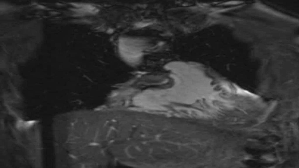
 demonstrates the coarctation of the aorta")
19 C 19 year old male with co-arctation of aorta. A. CEMRA. B. SSFP MRA. Note the uniform intravascular signal in the aorta and no motion artifact of the ascending aorta ( B). C: Volume rendered image (SSFP MRA) demonstrates the coarctation of the aorta (arrow). Krishnam et al, Investigative Radiology June 2008

20 SSFP 3D MRA DOLV Anomalous coronary
21 Non Contrast SSFP MRA Central Thoracic Veins

22 SSFP 3D MRA-central veins Non-contrast SSFP MRA demonstrates normal anatomy of the thoracic central veins.

23 34-year-old male with history of metastatic synovial sarcoma SVC tumor thrombosis. A- CE-MRA. B- non-contrast SSFP MRA Tomasian and Krishnam Invest Radiol May;43(5):306-13

24 CE-MRA B-SSFP 28-year-old male with history of Tetralogy of Fallot. Non-contrast SSFP MRA demonstrate extra-cardiac Fontan shunt and Glenn shunt from superior vena cava to main pulmonary artery confluence. Tomasian and Krishnam Invest Radiol May;43(5):306-13
25 NC SSFP 3D MRA Pulmonary veins Date can be acquired during systole in patients with controlled AF
26 Pulmonary veins Non-contrast SSFP MRA (Left) and CEMRA (Right) demonstrating conventional anatomy showing right and left superior pulmonary veins (A) and right and left inferior veins (B) (arrows). SSFP MRA CE-MRA Krishnam et al, Investigative Radiology June 2008
27 Pulmonary veins SSFP MRA CE-MRA

28 Pulmonary veins

29 Pulmonary veins. Volume rendered 3D image of the pulmonary veins and the left atrium from 3D SSFP MRA
30 Limitations Time consuming Requires consistent breathing pattern Off resonance artifacts Sensitive to different blood flow patterns Limited clinical data
31 SSFP Coronary MRA Whole heart MRA High spatial resolution Mainly coronary arteries Proximal and mid coronaries are better seen Preferred to rule out anomalous coronary arteries and proximal coronary aneurysm
32 Renal arteries
33 IR-SSFP Spatially selective inversion recovery pulse IR band extended caudally to suppress IVC signal Imaging FOV restricted to renal area Inflow of non inverted fresh blood into the imaging volume Data read out after appropriate TI( msec) Source images acquired in transverse orientation Navigator gated, EKG or peripheral pulse triggered

34 IR-SSFP Renal MRA
35 Limitations of IR SSFP Flow dependent Requires consistent breathing SSFP off resonance artifacts Background signal
36 Conclusions 3D SSFP MRA provides sufficient IQ for confident anatomical evaluation of thoracic vasculature, especially aorta. It is an alternative approach to CE-MRA especially in patients at increased risk for developing contrast-related complications and those with difficulty in holding breath. It relies on consistent breathing pattern and maybe associated with off resonance artifacts IR SSFP MRA is promising in the assessment of renal vessels More clinical data is needed for wider application
37 Thank You Acknowledgements Taka Natsuaki, Vibhas Deshpande,, Gerhard Laub- Siemens Medical solutions Paul Finn, Stefan Ruehm,, UCLA
Tips and Tricks of State of the art MRA
 Tips and Tricks of State of the art MRA Mayil Krishnam, MD,MBA, MRCP,FRCR(UK) Professor of Radiology Director, Cardiovascular and Thoracic Imaging University of California, Irvine Objectives Technical
MR Advance Techniques. Vascular Imaging. Class II
 MR Advance Techniques Vascular Imaging Class II 1 Vascular Imaging There are several methods that can be used to evaluate the cardiovascular systems with the use of MRI. MRI will aloud to evaluate morphology
CARDIAC MRI. Cardiovascular Disease. Cardiovascular Disease. Cardiovascular Disease. Overview
 CARDIAC MRI Dr Yang Faridah A. Aziz Department of Biomedical Imaging University of Malaya Medical Centre Cardiovascular Disease Diseases of the circulatory system, also called cardiovascular disease (CVD),
1Pulse sequences for non CE MRA
 MRI: Principles and Applications, Friday, 8.30 9.20 am Pulse sequences for non CE MRA S. I. Gonçalves, PhD Radiology Department University Hospital Coimbra Autumn Semester, 2011 1 Magnetic resonance angiography
Can SCMR CMR protocol recommendations
 Can SCMR CMR protocol recommendations V1.3 - April 2009 CanSCMR CMR Protocol and SOP Recommendation 2009 (15 minutes) 2 Planning of LV fct. real time multiple axes Realtime 3 cine long axis 6 long axes
Magnetic Resonance Angiography
 Magnetic Resonance Angiography 1 Magnetic Resonance Angiography exploits flow enhancement of GR sequences saturation of venous flow allows arterial visualization saturation of arterial flow allows venous
Objectives 8/17/2011. Challenges in Cardiac Imaging. Challenges in Cardiac Imaging. Basic Cardiac MRI Sequences
 8/17/2011 Traditional Protocol Model for Tomographic Imaging Cardiac MRI Sequences and Protocols Frandics Chan, M.D., Ph.D. Stanford University Medical Center Interpretation Lucile Packard Children s Hospital
Fulfilling the Promise
 Fulfilling the Promise of Cardiac MR Non-contrast, free-breathing technique generates comprehensive evaluation of the coronary arteries By Maggie Fung, MR Cardiovascular Clinical Development Manager; Wei
Non-Contrast MRA. How and When 1996! Why Non-Contrast MRA? Angiography: What are our goals? Inflow Techniques Differences in excitation hx
 A major teaching hospital of Harvard Medical School Angiography: What are our goals? Non-Contrast MRA: How and When Neil M. Rofsky, M.D. Professor of Radiology, Harvard Medical School Director of MRI &
MR Advance Techniques. Cardiac Imaging. Class IV
 MR Advance Techniques Cardiac Imaging Class IV Heart The heart is a muscular organ responsible for pumping blood through the blood vessels by repeated, rhythmic contractions. Layers of the heart Endocardium
Raja Muthupillai, PhD. Department of Diagnostic and Interventional Radiology St. Luke s Episcopal Hospital. Research Support: Philips Healthcare
 3D Cardiac Imaging Raja Muthupillai, PhD Department of Diagnostic and Interventional Radiology St. Luke s Episcopal Hospital Houston, TX Disclosures Research Support: Philips Healthcare This presentation
Essentials of Clinical MR, 2 nd edition. 99. MRA Principles and Carotid MRA
 99. MRA Principles and Carotid MRA As described in Chapter 12, time of flight (TOF) magnetic resonance angiography (MRA) is commonly utilized in the evaluation of the circle of Willis. TOF MRA allows depiction
How I do it: Non Contrast-Enhanced MR Angiography (syngo NATIVE)
") Clinical How-I-do-it Cardiovascular How I do it: Non Contrast-Enhanced MR Angiography (syngo NATIVE) Manuela Rick, Nina Kaarmann, Peter Weale, Peter Schmitt Siemens Healthcare, Erlangen, Germany Introduction
Clinical How-I-do-it Cardiovascular How I do it: Non Contrast-Enhanced MR Angiography (syngo NATIVE) Manuela Rick, Nina Kaarmann, Peter Weale, Peter Schmitt Siemens Healthcare, Erlangen, Germany Introduction
MRI protocol for post-repaired TOF
 2012 NASCI MRI protocol for post-repaired TOF Taylor Chung, M.D. Associate Director, Body and Cardiovascular Imaging Department of Diagnostic Imaging Children s Hospital & Research Center Oakland Oakland,
Magnetization Preparation Sequences
 Magnetization Preparation Sequences Acquisition method may not give desired contrast Prep block adds contrast (and/or encoding) MP-RAGE = Magnetization prepared rapid acquisition with gradient echo (Mugler,
9/8/2009 < 1 1,2 3,4 5,6 7,8 9,10 11,12 13,14 15,16 17,18 > 18. Tetralogy of Fallot. Complex Congenital Heart Disease.
 Current Indications for Pediatric CTA S Bruce Greenberg Professor of Radiology Arkansas Children s Hospital University of Arkansas for Medical Sciences greenbergsbruce@uams.edu 45 40 35 30 25 20 15 10
6/23/2009. Inversion Recovery (IR) Techniques and Applications. Variations of IR Technique. STIR, FLAIR, TI and TI Null. Applications of IR
 Techniques and Applications. Variations of IR Technique. STIR, FLAIR, TI and TI Null. Applications of IR") The Anatomy of Basic R Pulse Sequences Inversion Recovery () Techniques and Applications Chen Lin, PhD Indiana University School of edicine & Clarian Health Partners agnetization Preparation Section Chemical
The Anatomy of Basic R Pulse Sequences Inversion Recovery () Techniques and Applications Chen Lin, PhD Indiana University School of edicine & Clarian Health Partners agnetization Preparation Section Chemical
Functional Chest MRI in Children Hyun Woo Goo
 Functional Chest MRI in Children Hyun Woo Goo Department of Radiology and Research Institute of Radiology Asan Medical Center, University of Ulsan College of Medicine, Seoul, Korea No ionizing radiation
Cardiac MRI in ACHD What We. ACHD Patients
 Cardiac MRI in ACHD What We Have Learned to Apply to ACHD Patients Faris Al Mousily, MBChB, FAAC, FACC Consultant, Pediatric Cardiology, KFSH&RC/Jeddah Adjunct Faculty, Division of Pediatric Cardiology
CT angiography techniques. Boot camp
 CT angiography techniques Boot camp Overview Basic concepts Contrast administration arterial opacification Time scan acquisition during the arterial phase Protocol examples Helical non-gated CTA Pulmonary
A Magnetic Resonance Imaging Method for
 Journal of Cardiovascular Magnetic Resonance, 1(1), 59-64 (1999) INVITED PAPER Use of MRI in ASD Asessment A Magnetic Resonance Imaging Method for Evaluating Atrial Septa1 Defects Godtfred Holmvang Cardiac
Clinical Applications
 C H A P T E R 16 Clinical Applications In selecting pulse sequences and measurement parameters for a specific application, MRI allows the user tremendous flexibility to produce variations in contrast between
CT Versus MR for the Runoff
 CT Versus MR for the Runoff Robert R. Edelman, M.D. Dept. of Radiology NorthShore University HealthSystem Feinberg School of Medicine, Northwestern University Magnetic Resonance Computed Tomography Radio
A free-breathing non-contrast-enhanced pulmonary magnetic resonance angiography at 3 Tesla
 Chinese Medical Journal 2009;122(18):2111-2116 2111 Original article A free-breathing non-contrast-enhanced pulmonary magnetic resonance angiography at 3 Tesla YANG Jian, WANG Wei, WANG Ya-rong, NIU Gang,
ABSTRACT INTRODUCTION
 Journal of Cardiovascular Magnetic Resonance (2006) 8, 703 707 Copyright c 2006 Informa Healthcare ISSN: 1097-6647 print / 1532-429X online DOI: 10.1080/10976640600723706 Coronary Artery Magnetic Resonance
Department of Radiology University of California San Diego. MR Angiography. Techniques & Applications. John R. Hesselink, M.D.
 Department of Radiology University of California San Diego MR Angiography Techniques & Applications John R. Hesselink, M.D. Vascular Imaging Arterial flow void Flow enhancement Gadolinium enhancement Vascular
Non-contrast-enhanced MR portography and hepatic venography with time-spatial labeling inversion pulses: comparison at 1.5 Tesla and 3 Tesla
 Research Non-contrast-enhanced MR portography and hepatic venography with time-spatial labeling inversion pulses: comparison at 1.5 Tesla and 3 Tesla Acta Radiologica Open 4(5) 1 8! The Foundation Acta
High Field MR of the Spine
 Department of Radiology University of California San Diego 3T for MR Applications Advantages High Field MR of the Spine Increased signal-to-noise Better fat suppression Increased enhancement with gadolinium
New Cardiovascular Devices and Interventions: Non-Contrast MRI for TAVR Abhishek Chaturvedi Assistant Professor. Cardiothoracic Radiology
 New Cardiovascular Devices and Interventions: Non-Contrast MRI for TAVR Abhishek Chaturvedi Assistant Professor Cardiothoracic Radiology Disclosure I have no disclosure pertinent to this presentation.
Using Radial k-space Sampling and Steady-State Free Precession Imaging
 MRI of Coronary Vessel Walls Cardiac Imaging Original Research A C D E M N E U T R Y L I A M C A I G O F I N G Marcus Katoh 1 Elmar Spuentrup 1 Arno Buecker 1 Tobias Schaeffter 2 Matthias Stuber 3 Rolf
cardiac imaging planes planning basic cardiac & aortic views for MR
 cardiac imaging planes planning basic cardiac & aortic views for MR Dianna M. E. Bardo, M. D. Assistant Professor of Radiology & Cardiovascular Medicine Director of Cardiac Imaging cardiac imaging planes
Magnetic Resonance Imaging. Basics of MRI in practice. Generation of MR signal. Generation of MR signal. Spin echo imaging. Generation of MR signal
 Magnetic Resonance Imaging Protons aligned with B0 magnetic filed Longitudinal magnetization - T1 relaxation Transverse magnetization - T2 relaxation Signal measured in the transverse plane Basics of MRI
ACR MRI Accreditation: Medical Physicist Role in the Application Process
 ACR MRI Accreditation: Medical Physicist Role in the Application Process Donna M. Reeve, MS, DABR, DABMP Department of Imaging Physics University of Texas M.D. Anderson Cancer Center Educational Objectives
Cardiac CT Techniques in Neonates (and infants)
") Cardiac CT Techniques in Neonates (and infants) Siddharth P. Jadhav, MD Director, Body CT and MRI Edward B. Singleton Department of Pediatric Radiology Texas Children s Hospital Disclosures None Objectives
Cardiac CT Techniques in Neonates (and infants) Siddharth P. Jadhav, MD Director, Body CT and MRI Edward B. Singleton Department of Pediatric Radiology Texas Children s Hospital Disclosures None Objectives
Objectives. CMR Volumetric Analysis 8/25/11. CMR Volumetric Analysis Technique. Cardiac imaging plane acquisition. CMR Volumetric Analysis
 Objectives Cynthia K. Rigsby Children s Memorial Hospital Chicago, IL CMR volumetric analysis Techniques Normalized data Sources of error CMR phase contrast flow analysis Techniques What we can do with
Cardiovascular magnetic resonance artefacts
 Ferreira et al. Journal of Cardiovascular Magnetic Resonance 2013, 15:41 REVIEW Open Access Cardiovascular magnetic resonance artefacts Pedro F Ferreira 1,2*, Peter D Gatehouse 1,2, Raad H Mohiaddin 1,2
Pulmonary Embolism. Thoracic radiologist Helena Lauri
 Pulmonary Embolism Thoracic radiologist Helena Lauri 8.5.2017 Statistics 1-2 out of 1000 adults annually are diagnosed with deep vein thrombosis (DVT) and/or pulmonary embolism (PE) About half of patients
Time-Of-Flight MRA. Faculty Disclosures Vincent B. Ho, M.D. Presentation Objectives. MRA Techniques. Pros and Cons of MRA
 Faculty Disclosures Vincent B. Ho, M.D. MR Angiography Techniques and Pitfalls Financial Disclosure Grant/Research Support General Electric Medical Systems Off-Label/Investigational Drug Use Dr. Ho will
Congenital Heart Disease
 UNIT A10.1 One of the primary roles of cardiac MR imaging has been the assessment of congenital heart disease. Initially used to define cardiac anatomy as an adjunct to echocardiography, cardiac MR now
Initial Clinical Experience of TOSHIBA 3T MRI
 The 21st Conference of the Japanese Society of Cardiovascular Imaging & Dynamics Sponsored Seminar The Leading Edge of CT/MRI Diagnosis for the Cardiovascular System Initial Clinical Experience of TOSHIBA
Index. radiologic.theclinics.com. Note: Page numbers of article titles are in boldface type.
 Index Note: Page numbers of article titles are in boldface type. A ALCAPA. See Anomalous left coronary artery from the pulmonary artery. Angiosarcoma computed tomographic assessment of, 809 811 Anomalous
Cardiovascular MR Imaging at 3 T: Opportunities, Challenges, and Solutions 1
 TECHNICAL ADVANCEMENTS IN CARDIAC MR IMAGING 1612 Cardiovascular MR Imaging at 3 T: Opportunities, Challenges, and Solutions 1 Prabhakar Rajiah, MD, FRCR Michael A. Bolen, MD Abbreviations: BOLD = blood
Index. cardiology.theclinics.com. Note: Page numbers of article titles are in boldface type.
 Index Note: Page numbers of article titles are in boldface type. A ABI. See Ankle-brachial index (ABI). Afterload, deconstructing of, in ventricular vascular interaction in heart failure, 449 Air plethysmography
Introduction. Cardiac Imaging Modalities MRI. Overview. MRI (Continued) MRI (Continued) Arnaud Bistoquet 12/19/03
 MRI (Continued) Arnaud Bistoquet 12/19/03") Introduction Cardiac Imaging Modalities Arnaud Bistoquet 12/19/03 Coronary heart disease: the vessels that supply oxygen-carrying blood to the heart, become narrowed and unable to carry a normal amount
Introduction Cardiac Imaging Modalities Arnaud Bistoquet 12/19/03 Coronary heart disease: the vessels that supply oxygen-carrying blood to the heart, become narrowed and unable to carry a normal amount
Go With The Flow: Role of 4D Flow Imaging
 4D Flow Go With The Flow: Role of 4D Flow Imaging Niti R. Aggarwal, MD Associate Director of Cardiac MRI Assistant Professor of Medicine & Radiology University of Wisconsin Madison Disclosures GE Healthcare
Multiplane Magnetic Resonance Imaging of the Heart and Major Vessels:
 661 Charles B. Higgins1 David Stark Michael McNamara Peter Lanzer Lawrence E. Crooks Leon Kaufman Received October 25, 1983; accepted after revision January 5, 1984. This work was supported in part by
RECENT ADVANCES IN CLINICAL MR OF ARTICULAR CARTILAGE
 In Practice RECENT ADVANCES IN CLINICAL MR OF ARTICULAR CARTILAGE By Atsuya Watanabe, MD, PhD, Director, Advanced Diagnostic Imaging Center and Associate Professor, Department of Orthopedic Surgery, Teikyo
MR Advance Techniques. Cardiac Imaging. Class III
 MR Advance Techniques Cardiac Imaging Class III Black Blood Imaging & IR Blue= O2 poor blood Red=O2 rich blood Inversion pulses can produce black blood imaging in GRE pulse sequences. Specially on the
Abdominal applications of DWI
 Postgraduate course, SPR San Antonio (Texas), May 14-15, 2013 Abdominal applications of DWI Rutger A.J. Nievelstein Wilhelmina Children s s Hospital, Utrecht (NL) Outline What is DWI? How to perform? Challenges
醫用磁振學 MRM 肌肉骨骼磁振造影簡介 肌肉骨骼磁振造影. 本週課程內容 General Technical Considerations 肌肉骨骼磁振造影簡介 盧家鋒助理教授國立陽明大學生物醫學影像暨放射科學系
 本週課程內容 http://www.ym.edu.tw/~cflu 肌肉骨骼磁振造影簡介 醫用磁振學 MRM 肌肉骨骼磁振造影 盧家鋒助理教授國立陽明大學生物醫學影像暨放射科學系 alvin4016@ym.edu.tw MRI of the musculoskeletal system (5th/6th edition) Editor: Thomas H. Berquist MD 2 General
Essential tools for Clinical Cardiovascular MRI Raja Muthupillai, PhD,DABMP, DABR
 Essential tools for Clinical Cardiovascular MRI Raja Muthupillai, PhD,DABMP, DABR Director of Imaging Research Department of Diagnostic and Interventional Radiology Baylor St Luke s Medical Center, Houston,
Imaging Cardiovascular Disease in Pregnancy
 Imaging Cardiovascular Disease in Pregnancy Karen Ordovas MD, MAS Associate Professor of Radiology and Medicine Director of Cardiac Imaging University of California San Francisco Cardiac MRI during pregnancy
MR Angiography in the evaluation of Lower Extremity Arterial Disease
 March 2001 MR Angiography in the evaluation of Lower Extremity Arterial Disease Ted Mau, Harvard Medical School Year III Objectives We will cover: Indications for Magnetic Resonance Angiography (MRA) Basic
Neuroradiology MR Protocols
 Neuroradiology MR Protocols Brain protocols N 1: Brain MRI without contrast N 2: Pre- and post-contrast brain MRI N 3 is deleted N 4: Brain MRI without or pre-/post-contrast (seizure protocol) N 5: Pre-
Diagnostic Imaging
 www.fisiokinesiterapia.biz Diagnostic Imaging Diagnostic Imaging is no longer limited to radiography. Major technological advancements have lead to the use of new and improved imaging technologies. The
Assessment of Adipose Tissue from Whole Body 3T MRI Scans
 Assessment of Adipose Tissue from Whole Body 3T MRI Scans Ting Song 1, Jing An 2, Qun Chen 2, Vivian Lee 2, Andrew Laine 1 1 Department of Biomedical Engineering, Columbia University, New York, NY, USA
UK Biobank. Imaging modality Cardiovascular Magnetic Resonance (CMR) Version th Oct 2015
 Version th Oct 2015") Imaging modality Cardiovascular Magnetic Resonance (CMR) Version 1.0 http://www.ukbiobank.ac.uk/ 30 th Oct 2015 This document details the procedure for the CMR scan performed at an Imaging assessment centre
Imaging modality Cardiovascular Magnetic Resonance (CMR) Version 1.0 http://www.ukbiobank.ac.uk/ 30 th Oct 2015 This document details the procedure for the CMR scan performed at an Imaging assessment centre
Fully Refocused Gradient Recalled Echo (FRGRE): Factors Affecting Flow and Motion Sensitivity in Cardiac MRI
: Factors Affecting Flow and Motion Sensitivity in Cardiac MRI") Journal of Cardiovascular Magnetic Resonance w, 4(2), 211 222 (2002) METHODS Fully Refocused Gradient Recalled Echo (FRGRE): Factors Affecting Flow and Motion Sensitivity in Cardiac MRI Laurie B. Hildebrand
Journal of Cardiovascular Magnetic Resonance w, 4(2), 211 222 (2002) METHODS Fully Refocused Gradient Recalled Echo (FRGRE): Factors Affecting Flow and Motion Sensitivity in Cardiac MRI Laurie B. Hildebrand
Low-dose prospective ECG-triggering dual-source CT angiography in infants and children with complex congenital heart disease: first experience
 Low-dose prospective ECG-triggering dual-source CT angiography in infants and children with complex congenital heart disease: first experience Ximing Wang, M.D., Zhaoping Cheng, M.D., Dawei Wu, M.D., Lebin
MRI (AND CT) FOR REPAIRED TETRALOGY OF FALLOT
 FOR REPAIRED TETRALOGY OF FALLOT") MRI (AND CT) FOR REPAIRED TETRALOGY OF FALLOT Linda B Haramati MD, MS Departments of Radiology and Medicine Bronx, New York OUTLINE Pathogenesis Variants Initial surgical treatments Basic MR protocols
MRI (AND CT) FOR REPAIRED TETRALOGY OF FALLOT Linda B Haramati MD, MS Departments of Radiology and Medicine Bronx, New York OUTLINE Pathogenesis Variants Initial surgical treatments Basic MR protocols
8/4/2016. Optimizing Pediatric Cardiovascular MRI. Disclosure. Outline. Jie Deng, PhD, DABMP, Cynthia Rigsby, MD
 Optimizing Pediatric Cardiovascular MRI Jie Deng, PhD, DABMP, Cynthia Rigsby, MD Department of Medical Imaging Radiology, Feinberg School of Medicine, Northwestern University Aug 4 th, 2016 Disclosure
Isolated congenital coronary anomalies: Evaluation by multislice-ct or MRI
 Isolated congenital coronary anomalies: Evaluation by multislice-ct or MRI B.K. Velthuis, Dept. of Radiology UMC Utrecht, the Netherlands ESC 2010 Coronary artery anomalies CAA Uncommon 0.3-5% normal population
MRI Sequences: What to use for what
 MRI Sequences: What to use for what MRI basics T 1 and T 2 relaxation Common Imaging Protocols Mechanical function (cine) Tissue characterization LGE Edema imaging (T 2 weighted) T1 Special protocols MRA
, David Stultz, MD. Cardiac CT. David Stultz, MD Cardiology Fellow, PGY 6 March 28, 2006
 Cardiac CT David Stultz, MD Cardiology Fellow, PGY 6 March 28, 2006 Courtesy Tom Kracus Courtesy Kettering Tom Medical Kracus Cente Kettering Medical Center 2003-2006, David Stultz, MD Courtesy Tom Kracus
Guide to Small Animal Vascular Imaging using the Vevo 770 Micro-Ultrasound System
 Guide to Small Animal Vascular Imaging using the Vevo 770 Micro-Ultrasound System January 2007 Objectives: After completion of this module, the participant will be able to accomplish the following: Understand
MR coronary artery imaging with 3D motion adapted gating (MAG) in comparison to a standard prospective navigator technique
 in comparison to a standard prospective navigator technique") Journal of Cardiovascular Magnetic Resonance (2005) 7, 793 797 Copyright D 2005 Taylor & Francis Inc. ISSN: 1097-6647 print / 1532-429X online DOI: 10.1080/10976640500287547 ANGIOGRAPHY MR coronary artery
Journal of Cardiovascular Magnetic Resonance (2005) 7, 793 797 Copyright D 2005 Taylor & Francis Inc. ISSN: 1097-6647 print / 1532-429X online DOI: 10.1080/10976640500287547 ANGIOGRAPHY MR coronary artery
Cardiac Computed Tomography
 Cardiac Computed Tomography Authored and approved by Koen Nieman Stephan Achenbach Francesca Pugliese Bernard Cosyns Patrizio Lancellotti Anastasia Kitsiou Contents CARDIAC COMPUTED TOMOGRAPHY Page 1.
Methods. Yahya Paksoy, Bülent Oğuz Genç, and Emine Genç. AJNR Am J Neuroradiol 24: , August 2003
 AJNR Am J Neuroradiol 24:1364 1368, August 2003 Retrograde Flow in the Left Inferior Petrosal Sinus and Blood Steal of the Cavernous Sinus Associated with Central Vein Stenosis: MR Angiographic Findings
How to Learn MRI An Illustrated Workbook
 How to Learn MRI An Illustrated Workbook Exercise 8: Cine Imaging of the Heart Teaching Points: How to do cardiac gating? What is Steady State Free Precession (SSFP)? What are the basic cardiac views and
Applications of magnetic resonance imaging and magnetic resonance angiography to evaluate the hepatic vasculature in the pediatric patient
 Pediatr Radiol (1999) 29: 238±243 Ó Springer-Verlag 1999 Eu-Leong H. J. Teo Peter J.Strouse Martin R. Prince Applications of magnetic resonance imaging and magnetic resonance angiography to evaluate the
Renal artery stenosis (RAS) evaluation with Nonenhanced MR Angiography.
 evaluation with Nonenhanced MR Angiography.") Renal artery stenosis (RAS) evaluation with Nonenhanced MR Angiography. Poster No.: C-1329 Congress: ECR 2012 Type: Scientific Exhibit Authors: B. Corcioni, C. Gaudiano, F. Busato, M. G. Orrei, D. Valerio,
Renal artery stenosis (RAS) evaluation with Nonenhanced MR Angiography. Poster No.: C-1329 Congress: ECR 2012 Type: Scientific Exhibit Authors: B. Corcioni, C. Gaudiano, F. Busato, M. G. Orrei, D. Valerio,
Cover Page. The following handle holds various files of this Leiden University dissertation:
 Cover Page The following handle holds various files of this Leiden University dissertation: http://hdl.handle.net/1887/62047 Author: Bosch, H.C.M. van den Title: Clinical advances in cardiovascular magnetic
PHYSICS OF MRI ACQUISITION. Alternatives to BOLD for fmri
 PHYSICS OF MRI ACQUISITION Quick Review for fmri HST-583, Fall 2002 HST.583: Functional Magnetic Resonance Imaging: Data Acquisition and Analysis Harvard-MIT Division of Health Sciences and Technology
Cardiac Function Evaluation with Cine MRI of the Heart
 Cardiac Function Evaluation with Cine MRI of the Heart UNIT A11.4 MRI is considered to be the gold standard for the calculation of hemodynamic parameters of cardiac function such as ejection fraction (EF),
CARDIOVASCULAR SYSTEM
 CARDIOVASCULAR SYSTEM Overview Heart and Vessels 2 Major Divisions Pulmonary Circuit Systemic Circuit Closed and Continuous Loop Location Aorta Superior vena cava Right lung Pulmonary trunk Base of heart
Aortic Vessel Wall Imaging Using 3D Phase Sensitive Inversion Recovery in Children and Young Adults
 Aortic Vessel Wall Imaging Using 3D Phase Sensitive Inversion Recovery in Children and Young Adults Animesh Tandon, MD, MS 1,2, Tarique Hussain, MD, PhD 1,2, Andrew Tran, MD, MS 3, René M Botnar, PhD 4,
Cardiac Imaging Tests
 Cardiac Imaging Tests http://www.medpagetoday.com/upload/2010/11/15/23347.jpg Standard imaging tests include echocardiography, chest x-ray, CT, MRI, and various radionuclide techniques. Standard CT and
Robert R. Edelman, 1 * John J. Sheehan, 1 Eugene Dunkle, 1 Nancy Schindler, 2 James Carr, 3 and Ioannis Koktzoglou 1
 Magnetic Resonance in Medicine 63:951 958 (2010) Quiescent-Interval Single-Shot Unenhanced Magnetic Resonance Angiography of Peripheral Vascular Disease: Technical Considerations and Clinical Feasibility
MRI Abdomen Protocol Pancreas/MRCP with Contrast
 MRI Abdomen Protocol Pancreas/MRCP with Contrast Reviewed By: Brett Mollard, MD; Anna Ellermeier, MD Last Reviewed: July 2018 Contact: (866) 761-4200 Standard uses: 1. Characterization of cystic and solid
CFD Challenge: Simulation of Hemodynamics in a Patient-Specific Aortic Coarctation Model
 CFD Challenge: Simulation of Hemodynamics in a Patient-Specific Aortic Coarctation Model Background Coarctation of the aorta (CoA) accounts for 8%-11% of congenital heart defects, affecting tens of thousands
Adult Echocardiography Examination Content Outline
 Adult Echocardiography Examination Content Outline (Outline Summary) # Domain Subdomain Percentage 1 2 3 4 5 Anatomy and Physiology Pathology Clinical Care and Safety Measurement Techniques, Maneuvers,
High-Resolution MR Angiography: Results in Diseased Arteries
 IAGS Proceedings NEW IMAGING FOR NEW AND OLD DISEASES High-Resolution MR Angiography: Results in Diseased Arteries Peter Gonschior, M D, Ingo Pragst, M D, Gregor Valassis, M D, Claudia Vo g e l - Wiens,
Table 1. Summary of PET and fmri Methods. What is imaged PET fmri BOLD (T2*) Regional brain activation. Blood flow ( 15 O) Arterial spin tagging (AST)
 Regional brain activation. Blood flow ( 15 O) Arterial spin tagging (AST)") Table 1 Summary of PET and fmri Methods What is imaged PET fmri Brain structure Regional brain activation Anatomical connectivity Receptor binding and regional chemical distribution Blood flow ( 15 O)
Table 1 Summary of PET and fmri Methods What is imaged PET fmri Brain structure Regional brain activation Anatomical connectivity Receptor binding and regional chemical distribution Blood flow ( 15 O)
Orthopedic Hardware Imaging Part II: MRI v. Metal
 Orthopedic Hardware Imaging Trent Roth, MD And Lauren Ladd, MD Indiana University School of Medicine IU Health Physicians-Radiology Recap: Imaging Techniques Radiography Standard for initial and surveillance
8/4/2016. MRI for Radiotherapy: MRI Basics. Nuclear Magnetic Resonance. Nuclear Magnetic Resonance. Wilson Miller
 MRI for Radiotherap: MRI asics Wilson Miller Universit of Virginia Department of Radiolog & Medical Imaging AAPM 2016 August 4, 2016 Nuclear Magnetic Resonance Magnetic resonance images are created using
Complex Congenital Heart Disease in Adults
 Complex Congenital Heart Disease in Adults Linda B. Haramati, MD Disclosures Complex Congenital Heart Disease in Adults Linda B. Haramati MD, MS Jeffrey M. Levsky MD, PhD Meir Scheinfeld MD, PhD Department
ROLE OF CONTRAST ENHANCED MR ANGIOGRAPHY IN AORTIC COARCTATION
 ROLE OF CONTRAST ENHANCED MR ANGIOGRAPHY IN AORTIC COARCTATION By Adel El Badrawy, Ahmed Abdel Razek, Nermin Soliman, Hala El Marsafawy *, Sameh Amer** From Radiodiagnosis, Pediatric Cardiology* & Cardiothoracic
Cardiac CT - Coronary Calcium Basics Workshop II (Basic)
") Cardiac CT - Coronary Calcium Basics Workshop II (Basic) J. Jeffrey Carr, MD, MSCE Dept. of Radiology & Public Health Sciences Wake Forest University School of Medicine Winston-Salem, NC USA No significant
Cardiac CT - Coronary Calcium Basics Workshop II (Basic) J. Jeffrey Carr, MD, MSCE Dept. of Radiology & Public Health Sciences Wake Forest University School of Medicine Winston-Salem, NC USA No significant
Correlation of Cardiac CTA to Conventional Cardiac Angiography in Diagnosing Coronary Artery Stenosis in a Community Based Center
 Correlation of Cardiac CTA to Conventional Cardiac Angiography in Diagnosing Coronary Artery Stenosis in a Community Based Center Mathieu Sabbagh, R3 Michigan State University Radiology Garden City Hospital
非對比劑與對比劑增強 MRA. 血管攝影與對比劑 A Course of MRI. 本週課程內容 -MR Angiography (MRA) Unenhanced MRA
 Unenhanced MRA") 本週課程內容 -MR Angiography (MRA) 血管攝影與對比劑 A Course of MRI 盧家鋒助理教授國立陽明大學物理治療暨輔助科技學系 alvin4016@ym.edu.tw 非對比劑增強 MRA(Unenhanced MRA) Time-of-flight (TOF) angiography Phase-contrast (PC) angiography 對比劑增強 MRA(Contrast-enhanced
本週課程內容 -MR Angiography (MRA) 血管攝影與對比劑 A Course of MRI 盧家鋒助理教授國立陽明大學物理治療暨輔助科技學系 alvin4016@ym.edu.tw 非對比劑增強 MRA(Unenhanced MRA) Time-of-flight (TOF) angiography Phase-contrast (PC) angiography 對比劑增強 MRA(Contrast-enhanced
Case Report Sinus Venosus Atrial Septal Defect as a Cause of Palpitations and Dyspnea in an Adult: A Diagnostic Imaging Challenge
 Case Reports in Medicine Volume 2015, Article ID 128462, 4 pages http://dx.doi.org/10.1155/2015/128462 Case Report Sinus Venosus Atrial Septal Defect as a Cause of Palpitations and Dyspnea in an Adult:
Matthias Stuber, PhD Associate Professor Division of MRI Research Johns Hopkins University Baltimore, MD
 Coronary Magnetic Resonance Imaging Matthias Stuber, PhD Associate Professor Division of MRI Research Johns Hopkins University Baltimore, MD The Need for MRI Background X-ray coronary angiograpy (gold
Real-Time Black-Blood MRI Using Spatial Presaturation
 JOURNAL OF MAGNETIC RESONANCE IMAGING 13:807 812 (2001) Technical Note Real-Time Black-Blood MRI Using Spatial Presaturation Krishna S. Nayak, Ph.D., 1 * Pedro A. Rivas, M.D., 2 John M. Pauly, Ph.D., 1
Introduction to the Course and the Techniques. Jeffry R. Alger, PhD Ahmanson-Lovelace Brain Mapping Center Department of Neurology
 Introduction to the Course and the Techniques Jeffry R. Alger, PhD Ahmanson-Lovelace Brain Mapping Center Department of Neurology (jralger@ucla.edu) CTSI Neuroimaging April 2014 Rationale for the Course
Scientific Exhibit Authors: M. Sugiyama, Y. Takehara, T. Saito, N. Ooishi, M. Alley,
 Abnormal flow dynamics within the ascending aorta of the patients with aortic valve stenosis. Assessments with phase resolved three dimensional phase contrast MR image (4DFlow). Poster No.: C-2504 Congress:
Pediatric Echocardiography Examination Content Outline
 Pediatric Echocardiography Examination Content Outline (Outline Summary) # Domain Subdomain Percentage 1 Anatomy and Physiology Normal Anatomy and Physiology 10% 2 Abnormal Pathology and Pathophysiology
Normal values for cardiovascular magnetic resonance in adults and children
 Kawel-Boehm et al. Journal of Cardiovascular Magnetic Resonance (2015) 17:29 DOI 10.1186/s12968-015-0111-7 REVIEW Normal values for cardiovascular magnetic resonance in adults and children Nadine Kawel-Boehm
Magnetic Resonance Imaging Guided Coronary Interventions
 JOURNAL OF MAGNETIC RESONANCE IMAGING 19:734 749 (2004) Invited Review Magnetic Resonance Imaging Guided Coronary Interventions Nikolaos V. Tsekos, PhD, 1 * Ergin Atalar, PhD, 2 Debiao Li, PhD, 3 Reed
Computed tomography (CT) and magnetic resonance
 and magnetic resonance") Trends in Emergency ortic Imaging Determining optimal imaging techniques for each patient. Y PERRY CHOI, MD, ND HMID MOJIIN, MD Computed tomography (CT) and magnetic resonance imaging (MRI) play vital
Trends in Emergency ortic Imaging Determining optimal imaging techniques for each patient. Y PERRY CHOI, MD, ND HMID MOJIIN, MD Computed tomography (CT) and magnetic resonance imaging (MRI) play vital
Fat Suppression in the Abdomen
 Clinical How I do it? Fat Suppression in the Abdomen Wilhelm Horger Siemens Medical Solutions, Erlangen, Germany Introduction Due to the different chemical environment, hydrogen nuclei in - and in -tissue