Stephen G. Worthley. Cardiovascular Research Centre, Royal Adelaide Hospital and University of Adelaide, Adelaide, South Australia 5000, Australia
|
|
|
- Hillary Underwood
- 6 years ago
- Views:
Transcription
1 RIGHT VENTRICULAR SPECKLE TRACKING STRAIN HAS A CLOSER CORRELATION WITH RIGHT VENTRICULAR EJECTION FRACTION THAN OTHER ECHOCARDIOGRAPHIC INDICES OF RIGHT VENTRICULAR FUNCTION: A COMPARISON WITH CARDIAC MAGNETIC RESONANCE IMAGING 1,2 Rae F Duncan, 1 Adam J Nelson, 1 Ben K Dundon, 2 Julie Schuster, 1 Matthew I. Worthley, 1 Angelo Carbone, 1 Kerry Williams, 2 Robin Taylor, 2 Azfar Zaman, 1 Stephen G. Worthley. 1 Cardiovascular Research Centre, Royal Adelaide Hospital and University of Adelaide, Adelaide, South Australia 5000, Australia 2 Cardiology, The Freeman Hospital and Institute of Cellular Medicine, Newcastle University, Newcastle-upon-Tyne, UK
2 Disclosures: No authors have any relationships to disclose
3 BACKGROUND Accurate quantification of RV function is important for diagnostic and prognostic reasons. The superior spatial resolution of cardiac magnetic resonance (CMR) makes it the reference standard for RVEF calculation The complex geometry of the RV means 2D-echo (2DE) calculation of RVEF is inaccurate. Several non-volumetric 2DE indices of RV function have been developed It remains unclear which of these indices correlates best with CMR-derived RVEF and is the most reproducible.
4 STUDY AIM To perform a head-to-head comparison of eight 2DE non-volumetric indices of global RV function, indexed against CMR RVEF as the reference standard, in patients exhibiting a broad spectrum of RV function.
5 Methods: Patient Selection 60 patients (RVEF range: 24-73%) were recruited. Inclusion Criteria Sinus rhythm No CI to CMR Exclusion Criteria Atrial fibrillation CI to CMR Poor endocardial wall definition Severe TV annular calcification TV prosthesis/tv surgical ring
6 Methods: Study Design Study subjects underwent CMR (1.5T Siemens Sonata) & 2DE (GE Vivid 7) sequentially within two hours of each other to ensure similar cardiac loading conditions. CMR: RVEF was calculated in the standard manner. Echo: Standard 2D imaging, M-Mode, 2D strain, spectral Doppler & tissue Doppler (TDE) traces recorded for all patients. Eight 2DE indices of RV function were measured and then correlated against CMR-RVEF (using bivariate correlation), and compared against each other (using regression analysis).


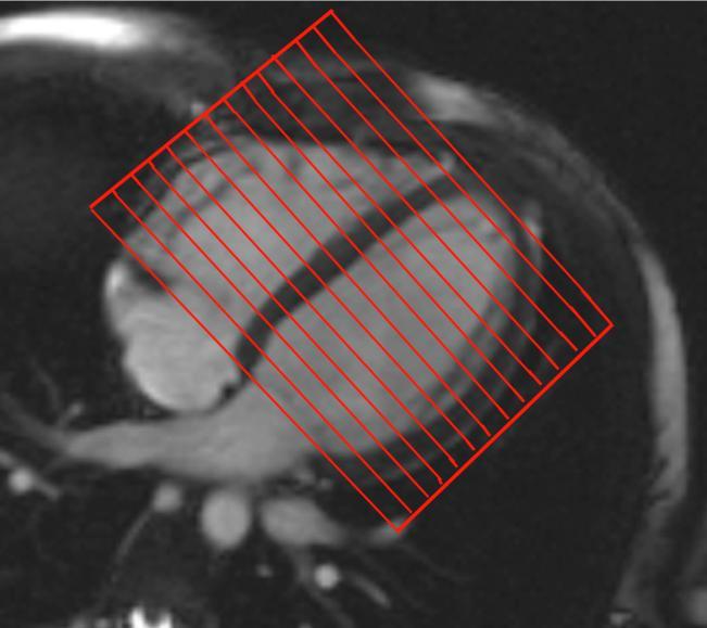
7 Methods: CMR Imaging Protocol Calculating RVEF by CMR

8 Methods: Echo Imaging Protocols Speckle tracking strain: RV global strain (RVGS)

9 Methods: Echo Imaging Protocols Speckle tracking strain: RV free wall strain (RVFWS)
")
10 Methods: Echo Imaging Protocols Tricuspid annular plane systolic excursion by M-Mode (M-Mode TAPSE)

11 Methods: Echo Imaging Protocols Tricuspid annular peak systolic myocardial tissue velocity by pulsed-wave tissue Doppler (RV PWTDE Sm)


12 Methods: Echo Imaging Protocols Tricuspid annular peak systolic tissue velocity by colour tissue velocity imaging (RV CTDE Sm)
.")
13 Methods: Echo Imaging Protocols Myocardial acceleration during isovolumic contraction of the right ventricle - Isovolumic acceleration (IVA). IVA = IVV/AT
.")
14 Methods: Echo Imaging Protocols Tricuspid annular plane systolic excursion by colour tissue Doppler (TDE TAPSE).
 Spectral Doppler")
/b <=>")
15 Methods: Echo Imaging Protocols RV Myocardial Performance Index (RV MPI) Spectral Doppler patterns of tricuspid inflow and pulmonary outflow were recorded RV MPI = (a-b)/b <=> (IVCT+IVRT)/RVET
16 Results:Patient Characteristics (N=50)
17 Results: Correlations with CMR (1/2) RVfwS RVGS MM TAPSE CTDE Sm
18 Results: Correlations with CMR (2/2) PWTDE Sm TDE TAPSE MPI IVA
 Sensitivity = 79% Sensitivity = 37% Specificity = 55% (80%) Specificity = 87% Specificity =")
19 Results:Receiver-operator characteristics (1/2) RVfwS RVGS MM-TAPSE Cut-off 25.50% ( 22.5%) Cut-off 19.15% Cut-off 1.6cm Sensitivity = 90% (80%) Sensitivity = 79% Sensitivity = 37% Specificity = 55% (80%) Specificity = 87% Specificity = 94%

20 Results:Receiver-operator characteristics (2/2) CTDE Sm PWTDE Sm TDE TAPSE Cut-off <8.5cm/s Cut-off <10cm/s Cut-off 1.8cm Sensitivity = 73% Sensitivity = 53% Sensitivity = 79% Specificity = 65% Specificity = 67% Specificity = 65%.
21 Results: Regression, Reproducibility. Acquisition & Analysis times (1/3) RV FREE WALL STRAIN: 1. was accurate: RVfwS had a stronger correlation with CMR-RVEF than all other RV echo indices. 2. was reproducible: 3. was clinically applicable:

22 Results: Regression, Reproducibility, Acquisition & Analysis times (2/3) 2ND: RVGS 1. Regression analysis: 3RD: MM TAPSE 1. Regression analysis: 2. Reproducibility: 2. Reproducibility: 3. Clinical application: 3. Clinical application:
23 Results: Regression, Reproducibility, Acquisition & Analysis times (3/3) Upon exclusion of RVfwS, RVGS and M-Mode TAPSE, no remaining echocardiographic index of RV systolic function exhibited a superior correlation over the other indices, when compared to CMR-RVEF.
24 Conclusions RVFwS had strongest correlation to CMR-derived RVEF. RVfwS is reproducible. RVfwS is sensitive & specific for detecting RV systolic dysfunction. RVfwS is quick and easy to use and has the potential to be used widely in clinical practice.
25 Thank you!
26
27 Results:Correlation of RV indices with visual assessment of RV function Comparison of CMR-RVEF and eight echo indices of RV function with visual assessment of RV systolic function. Each category of RV dysfunction is compared to the category above (i.e. Mild RV dysfunction is compared to normal RV function; moderatesevere RV dysfunction is compared to mild RV dysfunction).
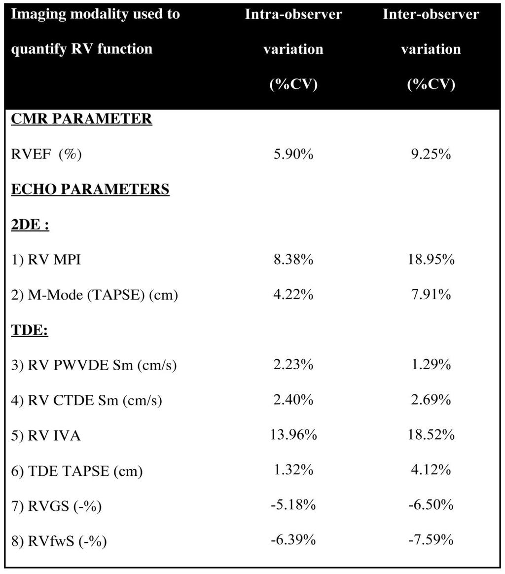
28 Results: Reproducibility
29 Study limitations & future work The normative cut-off values derived by ROC analysis in this study were for RV strain quantified using GE speckle tracking software. It is unclear if the same normative values can be applied to 2D strain analysed using software from other manufacturers and this warrants further investigation. The patient numbers in this pilot study are small, and the results of our research now need to be validated in a larger prospective study.
Incorporating the New Echo Guidelines Into Everyday Practice
 Incorporating the New Echo Guidelines Into Everyday Practice Clinical Case RIGHT VENTRICULAR FAILURE Gustavo Restrepo MD President Elect Interamerican Society of Cardiology Director Fellowship Training
Quantitation of right ventricular dimensions and function
 SCCS Basics of cardiac assessment Quantitation of right ventricular dimensions and function Tomasz Kukulski, MD PhD Dept of Cardiology, Congenital Heart Disease and Electrotherapy Silesian Medical University
RIGHT VENTRICULAR SIZE AND FUNCTION
 RIGHT VENTRICULAR SIZE AND FUNCTION Edwin S. Tucay, MD, FPCC, FPCC, FPSE Philippine Society of Echocardiography Quezon City, Philippines Echo Mission, BRTTH, Legaspi City, July 1-2, 2016 NO DISCLOSURE
Global left ventricular circumferential strain is a marker for both systolic and diastolic myocardial function
 Global left ventricular circumferential strain is a marker for both systolic and diastolic myocardial function Toshinari Onishi 1, Samir K. Saha 2, Daniel Ludwig 1, Erik B. Schelbert 1, David Schwartzman
Velocity Vector Imaging as a new approach for cardiac magnetic resonance: Comparison with echocardiography
 Velocity Vector Imaging as a new approach for cardiac magnetic resonance: Comparison with echocardiography Toshinari Onishi 1, Samir K. Saha 2, Daniel Ludwig 1, Erik B. Schelbert 1, David Schwartzman 1,
Right Ventricular Strain in Normal Healthy Adult Filipinos: A Retrospective, Cross- Sectional Pilot Study
 Right Ventricular Strain in Normal Healthy Adult Filipinos: A Retrospective, Cross- Sectional Pilot Study By Julius Caesar D. de Vera, MD Jonnah Fatima B. Pelat, MD Introduction Right ventricle contributes
Chamber Quantitation Guidelines - Update II
 Chamber Quantitation Guidelines - Update II Right Heart Measurements Steven A. Goldstein MD FACC FASE Professor of Medicine Georgetown University Medical Center MedStar Heart Institute Washington Hospital
Evaluation of the Right Ventricle and Risk Stratification for Sudden Cardiac Death
 Evaluation of the Right Ventricle and Risk Stratification for Sudden Cardiac Death Presenters: Sabrina Phillips, MD FACC FASE Director, Adult Congenital Heart Disease Services The University of Oklahoma
Fetal cardiac function: what to use and does it make a difference?
 17 th International Conference on Prenatal Diagnosis and Therapy Lisbon, June 2013 Fetal cardiac function: what to use and does it make a difference? Fàtima Crispi Department of Maternal-Fetal Medicine,
Pulmonary Hypertension: Echocardiographic Evaluation of Pulmonary Hypertension and Right Ventricular Function. Irmina Gradus-Pizlo, MD
 Pulmonary Hypertension: Echocardiographic Evaluation of Pulmonary Hypertension and Right Ventricular Function Irmina Gradus-Pizlo, MD Disclosures: Nothing to disclose Overview Is pulmonary hypertension
LV FUNCTION ASSESSMENT: WHAT IS BEYOND EJECTION FRACTION
 LV FUNCTION ASSESSMENT: WHAT IS BEYOND EJECTION FRACTION Jamilah S AlRahimi Assistant Professor, KSU-HS Consultant Noninvasive Cardiology KFCC, MNGHA-WR Introduction LV function assessment in Heart Failure:
Review Article Transthoracic Echocardiography in Children and Young Adults with Congenital Heart Disease
 International Scholarly Research Network ISRN Pediatrics Volume 2012, Article ID 753481, 15 pages doi:10.5402/2012/753481 Review Article Transthoracic Echocardiography in Children and Young Adults with
Right Ventricle Steven J. Lester MD, FACC, FRCP(C), FASE Mayo Clinic, Arizona
, FASE Mayo Clinic, Arizona") Right Ventricle Steven J. Lester MD, FACC, FRCP(C), FASE Mayo Clinic, Arizona 1. In which scenario will applying the simplified Bernoulli equation to the peak tricuspid regurgitation velocity and adding
Right Ventricle Steven J. Lester MD, FACC, FRCP(C), FASE Mayo Clinic, Arizona 1. In which scenario will applying the simplified Bernoulli equation to the peak tricuspid regurgitation velocity and adding
THE RIGHT VENTRICLE IN PULMONARY HYPERTENSION R. DRAGU
 THE RIGHT VENTRICLE IN PULMONARY HYPERTENSION R. DRAGU Cardiology Dept. Rambam Health Care Campus Rappaport Faculty of Medicine Technion, Israel Why the Right Ventricle? Pulmonary hypertension (PH) Right
10/7/2013. Systolic Function How to Measure, How Accurate is Echo, Role of Contrast. Thanks to our Course Director: Neil J.
 Systolic Function How to Measure, How Accurate is Echo, Role of Contrast Neil J. Weissman, MD MedStar Health Research Institute & Professor of Medicine Georgetown University Washington, D.C. No Disclosures
Research Article Changes in Mitral Annular Ascent with Worsening Echocardiographic Parameters of Left Ventricular Diastolic Function
 Scientifica Volume 216, Article ID 633815, 4 pages http://dx.doi.org/1.1155/216/633815 Research Article Changes in Mitral Annular Ascent with Worsening Echocardiographic Parameters of Left Ventricular
Fetal cardiovascular parameters for the prediction of postnatal cardiovascular risk in intrauterine growth-restriction?
 17 th International Conference on Prenatal Diagnosis and Therapy Lisbon, June 2013 Fetal cardiovascular parameters for the prediction of postnatal cardiovascular risk in intrauterine growth-restriction?
Myocardial Strain Imaging in Cardiac Diseases and Cardiomyopathies.
 Myocardial Strain Imaging in Cardiac Diseases and Cardiomyopathies. Session: Cardiomyopathy Tarun Pandey MD, FRCR. Associate Professor University of Arkansas for Medical Sciences Disclosures No relevant
Heart Failure in Women: Dr Goh Ping Ping Cardiologist Asian Heart & Vascular Centre
 Heart Failure in Women: More than EF? Dr Goh Ping Ping Cardiologist Asian Heart & Vascular Centre Overview Review pathophysiology as it relates to diagnosis and management Rational approach to workup:
Adel Hasanin Ahmed 1
 Adel Hasanin Ahmed 1 PERICARDIAL DISEASE The pericardial effusion ends anteriorly to the descending aorta and is best visualised in the PLAX. PSAX is actually very useful sometimes for looking at posterior
Pathophysiology and Current Evidence for Detection of Dyssynchrony
 Editorial Cardiol Res. 2017;8(5):179-183 Pathophysiology and Current Evidence for Detection of Dyssynchrony Michael Spartalis a, d, Eleni Tzatzaki a, Eleftherios Spartalis b, Christos Damaskos b, Antonios
General Cardiovascular Magnetic Resonance Imaging
 2 General Cardiovascular Magnetic Resonance Imaging 19 Peter G. Danias, Cardiovascular MRI: 150 Multiple-Choice Questions and Answers Humana Press 2008 20 Cardiovascular MRI: 150 Multiple-Choice Questions
Evaluation of the Right Ventricle in Candidates for Right Ventricular Assist Device Implantation.
 Evaluation of the Right Ventricle in Candidates for Right Ventricular Assist Device Implantation. Evaluation of RVAD Function. Ioannis A Paraskevaidis Attikon University Hospital Historical Perspective
Quantification of Cardiac Chamber Size
 2017 KSE 2017-11-25 Quantification of Cardiac Chamber Size Division of Cardiology Keimyung University Dongsan Medical Center In-Cheol Kim M.D., Ph.D. LV size and function Internal linear dimensions PLX
Echocardiographic assessment of the right ventricle in paediatric pulmonary hypertension.
 Echocardiographic assessment of the right ventricle in paediatric pulmonary hypertension. Mark K. Friedberg, MD No disclosures Outline RV response to increased afterload Echo assessment of RV function
Assessing the Impact on the Right Ventricle
 Advances in Tricuspid Regurgitation Congress of the European Society of Cardiology (ESC) Munich, August 25-29, 2012 Assessing the Impact on the Right Ventricle Stephan Rosenkranz, MD Clinic III for Internal
Echocardiography in Adult Congenital Heart Disease
 Echocardiography in Adult Congenital Heart Disease Michael Vogel Kinderherz-Praxis München CHD missed in childhood Subsequent lesions after repaired CHD Follow-up of cyanotic heart disease CHD missed in
Chamber Quantitation Guidelines II Right Heart Measurements
 Chamber Quantitation Guidelines II Right Heart Measurements Steven A. Goldstein MD FACC FASE Director, Noninvasive Cardiology MedStar Heart Institute Washington Hospital Center Sunday, October 9, 2016
Right Heart Evaluation ASE Guidelines Review. Chris Mann RDCS, RCS, FASE Faculty, Echocardiography Pitt Community College Greenville, NC
 Right Heart Evaluation ASE Guidelines Review Chris Mann RDCS, RCS, FASE Faculty, Echocardiography Pitt Community College Greenville, NC Objectives Briefly review right atrial and right ventricular anatomy
Index. K Knobology, TTE artifact, image resolution, ultrasound, 14
 A Acute aortic regurgitation (AR), 124 128 Acute aortic syndrome (AAS) classic aortic dissection diagnosis, 251 263 evolutive patterns, 253 255 pathology, 250 251 classifications, 247 248 incomplete aortic
Effect of Heart Rate on Tissue Doppler Measures of E/E
 Cardiology Department of Bangkok Metropolitan Administration Medical College and Vajira Hospital, Bangkok, Thailand Abstract Background: Our aim was to study the independent effect of heart rate (HR) on
HFPEF Echo with Strain vs. MRI T1 Mapping
 HFPEF Echo with Strain vs. MRI T1 Mapping Erik Schelbert, MD MS Director, Cardiovascular Magnetic Resonance Assistant Professor of Medicine Heart & Vascular Institute University of Pittsburgh Disclosures
Pediatric Echocardiographic Normal values. SIEC Firenze Febbraio 2016
 Pediatric Echocardiographic Normal values Massimiliano Cantinotti MD Fondazione Toscana G. Monasterio and Institute of Clinical Physiology (CNR) Massa and Pisa SIEC Firenze 18-20 Febbraio 2016 Background
Tissue Doppler Imaging in Congenital Heart Disease
 Tissue Doppler Imaging in Congenital Heart Disease L. Youngmin Eun, M.D. Department of Pediatrics, Division of Pediatric Cardiology, Kwandong University College of Medicine The potential advantage of ultrasound
Appendix II: ECHOCARDIOGRAPHY ANALYSIS
 Appendix II: ECHOCARDIOGRAPHY ANALYSIS Two-Dimensional (2D) imaging was performed using the Vivid 7 Advantage cardiovascular ultrasound system (GE Medical Systems, Milwaukee) with a frame rate of 400 frames
The background of the Cardiac Sonographer Network News masthead is a diagnostic image:
 Number 5 Welcome Number 5 Welcome to the newsletter created just for you: sonographers who perform pediatric echocardiograms in primarily adult echo labs. Each issue features tips on echocardiography of
Right Heart Hemodynamics: Echo-Cath Discrepancies
 Department of cardiac, thoracic and vascular sciences University of Padua, School of Medicine Padua, Italy Right Heart Hemodynamics: Echo-Cath Discrepancies Luigi P. Badano, MD, PhD, FESC, FACC **Dr. Badano
Evaluation of Left Ventricular Diastolic Dysfunction by Doppler and 2D Speckle-tracking Imaging in Patients with Primary Pulmonary Hypertension
 ESC Congress 2011.No 85975 Evaluation of Left Ventricular Diastolic Dysfunction by Doppler and 2D Speckle-tracking Imaging in Patients with Primary Pulmonary Hypertension Second Department of Internal
Mechanisms of False Positive Exercise Electrocardiography: Is False Positive Test Truly False?
 Mechanisms of False Positive Exercise Electrocardiography: Is False Positive Test Truly False? Masaki Izumo a, Kengo Suzuki b, Hidekazu Kikuchi b, Seisyo Kou b, Keisuke Kida b, Yu Eguchi b, Nobuyuki Azuma
Stress, strain and contrast. UK available agents. Safety 13/06/2018. Which enhancing agent do you use? Ultrasound enhancing agents.
 Stress, strain and contrast Stephen Glen Ultrasound enhancing agents Safety Effectiveness during stress Perfusion / myocardial contrast UK available agents Which enhancing agent do you use? Name Bubble
Strain and Strain Rate Imaging How, Why and When?
 Strain and Strain Rate Imaging How, Why and When? João L. Cavalcante, MD Advanced Cardiac Imaging Fellow Cleveland Clinic Foundation Disclosures: No conflicts of interest Movement vs Deformation Movement
Doppler-echocardiographic findings in a patient with persisting right ventricular sinusoids
 Zurich Open Repository and Archive University of Zurich Main Library Strickhofstrasse 39 CH-8057 Zurich www.zora.uzh.ch Year: 1990 Doppler-echocardiographic findings in a patient with persisting right
좌심실수축기능평가 Cardiac Function
 Basic Echo Review Course 좌심실수축기능평가 Cardiac Function Seonghoon Choi Cardiology Hallym university LV systolic function Systolic function 좌심실수축기능 - 심근의수축으로심실에서혈액을대동맥으로박출하는기능 실제임상에서 LV function 의의미 1Diagnosis
Coronary artery disease (CAD) risk factors
 risk factors") Background Coronary artery disease (CAD) risk factors CAD Risk factors Hypertension Insulin resistance /diabetes Dyslipidemia Smoking /Obesity Male gender/ Old age Atherosclerosis Arterial stiffness precedes
Background Coronary artery disease (CAD) risk factors CAD Risk factors Hypertension Insulin resistance /diabetes Dyslipidemia Smoking /Obesity Male gender/ Old age Atherosclerosis Arterial stiffness precedes
Tissue Doppler Imaging
 Cronicon OPEN ACCESS Hesham Rashid* Tissue Doppler Imaging CARDIOLOGY Editorial Department of Cardiology, Benha University, Egypt *Corresponding Author: Hesham Rashid, Department of Cardiology, Benha University,
Ιπποκράτειες μέρες καρδιολογίας Θεσσαλονίκη, 9-10 Μαρτίου Φωτεινή Α. Λαζαρίδου Επιμελήτρια Α Γενικό Νοσοκομείο Αγιος Παύλος, Θεσσαλονίκη
 Ιπποκράτειες μέρες καρδιολογίας Θεσσαλονίκη, 9-10 Μαρτίου 2018 Φωτεινή Α. Λαζαρίδου Επιμελήτρια Α Γενικό Νοσοκομείο Αγιος Παύλος, Θεσσαλονίκη RV shape Triangular shape in frontal plane crescent shape in
2019 Qualified Clinical Data Registry (QCDR) Performance Measures
 Performance Measures") 2019 Qualified Clinical Data Registry (QCDR) Performance Measures Description: This document contains the 18 performance measures approved by CMS for inclusion in the 2019 Qualified Clinical Data Registry
2019 Qualified Clinical Data Registry (QCDR) Performance Measures Description: This document contains the 18 performance measures approved by CMS for inclusion in the 2019 Qualified Clinical Data Registry
Right Ventricular Function
 Right Ventricular Function Jan Marek Professor of Cardiology Great Ormond Street Hospital & Institute of Cardiovascular Sciences, University College London No disclosures Right ventricular function: questions
PART II ECHOCARDIOGRAPHY LABORATORY OPERATIONS ADULT TRANSTHORACIC ECHOCARDIOGRAPHY TESTING
 PART II ECHOCARDIOGRAPHY LABORATORY OPERATIONS ADULT TRANSTHORACIC ECHOCARDIOGRAPHY TESTING STANDARD - Primary Instrumentation 1.1 Cardiac Ultrasound Systems SECTION 1 Instrumentation Ultrasound instruments
Københavns Universitet
 university of copenhagen Københavns Universitet Total average diastolic longitudinal displacement by colour tissue doppler imaging as an assessment of diastolic function de Knegt, Martina Chantal; Biering-Sørensen,
COMPLEX CONGENITAL HEART DISEASE: WHEN IS IT TOO LATE TO INTERVENE?
 COMPLEX CONGENITAL HEART DISEASE: WHEN IS IT TOO LATE TO INTERVENE? Aurora S. Gamponia, MD, FPPS, FPCC, FPSE OBJECTIVES Identify complex congenital heart disease at high risk or too late for intervention
Squeeze, Squeeze, Squeeze: The Importance of Right Ventricular Function and PH
 Squeeze, Squeeze, Squeeze: The Importance of Right Ventricular Function and PH Javier Jimenez MD PhD FACC Director, Advanced Heart Failure and Pulmonary Hypertension Miami Cardiac & Vascular Institute
Assesment of Right Ventricle Function with Speckle Tracking Echocardiography after the Percutaneous Closure of Atrial Septal Defect
 Original Article Acta Cardiol Sin 2017;33:523 529 doi: 10.6515/ACS20170106A Cardiac Image Assesment of Right Ventricle Function with Speckle Tracking Echocardiography after the Percutaneous Closure of
E/Ea is NOT an essential estimator of LV filling pressures
 Euroecho Kopenhagen Echo in Resynchronization in 2010 E/Ea is NOT an essential estimator of LV filling pressures Wilfried Mullens, MD, PhD December 10, 2010 Ziekenhuis Oost Limburg Genk University Hasselt
Conflict of interest: none declared
 The value of left ventricular global longitudinal strain assessed by three-dimensional strain imaging in the early detection of anthracycline-mediated cardiotoxicity C. Mornoş, A. Ionac, D. Cozma, S. Pescariu,
Echocardiographic Cardiovascular Risk Stratification: Beyond Ejection Fraction
 Echocardiographic Cardiovascular Risk Stratification: Beyond Ejection Fraction October 4, 2014 James S. Lee, M.D., F.A.C.C. Associates in Cardiology, P.A. Silver Spring, M.D. Disclosures Financial none
Hemodynamic Assessment. Assessment of Systolic Function Doppler Hemodynamics
 Hemodynamic Assessment Matt M. Umland, RDCS, FASE Aurora Medical Group Milwaukee, WI Assessment of Systolic Function Doppler Hemodynamics Stroke Volume Cardiac Output Cardiac Index Tei Index/Index of myocardial
Right ventricular adaptation in endurance athletes. António Freitas. No conflict of interest
 The role of echocardiography in sports cardiology Right ventricular adaptation in endurance athletes. António Freitas Cardiology Department - Fernando Fonseca Hospital Lisbon Sports Medicine Centre - Lisbon
Diastolic Function: What the Sonographer Needs to Know. Echocardiographic Assessment of Diastolic Function: Basic Concepts 2/8/2012
 Diastolic Function: What the Sonographer Needs to Know Pat Bailey, RDCS, FASE Technical Director Beaumont Health System Echocardiographic Assessment of Diastolic Function: Basic Concepts Practical Hints
4D Auto LAQ (Left Atrial Quantification)
") 4D Auto LAQ (Left Atrial Quantification) Introduction There has been an increased interest in quantification of the left atrium (LA) for various types of diseases; e.g. heart failure, heart valve diseases,
4D Auto LAQ (Left Atrial Quantification) Introduction There has been an increased interest in quantification of the left atrium (LA) for various types of diseases; e.g. heart failure, heart valve diseases,
Assessment of cardiac function with 3D echocardiography. Đánh giá chức năng tim bằng siêu âm tim 3D
 Assessment of cardiac function with 3D echocardiography Đánh giá chức năng tim bằng siêu âm tim 3D TS. BS. Nguyễn Thị Thu Hoài Viện Tim Mạch Quốc Gia Việt Nam TỪ SIÊU ÂM M-mode ĐẾN SIÊU ÂM 3D TỪ SIÊU ÂM
Myocardial performance index, Tissue Doppler echocardiography
 Value of Measuring Myocardial Performance Index by Tissue Doppler Echocardiography in Normal and Diseased Heart Tarkan TEKTEN, 1 MD, Alper O. ONBASILI, 1 MD, Ceyhun CEYHAN, 1 MD, Selim ÜNAL, 1 MD, and
Title:Relation Between E/e' ratio and NT-proBNP Levels in Elderly Patients with Symptomatic Severe Aortic Stenosis
 Author's response to reviews Title:Relation Between E/e' ratio and NT-proBNP Levels in Elderly Patients with Symptomatic Severe Aortic Stenosis Authors: Mihai Strachinaru (m.strachinaru@erasmusmc.nl) Bas
Ref 1. Ref 2. Ref 3. Ref 4. See graph
 Ref 1 Ref 2 Ref 3 1. Ages 6-23 y/o 2. Significant LVM differences by gender 3. For males 95 th percentiles: a. LVM/BSA = 103 b. LVM/height = 100 4. For females 95 th percentiles: a. LVM/BSA = 84 b. LVM/height
British Society of Echocardiography
 British Society of Echocardiography Affiliated to the British Cardiac Society A Minimum Dataset for a Standard Adult Transthoracic Echocardiogram From the British Society of Echocardiography Education
Vincent Wu 1, Janice Y Chyou 2, Sohae Chung 1, Sharath Bhagavatula 1 and Leon Axel 1,2*
 Wu et al. Journal of Cardiovascular Magnetic Resonance 2014, 16:71 RESEARCH Open Access Evaluation of diastolic function by three-dimensional volume tracking of the mitral annulus with cardiovascular magnetic
Cardiac MRI in ACHD What We. ACHD Patients
 Cardiac MRI in ACHD What We Have Learned to Apply to ACHD Patients Faris Al Mousily, MBChB, FAAC, FACC Consultant, Pediatric Cardiology, KFSH&RC/Jeddah Adjunct Faculty, Division of Pediatric Cardiology
Strain Imaging: Myocardial Mechanics Simplified and Applied
 9/28/217 Strain Imaging: Myocardial Mechanics Simplified and Applied John Gorcsan III, MD Professor of Medicine Director of Clinical Research Division of Cardiology VECTORS OF CONTRACTION Shortening Thickening
DOPPLER HEMODYNAMICS (1) QUANTIFICATION OF PRESSURE GRADIENTS and INTRACARDIAC PRESSURES
 QUANTIFICATION OF PRESSURE GRADIENTS and INTRACARDIAC PRESSURES") THORAXCENTRE DOPPLER HEMODYNAMICS (1) QUANTIFICATION OF PRESSURE GRADIENTS and INTRACARDIAC PRESSURES J. Roelandt DOPPLER HEMODYNAMICS Intracardiac pressures and pressure gradients Volumetric measurement
THORAXCENTRE DOPPLER HEMODYNAMICS (1) QUANTIFICATION OF PRESSURE GRADIENTS and INTRACARDIAC PRESSURES J. Roelandt DOPPLER HEMODYNAMICS Intracardiac pressures and pressure gradients Volumetric measurement
The rapid evolution of echocardiography during the past 25 years
 Evaluation of Myocardial Mechanics in the Fetus by Velocity Vector Imaging Adel K. Younoszai, MD, David E. Saudek, MD, Stephen P. Emery, MD, and James D. Thomas, MD, Denver, Colorado; Cleveland, Ohio;
Adult Echocardiography Examination Content Outline
 Adult Echocardiography Examination Content Outline (Outline Summary) # Domain Subdomain Percentage 1 2 3 4 5 Anatomy and Physiology Pathology Clinical Care and Safety Measurement Techniques, Maneuvers,
Restrictive Cardiomyopathy
 ESC Congress 2011, Paris Imaging Unusual Causes of Cardiomyopathy Restrictive Cardiomyopathy Kazuaki Tanabe, MD, PhD Professor of Medicine Chair, Division of Cardiology Izumo, Japan I Have No Disclosures
Functional Assessment for Congenital Heart Disease
 Review Print ISSN 1738-5520 On-line ISSN 1738-5555 Korean Circulation Journal Functional Assessment for Congenital Heart Disease Yiu-fai Cheung, MD Division of Paediatric Cardiology, Department of Paediatrics
Quantitative Assessment of Fetal Ventricular Function:
 Reprinted with permission from ECHOCARDIOGRAPHY, Volume 18, No. 1, January 2001 Copyright 2001 by Futura Publishing Company, Inc., Armonk, NY 1004-0418 Quantitative Assessment of Fetal Ventricular Function:
Objectives. CMR Volumetric Analysis 8/25/11. CMR Volumetric Analysis Technique. Cardiac imaging plane acquisition. CMR Volumetric Analysis
 Objectives Cynthia K. Rigsby Children s Memorial Hospital Chicago, IL CMR volumetric analysis Techniques Normalized data Sources of error CMR phase contrast flow analysis Techniques What we can do with
Echocardiography for the Electrophysiologist: Day-to-day practice. Emmanuel Fares, MD
 Echocardiography for the Electrophysiologist: Day-to-day practice Emmanuel Fares, MD EP and pacing service, Department of Cardiovascular Medicine, Cairo University Agenda Role of echo in arrhythmia management:
Cardiac Magnetic Resonance in pregnant women
 Cardiac Magnetic Resonance in pregnant women Chen SSM, Leeton L, Dennis AT Royal Women s Hospital and The University of Melbourne, Parkville, Australia alicia.dennis@thewomens.org.au Quantification of
VECTORS OF CONTRACTION
 1/3/216 Strain, Strain Rate, and Torsion: Myocardial Mechanics Simplified and Applied VECTORS OF CONTRACTION John Gorcsan, MD University of Pittsburgh, Pittsburgh, PA Shortening Thickening Twisting No
Cardiac ultrasound protocols
 Cardiac ultrasound protocols IDEXX Telemedicine Consultants Two-dimensional and M-mode imaging planes Right parasternal long axis four chamber Obtained from the right side Displays the relative proportions
Disclosures. Cardiac Ultrasound. Introductory Case. 80 y/o male Syncope at home Emesis x 3 in ambulance Looks sick. No pain.
 Disclosures Cardiac Ultrasound Justin A Davis, MD MPH RDMS Subchief for Emergency Ultrasound Kaiser Permanente East Bay Medical Center I have nothing to disclose. Introductory Case HR 118 BP 65/43 RR 27
ASCeXAM / ReASCE. Practice Board Exam Questions Monday Morning
 ASCeXAM / ReASCE Practice Board Exam Questions Monday Morning Ultrasound Physics Artifacts Doppler Physics Imaging, Knobology, and Artifacts Echocardiographic Evaluation of the RV Tricuspid and Pulmonary
COMPREHENSIVE EVALUATION OF FETAL HEART R. GOWDAMARAJAN MD
 COMPREHENSIVE EVALUATION OF FETAL HEART R. GOWDAMARAJAN MD Disclosure No Relevant Financial Relationships with Commercial Interests Fetal Echo: How to do it? Timing of Study -optimally between 22-24 weeks
Certificate in Clinician Performed Ultrasound (CCPU) Syllabus. Rapid Cardiac Echo (RCE)
 Syllabus. Rapid Cardiac Echo (RCE)") Certificate in Clinician Performed Ultrasound (CCPU) Syllabus Rapid Cardiac Echo (RCE) Purpose: Rapid Cardiac Echocardiography (RCE) This unit is designed to cover the theoretical and practical curriculum
Certificate in Clinician Performed Ultrasound (CCPU) Syllabus Rapid Cardiac Echo (RCE) Purpose: Rapid Cardiac Echocardiography (RCE) This unit is designed to cover the theoretical and practical curriculum
Objectives. Diastology: What the Radiologist Needs to Know. LV Diastolic Function: Introduction. LV Diastolic Function: Introduction
 Objectives Diastology: What the Radiologist Needs to Know. Jacobo Kirsch, MD Cardiopulmonary Imaging, Section Head Division of Radiology Cleveland Clinic Florida Weston, FL To review the physiology and
The new Guidelines: Focus on Chronic Heart Failure
 The new Guidelines: Focus on Chronic Heart Failure Petros Nihoyannopoulos MD, FRCP, FESC Professor of Cardiology Imperial College London and National & Kapodistrian University of Athens 2 3 4 The principal
DISCLOSURE. Myocardial Mechanics. Relevant Financial Relationship(s) Off Label Usage
 Off Label Usage") 7th Annual Team Echocardiography: The Heart of Cardiovascular Medicine Tissue Doppler, Strain, Speckle: What? How? Christopher J Kramer RDCS Aurora Medical Group Advanced Cardiovascular Services, Aurora
7th Annual Team Echocardiography: The Heart of Cardiovascular Medicine Tissue Doppler, Strain, Speckle: What? How? Christopher J Kramer RDCS Aurora Medical Group Advanced Cardiovascular Services, Aurora
22 nd Annual Conference of the Saudi Heart Association Riyadh, Saudi Arabia
 22 nd Annual Conference of the Saudi Heart Association Riyadh, Saudi Arabia New Echocardiographic Modalities to Evaluate Ventricular Function in Congenital Heart Disease: Tissue Doppler & Strain Rate Imaging
The Patient with Atrial Fibrilation
 Assessment of Diastolic Function The Patient with Atrial Fibrilation Assoc. Prof. Adriana Ilieşiu, FESC University of Medicine Carol Davila Bucharest, Romania Associated Conditions with Atrial Fibrillation
Vivid T8 Ultrasound. Cardiac and shared services. Together like never before from GE.
 Vivid T8 Ultrasound Cardiac and shared services. Together like never before from GE. Vivid cardiac. Shared services. Paired for excellent performance. In the Vivid T8, we ve taken the established cardiac
The Fontan circulation. Folkert Meijboom
 The Fontan circulation Folkert Meijboom What to expect? Why a Fontan-circulation Indications How does it work Types of Fontan circulation Historical overview Role of echocardiography What to expect? Why
evicore cardiology procedures and services requiring prior authorization
 evicore cardiology procedures and services requiring prior authorization Moda Health Commercial Group and Individual Members* *Check EBT to verify member enrollment in evicore program Radiology Advanced
EDITOR S PICK CURRENT STATUS OF FULLY AUTOMATED SOFTWARE WITH THREE-DIMENSIONAL ECHOCARDIOGRAPHY FOR THE QUANTIFICATION OF LEFT VENTRICULAR FUNCTION
 ITOR S PICK This paper, courtesy of Yang and Takeuchi, provides a timely and well-considered update on the current status of fully-automated software with three-dimensional echocardiography for quantifying
B-Mode measurements protocols:
 Application Note How to Perform the Most Commonly Used Measurements from the Cardiac Measurements Package associated with Calculations of Cardiac Function using the Vevo Lab Objective The Vevo LAB offline
Transthoracic echocardiography in the evaluation of pediatric pulmonary hypertension and ventricular dysfunction
 REVIEW ARTICLE Transthoracic echocardiography in the evaluation of pediatric pulmonary hypertension and ventricular dysfunction Martin Koestenberger, 1 Mark K. Friedberg, 2 Eirik Nestaas, 3 Ina Michel-Behnke,
OPTIMIZING ECHO ACQUISTION FOR STRAIN AND DIASTOLOGY
 OPTIMIZING ECHO ACQUISTION FOR STRAIN AND DIASTOLOGY October 8, 2017 Deborah Agler, ACS, RDCS, FASE Coordinator of Education and Training Cleveland Clinic General Principles Diastology Clinical Data Heart
Valvular Regurgitation: Can We Do Better Than Colour Doppler?
 Valvular Regurgitation: Can We Do Better Than Colour Doppler? A/Prof David Prior St Vincent s Hospital Melbourne Sports Cardiology Valvular Regurgitation Valve regurgitation volume loads the ventricles
Vivid T8 Ultrasound. Cardiac and shared services. Together like never before from GE. Take rugged reliability to challenging conditions.
 Vivid T8 Ultrasound Cardiac and shared services. Together like never before from GE. Vivid cardiac. Shared services. Paired for excellent performance. In the Vivid T8, we ve taken the established cardiac
2/2/2011. Strain and Strain Rate Imaging How, Why and When? Movement vs Deformation. Doppler Myocardial Velocities. Movement. Deformation.
 Strain and Strain Rate Imaging How, Why and When? João L. Cavalcante, MD Advanced Cardiac Imaging Fellow Cleveland Clinic Foundation Disclosures: No conflicts of interest Movement vs Deformation Movement
MITRAL STENOSIS. Joanne Cusack
 MITRAL STENOSIS Joanne Cusack BSE Breakdown Recognition of rheumatic mitral stenosis Qualitative description of valve and sub-valve calcification and fibrosis Measurement of orifice area by planimetry
Assessment of right ventricular contraction by speckle tracking echocardiography in pulmonary hypertension patients.
 Biomedical Research 2017; 28 (1): 173-177 ISSN 0970-938X www.biomedres.info Assessment of right ventricular contraction by speckle tracking echocardiography in pulmonary hypertension patients. Yudong Peng,