Echocardiographic Evaluation of the Cardiomyopathies. Stephanie Coulter, MD, FACC, FASE April, 2016
|
|
|
- Stephanie Stevenson
- 6 years ago
- Views:
Transcription
1 Echocardiographic Evaluation of the Cardiomyopathies Stephanie Coulter, MD, FACC, FASE April, 2016
2 Cardiomyopathies (CMP) primary disease intrinsic to cardiac muscle Dilated CMP Hypertrophic CMP Infiltrative CMP Left Ventricular Non-compaction Arrythmogenic Right Ventricular CMP Takotsubo Cardiomyopathy
3 Utility of Echo in CMP CMP presentation is clinically indistinct New onset CHF Arrhythmias Echo is the easiest and most utilized diagnostic test in the evaluation of CHF Etiology Valvular, congenital defects, ischemia Prognosis Systolic function Diastolic function Filling pressures Complications MR Thrombus
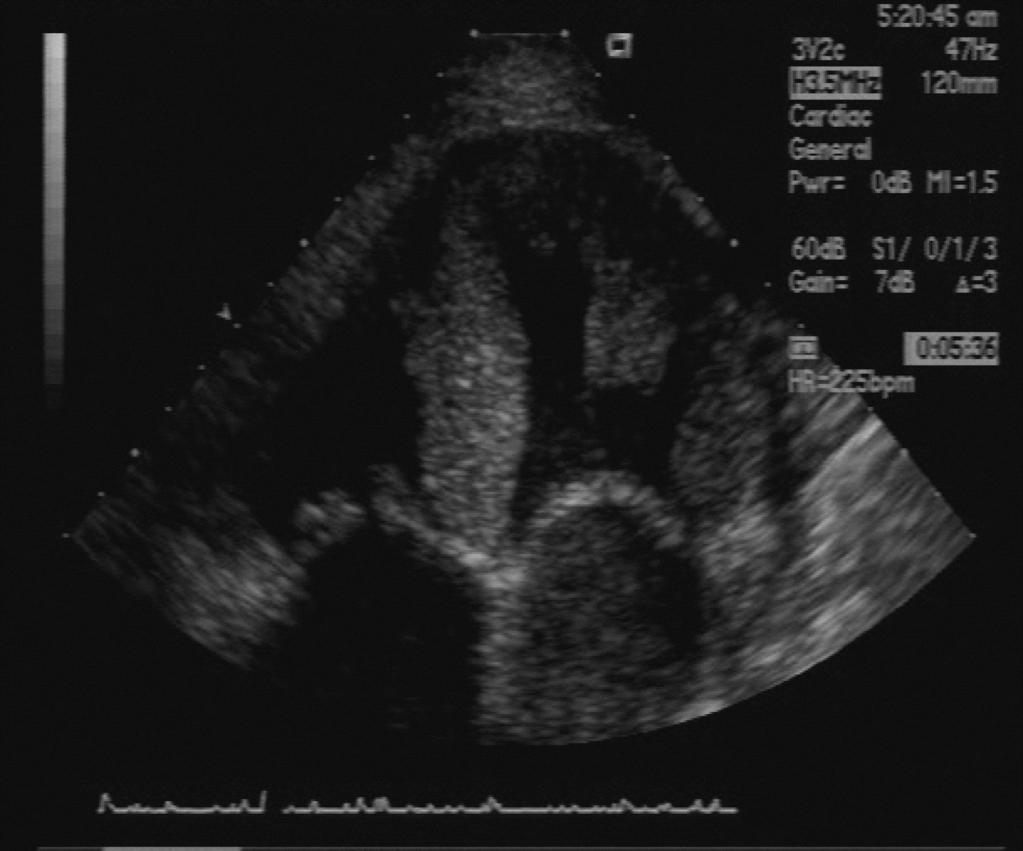
4 Dilated Cardiomyopathy Global decrease in systolic contractile function with eventual dilatation of the ventricles Echocardiographic features Dilated chambers, LV, LA RV, RA Usually normal LV wall thickness

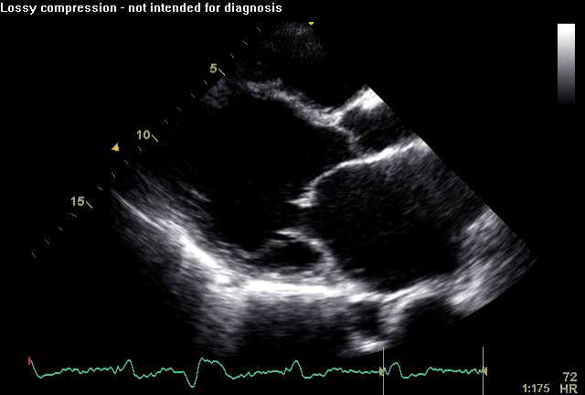
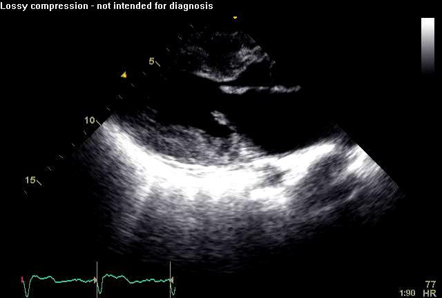
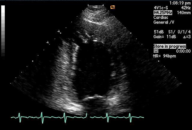
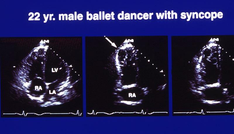
5 Dilated Non-Ischemic CMP- end stage RV LV LA

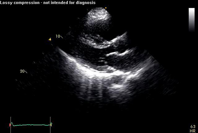
6 Apical 4 C Non-ischemic CMP

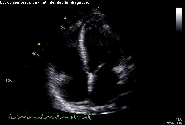
7 Mitral Regurgitation MV tethering LV MV annulus LA
8 LV LA

9 Mitral Regurgitation Altered LV geometry Apical tethering of leaflets Annular size increases Decreased LV closing force Decreases survival

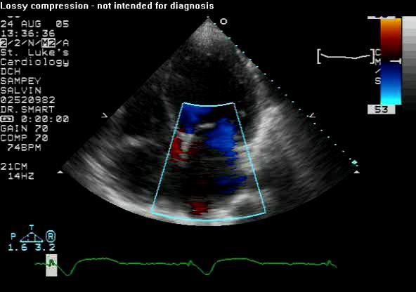
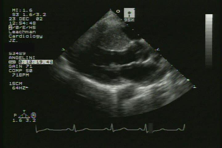
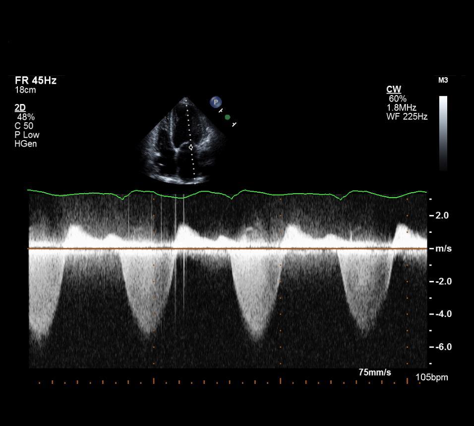
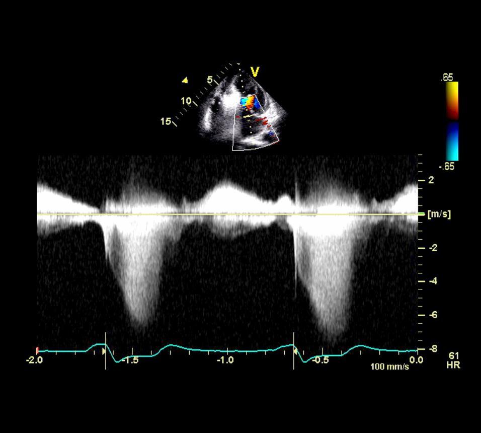
10 Dynamic Mitral Regurgitation
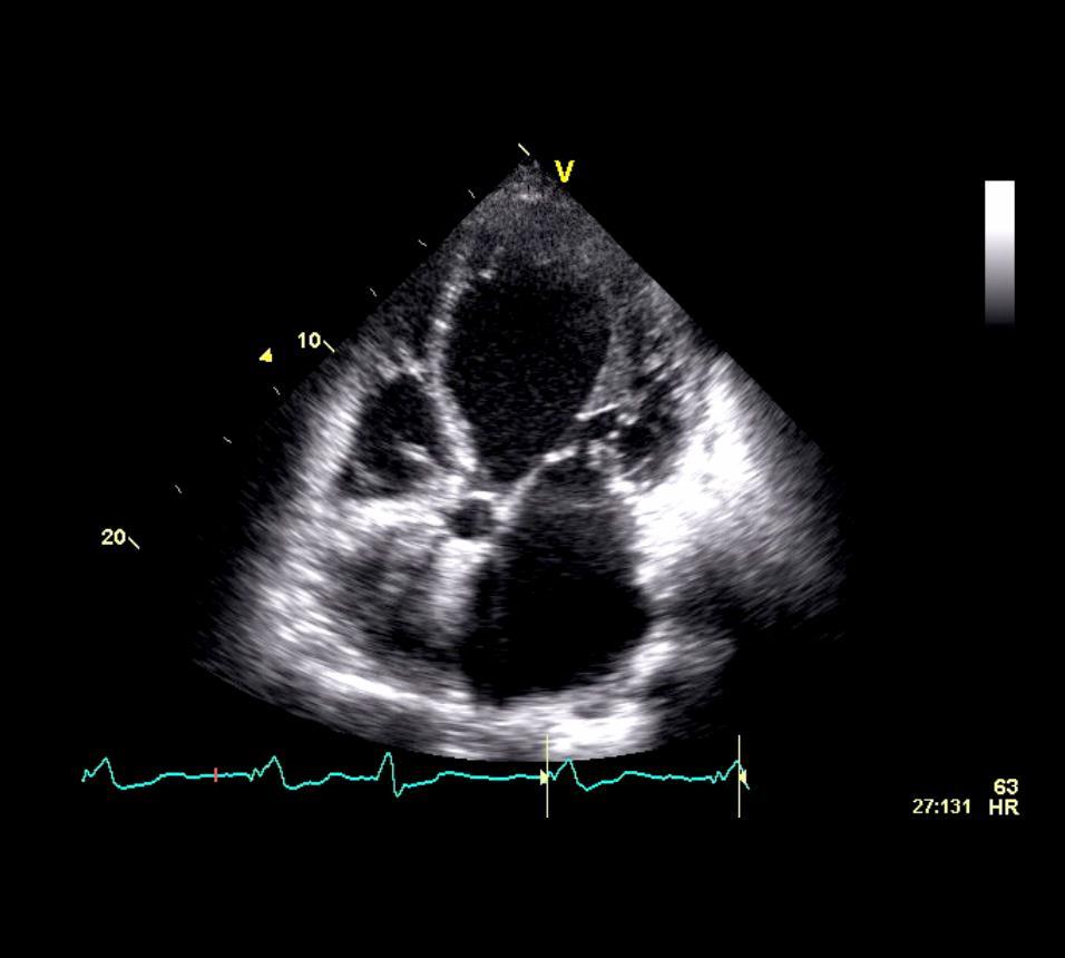
11

12 Apical Thrombus

13 Contrast Enhances Apical Thrombus
14 Echocardiographic Predictors in CMP LV size and function RV involvement Diastolic indices DT Best predictor of death in symptomatic patients EF< 25%: DT < 130ms mortality in 2 yrs of 65% vs 26% DT >125 Filling pressures LA size CRT another lecture
15 Hypertrophic Cardiomyopathy Unexplained hypertrophy (no HTN, AS ) Any degree or pattern: diffuse, septal, apical Abnormalities of diastolic function Always present Not a consequence of hypertrophy Precedes hypertrophy!! Correlates with symptoms Early diagnosis in genetically at risk Increased risk of sudden death Athletes Marked septal hypertrophy Family history of sudden death LVOT obstruction with mild or no symptoms
16 Hypertrophic CMP Screening Autosomal dominant Phenotypic expression variable 1/500 First degree relatives Ages yrs annual clinical, ecg, echo > 18 yrs ASX every 5 years <12 yrs evaluate if symptomatic or intense sports Affected family members need repeat screening every months
 LV wall thickness >13 mm Does not require assymmetry (not specific) ASH leads to earlier diagnosis---sam murmur and")
17 Echocardiographic use in HCM Best screening test Defines magnitude and location of hypertrophy Excludes other causes of hypertrophy (AS, VSD) LV wall thickness >13 mm Does not require assymmetry (not specific) ASH leads to earlier diagnosis---sam murmur and symptoms
18 Hypertrophic Cardiomyopathy
19
20 LVOT Obstruction Systolic Anterior Motion (SAM) most common cause of LVOT obstruction in HCM Only 25% of HCM at rest have LVOTo > 30 mmhg Dynamic Recent study found 70% of patients without LVOTo at rest developed LVOTo with exercise Increases with increases in contractility and HR Decreases with increases in LV volume
21 LVOT Obstruction Independently predicts More severe diastolic dysfunction and symptoms Stroke Atrial fibrillation Arrythymia-related death Most common in Females Elderly Greater degree of hypertrophy
22 Systolic Anterior Motion Abnormal independent anterior motion of the mitral leaflets into the LVOT during systole Either leaflet; anterior, posterior or both Requires Narrow LVOT Hypercontractile LV Redudant MV leaflets Severity of LVOT o is related to the duration and degree of SAM and to the reduction in LVOT size Decreases stroke volume Raises ventricular pressures creating more hypertrophy

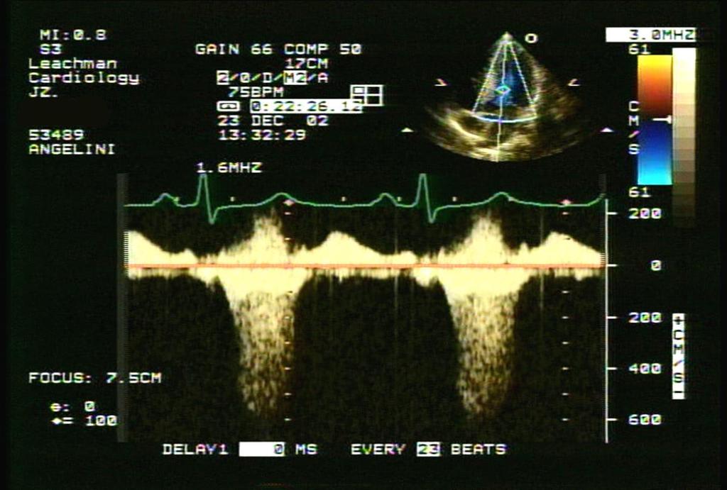
23 Systolic Anterior Motion by M mode

24 HCM and SAM still

25 Color alias pattern of SAM, suggests area of LVOTo still

26 HCM and SAM

27 HCM, SAM and MR
28 Doppler Echocardiography
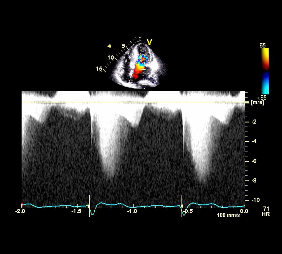
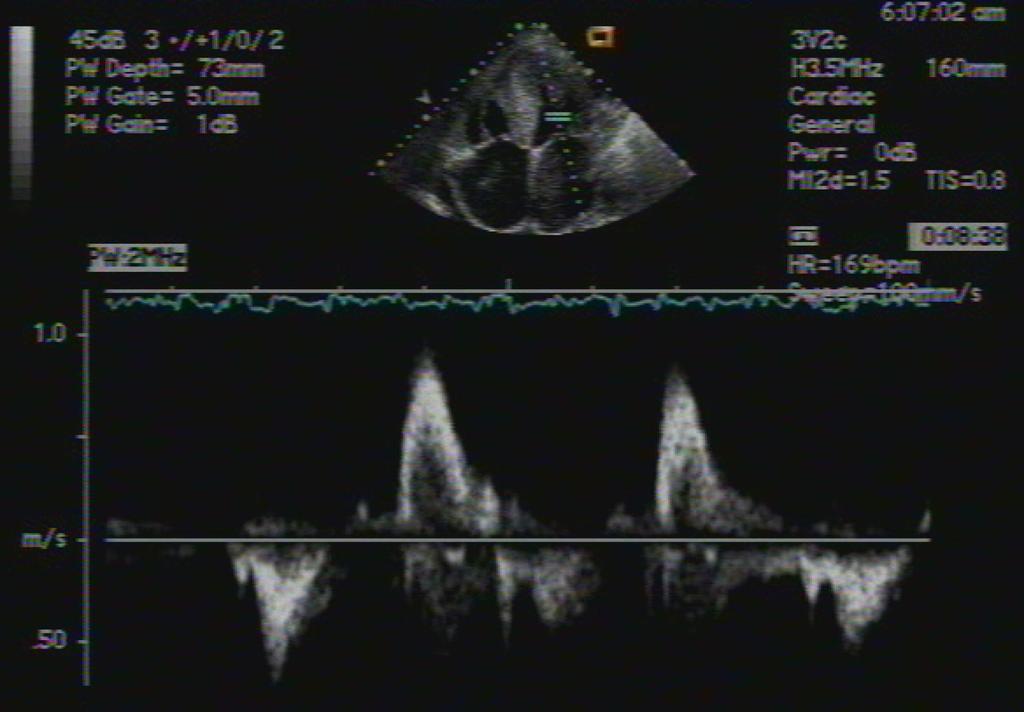
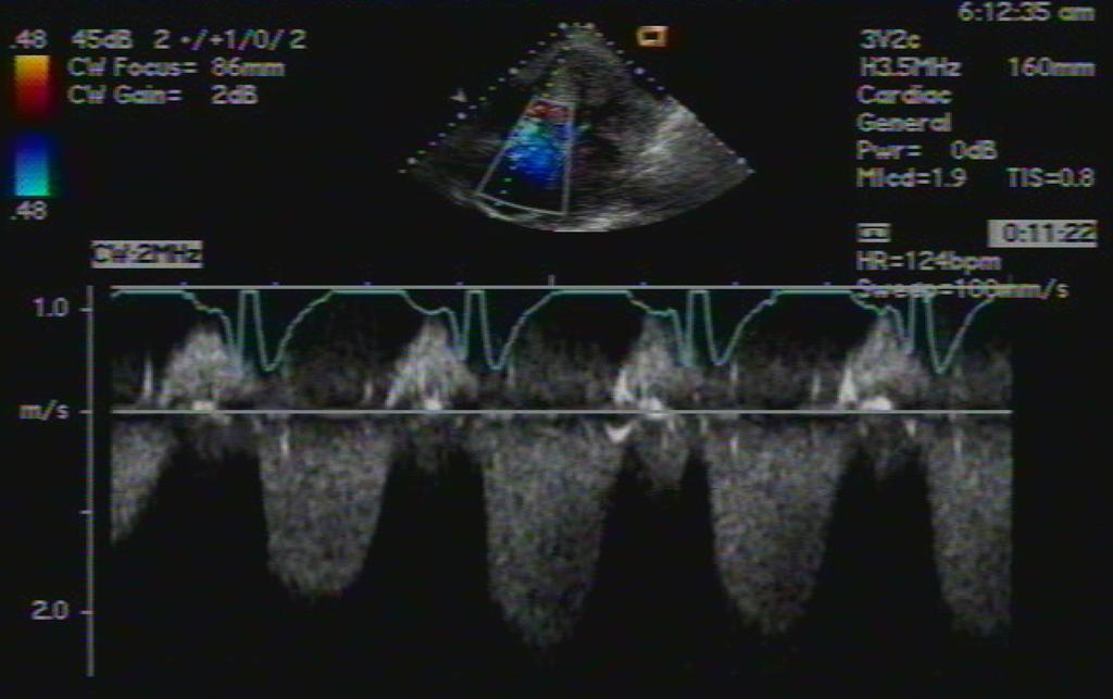
29 Doppler Profiles in HCM LVOTo from SAM Later-peaking than MR jet Delayed onset Estimate LVOTo Peak LVOTo = 4 (MR systolic jet velocity)2 +LA p - SBP Mid-cavity obstruction Steep, dagger shaped, Latest peak Short duration Mitral Regurgitation Posteriorly directed Earlier onset Longer duration Greater magnitude than SAM jet Earlier peak (first third of systole)
30
31 MR CW

32 Doppler Trio in HCM
33

34 Intracoronary Coronary Contrast Alcohol Septal Ablation

35 Septal Motion Following ASA

36 Restrictive CMP: Infiltrative or Endomyocardial Diseases
37 Hypertrophy Enlarged atria Restrictive CMP Echo Findings Consequence of poor ventricular compliance Atrial fibrillation common Restricted ventricular filling Remember the myocardium is involved primarily Annular E is markedly reduced (always < 8) Elevated filling pressures
38 Echocardiographic Findings in Amyloid Normal to small LV cavity size Thickend LV and often RV walls Speckling not specific Infiltration into valve structures Enlarged atria Pericardial effusion in up to a third High EF but low stroke volume Elevated filling pressures Restrictive filling

39 Amyloid Most Common
40 Amyloid
41 Amyloid
42
43
44
45 Significant TR / MR
46 LV Non-Compaction Not yet in WHO as a form of CMP Abnormality of ventricular morphogenesis Unique, rare cause of CHF, lethal arryhthmias, and systemic embolization Identifiable non-invasively
47 Echocardiographic diagnostic Criteria in LVNC Absence of other coexisting cardiac abnormalities Thin, compacted, epicardial tissue layer and a thick spongy, endocardial, non compacted layer with extensive trabeculations and sinusoids End-systolic ratio of compacted to non-compacted layers of > 2 discrimates LVNC from HCM, HTN, etc Deep intratrabecular recesses that communicate with LV cavity Localization in the apex, mid lateral, or mid-inferior walls Local or global LV dysfunction RV can be involved but difficult to diagnose (normally trabeculated)
48 LV Non-Compaction
49
50 Arrhythmogenic Right Ventricular Cardiomyopathy (ARVC) Genetic CMP: desmosomes fail with mechanical stress M:F 1:3 Familial occurence 30-50% Dominant: desmoplakin, ryanodine Recessive: Naxos-keratosis Pathologic fibrofatty infiltration of the RV free wall Lethal arrhythmias in young patients Clinical Criteria for diagnosis No gold standard Clinical symptoms, ecg, structural changes
 Moderator band hyperreflectivity (34%) Focal RV saculations or aneurysms (17%) Abnormalities in Regional RV function")
51 Echocardiographic Findings in ARVC Regional RV dilatation (89%) PSLA view RVOT > 3.2 cm PSAX RVOT >3.6 cm Morphologic RV abnormalities Trabecular derangement (54%) Moderator band hyperreflectivity (34%) Focal RV saculations or aneurysms (17%) Abnormalities in Regional RV function (62%) 60% of patients with normal RV function had identifiable structural abnormalities
52 ARVC

53 ARVC
54
55 ARVC
56
57
How NOT to miss Hypertrophic Cardiomyopathy? Adaya Weissler-Snir, MD University Health Network, University of Toronto
 How NOT to miss Hypertrophic Cardiomyopathy? Adaya Weissler-Snir, MD University Health Network, University of Toronto Introduction Hypertrophic cardiomyopathy is the most common genetic cardiomyopathy,
Utility of Echocardiography
 Hypertrophic Cardiomyopathy and Beyond- Echo Hawaii 2018 Lawrence Rudski MD FRCPC FACC FASE Professor of Medicine Director, Division of Cardiology and Azrieli Heart Center Jewish General Hospital, McGill
Managing Hypertrophic Cardiomyopathy with Imaging. Gisela C. Mueller University of Michigan Department of Radiology
 Managing Hypertrophic Cardiomyopathy with Imaging Gisela C. Mueller University of Michigan Department of Radiology Disclosures Gadolinium contrast material for cardiac MRI Acronyms Afib CAD Atrial fibrillation
LV FUNCTION ASSESSMENT: WHAT IS BEYOND EJECTION FRACTION
 LV FUNCTION ASSESSMENT: WHAT IS BEYOND EJECTION FRACTION Jamilah S AlRahimi Assistant Professor, KSU-HS Consultant Noninvasive Cardiology KFCC, MNGHA-WR Introduction LV function assessment in Heart Failure:
Cardiac MRI: Cardiomyopathy
 Cardiac MRI: Cardiomyopathy Laura E. Heyneman, MD I do not have any relevant financial relationships with any commercial interests Cardiac MRI: Cardiomyopathy Laura E. Heyneman, MD Duke University Medical
HYPERTROPHY: Behind the curtain. V. Yotova St. Radboud Medical University Center, Nijmegen
 HYPERTROPHY: Behind the curtain V. Yotova St. Radboud Medical University Center, Nijmegen Disclosure of interest: none Relative wall thickness (cm) M 0.22 0.42 0.43 0.47 0.48 0.52 0.53 F 0.24 0.42 0.43
Echocardiography for the Electrophysiologist: Day-to-day practice. Emmanuel Fares, MD
 Echocardiography for the Electrophysiologist: Day-to-day practice Emmanuel Fares, MD EP and pacing service, Department of Cardiovascular Medicine, Cairo University Agenda Role of echo in arrhythmia management:
Adel Hasanin Ahmed 1 CARDIOMYOPATHIES
 Adel Hasanin Ahmed 1 CARDIOMYOPATHIES 1. DILATED CARDIOMYOPATHY DCM is characterized by dilatation and systolic impairment of the LV, usually accompanied by dilatation of the RV and the atria. Diagnostic
Anaesthesia for non-cardiac surgery in patients left ventricular outflow tract obstruction (LVOTO)
") Anaesthesia for non-cardiac surgery in patients left ventricular outflow tract obstruction (LVOTO) Dr. Siân Jaggar Consultant Anaesthetist Royal Brompton Hospital London UK Congenital Cardiac Services
Anaesthesia for non-cardiac surgery in patients left ventricular outflow tract obstruction (LVOTO) Dr. Siân Jaggar Consultant Anaesthetist Royal Brompton Hospital London UK Congenital Cardiac Services
Cardiac MRI: Clinical Application to Disease
 Cardiac MRI: Clinical Application to Disease Jessi Smith, MD Cardiothoracic imaging, Indiana University Slides courtesy of Stacy Rissing, MD Outline Imaging planes Disease findings Pulse sequences used
Cardiomyopathy. Cardiomyopathies HOCM. Hypertrophic Obstructive Cardiomyopathy. Systolic Anterior Movement (SAM) of Mitral Valve (Venturi Effect) Cine
 of Mitral Valve (Venturi Effect) Cine") Jens Bremerich Radiology University Hospital Basel Hypertrophic Obstructive Cine VENC Cine (5m/s) Modified Bernoulli Equation: P (in mmhg) = 4 x (Vmax)2 Vmax= 4.2 m/s, P = 70mm Hg Hydrodynamica 738 HOCM
Jens Bremerich Radiology University Hospital Basel Hypertrophic Obstructive Cine VENC Cine (5m/s) Modified Bernoulli Equation: P (in mmhg) = 4 x (Vmax)2 Vmax= 4.2 m/s, P = 70mm Hg Hydrodynamica 738 HOCM
Etiology, Classification & Management. Sheba Medical Center Cardiology Department Matthew Wright St. George s University of London
 Etiology, Classification & Management Sheba Medical Center Cardiology Department Matthew Wright St. George s University of London Introduction World Health Organization (1995): Diseases of myocardium (heart
Cardiac MRI: Clinical Application to Disease
 Cardiac MRI: Clinical Application to Disease Stacy Rissing, MD! Cardiothoracic imaging, Indiana University! Outline Imaging planes Disease findings Pulse sequences used for each indication Pathophysiology
Highlights from EuroEcho 2009 Echo in cardiomyopathies
 Highlights from EuroEcho 2009 Echo in cardiomyopathies Bogdan A. Popescu University of Medicine and Pharmacy, Bucharest, Romania ESC Congress 2010 Hypertrophic cardiomyopathy To determine the differences
Imaging in Heart Failure: A Multimodality Approach. Thomas Ryan, MD
 Imaging in Heart Failure: A Multimodality Approach Thomas Ryan, MD Heart Failure HFrEF HFpEF EF50% Lifetime risk 20% Prevalence 6M Americans Societal costs - $30B 50% 5-year survival 1 Systolic
Cardiomyopathy. ACOI IM Board Review 2018 Martin C. Burke DO, FACOI
 Cardiomyopathy ACOI IM Board Review 2018 Martin C. Burke DO, FACOI No Disclosures Cardiomyopathies Definition: diseases of heart muscle 1980 WHO: unknown causes Not clinically relevant 1995 WHO: diseases
Basic Approach to the Echocardiographic Evaluation of Ventricular Diastolic Function
 Basic Approach to the Echocardiographic Evaluation of Ventricular Diastolic Function J A F E R A L I, M D U N I V E R S I T Y H O S P I T A L S C A S E M E D I C A L C E N T E R S T A F F C A R D I O T
Nancy Goldman Cutler, MD Beaumont Children s Hospital Royal Oak, Mi
 Nancy Goldman Cutler, MD Beaumont Children s Hospital Royal Oak, Mi Identify increased LV wall thickness (WT) Understand increased WT in athletes Understand hypertrophic cardiomyopathy (HCM) Enhance understanding
Hypertrophic Cardiomyopathy: beyond gradient and wall thickness
 Hypertrophic Cardiomyopathy: beyond gradient and wall thickness Michael H. Picard, M.D. Massachusetts General Hospital Harvard Medical School no disclosures special thanks to A. Baggish 1 Hypertrophic
Left ventricular non-compaction: the New Cardiomyopathy on the Block
 Left ventricular non-compaction: the New Cardiomyopathy on the Block Aamir Jeewa MB BCh, FAAP, FRCPC Section Head, Cardiomyopathy & Heart Function Program The Hospital for Sick Children Assistant Professor
8/31/2016. Mitraclip in Matthew Johnson, MD
 Mitraclip in 2016 Matthew Johnson, MD 1 Abnormal Valve Function Valve Stenosis Obstruction to valve flow during that phase of the cardiac cycle when the valve is normally open. Hemodynamic hallmark - pressure
Adel Hasanin Ahmed 1
 Adel Hasanin Ahmed 1 PERICARDIAL DISEASE The pericardial effusion ends anteriorly to the descending aorta and is best visualised in the PLAX. PSAX is actually very useful sometimes for looking at posterior
Steel vs Alcohol. Or Neither. Management of Hypertrophic Cardiomyopathy. Josh Doll, MD January 24, 2015
 Steel vs Alcohol Or Neither Management of Hypertrophic Cardiomyopathy Josh Doll, MD January 24, 2015 47yo Male, Mr. L Severe progressive dyspnea on exertion and weight gain Previous avid Cross-Fit participant
Rest and Exercise Echocardiography in Hypertrophic Cardiomyopathy: Determinants of Exercise Peak Gradient and Predictors of Outcome
 Rest and Exercise Echocardiography in Hypertrophic Cardiomyopathy: Determinants of Exercise Peak Gradient and Predictors of Outcome G. Deswarte, AS. Polge, N. Lamblin, A. Millaire, M. Richardson, C. Bauters,
6/12/2017. Isolated Noncompaction of the Left Ventricle. Objectives. Pathophysiology
 Isolated Noncompaction of the Left Ventricle Beth Davidson DNP, ACNP, CHFN, CCRN AAHFN June 23, 2017 Objectives 1. Describe the pathophysiology of LVNC 2. Identify the key clinical findings/manifestations
Global left ventricular circumferential strain is a marker for both systolic and diastolic myocardial function
 Global left ventricular circumferential strain is a marker for both systolic and diastolic myocardial function Toshinari Onishi 1, Samir K. Saha 2, Daniel Ludwig 1, Erik B. Schelbert 1, David Schwartzman
What s New in Cardiac MRI
 What s New in Cardiac MRI Katie M. Hawthorne, MD Director, Cardiac MRI Main Line Health Philadelphia Cardiovascular Summit November 18, 2017 Cardiac MRI: Disclosure 2 Disclosures No financial disclosures
9/23/2011. Cardiac MRI Evaluation of Cardiomyopathy and Myocarditis. Primary Hypertrophic Cardiomyopathy. Cardiomyopathy.
 Cardiomyopathy Cardiac MRI Evaluation of Cardiomyopathy and Myocarditis Laureen Sena Children s Hospital Boston, MA NASCI 2011 Baltimore, Maryland Primary Hypertrophic ARVD Dilated Restrictive Unclassified
Proceedings of the 34th World Small Animal Veterinary Congress WSAVA 2009
 www.ivis.org Proceedings of the 34th World Small Animal Veterinary Congress WSAVA 2009 São Paulo, Brazil - 2009 Next WSAVA Congress : Reprinted in IVIS with the permission of the Congress Organizers MANAGEMENT
Index of subjects. effect on ventricular tachycardia 30 treatment with 101, 116 boosterpump 80 Brockenbrough phenomenon 55, 125
 145 Index of subjects A accessory pathways 3 amiodarone 4, 5, 6, 23, 30, 97, 102 angina pectoris 4, 24, 1l0, 137, 139, 140 angulation, of cavity 73, 74 aorta aortic flow velocity 2 aortic insufficiency
Echocardiographic Cardiovascular Risk Stratification: Beyond Ejection Fraction
 Echocardiographic Cardiovascular Risk Stratification: Beyond Ejection Fraction October 4, 2014 James S. Lee, M.D., F.A.C.C. Associates in Cardiology, P.A. Silver Spring, M.D. Disclosures Financial none
Congestive Heart Failure or Heart Failure
 Congestive Heart Failure or Heart Failure Dr Hitesh Patel Ascot Cardiology Group Heart Failure Workshop April, 2014 Question One What is the difference between congestive heart failure and heart failure?
Normal TTE/TEE Examinations
 Normal TTE/TEE Examinations Geoffrey A. Rose, MD FACC FASE Sanger Heart & Vascular Institute Before you begin imaging... Obtain the patient s Height Weight BP PLAX View PLAX View Is apex @ 9-10 o clock?
Acute Pulmonary edema Secondary to CARDIAC FAILURE NON COMPACTED LEFT VENTRICLE in Pregnancy
 Acute Pulmonary edema Secondary to CARDIAC FAILURE NON COMPACTED LEFT VENTRICLE in Pregnancy Dr Anisha Gala, Consultant Obstetrician Dr. Tarakeswari S HOD, Dept of Obstetric Medicine, Fernandez Hospitals
Evalua&on)of)Le-)Ventricular)Diastolic) Dysfunc&on)by)Echocardiography:) Role)of)Ejec&on)Frac&on)
of)Le-)Ventricular)Diastolic) Dysfunc&on)by)Echocardiography:) Role)of)Ejec&on)Frac&on)") Evalua&on)of)Le-)Ventricular)Diastolic) Dysfunc&on)by)Echocardiography:) Role)of)Ejec&on)Frac&on) N.Koutsogiannis) Department)of)Cardiology) University)Hospital)of)Patras)! I have no conflicts of interest
Evalua&on)of)Le-)Ventricular)Diastolic) Dysfunc&on)by)Echocardiography:) Role)of)Ejec&on)Frac&on) N.Koutsogiannis) Department)of)Cardiology) University)Hospital)of)Patras)! I have no conflicts of interest
27-year-old professionnal rugby player: asymptomatic
 27-year-old professionnal rugby player: asymptomatic Benefits and limits of cardiac MRI in the young athlete with a suspected heart disease. Philippe PAULE Service de Cardiologie, HIA Clermont Tonnerre,
Review of Cardiac Imaging Modalities in the Renal Patient. George Youssef
 Review of Cardiac Imaging Modalities in the Renal Patient George Youssef ECHO Left ventricular hypertrophy (LVH) assessment Diastolic dysfunction Stress ECHO Cardiac CT angiography Echocardiography - positives
Cardiomyopathy: The Good, the Bad.and the Insurable?
 Cardiomyopathy: The Good, the Bad.and the Insurable? WAHLU Spring Seminar 2014 Joy Geiger, RN, BSN, ALMI Medical Consultant The Northwestern Mutual Life Insurance Company Milwaukee, WI Objectives Overview
British Society of Echocardiography
 British Society of Echocardiography Affiliated to the British Cardiac Society A Minimum Dataset for a Standard Adult Transthoracic Echocardiogram From the British Society of Echocardiography Education
Cardiomyopathy. Jeff Grubbe MD FACP, Chief Medical Director, Allstate Life & Retirement
 Cardiomyopathy Jeff Grubbe MD FACP, Chief Medical Director, Allstate Life & Retirement Nebraska Home Office Life Underwriters Association March 20, 2018 1 Cardiomyopathy A myocardial disorder in which
ESC Guidelines on Hypertrophic Cardiomyopathy
 2014 version ES Guidelines on Hypertrophic ardiomyopathy Pr Michel KOMAJDA Dept of ardiology HU PTE SALPETRERE University Pierre et Marie urie PARS FRANE European Heart Journal (2014):doi:10.1093/eurheartj/ehu284
Right Heart Evaluation ASE Guidelines Review. Chris Mann RDCS, RCS, FASE Faculty, Echocardiography Pitt Community College Greenville, NC
 Right Heart Evaluation ASE Guidelines Review Chris Mann RDCS, RCS, FASE Faculty, Echocardiography Pitt Community College Greenville, NC Objectives Briefly review right atrial and right ventricular anatomy
Cardiac Issues in the Adolescent Athlete. Sean Levchuck, M.D. St. Francis Hospital- The Heart Center
 Cardiac Issues in the Adolescent Athlete Sean Levchuck, M.D. St. Francis Hospital- The Heart Center Sudden Cardiac Death Incidence is.6-6.2 % per 100,000 children in the US 20-25 % of the deaths occur
Cases in Adult Congenital Heart Disease
 Cases in Adult Congenital Heart Disease Sabrina Phillips, MD FACC FASE Associate Professor of Medicine The University of Oklahoma Health Sciences Center No Disclosures I Have Palpitations 18 Year old Man
Update on use of cardiac MRI in ARVC/D. Stefan L. Zimmerman, MD Johns Hopkins University Department of Radiology
 Update on use of cardiac MRI in ARVC/D Stefan L. Zimmerman, MD Johns Hopkins University Department of Radiology Outline Background Diagnosis Characteristic imaging findings Genetics of ARVC Genotype phenotype
INTRODUCTION. left ventricular non-compaction is a sporadic or familial cardiomyopathy characterized by
 A Rare Case of Arrhythmogenic Right Ventricular Cardiomyopathy Co-existing with Isolated Left Ventricular Non-compaction NS Yelgeç, AT Alper, Aİ Tekkeşin, C Türkkan INTRODUCTION Arrhythmogenic right ventricular
PROSTHETIC VALVE BOARD REVIEW
 PROSTHETIC VALVE BOARD REVIEW The correct answer D This two chamber view shows a porcine mitral prosthesis with the typical appearance of the struts although the leaflets are not well seen. The valve
Implications of the new diagnostic criteria for ARVC
 EUROECHO 2010 Echocardiographic assessment of cardiomyopathies Implications of the new diagnostic criteria for ARVC Barbara Bauce, MD, PhD Department of Cardiac, Thoracic and Vascular Sciences University
좌심실수축기능평가 Cardiac Function
 Basic Echo Review Course 좌심실수축기능평가 Cardiac Function Seonghoon Choi Cardiology Hallym university LV systolic function Systolic function 좌심실수축기능 - 심근의수축으로심실에서혈액을대동맥으로박출하는기능 실제임상에서 LV function 의의미 1Diagnosis
Pulmonary Hypertension. Echocardiography: Pearls & Pitfalls
 Pulmonary Hypertension Echocardiography: Pearls & Pitfalls Αθανάσιος Γ. Κουτσάκης Ειδικευόμενος Καρδιολογίας Α Καρδιολογική Κλινική ΑΠΘ Σεμινάρια Ομάδων Εργασίας Ελληνικής Καρδιολογικής Εταιρείας Ιωάννινα,
Historical Notes: Clinical Exercise Testing in the Athlete. An Efficient Heart. Structural Changes, Cont. Coronary Arteries
 Clinical Exercise Testing in the Athlete The athlete s heart Sudden cardiac death in athletes Screening athletes for cardiovascular disease Historical Notes: Giovanni Lancisi (father of cardiology), 17
Comprehensive Hemodynamics By Doppler Echocardiography. The Echocardiographic Swan-Ganz Catheter.
 Comprehensive Hemodynamics By Doppler Echocardiography. The Echocardiographic Swan-Ganz Catheter. Itzhak Kronzon, MD, FASE, FACC, FESC, FAHA, FACP, FCCP North Shore HS, LIJ/Lenox Hill Hospital, New York
Aortic Stenosis: Spectrum of Disease, Low Flow/Low Gradient and Variants
 Aortic Stenosis: Spectrum of Disease, Low Flow/Low Gradient and Variants Martin G. Keane, MD, FASE Professor of Medicine Lewis Katz School of Medicine at Temple University Basic root structure Parasternal
(Ann Thorac Surg 2008;85:845 53)
") I Made Adi Parmana The utility of intraoperative TEE has become increasingly more evident as anesthesiologists, cardiologists, and surgeons continue to appreciate its potential application as an invaluable
I Made Adi Parmana The utility of intraoperative TEE has become increasingly more evident as anesthesiologists, cardiologists, and surgeons continue to appreciate its potential application as an invaluable
MITRAL STENOSIS. Joanne Cusack
 MITRAL STENOSIS Joanne Cusack BSE Breakdown Recognition of rheumatic mitral stenosis Qualitative description of valve and sub-valve calcification and fibrosis Measurement of orifice area by planimetry
Treatment of Hypertrophic Cardiomyopathy in Bruce B. Reid, MD
 Treatment of Hypertrophic Cardiomyopathy in 2017 Bruce B. Reid, MD Disclosures I have no conflicts of interest to disclose I will not be discussing any off label medications and/or devices Objectives 1)
Interventions in Adult Congenital Heart Disease: Role of CV Imaging. Associate Professor. ACHD mortality. Pillutla. Am Heart J 2009;158:874-9
 Interventions in Adult Congenital Heart Disease: Role of CV Imaging Sangeeta Shah MD, FACC, FASE Associate Professor ACHD mortality Pillutla. Am Heart J 2009;158:874-9 Adult Congenital Heart Disease Heterogenity
Tricuspid and Pulmonary Valve Disease
 Tricuspid and Pulmonary Valve Disease Lawrence Rudski MD FRCPC FACC FASE Professor of Medicine Director, Division of Cardiology Jewish General Hospital McGill University Right Sided Failure Edema Gut congestion
Dr Navin Chandra Clinical Research Fellow in Cardiology St George s University of London
 Dr Navin Chandra Clinical Research Fellow in Cardiology St George s University of London Cardiac Adaptation in Athletes Athletic training for prolonged periods may result in physiological adaptations of
How to assess ischaemic MR?
 ESC 2012 How to assess ischaemic MR? Luc A. Pierard, MD, PhD, FESC, FACC Professor of Medicine Head, Department of Cardiology University Hospital Sart Tilman, Liège ESC 2012 No conflict of interest Luc
Echo Doppler Assessment of Right and Left Ventricular Hemodynamics.
 Echo Doppler Assessment of Right and Left Ventricular Hemodynamics. Itzhak Kronzon, MD, FASE, FACC, FESC, FAHA, FACP, FCCP Northwell, Lenox Hill Hospital, New York Professor of Cardiology Hofstra University
How to Assess and Treat Obstructive Lesions
 How to Assess and Treat Obstructive Lesions Erwin Oechslin, MD, FESC, FRCPC, Director, Congenital Cardiac Centre for Adults Peter Munk Cardiac Centre University Health Network/Toronto General Hospital
Management of HOCM: Non-Surgical Options
 Management of HOCM: Non-Surgical Options Howard C. Herrmann, MD, FACC, MSCAI John Bryfogle Professor of Cardiovascular Medicine and Surgery Health System Director for Interventional Cardiology Director,
NEW GUIDELINES MAJOR ECHOCARDIOGRAPHIC CRITERIA FOR ARVC MINOR ECHOCARDIOGRAPHIC CRITERIA FOR ARVC
 NEW GUIDELINES The Echocardiographic Assessment of the Right Ventricle with particular reference to Arrhythmogenic Right Ventricular Cardiomyopathy A Protocol of the British Society of Echocardiography
Evaluation of the Right Ventricle and Risk Stratification for Sudden Cardiac Death
 Evaluation of the Right Ventricle and Risk Stratification for Sudden Cardiac Death Presenters: Sabrina Phillips, MD FACC FASE Director, Adult Congenital Heart Disease Services The University of Oklahoma
P = 4V 2. IVC Dimensions 10/20/2014. Comprehensive Hemodynamic Evaluation by Doppler Echocardiography. The Simplified Bernoulli Equation
 Comprehensive Hemodynamic Evaluation by Doppler Echocardiography Itzhak Kronzon, MD North Shore LIJ/ Lenox Hill Hospital New York, NY Disclosure: Philips Healthcare St. Jude Medical The Simplified Bernoulli
Takotsubo syndrome. Ευτυχία Σμπαρούνη, FACC, FESC
 Takotsubo syndrome Ευτυχία Σμπαρούνη, FACC, FESC Definition Takotsubo Apical ballooning Broken heart syndrome Stress cardiomyopathy Cathecholaminergic cardiomyopathy Epidemiology 1990 first report by Japanese
Athlete s Heart vs. Cardiomyopathy
 Athlete s Heart vs. Cardiomyopathy Linda D. Gillam, MD, MPH, FASE Chair, Department of Cardiovascular Medicine Medical Director, Cardiovascular Service Line Former Team Cardiologist to the New York Jets
Two Cardiology Zebras ERIC MARTIN MD
 Two Cardiology Zebras ERIC MARTIN MD Disclosures Bayer Gilead Sciences NIH Vascular Dynamics, In. Employer Iowa Heart Center/Mercy Des Moines Zebra # 1 History CC: 52-year-old man seen in consultation
Evaluation of Left Ventricular Diastolic Dysfunction by Doppler and 2D Speckle-tracking Imaging in Patients with Primary Pulmonary Hypertension
 ESC Congress 2011.No 85975 Evaluation of Left Ventricular Diastolic Dysfunction by Doppler and 2D Speckle-tracking Imaging in Patients with Primary Pulmonary Hypertension Second Department of Internal
Tricuspid and Pulmonary Valve Disease
 Tricuspid and Pulmonary Valve Disease Lawrence Rudski MD FRCPC FACC FASE Professor of Medicine Director, Division of Cardiology Jewish General Hospital McGill University Question 1 All of the following
Giovanni Di Salvo MD, PhD, FESC Second University of Naples Monaldi Hospital
 Giovanni Di Salvo MD, PhD, FESC Second University of Naples Monaldi Hospital VSD is one of the most common congenital cardiac abnormalities in the newborn. It can occur as an isolated finding or in combination
Mitral Valve Disease. Prof. Sirchak Yelizaveta Stepanovna
 Mitral Valve Disease Prof. Sirchak Yelizaveta Stepanovna Fall 2008 Mitral Valve Stenosis Lecture Outline Mitral Stenosis Mitral Regurgitation Etiology Pathophysiology Clinical features Diagnostic testing
M-Mode Echocardiography Is it still Alive? Itzhak Kronzon, MD,FASE. Sampling Rate M-Mode: 1800 / sec 2D: 30 / sec
 M-Mode Echocardiography Is it still Alive? Itzhak Kronzon, MD,FASE Honoraria: Philips Classical M-mode Echocardiography M-Mode offers better time and image resolution. Sampling Rate M-Mode: 1800 / sec
Novel echocardiographic modalities: 3D echo, speckle tracking and strain rate imaging. Potential roles in sports cardiology. Stefano Caselli, MD, PhD
 Novel echocardiographic modalities: 3D echo, speckle tracking and strain rate imaging. Potential roles in sports cardiology. Stefano Caselli, MD, PhD Ospedale San Pietro Fatebenefratelli Rome, Italy Differential
MR echo case. N.Koutsogiannis Department of Cardiology University Hospital Of Patras
 MR echo case N.Koutsogiannis Department of Cardiology University Hospital Of Patras Case A 35 years old male came to the echo lab for a third opinion for his valvulopathy. He reports a long standing MR
ASCeXAM / ReASCE. Practice Board Exam Questions. Monday Afternoon
 ASCeXAM / ReASCE Practice Board Exam Questions Monday Afternoon Aortic Valve Prostheses Aortic Regurgitation Stress Echocardiography Ischemic Heart Disease and Myocardial Infarction 3D Echocardiography
The background of the Cardiac Sonographer Network News masthead is a diagnostic image:
 Number 5 Welcome Number 5 Welcome to the newsletter created just for you: sonographers who perform pediatric echocardiograms in primarily adult echo labs. Each issue features tips on echocardiography of
Objectives. Diastology: What the Radiologist Needs to Know. LV Diastolic Function: Introduction. LV Diastolic Function: Introduction
 Objectives Diastology: What the Radiologist Needs to Know. Jacobo Kirsch, MD Cardiopulmonary Imaging, Section Head Division of Radiology Cleveland Clinic Florida Weston, FL To review the physiology and
The different clinical scenarios of left ventricular non-compaction: report of three cases
 Page 1 of 5 Cardiovascular Medicine The different clinical scenarios of left ventricular non-compaction: report of three cases M Bolognesi 1 *, D Bolognesi 2 Abstract Introduction Left ventricular non-compaction
DELAYED ENHANCEMENT IMAGING IN CHILDREN
 NASCI 38 TH ANNUAL MEENG, SEATLE October 3-5, 21 1. DELAYED ENHANCEMENT IN CHILDREN Shi-Joon Yoo, MD Lars Grosse-Wortmann, MD University of Toronto Canada -1. 1. 1. Magnitude image Magnitude images -1.
RIGHT VENTRICULAR SIZE AND FUNCTION
 RIGHT VENTRICULAR SIZE AND FUNCTION Edwin S. Tucay, MD, FPCC, FPCC, FPSE Philippine Society of Echocardiography Quezon City, Philippines Echo Mission, BRTTH, Legaspi City, July 1-2, 2016 NO DISCLOSURE
Ablative Therapy for Ventricular Tachycardia
 Ablative Therapy for Ventricular Tachycardia Nitish Badhwar, MD, FACC, FHRS 2 nd Annual UC Davis Heart and Vascular Center Cardiovascular Nurse / Technologist Symposium May 5, 2012 Disclosures Research
Μαρία Μπόνου Διευθύντρια ΕΣΥ, ΓΝΑ Λαϊκό
 Μαρία Μπόνου Διευθύντρια ΕΣΥ, ΓΝΑ Λαϊκό Diastolic HF DD: Diastolic Dysfunction DHF: Diastolic HF HFpEF: HF with preserved EF DD Pathophysiologic condition: impaired relaxation, LV compliance, LV filling
Το ΗΚΓ στις Μυοκαρδιοπάθειες και στην Περικαρδίτιδα
 4 ο ΠΑΝΕΛΛΗΝΙΟ ΑΡΡΥΘΜΙΟΛΟΓΙΚΟ ΣΥΝΕΔΡΙΟ Φροντιστηριακό Μάθημα ΗΚΓ Το ΗΚΓ στις Μυοκαρδιοπάθειες και στην Περικαρδίτιδα Γ.Ν.Α. «Ο Ευαγγελισμός» Ξυδώνας Σωτήριος, MD, PhD, FESC Εργαστήριο Ηλεκτροφυσιολογίας
DOPPLER HEMODYNAMICS (1) QUANTIFICATION OF PRESSURE GRADIENTS and INTRACARDIAC PRESSURES
 QUANTIFICATION OF PRESSURE GRADIENTS and INTRACARDIAC PRESSURES") THORAXCENTRE DOPPLER HEMODYNAMICS (1) QUANTIFICATION OF PRESSURE GRADIENTS and INTRACARDIAC PRESSURES J. Roelandt DOPPLER HEMODYNAMICS Intracardiac pressures and pressure gradients Volumetric measurement
THORAXCENTRE DOPPLER HEMODYNAMICS (1) QUANTIFICATION OF PRESSURE GRADIENTS and INTRACARDIAC PRESSURES J. Roelandt DOPPLER HEMODYNAMICS Intracardiac pressures and pressure gradients Volumetric measurement
Congenital Heart Disease Cases
 Congenital Heart Disease Cases Sabrina Phillips, MD FACC FASE Mayo Clinic Congenital Heart Disease Center 2013 MFMER slide-1 No Disclosures 2013 MFMER slide-2 1 CASE 1 2013 MFMER slide-3 63 year old Woman
Case based learning: CMR in Heart Failure
 Case based learning: CMR in Heart Failure Milind Y Desai, MD FACC FAHA FESC Associate Professor of Medicine Heart and Vascular Institute, Cleveland Clinic Cleveland, OH Disclosures: none Use of Gadolinium
Adult Echocardiography Examination Content Outline
 Adult Echocardiography Examination Content Outline (Outline Summary) # Domain Subdomain Percentage 1 2 3 4 5 Anatomy and Physiology Pathology Clinical Care and Safety Measurement Techniques, Maneuvers,
Diastolic Heart Function: Applying the New Guidelines Case Studies
 Diastolic Heart Function: Applying the New Guidelines Case Studies Mitral Regurgitation The New ASE William Guidelines: A. Zoghbi Role MD, of FASE, 2D/3D MACCand CMR Professor and Chairman, Department
Stephen Glen ISCHAEMIC HEART DISEASE AND LEFT VENTRICULAR FUNCTION
 Stephen Glen ISCHAEMIC HEART DISEASE AND LEFT VENTRICULAR FUNCTION Overview Coronary arteries Terminology to describe contractility Measuring ventricular function Systolic dysfunction Practice cases- LV
Case Reviews: Hemodynamic Calculations in Valvular Regurgitation
 Case Reviews: Hemodynamic Calculations in Valvular Regurgitation Case 5 History: 69-year-old man with orthotopic heart transplant 15 years ago. Inferior MI several years ago. Recurrent CHF. Currently dyspneic
Sudden Death (SD) and hypertrophic cardiomyopathy (HCM) Attempt of risk stratification
 and hypertrophic cardiomyopathy (HCM) Attempt of risk stratification") Sudden Death (SD) and hypertrophic cardiomyopathy (HCM) Attempt of risk stratification 84th Annual Scientific Meeting of the Aerospace Medical Association May 12-16, 2013 Sheraton Chicago Hotel & Towers,
Sudden Death (SD) and hypertrophic cardiomyopathy (HCM) Attempt of risk stratification 84th Annual Scientific Meeting of the Aerospace Medical Association May 12-16, 2013 Sheraton Chicago Hotel & Towers,
Alicia Armour, MA, BS, RDCS
 Alicia Armour, MA, BS, RDCS No disclosures Review 2D Speckle Strain (briefly) Discuss some various patient populations & disease pathways where Strain can be helpful Discuss how to acquire images for Strain
Apical Hypertrophic Cardiomyopathy With Hemodynamically Unstable Ventricular Arrhythmia Atypical Presentation
 Cronicon OPEN ACCESS Hemant Chaturvedi* Department of Cardiology, Non-Invasive Cardiology, Eternal Heart Care Center & research Institute, Rajasthan, India Received: September 15, 2015; Published: October
Hypertrophic Cardiomyopathy
 019-CardioCase:019-CardioCase 4/16/07 1:39 PM Page 19 Hypertrophic Cardiomyopathy Abdullah Alshehri, MD; and Andrew Ignaszewski, MD, FRCPC CardioCase presentation Presley s check-up Presley, 37, discovered
Valvular Regurgitation: Can We Do Better Than Colour Doppler?
 Valvular Regurgitation: Can We Do Better Than Colour Doppler? A/Prof David Prior St Vincent s Hospital Melbourne Sports Cardiology Valvular Regurgitation Valve regurgitation volume loads the ventricles
DISCLOSURE. Echocardiography in Systemic Diseases: Questions. Relevant Financial Relationship(s) None. Off Label Usage None 5/7/2018
 None. Off Label Usage None 5/7/2018") Echocardiography in Systemic Diseases: Questions Sunil Mankad, MD, FACC, FCCP, FASE Associate Professor of Medicine Mayo Clinic College of Medicine Director, Transesophageal Echocardiography Associate
Echocardiography in Systemic Diseases: Questions Sunil Mankad, MD, FACC, FCCP, FASE Associate Professor of Medicine Mayo Clinic College of Medicine Director, Transesophageal Echocardiography Associate
The Management of HOCM: What are the Surgical Options
 The Management of HOCM: What are the Surgical Options Konstadinos A Plestis, MD System Chief of Cardiac Thoracic and Vascular Surgery Main Line Health Care System Professor Sidney Kimmel Medical College
Quantitation of right ventricular dimensions and function
 SCCS Basics of cardiac assessment Quantitation of right ventricular dimensions and function Tomasz Kukulski, MD PhD Dept of Cardiology, Congenital Heart Disease and Electrotherapy Silesian Medical University