Pulmonary Hypertension. Echocardiography: Pearls & Pitfalls
|
|
|
- Henry Gibson
- 6 years ago
- Views:
Transcription

1 Pulmonary Hypertension Echocardiography: Pearls & Pitfalls Αθανάσιος Γ. Κουτσάκης Ειδικευόμενος Καρδιολογίας Α Καρδιολογική Κλινική ΑΠΘ Σεμινάρια Ομάδων Εργασίας Ελληνικής Καρδιολογικής Εταιρείας Ιωάννινα, 13/02/2016
2 Nothing to declare
3 Transthoracic echocardiography is used to: estimate PAP from continuous wave Doppler measurements image the effects of PH on the heart infer a diagnosis of PH in patients in whom multiple different echocardiographic measurements are consistent with this diagnosis Echocardiography should always be performed when PH is suspected When treatment of PH itself is being considered, echocardiography alone is not sufficient to support a treatment decision and cardiac catheterization is required.
4
5 What info are we getting from echo 1. PASP 2. RV size and function 3. Causes of PH 4. Prognosis
6 48% of the echocardiographic estimates were accurate (within 10 mm Hg of the RHC value). DE within 1 hour of RHC Fisher MR et al. Am J Respir Crit Care Med 2009; 179:


7 PASP Estimation Pitfalls PASP=4V²+RAP Pitfalls: TR Related RAP Related
8 PASP Estimation Pitfalls I TR related: The severity of tricuspid regurgitation does not correlate with degree of PH Overestimation Underestimation RVOT Stenosis
")
9 Shadow (Overestimation) Reduce the Gain!
")
10 Non parallel position of the transducer (Underestimation)


11 Little things make a big difference! 4 chambers 4 chambers RV focused view

12 Suboptimal signal (Underestimation) Contrast echocardiography (agitated saline administered i.v.) can improve the signal


13 Use all the available views! PLAX Right Chambers focused view PSAX view 4 chambers & focused view Subcostal view The maximal velocity depends on optimal alignment with the jet
")

14 Severe TR (underestimation) Triangular protosystolic shape (early equalization of RV and RA pressures)

")

15 RVOT Stenosis (Non Reliable) 42 yo, male, Congenital PV Stenosis
16 PASP Estimation Pitfalls II RAP related (IVC Diameter): Underestimation Left lateral position: increased intra-abdominal pressure and compression of the IVC by the liver Overestimation Measurement not perpendicular to the IVC long axis Dilated and may not collapse in patients on ventilators IVC Diameter (cm) Right Atrial Pressure Estimation Collapse (%) Estimated RA Pressure (mmhg) Range (mmhg) 2.1 cm >50% > 2.1 cm <50% Rest of cases
 as the main variable for assigning the echocardiographic")
17 ..given the inaccuracies of RAP estimation and the amplification of measurement errors by using derived variables, we recommend using the continuous wave Doppler measurement of peak TRV (and not the estimated PASP) as the main variable for assigning the echocardiographic probability of PH.
18 The TR Velocity only is not sufficient! Search for other echo findings that raise or reinforce suspicion of PH


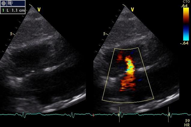
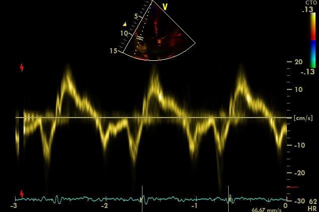
19 Short AccT & Notching 56 yo, female, NYHA II FC, with scleroderma RHC: mean PAP 38 mmhg, spap: 67 mmhg

20 Right Ventricle Assessment Structurally complex cavity: complex shape irregular endocardial surface due to heavy trabeculation: difficult to delineate endocardial border location behind the sternum: inadequate image quality Fits no simple geometric figure: failure to standardize RV volume determination

21 Right ventricle focused view

22 Use all the available views! RV size estimation requires integration of multiple views and qualitative and quantitative assessment

23 RV:LV Ratio - Foreshortening


24 LV eccentricity index Volume vs Pressure Overload RV volume overload the flattening of the septum is seen only in diastole Ecc Index >1 at end diastole RV pressure overload the septum maintains the altered shape during the entire cardiac cycle Ecc Index >1 at end systole and end diastole
 ventricle LV systolic performance may")

25 TAPSE Longitudinal function of the right angle dependency load dependency (significant tricuspid regurgitation) ventricle LV systolic performance may influence TAPSE value due to ventricular interdependence Tissue Doppler Ensure optimal image orientation to avoid underestimation Insufficient data in the elderly

26 Fractional Area Change Quantitative estimation of RV function Good correlation with RVEF Take Care: Accurate definition of endocardial limits Trace the free wall beneath the trabeculations
27 Right Heart Dysfunction PH RV Infarction Arrhythmogenic right ventricular dysplasia (ARVC) Congenital heart disease


28 Normal RV Function does not exclude PH
29 Causes of PH
")

30 Be careful! LHD (group2) is the most common cause of PH

31 S. Rosenkranz et al. Eur. Heart J. 2015


32 81 yo, Female, AF, LVH, LA/RA = 1, E/ e >15, TR = 3,3 m/sec PH due to LHD


33 64 yo, Female, E<A, RV/LV > 1, RA/LA > 1, Interatrial septum bows from RA to LA, TR = 5,06 m/sec PH due to PAH
34 Prognosis
35

36 Επιπολασμός Περικαρδιακής Συλλογής στην ΠΥ


37 Tamponade? 71 yo, female, WHO-FC IV, Group I PH BP 90/60mmHg RVSP 105 mmhg
38 Pericardial involvement in PAH: Diagnosis of cardiac tamponade in a patient with severe PAH is challenging. High rightsided pressures can mask many of the typical right-sided clinical and echocardiographic findings of tamponade. Right-sided chamber collapse is uncommon Left atrial early diastolic collapse is more commonly seen.
39 Conclusions A detailed echocardiographic assessment must be performed in all patients with suspected PH TR velocity and other echocardiographic variables might raise or reinforce suspicion of PH Heart chambers must be evaluated for causative and prognostic information
40 Thank You!
41 Pericardial Effusion
42 Prognostic information right atrial area eccentricity index Pericardial effusion Tei index TAPSE
43 Right Heart Dysfunction PH Ischaemic right ventricular disease and right ventricular failure RV ischaemia complicates up to 50% of inferior myocardial infarction RV free wall hypokinesia or akinesia detected by echocardiography is a qualitative and sensitive parameter for RV dysfunction
44 Right Heart Dysfunction PH Arrhythmogenic right ventricular dysplasia (ARVC) Regional RV akinesia, dyskinesia, or aneurysm The triangle of dysplasia: a region including the subtricuspidal RV wall, the RV apex, and the RV outflow tract
45 Right Heart Dysfunction PH Congenital heart disease chronic volume overload intracardiac shunts (atrial septal defect), anomalies of the pulmonary valve, and arteries (pulmonary atresia) RV is pumping in the systemic circulation (transposition of the great arteries, single ventricle).
46 Also evaluate Pulmonary Artery AcT (<105 msec) (HR b/min) Notching (midsystolic) Diameter (> 25 mm) Early diastolic Pulmonary Velocity Regurgitation (>2,2m/sec) Mean PAP IVC diameter and Respiratory Variation Right Atrial Dimensions
47 Regional RV Strain and Strain Rate identify the early signs of RV dysfunction follow patients under therapy probably accounts for the RV global dysfunction, given the fibre arrangement RV longitudinal strain <19% has been described as an independent predictor of allcause mortality

48 PAH vs PVH: Echocardiographic Differentiation Mc Laughlin et al. JACC 2015; 65. NO 18.

49 Checklist for the Echocardiographic Evaluation of PH Mc Laughlin et al. JACC 2015; 65. NO 18.
50
51 Raymond et al. JACC 2002; 39, No 7:
52 Non Conventional echocardiography RT3DE Accurate volume analysis independent of RV size and shape enables unique views to better understand specific causes of PH (i.e., septal defects, complex congenital pathology, left-sided valvular or ventricular heart disease) investigate RV functional and morphologic changes Two-Dimensional Strain identify the early signs of RV dysfunction follow patients under therapy <19% an independent predictor of all-cause mortality Bossone et al. JACC 2013; 26. NO 1.

53 TR occurs as the result of the effect of increased RV afterload on RV dilatation and function. TR is usually caused by tricuspid annular dilation, altered RV geometry, and apical displacement of the tricuspid leaflets. The degree of TR cannot be used as a surrogate for the degree of PAP elevation TR PH
54 Evaluation of the RV Function Subjective: Thickening of the RV wall Motion (Inward/radial/longitudinal) Regional dysfunction Integrate findings from various views RV fractional area change (FAC) TAPSE Tissue Doppler Tei Index
 Farber et al. Congest Heart Fail. 2011 Mar-Apr;17(2):56-64")
55 Echo inaccurate in 57,4% of PASP estimates (>10 mmhg higher or lower than RHC) Farber et al. Congest Heart Fail Mar-Apr;17(2):56-64
56 Use all the available views! PLAX Right Chambers focused view PSAX view 4 chambers & focused view Subcostal view The maximal velocity depends on optimal alignment with the jet
57 PASP Estimation Pitfalls The volume of tricuspid regurgitation does not correlate with the velocity The absence of TR (moderate or severe) does not exclude PH Severity of TR does not predict severity of PH Overestimation Detecting shadow Underestimation Non parallel position of the transducer Suboptimal signal Free TR Eccentric tricuspid regurgitation jet RVOT Stenosis
58 IVC Diameter Not reliable in: Normal young athletes (dilated in the presence of normal pressure) In patients on ventilators (TEE at the cavoatrial junction)
59 PH Pathophysiology RV function and size are indicators of the severity and chronicity of pulmonary hypertension impose an additional cause of symptoms and reduced longevity. Right ventricular function is the most important determinant of longevity in patients with pulmonary arterial hypertension

60 Right Atrium Evaluation Apical 4-chamber view End-systole Significant dilatation if > 18cm² Right atrial enlargement is a manifestation of high right atrial pressure due to functional tricuspid regurgitation or elevated right ventricular diastolic pressure, both consequences of right ventricular failure
61 65patients, various forms of PH, DE within 1 hour of a clinically indicated right-heart catheterization to compare noninvasive hemodynamic estimates with invasively measured values 48% of the echocardiographic estimates were accurate (within 10 mm Hg of the RHC value). the magnitude of underestimation was greater than the overestimation (38% pressure overestimates > 20 mm Hg 80% pressure underestimates > 20 mm Hg). 83% underestimates were associated with a fair or poor quality Doppler jet. Fisher MR et al. Am J Respir Crit Care Med 2009; 179:
62 TAPSE Longitudinal function of the right ventricle Several limitations angle dependency load dependency (significant tricuspid regurgitation) 1D (Regional RV myocardial abnormalities are neglected) LV systolic performance may influence TAPSE value due to ventricular interdependence
63 Tissue Doppler Assumes that the function of a single segment represents the function of the entire right ventricle Normal value S > 10cm/sec

64 The TR Velocity only is not sufficient! Search for other echo findings that raise or reinforce suspicion of PH
Right Heart Evaluation ASE Guidelines Review. Chris Mann RDCS, RCS, FASE Faculty, Echocardiography Pitt Community College Greenville, NC
 Right Heart Evaluation ASE Guidelines Review Chris Mann RDCS, RCS, FASE Faculty, Echocardiography Pitt Community College Greenville, NC Objectives Briefly review right atrial and right ventricular anatomy
Adel Hasanin Ahmed 1
 Adel Hasanin Ahmed 1 PERICARDIAL DISEASE The pericardial effusion ends anteriorly to the descending aorta and is best visualised in the PLAX. PSAX is actually very useful sometimes for looking at posterior
Pulmonary Hypertension: Echocardiographic Evaluation of Pulmonary Hypertension and Right Ventricular Function. Irmina Gradus-Pizlo, MD
 Pulmonary Hypertension: Echocardiographic Evaluation of Pulmonary Hypertension and Right Ventricular Function Irmina Gradus-Pizlo, MD Disclosures: Nothing to disclose Overview Is pulmonary hypertension
RIGHT VENTRICULAR SIZE AND FUNCTION
 RIGHT VENTRICULAR SIZE AND FUNCTION Edwin S. Tucay, MD, FPCC, FPCC, FPSE Philippine Society of Echocardiography Quezon City, Philippines Echo Mission, BRTTH, Legaspi City, July 1-2, 2016 NO DISCLOSURE
ASCeXAM / ReASCE. Practice Board Exam Questions Monday Morning
 ASCeXAM / ReASCE Practice Board Exam Questions Monday Morning Ultrasound Physics Artifacts Doppler Physics Imaging, Knobology, and Artifacts Echocardiographic Evaluation of the RV Tricuspid and Pulmonary
Echo in Pulmonary HTN
 Echo in Pulmonary HTN Steven A. Goldstein MD FACC FASE Professor of Medicine Georgetown University Medical Center MedStar Heart Institute Washington Hospital Center Monday, October 10, 2017 Pulmonary Artery
Quantitation of right ventricular dimensions and function
 SCCS Basics of cardiac assessment Quantitation of right ventricular dimensions and function Tomasz Kukulski, MD PhD Dept of Cardiology, Congenital Heart Disease and Electrotherapy Silesian Medical University
Appendix II: ECHOCARDIOGRAPHY ANALYSIS
 Appendix II: ECHOCARDIOGRAPHY ANALYSIS Two-Dimensional (2D) imaging was performed using the Vivid 7 Advantage cardiovascular ultrasound system (GE Medical Systems, Milwaukee) with a frame rate of 400 frames
Right Heart Hemodynamics: Echo-Cath Discrepancies
 Department of cardiac, thoracic and vascular sciences University of Padua, School of Medicine Padua, Italy Right Heart Hemodynamics: Echo-Cath Discrepancies Luigi P. Badano, MD, PhD, FESC, FACC **Dr. Badano
Fig.1 Normal appearance of RV in SAX:
 Tutorial 7 - Assessment of the right heart Assessment of the Right heart The right heart assessment clinically and echocardiographically is not a very important part of mainstream cardiology. In the ICU,
Transthoracic echocardiography in the evaluation of pediatric pulmonary hypertension and ventricular dysfunction
 REVIEW ARTICLE Transthoracic echocardiography in the evaluation of pediatric pulmonary hypertension and ventricular dysfunction Martin Koestenberger, 1 Mark K. Friedberg, 2 Eirik Nestaas, 3 Ina Michel-Behnke,
Right Ventricle Steven J. Lester MD, FACC, FRCP(C), FASE Mayo Clinic, Arizona
, FASE Mayo Clinic, Arizona") Right Ventricle Steven J. Lester MD, FACC, FRCP(C), FASE Mayo Clinic, Arizona 1. In which scenario will applying the simplified Bernoulli equation to the peak tricuspid regurgitation velocity and adding
Right Ventricle Steven J. Lester MD, FACC, FRCP(C), FASE Mayo Clinic, Arizona 1. In which scenario will applying the simplified Bernoulli equation to the peak tricuspid regurgitation velocity and adding
Ιπποκράτειες μέρες καρδιολογίας Θεσσαλονίκη, 9-10 Μαρτίου Φωτεινή Α. Λαζαρίδου Επιμελήτρια Α Γενικό Νοσοκομείο Αγιος Παύλος, Θεσσαλονίκη
 Ιπποκράτειες μέρες καρδιολογίας Θεσσαλονίκη, 9-10 Μαρτίου 2018 Φωτεινή Α. Λαζαρίδου Επιμελήτρια Α Γενικό Νοσοκομείο Αγιος Παύλος, Θεσσαλονίκη RV shape Triangular shape in frontal plane crescent shape in
Evaluation of Left Ventricular Diastolic Dysfunction by Doppler and 2D Speckle-tracking Imaging in Patients with Primary Pulmonary Hypertension
 ESC Congress 2011.No 85975 Evaluation of Left Ventricular Diastolic Dysfunction by Doppler and 2D Speckle-tracking Imaging in Patients with Primary Pulmonary Hypertension Second Department of Internal
Hemodynamic Assessment. Assessment of Systolic Function Doppler Hemodynamics
 Hemodynamic Assessment Matt M. Umland, RDCS, FASE Aurora Medical Group Milwaukee, WI Assessment of Systolic Function Doppler Hemodynamics Stroke Volume Cardiac Output Cardiac Index Tei Index/Index of myocardial
Assessing the Impact on the Right Ventricle
 Advances in Tricuspid Regurgitation Congress of the European Society of Cardiology (ESC) Munich, August 25-29, 2012 Assessing the Impact on the Right Ventricle Stephan Rosenkranz, MD Clinic III for Internal
The Doppler Examination. Katie Twomley, MD Wake Forest Baptist Health - Lexington
 The Doppler Examination Katie Twomley, MD Wake Forest Baptist Health - Lexington OUTLINE Principles/Physics Use in valvular assessment Aortic stenosis (continuity equation) Aortic regurgitation (pressure
ECHO HAWAII. My home. Pulmonary Hypertension and Pulmonary Embolism: Role of Echo U.S.A. Japan. Hawaii Island 1/9/2018
 Pulmonary Hypertension and Pulmonary Embolism: Role of Echo ECHO HAWAII January 15 19, 2018 Kenya Kusunose, MD, PhD, FASE Tokushima University Hospital Japan My home Japan U.S.A Hawaii Island 1 Economy
Echocardiographic Evaluation of the Cardiomyopathies. Stephanie Coulter, MD, FACC, FASE April, 2016
 Echocardiographic Evaluation of the Cardiomyopathies Stephanie Coulter, MD, FACC, FASE April, 2016 Cardiomyopathies (CMP) primary disease intrinsic to cardiac muscle Dilated CMP Hypertrophic CMP Infiltrative
Adult Echocardiography Examination Content Outline
 Adult Echocardiography Examination Content Outline (Outline Summary) # Domain Subdomain Percentage 1 2 3 4 5 Anatomy and Physiology Pathology Clinical Care and Safety Measurement Techniques, Maneuvers,
2/4/2011. Nathan Kerner, M.D.
 Nathan Kerner, M.D. Definition Elevated pressures - cut off usually >40 mmhg pulmonary artery systolic pressure (PASP) Usually associated with elevated pulmonary vascular resistance (PVR) measured in dynessec/cm
Comprehensive Hemodynamics By Doppler Echocardiography. The Echocardiographic Swan-Ganz Catheter.
 Comprehensive Hemodynamics By Doppler Echocardiography. The Echocardiographic Swan-Ganz Catheter. Itzhak Kronzon, MD, FASE, FACC, FESC, FAHA, FACP, FCCP North Shore HS, LIJ/Lenox Hill Hospital, New York
P = 4V 2. IVC Dimensions 10/20/2014. Comprehensive Hemodynamic Evaluation by Doppler Echocardiography. The Simplified Bernoulli Equation
 Comprehensive Hemodynamic Evaluation by Doppler Echocardiography Itzhak Kronzon, MD North Shore LIJ/ Lenox Hill Hospital New York, NY Disclosure: Philips Healthcare St. Jude Medical The Simplified Bernoulli
Right Heart Catheterization. Franz R. Eberli MD Chief of Cardiology Stadtspital Triemli, Zurich
 Right Heart Catheterization Franz R. Eberli MD Chief of Cardiology Stadtspital Triemli, Zurich Right Heart Catheterization Pressure measurements Oxygen saturation measurements Cardiac output, Vascular
Quantification of Cardiac Chamber Size
 2017 KSE 2017-11-25 Quantification of Cardiac Chamber Size Division of Cardiology Keimyung University Dongsan Medical Center In-Cheol Kim M.D., Ph.D. LV size and function Internal linear dimensions PLX
Echo Doppler Assessment of Right and Left Ventricular Hemodynamics.
 Echo Doppler Assessment of Right and Left Ventricular Hemodynamics. Itzhak Kronzon, MD, FASE, FACC, FESC, FAHA, FACP, FCCP Northwell, Lenox Hill Hospital, New York Professor of Cardiology Hofstra University
Certificate in Clinician Performed Ultrasound (CCPU) Syllabus. Rapid Cardiac Echo (RCE)
 Syllabus. Rapid Cardiac Echo (RCE)") Certificate in Clinician Performed Ultrasound (CCPU) Syllabus Rapid Cardiac Echo (RCE) Purpose: Rapid Cardiac Echocardiography (RCE) This unit is designed to cover the theoretical and practical curriculum
Certificate in Clinician Performed Ultrasound (CCPU) Syllabus Rapid Cardiac Echo (RCE) Purpose: Rapid Cardiac Echocardiography (RCE) This unit is designed to cover the theoretical and practical curriculum
Chamber Quantitation Guidelines - Update II
 Chamber Quantitation Guidelines - Update II Right Heart Measurements Steven A. Goldstein MD FACC FASE Professor of Medicine Georgetown University Medical Center MedStar Heart Institute Washington Hospital
Diagnosis and assessment of pulmonary vascular disease by Doppler echocardiography
 Review Article Diagnosis and assessment of pulmonary vascular disease by Doppler echocardiography Justin D. Roberts and Paul R. Forfia Department of Medicine, Division of Cardiovascular Medicine, University
THE RIGHT VENTRICLE IN PULMONARY HYPERTENSION R. DRAGU
 THE RIGHT VENTRICLE IN PULMONARY HYPERTENSION R. DRAGU Cardiology Dept. Rambam Health Care Campus Rappaport Faculty of Medicine Technion, Israel Why the Right Ventricle? Pulmonary hypertension (PH) Right
Echocardiographic Cardiovascular Risk Stratification: Beyond Ejection Fraction
 Echocardiographic Cardiovascular Risk Stratification: Beyond Ejection Fraction October 4, 2014 James S. Lee, M.D., F.A.C.C. Associates in Cardiology, P.A. Silver Spring, M.D. Disclosures Financial none
NEW GUIDELINES MAJOR ECHOCARDIOGRAPHIC CRITERIA FOR ARVC MINOR ECHOCARDIOGRAPHIC CRITERIA FOR ARVC
 NEW GUIDELINES The Echocardiographic Assessment of the Right Ventricle with particular reference to Arrhythmogenic Right Ventricular Cardiomyopathy A Protocol of the British Society of Echocardiography
Φωτεινή Α. Λαζαρίδου Επιμελήτρια Α Γενικό Νοσοκομείο Αγιος Παύλος, Θεσσαλονίκη
 17 ο Πανελλήνιο Καρδιολογικό Συνέδριο ΚΕΒΕ Θεσσαλονίκη, 24-26 Μαϊου 2018 Στρογγυλό τραπέζι: Κλινικά προβλήματα στην πνευμονική υπέρταση Φωτεινή Α. Λαζαρίδου Επιμελήτρια Α Γενικό Νοσοκομείο Αγιος Παύλος,
좌심실수축기능평가 Cardiac Function
 Basic Echo Review Course 좌심실수축기능평가 Cardiac Function Seonghoon Choi Cardiology Hallym university LV systolic function Systolic function 좌심실수축기능 - 심근의수축으로심실에서혈액을대동맥으로박출하는기능 실제임상에서 LV function 의의미 1Diagnosis
COMPLEX CONGENITAL HEART DISEASE: WHEN IS IT TOO LATE TO INTERVENE?
 COMPLEX CONGENITAL HEART DISEASE: WHEN IS IT TOO LATE TO INTERVENE? Aurora S. Gamponia, MD, FPPS, FPCC, FPSE OBJECTIVES Identify complex congenital heart disease at high risk or too late for intervention
HEMODYNAMIC ASSESSMENT
 HEMODYNAMIC ASSESSMENT INTRODUCTION Conventionally hemodynamics were obtained by cardiac catheterization. It is possible to determine the same by echocardiography. Methods M-mode & 2D echo alone can provide
Disclosure. RV is not the innocent bystander 10/1/16. Assessment and Management of Pulmonary Heart Disease in the Female Patient
 Assessment and Management of Pulmonary Heart Disease in the Female Patient Oct 1, 2016 Deborah Women s Heart Center Susan E Wiegers, MD, FASE, FACC Professor of Medicine Senior Associate Dean of Faculty
Giovanni Di Salvo MD, PhD, FESC Second University of Naples Monaldi Hospital
 Giovanni Di Salvo MD, PhD, FESC Second University of Naples Monaldi Hospital VSD is one of the most common congenital cardiac abnormalities in the newborn. It can occur as an isolated finding or in combination
The right ventricle in chronic heart failure
 The right ventricle in chronic heart failure ESC 2012 Christian Opitz, Berlin There are no conflicts of interest relevant to this presentation Percent of Population Prevalence of Heart Failure by Age and
Mechanical Cardiac Support and Cardiac Transplant: The Role for Echocardiography
 Mechanical Cardiac Support and Cardiac Transplant: The Role for Echocardiography David Langholz, M.D., F.A.C.C. Co-Director Cardiovascular Imaging Fredrick Meijer Heart and Vascular Institute Spectrum
MITRAL STENOSIS. Joanne Cusack
 MITRAL STENOSIS Joanne Cusack BSE Breakdown Recognition of rheumatic mitral stenosis Qualitative description of valve and sub-valve calcification and fibrosis Measurement of orifice area by planimetry
Atrial Septal Defects
 Supplementary ACHD Echo Acquisition Protocol for Atrial Septal Defects The following protocol for echo in adult patients with atrial septal defects (ASDs) is a guide for performing a comprehensive assessment
PROSTHETIC VALVE BOARD REVIEW
 PROSTHETIC VALVE BOARD REVIEW The correct answer D This two chamber view shows a porcine mitral prosthesis with the typical appearance of the struts although the leaflets are not well seen. The valve
Echocardiography Conference
 Echocardiography Conference David Stultz, MD Cardiology Fellow, PGY-6 September 20, 2005 Atrial Septal Aneurysm Bulging of Fossa Ovalis Associated commonly with Atrial septal defect or small perforations
Bogdan A. Popescu. University of Medicine and Pharmacy Bucharest, Romania. EAE Course, Bucharest, April 2010
 Bogdan A. Popescu University of Medicine and Pharmacy Bucharest, Romania EAE Course, Bucharest, April 2010 This is how it started Mitral stenosis at a glance 2D echo narrow diastolic opening of MV leaflets
Assessment of LV systolic function
 Tutorial 5 - Assessment of LV systolic function Assessment of LV systolic function A knowledge of the LV systolic function is crucial in the undertanding of and management of unstable hemodynamics or a
LV FUNCTION ASSESSMENT: WHAT IS BEYOND EJECTION FRACTION
 LV FUNCTION ASSESSMENT: WHAT IS BEYOND EJECTION FRACTION Jamilah S AlRahimi Assistant Professor, KSU-HS Consultant Noninvasive Cardiology KFCC, MNGHA-WR Introduction LV function assessment in Heart Failure:
Evaluation of the Right Ventricle in Candidates for Right Ventricular Assist Device Implantation.
 Evaluation of the Right Ventricle in Candidates for Right Ventricular Assist Device Implantation. Evaluation of RVAD Function. Ioannis A Paraskevaidis Attikon University Hospital Historical Perspective
Incorporating the New Echo Guidelines Into Everyday Practice
 Incorporating the New Echo Guidelines Into Everyday Practice Clinical Case RIGHT VENTRICULAR FAILURE Gustavo Restrepo MD President Elect Interamerican Society of Cardiology Director Fellowship Training
Index. K Knobology, TTE artifact, image resolution, ultrasound, 14
 A Acute aortic regurgitation (AR), 124 128 Acute aortic syndrome (AAS) classic aortic dissection diagnosis, 251 263 evolutive patterns, 253 255 pathology, 250 251 classifications, 247 248 incomplete aortic
3/27/2014. Introduction.
 Introduction. Myocardial perfusion & contractility becomes abnormal immediately after the onset of ischaemia, even before the development of the symptoms & ST segment changes. 1 Myocardial Wall Motion
ΠΝΔΤΜΟΝΙΚΗ ΔΜΒΟΛΗ Ο ΡΟΛΟ ΣΗ ΓΔΞΙΑ ΚΟΙΛΙΑ
 ΠΝΔΤΜΟΝΙΚΗ ΔΜΒΟΛΗ Ο ΡΟΛΟ ΣΗ ΓΔΞΙΑ ΚΟΙΛΙΑ Γ. Καρατασάκης ΔΚΔ 02/11/12 FACTS ON PE Related to DVT High morbidity Mortality is (still) high Low detection rate before death Frequent overdiagnosis and overtreatment
Tricuspid and Pulmonary Valve Disease
 Tricuspid and Pulmonary Valve Disease Lawrence Rudski MD FRCPC FACC FASE Professor of Medicine Director, Division of Cardiology Jewish General Hospital McGill University Question 1 All of the following
DOPPLER HEMODYNAMICS (1) QUANTIFICATION OF PRESSURE GRADIENTS and INTRACARDIAC PRESSURES
 QUANTIFICATION OF PRESSURE GRADIENTS and INTRACARDIAC PRESSURES") THORAXCENTRE DOPPLER HEMODYNAMICS (1) QUANTIFICATION OF PRESSURE GRADIENTS and INTRACARDIAC PRESSURES J. Roelandt DOPPLER HEMODYNAMICS Intracardiac pressures and pressure gradients Volumetric measurement
THORAXCENTRE DOPPLER HEMODYNAMICS (1) QUANTIFICATION OF PRESSURE GRADIENTS and INTRACARDIAC PRESSURES J. Roelandt DOPPLER HEMODYNAMICS Intracardiac pressures and pressure gradients Volumetric measurement
Normal TTE/TEE Examinations
 Normal TTE/TEE Examinations Geoffrey A. Rose, MD FACC FASE Sanger Heart & Vascular Institute Before you begin imaging... Obtain the patient s Height Weight BP PLAX View PLAX View Is apex @ 9-10 o clock?
CMR for Congenital Heart Disease
 CMR for Congenital Heart Disease * Second-line tool after TTE * Strengths of CMR : tissue characterisation, comprehensive access and coverage, relatively accurate measurements of biventricular function/
How to Assess and Treat Obstructive Lesions
 How to Assess and Treat Obstructive Lesions Erwin Oechslin, MD, FESC, FRCPC, Director, Congenital Cardiac Centre for Adults Peter Munk Cardiac Centre University Health Network/Toronto General Hospital
The background of the Cardiac Sonographer Network News masthead is a diagnostic image:
 Number 5 Welcome Number 5 Welcome to the newsletter created just for you: sonographers who perform pediatric echocardiograms in primarily adult echo labs. Each issue features tips on echocardiography of
Doppler Basic & Hemodynamic Calculations
 Doppler Basic & Hemodynamic Calculations August 19, 2017 Smonporn Boonyaratavej MD Division of Cardiology, Department of Medicine Chulalongkorn University Cardiac Center, King Chulalongkorn Memorial Hospital
ECHOCARDIOGRAPHY DATA REPORT FORM
 Patient ID Patient Study ID AVM - - Date of form completion / / 20 Initials of person completing the form mm dd yyyy Study period Preoperative Postoperative Operative 6-month f/u 1-year f/u 2-year f/u
An Inconvenient Choice Pulmonary Artery Systolic Pressure of 43 mmhg: Is a Work Up for Pulmonary Hypertension Warranted?
 An Inconvenient Choice Pulmonary Artery Systolic Pressure of 43 mmhg: Is a Work Up for Pulmonary Warranted? Michael D. McGoon, MD Professor of Medicine Consultant in Cardiovascular Diseases Mayo Clinic
PART II ECHOCARDIOGRAPHY LABORATORY OPERATIONS ADULT TRANSTHORACIC ECHOCARDIOGRAPHY TESTING
 PART II ECHOCARDIOGRAPHY LABORATORY OPERATIONS ADULT TRANSTHORACIC ECHOCARDIOGRAPHY TESTING STANDARD - Primary Instrumentation 1.1 Cardiac Ultrasound Systems SECTION 1 Instrumentation Ultrasound instruments
Echocardiography as a diagnostic and management tool in medical emergencies
 Echocardiography as a diagnostic and management tool in medical emergencies Frank van der Heusen MD Department of Anesthesia and perioperative Care UCSF Medical Center Objective of this presentation Indications
Chamber Quantitation Guidelines II Right Heart Measurements
 Chamber Quantitation Guidelines II Right Heart Measurements Steven A. Goldstein MD FACC FASE Director, Noninvasive Cardiology MedStar Heart Institute Washington Hospital Center Sunday, October 9, 2016
Little is known about the degree and time course of
 Differential Changes in Regional Right Ventricular Function Before and After a Bilateral Lung Transplantation: An Ultrasonic Strain and Strain Rate Study Virginija Dambrauskaite, MD, Lieven Herbots, MD,
Pulmonary Hypertension: A Cardiology Perspective
 Pulmonary Hypertension: A Cardiology Perspective Tarek Kashour, MBChB, FRCPC, FACC Head, Cath Lab King Khalid University Hospital Professor, Cardiac Sciences King Saud Unversity, Riyadh The most common
LV geometric and functional changes in VHD: How to assess? Mi-Seung Shin M.D., Ph.D. Gachon University Gil Hospital
 LV geometric and functional changes in VHD: How to assess? Mi-Seung Shin M.D., Ph.D. Gachon University Gil Hospital LV inflow across MV LV LV outflow across AV LV LV geometric changes Pressure overload
British Society of Echocardiography
 British Society of Echocardiography Affiliated to the British Cardiac Society A Minimum Dataset for a Standard Adult Transthoracic Echocardiogram From the British Society of Echocardiography Education
Cardiac ultrasound protocols
 Cardiac ultrasound protocols IDEXX Telemedicine Consultants Two-dimensional and M-mode imaging planes Right parasternal long axis four chamber Obtained from the right side Displays the relative proportions
Right ventricular adaptation in endurance athletes. António Freitas. No conflict of interest
 The role of echocardiography in sports cardiology Right ventricular adaptation in endurance athletes. António Freitas Cardiology Department - Fernando Fonseca Hospital Lisbon Sports Medicine Centre - Lisbon
TRANSTHORACIC ECHOCARDIOGRAPHY (TTE) An overview for Perioperative Care Dr Andrew Cluer, Sydney, Australia 2015
 An overview for Perioperative Care Dr Andrew Cluer, Sydney, Australia 2015") TRANSTHORACIC ECHOCARDIOGRAPHY (TTE) An overview for Perioperative Care Dr Andrew Cluer, Sydney, Australia 2015 This piece of work is not meant to teach students echo interpretation, but instead offers
TRANSTHORACIC ECHOCARDIOGRAPHY (TTE) An overview for Perioperative Care Dr Andrew Cluer, Sydney, Australia 2015 This piece of work is not meant to teach students echo interpretation, but instead offers
Outline. Echocardiographic Assessment of Pericardial Effusion/Tamponade: The Essentials
 Echocardiographic Assessment of Pericardial Effusion/Tamponade: The Essentials John R Schairer DO FACC Henry Ford Heart and Vascular Institute No Disclosures Outline Normal Anatomy and Physiology Pathophysiology
Evaluation of the Right Ventricle and Risk Stratification for Sudden Cardiac Death
 Evaluation of the Right Ventricle and Risk Stratification for Sudden Cardiac Death Presenters: Sabrina Phillips, MD FACC FASE Director, Adult Congenital Heart Disease Services The University of Oklahoma
Cardiac MRI in ACHD What We. ACHD Patients
 Cardiac MRI in ACHD What We Have Learned to Apply to ACHD Patients Faris Al Mousily, MBChB, FAAC, FACC Consultant, Pediatric Cardiology, KFSH&RC/Jeddah Adjunct Faculty, Division of Pediatric Cardiology
Tricuspid and Pulmonary Valve Disease
 Tricuspid and Pulmonary Valve Disease Lawrence Rudski MD FRCPC FACC FASE Professor of Medicine Director, Division of Cardiology Jewish General Hospital McGill University Right Sided Failure Edema Gut congestion
Copyright 2017 American College of Emergency Physicians. All rights reserved.
 POLICY Approved April 2017 Guidelines for the Use of Transesophageal Echocardiography (TEE) in the ED for Cardiac Arrest Approved by the ACEP Board of Directors April 2017 1. Introduction The American
Echocardiography for the Electrophysiologist: Day-to-day practice. Emmanuel Fares, MD
 Echocardiography for the Electrophysiologist: Day-to-day practice Emmanuel Fares, MD EP and pacing service, Department of Cardiovascular Medicine, Cairo University Agenda Role of echo in arrhythmia management:
Echocardiographic assessment of the right ventricle in paediatric pulmonary hypertension.
 Echocardiographic assessment of the right ventricle in paediatric pulmonary hypertension. Mark K. Friedberg, MD No disclosures Outline RV response to increased afterload Echo assessment of RV function
Squeeze, Squeeze, Squeeze: The Importance of Right Ventricular Function and PH
 Squeeze, Squeeze, Squeeze: The Importance of Right Ventricular Function and PH Javier Jimenez MD PhD FACC Director, Advanced Heart Failure and Pulmonary Hypertension Miami Cardiac & Vascular Institute
Πνευμονική υπέρταση και περικαρδιακή συλλογή. Τρόποι αντιμετώπισης
 Πνευμονική υπέρταση και περικαρδιακή συλλογή. Τρόποι αντιμετώπισης Γεώργιος Λάζαρος Καρδιολόγος, Διευθυντής ΕΣΥ Α Πανεπιστημιακή Καρδιολογική Κλινική Ιπποκράτειο Γ.Ν. Αθηνών Pericardial syndromes o Acute
Prof. JL Zamorano Hospital Universitario Ramón y Cajal
 Prof. JL Zamorano Hospital Universitario Ramón y Cajal Should we forget TR? Nath J et al. Impact of tricuspid regurgitation on long-term survival. J Am Coll Cardiol. 2004; 43:405-409 Why is it difficult
Pulmonary valve echo motion in pulmonary
 British HeartJournal, I975, 37, ii84-ii90. Pulmonary valve echo motion in pulmonary regurgitation' Arthur E. Weyman, James C. Dillon, Harvey Feigenbaum, and Sonia Chang From the Department of Medicine,
Evalua&on)of)Le-)Ventricular)Diastolic) Dysfunc&on)by)Echocardiography:) Role)of)Ejec&on)Frac&on)
of)Le-)Ventricular)Diastolic) Dysfunc&on)by)Echocardiography:) Role)of)Ejec&on)Frac&on)") Evalua&on)of)Le-)Ventricular)Diastolic) Dysfunc&on)by)Echocardiography:) Role)of)Ejec&on)Frac&on) N.Koutsogiannis) Department)of)Cardiology) University)Hospital)of)Patras)! I have no conflicts of interest
Evalua&on)of)Le-)Ventricular)Diastolic) Dysfunc&on)by)Echocardiography:) Role)of)Ejec&on)Frac&on) N.Koutsogiannis) Department)of)Cardiology) University)Hospital)of)Patras)! I have no conflicts of interest
Brief View of Calculation and Measurement of Cardiac Hemodynamics
 Cronicon OPEN ACCESS EC CARDIOLOGY Review Article Brief View of Calculation and Measurement of Cardiac Hemodynamics Samah Alasrawi* Pediatric Cardiologist, Al Jalila Children Heart Center, Dubai, UAE *
Case # 1. Page: 8. DUKE: Adams
 Case # 1 Page: 8 1. The cardiac output in this patient is reduced because of: O a) tamponade physiology O b) restrictive physiology O c) coronary artery disease O d) left bundle branch block Page: 8 1.
가천의대길병원소아심장과최덕영 PA C IVS THE EVALUATION AND PRINCIPLES OF TREATMENT STRATEGY
 가천의대길병원소아심장과최덕영 PA C IVS THE EVALUATION AND PRINCIPLES OF TREATMENT STRATEGY PA c IVS (not only pulmonary valve disease) Edwards JE. Pathologic Alteration of the right heart. In: Konstam MA, Isner M, eds.
JFICMI Basic Critical Care Echocardiography (BCCE)
") JFICMI Basic Critical Care Echocardiography (BCCE) 2017 Introduction The International expert statement on training standards for critical care ultrasonography position paper published in Intensive Care
JFICMI Basic Critical Care Echocardiography (BCCE) 2017 Introduction The International expert statement on training standards for critical care ultrasonography position paper published in Intensive Care
Choose the grading of diastolic function in 82 yo woman
 Question #1 Choose the grading of diastolic function in 82 yo woman E= 80 cm/s A= 70 cm/s LAVI < 34 ml/m 2 1= Grade 1 2= Grade 2 3= Grade 3 4= Normal 5= Indeterminate 2018 MFMER 3712003-1 Choose the grading
ANATOMICAL IMAGING USING 3D ECHOCARDIOGRAPHY IN PATIENTS WITH MECHANICAL COMPLICATIONS POST MYOCARDIAL INFARCTION
 Revista Română de Anatomie funcţională şi clinică, macro- şi microscopică şi de Antropologie Vol. XIV Nr. 2 2015 UPDATES ANATOMICAL IMAGING USING 3D ECHOCARDIOGRAPHY IN PATIENTS WITH MECHANICAL COMPLICATIONS
RVOTO adult and post-op
 Right ventricular outflow tract obstruction in the adult: native and post-op Helmut Baumgartner Westfälische Wilhelms-Universität Münster Adult Congenital and Valvular Heart Disease Center University of
Right Ventricular Failure and Pulmonary Hypertension 2011
 Right Ventricular Failure and Pulmonary Hypertension 2011 George G. Sokos, DO FACC Assistant Professor of Medicine, Temple University Director, Advanced Heart Failure and Cardiac Transplant Fellowship
Echocardiography in Adult Congenital Heart Disease
 Echocardiography in Adult Congenital Heart Disease Michael Vogel Kinderherz-Praxis München CHD missed in childhood Subsequent lesions after repaired CHD Follow-up of cyanotic heart disease CHD missed in
The right heart: the Cinderella of heart failure
 The right heart: the Cinderella of heart failure Piotr Ponikowski, MD, PhD, FESC Medical University, Centre for Heart Disease Clinical Military Hospital Wroclaw, Poland none Disclosure Look into the Heart
Basic Approach to the Echocardiographic Evaluation of Ventricular Diastolic Function
 Basic Approach to the Echocardiographic Evaluation of Ventricular Diastolic Function J A F E R A L I, M D U N I V E R S I T Y H O S P I T A L S C A S E M E D I C A L C E N T E R S T A F F C A R D I O T
10/7/2013. Systolic Function How to Measure, How Accurate is Echo, Role of Contrast. Thanks to our Course Director: Neil J.
 Systolic Function How to Measure, How Accurate is Echo, Role of Contrast Neil J. Weissman, MD MedStar Health Research Institute & Professor of Medicine Georgetown University Washington, D.C. No Disclosures
Questions on Chamber Quantitation
 Questions on Chamber Quantitation @RobertoMLang Which of the following statements is true? 1. The aortic annulus should be measured in midsystole. 2. The aortic annulus should be measured in enddiastole.
Diastolic Heart Function: Applying the New Guidelines Case Studies
 Diastolic Heart Function: Applying the New Guidelines Case Studies Mitral Regurgitation The New ASE William Guidelines: A. Zoghbi Role MD, of FASE, 2D/3D MACCand CMR Professor and Chairman, Department
Das recht Ventrikel ist auch noch da! RV function The RV operates as. Physiology Not very sensitive to preload Good compliance of the free wall
 Das recht Ventrikel ist auch noch da! I.Michaux Intensive Care Medicine University Hospital CHU UCL Namur Mont-Godinne Belgium RV function The RV operates as a low pressure, volume pump, moving the blood
Radiology of the respiratory/cardiac diseases (part 2)
") Cardiology Cycle - Lecture 6 436 Teams Radiology of the respiratory/cardiac diseases (part 2) Objectives Done By Team Leaders: Khalid Alshehri Hanin Bashaikh Team Members: Leena Alwakeel Aroob Alhuthail
Cardiology Cycle - Lecture 6 436 Teams Radiology of the respiratory/cardiac diseases (part 2) Objectives Done By Team Leaders: Khalid Alshehri Hanin Bashaikh Team Members: Leena Alwakeel Aroob Alhuthail
Diastolic Function: What the Sonographer Needs to Know. Echocardiographic Assessment of Diastolic Function: Basic Concepts 2/8/2012
 Diastolic Function: What the Sonographer Needs to Know Pat Bailey, RDCS, FASE Technical Director Beaumont Health System Echocardiographic Assessment of Diastolic Function: Basic Concepts Practical Hints