2017 EKG Workshop Basic. Family Medicine Review Course Lou Mancano, MD, FAAFP Reading Health System Family and Community Medicine Reading, PA
|
|
|
- Alvin Stephens
- 6 years ago
- Views:
Transcription
1 2017 EKG Workshop Basic Family Medicine Review Course Lou Mancano, MD, FAAFP Reading Health System Family and Community Medicine Reading, PA
2 Part I - Objectives Discuss a systematic approach to EKG interpretation Explain EKG criteria used to diagnose various cardiac conditions Through case presentations, illustrate cardiac etiologies of some nonspecific symptoms
3 Disclosure The speaker has nothing to disclose
4 EKG Review Theory Rate Rhythm Axis Intervals (PR, QRS, QT) Chamber sizes QRST changes
5 Screening EKGs in Adults (last update July 2012) The USPSTF recommends against screening with resting or exercise electrocardiography (ECG) for the prediction of coronary heart disease (CHD) events in asymptomatic adults at low risk for CHD events. Grade: D Recommendation
6 Screening EKGs in Adults (last update July 2012) The USPSTF concludes that the current evidence is insufficient to assess the balance of benefits and harms of screening with resting or exercise ECG for the prediction of CHD events in asymptomatic adults at intermediate or high risk for CHD events. Grade: I Statement
7 Theory

8 Limb leads record coronal plane forces Anatomy and EKG Leads Chest leads record transverse plane forces

9 Anatomy and EKG Lead +/- Poles


10 Limb Leads + Lead I When a wave of depolarization moves through the heart toward a lead s positive electrode, there s an upward deflection recorded in that lead; toward the negative electrode, a downward deflection
11 ISOELECTRIC

12 J point - o 180 o 0 + I

13 o 0 o I atria septum SA AV ventricles
14 Waves
15 Intervals
16 Chest (Precordial) Lead Placement
17 Chest (Precordial) Lead Transverse Plane
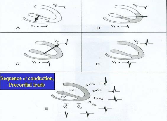
18 R wave progression
19 Rate
 If there is 1 beat/large box [5 beats per second] X 60 sec/minute, then there are (5 X 60) 300 beats/min If there is 1 beat/every 2 nd box [2.")
20 Rate with Regular Rhythm rule 5 small boxes (0.04 sec) = 1 large box (0.2 sec) If there is 1 beat/large box [5 beats per second] X 60 sec/minute, then there are (5 X 60) 300 beats/min If there is 1 beat/every 2 nd box [2.5 beats per sec], then there are (2.5 X 60) 150 beats/min Thus, the rule
 and multiply by 10 = avg # beats/60 sec.")
21 Rate with Irregular Rhythm For irregular rhythms, check the EKG s 3 second markers (15 large boxes). Count the complexes beginning with zero within a 30 large boxes (6 seconds) and multiply by 10 = avg # beats/60 sec. or estimated beats per minute large boxes
22 Rhythm
23 Common Rhythms Regular Sinus rhythm Atrial flutter (with consistent block) AV Nodal Reentrant Tachycardia (AVNRT or PSVT) Junctional rhythm Irregular Atrial fibrillation Atrial flutter (with variable block) Sinus arrhythmia Wandering Atrial Pacemaker Multifocal Atrial Tachycardia Premature beats (PVCs & PACs with and w/o aberrancy)
 Rate")
24 Sinus Rhythm Normal Sinus Rate of 60 to 100 Same P wave Upright P wave in lead II Sinus Bradycardia Rate < 60 Sinus Tachycardia (intrinsic rate of SA node is 100 to 110) Rate >100 SA

25 Sinus Rhythm Rate ~84

26 Consistent block segment Atrial Flutter w/ Consistent Block Consistent block Variable block segment Variable block
, usually narrow")
27 AV Nodal Re-entrant Tachycardia No definite p waves, rate ~ 150 to 200 and regular w/o variability May see retrograde p waves (seen as pseudo-terminal S waves in II & avf, and pseudo-r in V1), usually narrow QRS
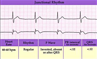
28 Junctional Rhythm
29 Case #1 67 year old male with history of hypertension presented with 1 month of one flight or one-half block DOE. Denied chest pain and the ROS was otherwise negative. Only medication is HCTZ. Physical exam was normal Labs in his chart from 2 months ago show a normal CBC, a mildly abnormal lipid panel, and normal basic chemistries. Pulse Ox 94% on RA
30 Case #1
31 Case #1 Sinus Tachycardia (common causes) Fever Anemia Hypovolemia Heart Failure Anxiety Hyperthyroidism Medications Pulmonary Emboli
32 Irregular Rhythms Atrial fibrillation Atrial flutter w/ variable block Sinus arrhythmia Wandering Atrial Pacemaker Multifocal Atrial Tachycardia Premature beats (PVCs & PACs with and w/o aberrancy)
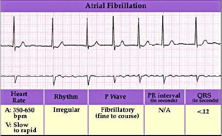
33 Atrial Fibrillation

34 Atrial Fibrillation

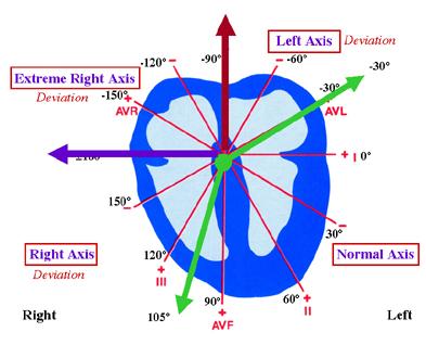
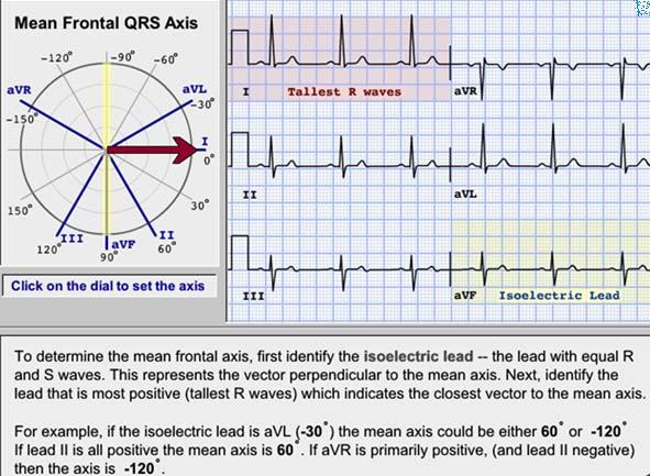
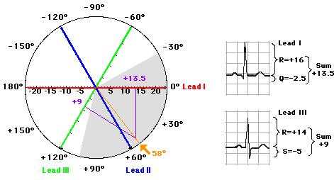
35 Sinus arrhythmia Rate increases with inspiration and decreases with expiration

36 Wandering Atrial Pacemaker
 An atrial rate of > 100 beats/min is the classic definition (> 90 bpm has been proposed)) The P-P intervals, the P-R duration, and the R-R intervals")
37 Multifocal Atrial Tachycardia Discrete P waves with at least three different morphologies (P wave morphology is generally best seen in leads II, III and V1) An atrial rate of > 100 beats/min is the classic definition (> 90 bpm has been proposed)) The P-P intervals, the P-R duration, and the R-R intervals vary

38 Ventricular Premature Beats (VPBs or PVCs) QRS > 120 ms VPBs have early widened QRS complexes with a bizarre morphology and repolarization abnormalities markedly different from the sinus QRS complexes. A full compensatory pause usually follows the VPB (the interval between the QRS complexes before and after the VPB is 2X the interval of the 2 successive sinus beats).

39 Premature Atrial Beats QRS < 120 ms The interval between the second sinus beat and the ectopic beat is shorter than the interval between the first two sinus beats. The P wave morphology differs from that of the sinus beats. The PR interval differs from that of the sinus beat. Activation of the ventricular myocardium occurs in a normal fashion so the QRS complex is unchanged from that of sinus rhythm.

40 PAC with Aberrancy vs VPB RBBB pattern (rsr ) of the anomalous beats in a right sided monitor lead (V1) An initial deflection similar in the same direction as that of normally conducted beats Identification of a premature P wave Premature P wave rsr = RBBB pattern
41 Case #2 36 year old female with PMH of diabetes mellitus and pernicious anemia presents with fatigue. Further questioning reveals a 12 lb weight loss in the past 3 months. Her glucose diary shows blood sugars ranging from 80 to 200 and a HgbA1c from 4 months ago was 6.9%. Today s in-office fingerstick glucose is 142. Her ROS is otherwise unremarkable.
42 Case #2 Vital signs recorded as BP 136/74 - P 120 RR 20 T 37.4 C. Your exam discovers a faster heart rate, normal lung, abdominal, extremity and HEENT exams. She seems anxious. DTRs are +3 and symmetric. EKG

43 Case #2 Aberrant conduction Rapid Atrial fibrillation at about 160 bpm
44 Atrial Fibrillation Mitral Valve disease Ischemic Heart disease Hypertension Thyrotoxicosis Lone Atrial fibrillation Advanced age Pulmonary Embolus Pericarditis Case #2 Due to new onset of Graves Disease
45 Axis and Intervals
46 QRS Axis
47 QRS Axis
48 QRS Axis
49 Left QRS Axis Deviation Negative 30 to negative 90 degrees Mechanical shifts (elevated diaphragm) LBBB LAHB or LAFB WPW syndrome (sometimes) Inferior wall MI Sometimes LVH

50 Left Axis Deviation - + Axis = -40 degrees (LVH)

51 Wolf-Parkinson-White QRS axis = -32 degrees PR interval = 0.10 s QRS duration = s
52 Right QRS Axis Deviation > degrees to degrees Mechanical shifts (emphysema) RBBB RVH LPHB WPW syndrome Dextrocardia Lateral wall MI Acute right heart strain (massive PE) Children under age 2


53 Right Axis Deviation _ + Axis = +115 degrees (anterolateral MI)

54 Case #3 What is the QRS axis? QRS Axis +110 Does avr then appear as expected? Should avr be closer to isoelectric?

55 Left and right arm limb leads are reversed If negative p wave and negative QRS complex in Lead I, the most common cause is limb lead reversal Lead III is recorded as lead II and vice versa Case #3
56 Intervals PR Intervals and AV Blocks QRS Duration - Bundle Branch Blocks and hemiblocks QT intervals

57 sec **Males < 0.45 sec **Females < 0.46 sec ** - Hyperkalemia - Digitalis ** Some expert opinions do not agree with this QT interval upper limit of normal
58 Atrioventricular Blocks 1 st Degree PR interval > 0.20 seconds 7.5 blocks or 0.3 sec

59 Atrioventricular Blocks 2 nd Degree AV Block Type 1 (Wenckebach) In classic Wenkebach, the PR interval gets longer until a nonconducted P wave occurs. The RR interval of the pause is shorter than the sum of the 2 preceding RR intervals, and the RR interval after the pause is longer than the RR interval before the pause. The block is located in the AV node.

60 2 nd Degree AV Block (Wenkebach) R-R interval after pause longer than R-R interval before pause R-R interval of the pause is shorter than the sum of the 2 R-R intervals before the pause PR intervals lengthen

61 Atrioventricular Blocks 2 nd Degree AV Block (Mobitz Type 2)


62 Atrioventricular Blocks 3 rd Degree AV Block with Junctional Escape Beats 3 rd Degree AV Block with Ventricular Escape Beats
63 QRS Duration Normal is 0.06 to 0.10 sec Bundle Branch Block = 0.12 sec or more Upper limits of normal to just under BBB (0.10 & 0.11 sec) sometimes seen in hemiblocks and with some medications

64 Left Bundle Branch Block
65 Left Bundle Branch Blocks Complete LBBB has QRS > 0.12 sec Terminal forces (2 nd half of QRS) are oriented leftward and posteriorly because the LV is depolarized after the RV Terminal S wave in V1 indicating late posterior forces (QS or rs in V1) Terminal R wave in leads I, avl, V6 indicating late leftward forces Late left ventricular deflection in V6 No Q wave & Monophasic R wave in V6
 Terminal R wave in leads I, avl, V6 indicating late leftward")
66 Left Bundle Branch Block rs pattern Terminal S wave in V1 indicating late posterior forces (QS or rs in V1) Terminal R wave in leads I, avl, V6 indicating late leftward forces
 Terminal R wave in leads I, avl, V6 indicating late leftward")
67 Left Bundle Branch Block QS pattern Terminal S wave in V1 indicating late posterior forces (QS or rs in V1) Terminal R wave in leads I, avl, V6 indicating late leftward forces

68 Right Bundle Branch Block
69 Right Bundle Branch Block Complete RBBB has QRS > 0.12 sec Terminal forces (2 nd half of QRS) are oriented rightward and anteriorly because the RV is depolarized after the LV Late Ventricular Deflection in V1, rsr variant, indicating late anterior forces, or wide R or qr in V1 Early Ventricular Deflection and wide S wave in V6 Wide terminal S wave in leads I, avl, V6 indicating late rightward forces

70 Right Bundle Branch Block 120 ms qr Late Ventricular Deflection in V1, rsr variant, indicating late anterior forces, or wide R or qr in V1 2-3 Early Ventricular Deflection and wide S wave in V6 Wide terminal S wave in leads I, avl, V6 indicating late rightward forces

71 Left Anterior Fascicular Block I avf

72 Left Anterior Fascicular Block Left Axis Deviation (usually < - 35 degrees) Small Q waves in leads I and avl; S > R waves in leads II, III, and avf QRS duration < 0.12 sec Small Q, Larger R in I & AVL S > R in II, III, AVF Axis < -35 Left QRS Axis Deviation ~ - 40 degrees

73 Left Anterior Fascicular Block Sometimes a deep S wave in V6

74 Left Posterior Fascicular Block I avf

75 Left Posterior Hemiblock Right axis deviation (> +105) S > R waves in leads I and avl Small Q wave in leads II, III, avf QRS duration < 0.12 sec No evidence of RVH

76 Left Posterior Fascicular Block Small R wave (S > R) in lead I and avl Small Q wave in lead II, III, avf Axis +120 Sinus tachycardia, possible anterior infarct, left posterior fascicular block
77 Case #4 52 year old male with hyperlipidemia presents with dizziness and feeling tired for one day. He takes his statin regularly and has no other complaints. His exam reveals normal lung, HEENT, and neurological exams. BP is 126/75. Pulse is 36.

78 Case #4 Diagnosis = 3rd Degree AV Block
79 QT represents ventricular depolarization/repolarization Longer at slower heart rates and shorter at faster heart rates, therefore the QTc (Bazzett s Formula) Shortened by hypercalcemia, hyperkalemia, and digitalis Prolonged by hypocalcemia, hypokalemia, hypomagnesemia, congenital causes, myocarditis, and many medications QT Intervals
80 QT Intervals QTc-interval between 360 msec up to 440 msec was traditionally considered to be normal. Many experts believe 450 msec in males and 460 msec in females are the upper limits of normal. Some consider up to 470 msec to be borderline prolonged, but not prolonged enough to clearly make the prolonged QTc diagnosis. A QTc at or above 470 milliseconds in males or 480 milliseconds in females is clearly prolonged. The average QTc for someone who has prolonged-qt syndrome is 490 milliseconds.
 was more than double (odds ratio 2.20) that of patients with a QTC of 0.44, whereas for those arriving at the ED 1.5 hours after chest pain onset, the risk was 79% lower (OR 0.21). http://www.")
81 Acute Septal Infarct with prolonged QT interval QTc = 0.51 For patients arriving at the ED 30 minutes after chest pain onset, risk for cardiac arrest for patients with a prolonged QTc (0.50) was more than double (odds ratio 2.20) that of patients with a QTC of 0.44, whereas for those arriving at the ED 1.5 hours after chest pain onset, the risk was 79% lower (OR 0.21). Evidence Level B
82 Chamber Sizes
83 Hypertension JNC8 Recommendations Routine tests recommended before initiating therapy include: 12-lead ECG Looking for LVH and Conduction Abnormalities Serum potassium, creatinine, and consider sodium Diabetes screening Cholesterol screening JNC8+Hypertension+laboratory+and+ECG+testing+recommendations&gs_l=serp i j19j msedr c.1.60.serp bqadzlrhujg Evidence Level B
84 Left Atrial Abnormality Delayed broadening of the latter part of the P wave of at least 0.12 sec in leads I or II resulting in a notched P wave A negative > 1 mm X 1 mm P wave deflection in V1 II V1

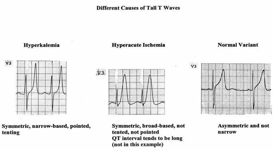
85 Left Atrial Enlargement
86 Right Atrial Enlargement P wave amplitude >2.5 mm in lead II and/or Positive P wave deflection >1.5 mm in V1 II V1

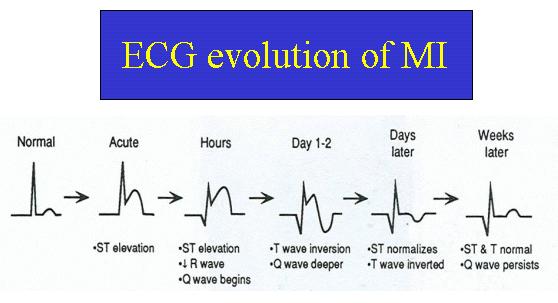
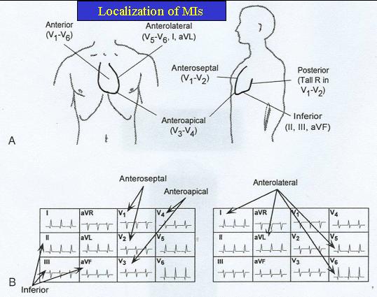
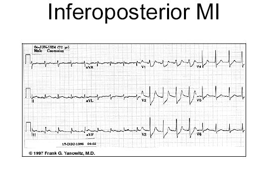
87 Right Atrial Enlargement

88 Left Ventricular Hypertrophy Increased QRS amplitude Leftward shift in frontal plane QRS axis Delayed intrinsicoid deflection in V6 (time from QRS onset to peak R is > 0.05 sec) Widened QRS/T angle (LV strain pattern) with slow downstroke-rapid upstroke T wave inversion in V5-V6 Evidence of left atrial enlargement

89 Left Ventricular Hypertrophy Sokolow-Lyons indices Height of R wave in avl > 11 mm or S wave in lead V1 plus R wave in either V5 or V6 > 35 mm Cornell s Voltage Criteria S in V3 + R in avl > 20 mm in women S in V3 + R in avl > 28 mm in men Cornell s sensitivity = 51%, specificity = 95%. All EKG criteria are inferior to Echocardiogram for diagnosing LVH

90 Left Ventricular Hypertrophy
 Might have deep S wave in V5 and V6 Right Axis")
91 Right Ventricular Hypertrophy Negative QRS in lead I (S > R) Positive QRS in V1 (R > S) Might have deep S wave in V5 and V6 Right Axis Deviation

92 Right Ventricular Hypertrophy Axis = +122 degrees
93 Case #5 45 year old male presents for a sore throat. On exam, you notice a BP of 152/92. You review BPs on other visits and they consistently range 140 to 160/85 to 98. When you bring this to his attention and recommend an initial hypertensive workup and treatment, he responds, I get nervous in the doctors office. My pressure is always normal when I check it at the local pharmacy. You check an EKG

94 Case #5 Left Ventricular Hypertrophy This patient likely has long-standing hypertension and should be treated
95 QRST Changes
96 ST Segment Depression >1 mm below baseline measured at 0.08 sec after the J point Ischemia Strain pattern Hypokalemia or hypomagnesemia Rate related Digitalis effect Pericarditis Any combination of above

97 ST Segment Depression
98 ST Segment Elevation At least 1 mm (0.1 mv) in limb leads, V4, V5 or V6 At least 2 mm elevation in males (1.5 mm in females) at the V2 and V3 J- point AMI or cardiac contusion LBBB early repolarization ventricular aneurysm Osborne J waves (hypothermia) Prinzmetal s vasospasm pericarditis
99 T Waves

100 T Waves NSTEMI myocarditis pericarditis cerebral T waves

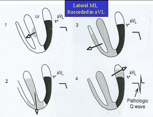
101 HypoKalemia U wave EKG shows a long QT interval, ST depression, low T waves, and TU wave fusion characteristic of hypokalemia U waves are thought to represent repolarization of the papillary muscles or Purkinje fibers
102 Acute Myocardial Infarction ECG diagnosis requires at least 1 mm (0.1 mv) of new ST segment elevation in corresponding anatomical limb leads, V4, V5 or V6 At least 2 mm elevation in males (1.5 mm in females) at the V2 and V3 J-point These elevations must be anatomically contiguous I, avl, V5, V6 = lateral wall V1-V4 = septal/anterior wall II, III, avf = inferior wall True posterior MI pattern (V1-V2) New LBBB in suspicious setting

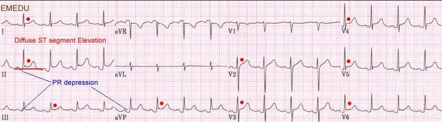
103 Myocardial Infarction --V3
104

105 Q waves Because the septum depolarizes left to right, small insignificant Q waves are normal in leads I, avl, V5, and V6. Significant Q waves may indicate a myocardial infarction and are 1/4 the height of the QRS complex or 0.04 sec or more wide
106
107 Infarct Patterns
108
109

110 Acute Inferior Wall MI
111
112
113

114 Acute Anteroseptal MI V1-2-3

115 Acute Anteroapical Infarct
116 Acute Anterolateral Wall MI

117 Acute Lateral or High Lateral Infarct ST elevation in lead avl and reciprocal ST depression in leads II, III, and avf
118 Case #6 A 38 year old construction worker is driven from work to your office complaining of sharp chest pain. He denies shortness of breath, nausea, vomiting, or palpitations. He is diaphoretic but was just working outside on a hot day. His exam reveals normal VS, clear lungs, RRR and you describe a III/VI systolic murmur in your note. No gallop. No JVD. You check an EKG
119 Acute Pericarditis
120 References Stein, E. Rapid Analysis of Electrocardiograms. Lippincott, Williams & Wilkins Marriott, H. Practical Electrocardiography. Williams & Wilkins Accessed January t nt/documents/ekg.pdf
121 Thank You
Pennsylvania Academy of Family Physicians Foundation & UPMC 43rd Refresher Course in Family Medicine CME Conference March 10-13, 2016
 Pennsylvania Academy of Family Physicians Foundation & UPMC 43rd Refresher Course in Family Medicine CME Conference March 10-13, 2016 Disclosures: EKG Workshop Louis Mancano, MD Speaker has no disclosures
2017 EKG Workshop Advanced. Family Medicine Review Course Lou Mancano, MD, FAAFP Reading Health System Family and Community Medicine Reading, PA
 2017 EKG Workshop Advanced Family Medicine Review Course Lou Mancano, MD, FAAFP Reading Health System Family and Community Medicine Reading, PA Part II - Objective Describe a useful approach to interpreting
REtrive. REpeat. RElearn Design by. Test-Enhanced Learning based ECG practice E-book
 Test-Enhanced Learning Test-Enhanced Learning Test-Enhanced Learning Test-Enhanced Learning based ECG practice E-book REtrive REpeat RElearn Design by S I T T I N U N T H A N G J U I P E E R I Y A W A
Please check your answers with correct statements in answer pages after the ECG cases.
 ECG Cases ECG Case 1 Springer International Publishing AG, part of Springer Nature 2018 S. Okutucu, A. Oto, Interpreting ECGs in Clinical Practice, In Clinical Practice, https://doi.org/10.1007/978-3-319-90557-0
12-Lead ECG Interpretation. Kathy Kuznar, RN, ANP
 12-Lead ECG Interpretation Kathy Kuznar, RN, ANP The 12-Lead ECG Objectives Identify the normal morphology and features of the 12- lead ECG. Perform systematic analysis of the 12-lead ECG. Recognize abnormalities
Basic electrocardiography reading. R3 lee wei-chieh
 Basic electrocardiography reading R3 lee wei-chieh The Normal Conduction System Lead Placement avf Limb Leads Precordial Leads Interpretation Rate Rhythm Interval Axis Chamber abnormality QRST change What
Appendix D Output Code and Interpretation of Analysis
 Appendix D Output Code and Interpretation of Analysis 8 Arrhythmia Code No. Description 8002 Marked rhythm irregularity 8110 Sinus rhythm 8102 Sinus arrhythmia 8108 Marked sinus arrhythmia 8120 Sinus tachycardia
Study methodology for screening candidates to athletes risk
 1. Periodical Evaluations: each 2 years. Study methodology for screening candidates to athletes risk 2. Personal history: Personal history of murmur in childhood; dizziness, syncope, palpitations, intolerance
ECG Interpretation Made Easy
 ECG Interpretation Made Easy Dr. A Tageldien Abdellah, MSc MD EBSC Lecturer of Cardiology- Hull University Hull York Medical School 2007-2008 ECG Interpretation Made Easy Synopsis Benefits Objectives Process
Family Medicine for English language students of Medical University of Lodz ECG. Jakub Dorożyński
 Family Medicine for English language students of Medical University of Lodz ECG Jakub Dorożyński Parts of an ECG The standard ECG has 12 leads: six of them are considered limb leads because they are placed
, David Stultz, MD.
 http://www.dilbert.com EKG Rounds Handouts available at http://www.drstultz.com January 5, 2004 David Stultz, MD Cardiology Fellow, PGY 4 Overview of Topics How to read an EKG Normal EKG Determination
ECG Interpretation Cat Williams, DVM DACVIM (Cardiology)
") ECG Interpretation Cat Williams, DVM DACVIM (Cardiology) Providing the best quality care and service for the patient, the client, and the referring veterinarian. GOAL: Reduce Anxiety about ECGs Back to
ECG Interpretation Cat Williams, DVM DACVIM (Cardiology) Providing the best quality care and service for the patient, the client, and the referring veterinarian. GOAL: Reduce Anxiety about ECGs Back to
ECG CONVENTIONS AND INTERVALS
 1 ECG Waveforms and Intervals ECG waveforms labeled alphabetically P wave== represents atrial depolarization QRS complex=ventricular depolarization ST-T-U complex (ST segment, T wave, and U wave)== V repolarization.
General Introduction to ECG. Reading Assignment (p2-16 in PDF Outline )
") General Introduction to ECG Reading Assignment (p2-16 in PDF Outline ) Objectives 1. Practice the 5-step Method 2. Differential Diagnosis: R & L axis deviation 3. Differential Diagnosis: Poor R-wave progression
General Introduction to ECG Reading Assignment (p2-16 in PDF Outline ) Objectives 1. Practice the 5-step Method 2. Differential Diagnosis: R & L axis deviation 3. Differential Diagnosis: Poor R-wave progression
Supraventricular Arrhythmias. Reading Assignment. Chapter 5 (p17-30)
") Supraventricular Arrhythmias Reading Assignment Chapter 5 (p17-30) The Supraventricular Rhythms In Our Lives Site of Origin Single Events Slow Rates Intermediate Rates Fast Rates (>100 bpm) Sinus Sinus
Supraventricular Arrhythmias Reading Assignment Chapter 5 (p17-30) The Supraventricular Rhythms In Our Lives Site of Origin Single Events Slow Rates Intermediate Rates Fast Rates (>100 bpm) Sinus Sinus
Chapter 2 Practical Approach
 Chapter 2 Practical Approach There are beginners in electrocardiogram (ECG) analysis who are fascinated by a special pattern (e.g., a bundle-branch block or a striking Q wave) and thereby overlook other
Blocks & Dissociations. Reading Assignment (p47-52 in Outline )
") Blocks & Dissociations Reading Assignment (p47-52 in Outline ) Objectives Who are Wenckebach and Mobitz? Review SA and AV Blocks AV Dissociations: learning who s the boss and why 2 nd degree SA Block:
Blocks & Dissociations Reading Assignment (p47-52 in Outline ) Objectives Who are Wenckebach and Mobitz? Review SA and AV Blocks AV Dissociations: learning who s the boss and why 2 nd degree SA Block:
Electrocardiography Abnormalities (Arrhythmias) 7. Faisal I. Mohammed, MD, PhD
 7. Faisal I. Mohammed, MD, PhD") Electrocardiography Abnormalities (Arrhythmias) 7 Faisal I. Mohammed, MD, PhD 1 Causes of Cardiac Arrythmias Abnormal rhythmicity of the pacemaker Shift of pacemaker from sinus node Blocks at different
Electrocardiography Abnormalities (Arrhythmias) 7 Faisal I. Mohammed, MD, PhD 1 Causes of Cardiac Arrythmias Abnormal rhythmicity of the pacemaker Shift of pacemaker from sinus node Blocks at different
Ekg pra pr c a tice D.HAMMOUDI.MD
 Ekg practice D.HAMMOUDI.MD Anatomy Revisited RCA (Right Coronary Artery) Right ventricle Inferior wall of LV Posterior wall of LV (75%) SA Node (60%) AV Node (>80%) LCA (Left Coronary Artery) Septal wall
ECG Basics Sonia Samtani 7/2017 UCI Resident Lecture Series
 ECG Basics Sonia Samtani 7/2017 UCI Resident Lecture Series Agenda I. Introduction II.The Conduction System III.ECG Basics IV.Cardiac Emergencies V.Summary The Conduction System Lead Placement avf Precordial
12 Lead ECG. Presented by Rebecca Sevigny BSN, RN Professional Practice & Development Dept.
 12 Lead ECG Presented by Rebecca Sevigny BSN, RN Professional Practice & Development Dept. Two Main Coronary Arteries RCA LCA which branches into Left Anterior Descending Circumflex Artery Two Main Coronary
ECG (MCQs) In the fundamental rules of the ECG all the following are right EXCEP:
 In the fundamental rules of the ECG all the following are right EXCEP:") ECG (MCQs) 2010 1- In the fundamental rules of the ECG all the following are right EXCEP: a- It is a biphasic record of myocardial action potential fluctuations. b- Deflection record occurs only during
ECG (MCQs) 2010 1- In the fundamental rules of the ECG all the following are right EXCEP: a- It is a biphasic record of myocardial action potential fluctuations. b- Deflection record occurs only during
ECG ABNORMALITIES D R. T AM A R A AL Q U D AH
 ECG ABNORMALITIES D R. T AM A R A AL Q U D AH When we interpret an ECG we compare it instantaneously with the normal ECG and normal variants stored in our memory; these memories are stored visually in
The Electrocardiogram part II. Dr. Adelina Vlad, MD PhD
 The Electrocardiogram part II Dr. Adelina Vlad, MD PhD Basic Interpretation of the ECG 1) Evaluate calibration 2) Calculate rate 3) Determine rhythm 4) Determine QRS axis 5) Measure intervals 6) Analyze
402 Index. B β-blockers, 4, 5 Bradyarrhythmias, 76 77
 Index A Acquired immunodeficiency syndrome (AIDS), 126, 163 Action potentials, 1, 5, 27 Acute coronary syndromes, 123t, 129 Adenosine, intravenous, 277 Alcohol abuse, as T wave inversion cause, 199 Aneurysm,
Paediatric ECG Interpretation
 Paediatric ECG Interpretation Dr Sanj Fernando (thanks to http://lifeinthefastlane.com/ecg-library/paediatric-ecginterpretation/) 3 yo boy complaining of abdominal pain and chest pain Child ECG vs Adult
If the P wave > 0.12 sec( 3 mm) usually in any lead. Notched P wave usually in lead I,aVl may be lead II Negative terminal portion of P wave in V1, 1
 usually in any lead. Notched P wave usually in lead I,aVl may be lead II Negative terminal portion of P wave in V1, 1") If the P wave > 0.12 sec( 3 mm) usually in any lead. Notched P wave usually in lead I,aVl may be lead II Negative terminal portion of P wave in V1, 1 mm depth and 3 mm width( most specific) Since Mitral
If the P wave > 0.12 sec( 3 mm) usually in any lead. Notched P wave usually in lead I,aVl may be lead II Negative terminal portion of P wave in V1, 1 mm depth and 3 mm width( most specific) Since Mitral
ECG Practice Strips Discussion part 1:
 ECG Practice Strips Discussion part 1: The first 20 strips are for teaching various abnormalities of the morphology of the waves of the ECG. Strips 21 and following are for teaching some abnormalities
Understanding the 12-lead ECG, part II
 Bundle-branch blocks Understanding the 12-lead ECG, part II Most common electrocardiogram (ECG) abnormality Appears as a wider than normal S complex Occurs when one of the two bundle branches can t conduct
ELECTROCARDIOGRAPH. General. Heart Rate. Starship Children s Health Clinical Guideline
 General Heart Rate QRS Axis T Wave Axis PR Interval according to Heart Rate & Age P Wave Duration and Amplitude QRS Duration according to Age QT Interval R & S voltages according to Lead & Age R/S ratio
Cardiology Flash Cards
 Cardiology Flash Cards EKG in a nut shell www.brain101.info Conduction System www.brain101.info 2 Analyzing EKG Step by step Steps in Analyzing ECG'S 1. Rhythm: - Regular _ Sinus, Junctional or Ventricular.
Myocardial Infarction. Reading Assignment (p66-78 in Outline )
") Myocardial Infarction Reading Assignment (p66-78 in Outline ) Objectives 1. Why do ST segments go up or down in ischemia? 2. STEMI locations and culprit vessels 3. Why 15-lead ECGs? 4. What s up with avr?
Myocardial Infarction Reading Assignment (p66-78 in Outline ) Objectives 1. Why do ST segments go up or down in ischemia? 2. STEMI locations and culprit vessels 3. Why 15-lead ECGs? 4. What s up with avr?
10 ECGs No Practitioner Can Afford to Miss. Objectives
 10 ECGs No Practitioner Can Afford to Miss Mary L. Dohrmann, MD Professor of Clinical Medicine Division of Cardiovascular Medicine University of Missouri School of Medicine No disclosures Objectives 1.
FLB s What Are Those Funny-Looking Beats?
 FLB s What Are Those Funny-Looking Beats? Reading Assignment (pages 27-45 in Outline ) The 5-Step Method ECG #: Mearurements: Rhythm (s): Conduction: Waveform: Interpretation: A= V= PR= QRS= QT= Axis=
Electrical System Overview Electrocardiograms Action Potentials 12-Lead Positioning Values To Memorize Calculating Rates
 Electrocardiograms Electrical System Overview James Lamberg 2/ 74 Action Potentials 12-Lead Positioning 3/ 74 4/ 74 Values To Memorize Inherent Rates SA: 60 to 100 AV: 40 to 60 Ventricles: 20 to 40 Normal
also aid the clinician in recognizing both the obvious and subtle abnormalities that may help guide therapy.
 Karen Lieberman, MS, CRNP f the many diagnostic tools used to screen for and evaluate cardiac abnormalities, the 12-lead electrocardiogram (ECG) is among the most basic. This inexpensive and noninvasive
Pathologic ECG. Adelina Vlad, MD PhD
 Pathologic ECG Adelina Vlad, MD PhD Basic Interpretation of the ECG 1) Evaluate calibration 2) Calculate rate 3) Determine rhythm 4) Determine QRS axis 5) Measure intervals 6) Analyze the morphology and
Acute Coronary Syndromes Unstable Angina Non ST segment Elevation MI (NSTEMI) ST segment Elevation MI (STEMI)
 ST segment Elevation MI (STEMI)") Leanna R. Miller, RN, MN, CCRN-CSC, PCCN-CMC, CEN, CNRN, CMSRN, NP Education Specialist LRM Consulting Nashville, TN Objectives Evaluate common abnormalities that mimic myocardial infarction. Identify
Leanna R. Miller, RN, MN, CCRN-CSC, PCCN-CMC, CEN, CNRN, CMSRN, NP Education Specialist LRM Consulting Nashville, TN Objectives Evaluate common abnormalities that mimic myocardial infarction. Identify
HR: 50 bpm (Sinus) PR: 280 ms QRS: 120 ms QT: 490 ms Axis: -70. Sinus bradycardia with one ventricular escape (*)
 PR: 280 ms QRS: 120 ms QT: 490 ms Axis: -70. Sinus bradycardia with one ventricular escape (*)") 1? HR: 50 bpm (Sinus) PR: 280 ms QRS: 120 ms QT: 490 ms Axis: -70 1 Sinus P waves? 2 sinus cycles The pause (2 sinus cycles) suggests that the sinus fired (?) but did not conduct to the atria (i.e., missing
1? HR: 50 bpm (Sinus) PR: 280 ms QRS: 120 ms QT: 490 ms Axis: -70 1 Sinus P waves? 2 sinus cycles The pause (2 sinus cycles) suggests that the sinus fired (?) but did not conduct to the atria (i.e., missing
Section V. Objectives
 Section V Landscape of an MI Objectives At the conclusion of this presentation the participant will be able to Outline a systematic approach to 12 lead ECG interpretation Demonstrate the process for determining
12 Lead ECG Skills: Building Confidence for Clinical Practice. Presented By: Cynthia Webner, BSN, RN, CCRN-CMC. Karen Marzlin, BSN, RN,CCRN-CMC
 12 Lead ECG Skills: Building Confidence for Clinical Practice NTI 2009 Preconference Session 803 Presented By: Karen Marzlin, BSN, RN,CCRN-CMC 1 12 Lead ECG Fundamentals: The Starting Place for Linking
UNDERSTANDING YOUR ECG: A REVIEW
 UNDERSTANDING YOUR ECG: A REVIEW Health professionals use the electrocardiograph (ECG) rhythm strip to systematically analyse the cardiac rhythm. Before the systematic process of ECG analysis is described
Bundle Branch & Fascicular Blocks. Reading Assignment (p53-58 in Outline )
") Bundle Branch & Fascicular Blocks Reading Assignment (p53-58 in Outline ) Objectives 1. QRS analysis of Right and Left BBB 2. Uncomplicated vs complicated BBB 3. Diagnosis of RBBB with LAFB and LPFB 4.
Bundle Branch & Fascicular Blocks Reading Assignment (p53-58 in Outline ) Objectives 1. QRS analysis of Right and Left BBB 2. Uncomplicated vs complicated BBB 3. Diagnosis of RBBB with LAFB and LPFB 4.
Acute Coronary Syndromes. Disclosures
 Acute Coronary Syndromes Disclosures I work for Virginia Garcia Memorial Health Center, Beaverton, OR. Jon Tardiff, BS, PA-C OHSU Clinical Assistant Professor And I am a medical editor for Jones & Bartlett
Introduction to Electrocardiography
 Introduction to Electrocardiography Class Objectives: Introduction to ECG monitoring Discuss principles of interpretation Identify the components and measurements of the ECG ECG analysis ECG Monitoring
DR QAZI IMTIAZ RASOOL OBJECTIVES
 PRACTICAL ELECTROCARDIOGRAPHY DR QAZI IMTIAZ RASOOL OBJECTIVES Recording of electrical events in heart Established electrode pattern results in specific tracing pattern Health of heart i. e. Anatomical
1 st Degree Block Prolonged P-R interval caused by first degree heart block (lead II)
") AV Heart Blocks 1 st degree A condition of a rhythm, not a true rhythm Need to always state underlying rhythm 2 nd degree Type I - Wenckebach Type II Classic dangerous to the patient Can be variable (periodic)
AV Heart Blocks 1 st degree A condition of a rhythm, not a true rhythm Need to always state underlying rhythm 2 nd degree Type I - Wenckebach Type II Classic dangerous to the patient Can be variable (periodic)
Dr. Schroeder has no financial relationships to disclose
 Valerie A Schroeder MD MS Assistant Professor University of Kansas Medical Center READING THE WAVES- THE HEART S ELECTRICAL MESSAGE FINANCIAL DISCLOSURE Dr. Schroeder has no financial relationships to
Miscellaneous Stuff Keep reading the Outline
 Miscellaneous Stuff Keep reading the Outline Welcome to the 5-Step Method ECG #: Mearurements: Rhythm (s): Conduction: Waveform: Interpretation: A= V= PR= QRS= QT= Axis= 1. Compute the 5 basic measurements:
Electrocardiography for Healthcare Professionals. Chapter 14 Basic 12-Lead ECG Interpretation
 Electrocardiography for Healthcare Professionals Chapter 14 Basic 12-Lead ECG Interpretation 2012 The Companies, Inc. All rights reserved. Learning Outcomes 14.1 Discuss the anatomic views seen on a 12-lead
Introduction to ECG Gary Martin, M.D.
 Brief review of basic concepts Introduction to ECG Gary Martin, M.D. The electrical activity of the heart is caused by a sequence of rapid ionic movements across cell membranes resulting first in depolarization
12 LEAD EKG BASICS. By: Steven Jones, NREMT P CLEMC
 12 LEAD EKG BASICS By: Steven Jones, NREMT P CLEMC ECG Review Waves and Intervals P wave: the sequential activation (depolarization) of the right and left atria QRS complex: right and left ventricular
Ben Taylor, PhD, PA-C
 Ben Taylor, PhD, PA-C The patient is a 23-year-old white male with a history of polysubstance abuse who was found unresponsive, last seen the day before. Classic signs of systemic hypothermia with prominent
Huseng Vefali MD St. Luke s University Health Network Department of Cardiology
 Huseng Vefali MD St. Luke s University Health Network Department of Cardiology Learning Objectives Establish Consistent Approach to Interpreting ECGs Review Essential Cases for Paramedics and first responders
ELECTROCARDIOGRAPHY KEVIN REBECK PA-C. For more presentations
 ELECTROCARDIOGRAPHY KEVIN REBECK PA-C For more presentations www.medicalppt.blogspot.com Objectives ECG History Pathophysiology Basics Case Historys Electrical activation of the heart In the heart
This presentation will deal with the basics of ECG description as well as the physiological basics of
 Snímka 1 Electrocardiography basics This presentation will deal with the basics of ECG description as well as the physiological basics of Snímka 2 Lecture overview 1. Cardiac conduction system functional
Reading Assignment (p1-91 in Outline ) Objectives What s in an ECG?
 Objectives What s in an ECG?") Reading Assignment (p1-91 in Outline ) Objectives What s in an ECG? The 5-Step Method ECG #: Mearurements: Rhythm (s): Conduction: Waveform: Interpretation: A= V= PR= QRS= QT= Axis= 1. Compute the 5 basic
Reading Assignment (p1-91 in Outline ) Objectives What s in an ECG? The 5-Step Method ECG #: Mearurements: Rhythm (s): Conduction: Waveform: Interpretation: A= V= PR= QRS= QT= Axis= 1. Compute the 5 basic
CASE 10. What would the ST segment of this ECG look like? On which leads would you see this ST segment change? What does the T wave represent?
 CASE 10 A 57-year-old man presents to the emergency center with complaints of chest pain with radiation to the left arm and jaw. He reports feeling anxious, diaphoretic, and short of breath. His past history
ECG Cases and Questions. Ashish Sadhu, MD, FHRS, FACC Electrophysiology/Cardiology
 ECG Cases and Questions Ashish Sadhu, MD, FHRS, FACC Electrophysiology/Cardiology 32 yo female Life Insurance Physical 56 yo male with chest pain Terminology Injury ST elevation Ischemia T wave inversion
SIMPLY ECGs. Dr William Dooley
 SIMPLY ECGs Dr William Dooley 1 No anatomy just interpretation 2 Setting up an ECG 3 Setting up an ECG 1 V1-4 th Right intercostal space at sternal border 2 V2-4 th Left intercostal space at sternal border
ECGs: Everything a finalist needs to know. Dr Amy Coulden As part of the Simply Finals series
 ECGs: Everything a finalist needs to know Dr Amy Coulden As part of the Simply Finals series Aims and objectives To be able to interpret basic ECG abnormalities To be able to recognise commonly tested
SIMPLY ECGs. Dr William Dooley
 SIMPLY ECGs Dr William Dooley Content Basic ECG interpretation pattern Some common (examined) abnormalities Presenting ECGs in context Setting up an ECG Setting up an ECG 1 V1-4 th Right intercostal space
Relax and Learn At the Farm 2012
 Relax and Learn At the Farm 2012 Session 2: 12 Lead ECG Fundamentals 101 Cynthia Webner DNP, RN, CCNS, CCRN-CMC, CHFN Though for Today Mastery is not something that strikes in an instant, like a thunderbolt,
ELECTROCARDIOGRAPHY (III) THE ANALYSIS OF THE ELECTROCARDIOGRAM
 THE ANALYSIS OF THE ELECTROCARDIOGRAM") ELECTROCARDIOGRAPHY (III) THE ANALYSIS OF THE ELECTROCARDIOGRAM Scridon Alina, Șerban Răzvan Constantin Recording and analysis of the 12-lead ECG is part of the basic medical assessment performed for every
ELECTROCARDIOGRAPHY (III) THE ANALYSIS OF THE ELECTROCARDIOGRAM Scridon Alina, Șerban Răzvan Constantin Recording and analysis of the 12-lead ECG is part of the basic medical assessment performed for every
ECGs and Arrhythmias: Family Medicine Board Review 2009
 Rate Rhythm Intervals Hypertrophy ECGs and Arrhythmias: Family Medicine Board Review 2009 Axis Jess (Fogler) Waldura, MD University of California, San Francisco walduraj@nccc.ucsf.edu Ischemia Overview
PATIENT S NAME, DATE/TIME,
 ECG and Arrhythmias Dec, 1 st 2014 Doctor Mohammad Jarrah References: - Lecture and Slides - ECG Made Easy - Davidson Principles of Medicine - First Aid Cases for the USMLE Step 1 - Mini-OSCE Archive ECG
Appendix. Table 1: Causes for abnormal axis deviation Left axis deviation
 Appendix Table 1: Causes for abnormal axis deviation Left axis deviation Normal variant (2 5%) Left anterior fascicular block Left ventricular hypertrophy Inferior wall myocardial infarction Primum atrial
Return to Basics. ECG Rate and Rhythm. Management of the Hospitalized Patient September 25, 2009
 Management of the Hospitalized Patient September 25, 2009 ECG Refresher and Update 2009 Return to Basics Determine rate and rhythm Determine intervals and axes Define morphology of P-QRS-T-U Compare with
ECG Interpretation. Introduction to Cardiac Telemetry. Michael Peters, RN, CCRN, CFRN CALSTAR Air Medical Services
 ECG Interpretation Introduction to Cardiac Telemetry Michael Peters, RN, CCRN, CFRN CALSTAR Air Medical Services Disclosures Nothing to disclose Objectives Describe the electrical conduction pathway in
PAEDIATRIC ECG Dimosthenis Avramidis, MD.
 PAEDIATRIC ECG Dimosthenis Avramidis, MD. Consultant Mitera Children s Hospital Athens Greece S. Associate 1st Cardiology Dpt Evangelismos Hospital Athens Greece 5 y/o with sinus tach Background ECG changes
Conduction Problems / Arrhythmias. Conduction
 Conduction Problems / Arrhythmias Conduction Wolf-Parkinson White Syndrome (WPW) and Lown-Ganong-Levine (LGL): Atrial impulses bypass the AV node through an accessory pathway or bypass tract (bundle of
Diploma in Electrocardiography
 The Society for Cardiological Science and Technology Diploma in Electrocardiography The Society makes this award to candidates who can demonstrate the ability to accurately record a resting 12-lead electrocardiogram
Cardiac Arrhythmia How to approach นพ.พ น จ แกวส วรรณะ หน วยโรคห วใจและหลอดเล อด
 Cardiac Arrhythmia How to approach นพ.พ น จ แกวส วรรณะ หน วยโรคห วใจและหลอดเล อด EKG paper is a grid where time is measured along the horizontal axis. Each small square is 1 mm in length and represents
1/22/2007 Fernald Medical Monitoring Program Sort Code EKG coding
 1/22/2007 Fernald Medical Monitoring Program Sort Code EKG coding PLEASE NOTE THAT ALL EKG CODES MUST RANGE FROM 500-599 OR FROM 900-999. PLEASE DO NOT ASSIGN NUMBERS OUTSIDE THAT RANGE FOR EKG CODES.
Normal ECG And ECHO Findings in Athletes
 Normal ECG And ECHO Findings in Athletes Dr.Yahya Kiwan Consultant Interventional Cardiologist Head Of Departement Of Cardiology Canadian Specialist Hospital Sinus Bradycardia The normal heartbeat is initiated
Office ECG Interpretation
 Office ECG Interpretation Jason Evanchan, DO Assistant Professor of Medicine Division of Cardiovascular Medicine The Ohio State University Wexner Medical Center Outline of topics High risk ischemia T wave
Return to Basics. Normal Intervals & Axes. ECG Rate and Rhythm
 Return to Basics Management of the Hospitalized Patient October 15, 2010 ECG Refresher and Update 2010 Determine rate and rhythm Determine intervals and axes Define morphology of P-QRS-T-U Compare with
Lab Activity 24 EKG. Portland Community College BI 232
 Lab Activity 24 EKG Reference: Dubin, Dale. Rapid Interpretation of EKG s. 6 th edition. Tampa: Cover Publishing Company, 2000. Portland Community College BI 232 Graph Paper 1 second equals 25 little boxes
Understanding basics of EKG
 Understanding basics of EKG By Alula A.(R III) www.le.ac.uk Topic for discussion Understanding of cellular electrophysiology Basics Rate Rhythm Axis Intervals P wave QRS ST/T wave Abnormal EKGs Understanding
2) Heart Arrhythmias 2 - Dr. Abdullah Sharif
 Heart Arrhythmias 2 - Dr. Abdullah Sharif") 2) Heart Arrhythmias 2 - Dr. Abdullah Sharif Rhythms from the Sinus Node Sinus Tachycardia: HR > 100 b/m Causes: o Withdrawal of vagal tone & Sympathetic stimulation (exercise, fight or flight) o Fever
2) Heart Arrhythmias 2 - Dr. Abdullah Sharif Rhythms from the Sinus Node Sinus Tachycardia: HR > 100 b/m Causes: o Withdrawal of vagal tone & Sympathetic stimulation (exercise, fight or flight) o Fever
CRC 431 ECG Basics. Bill Pruitt, MBA, RRT, CPFT, AE-C
 CRC 431 ECG Basics Bill Pruitt, MBA, RRT, CPFT, AE-C Resources White s 5 th ed. Ch 6 Electrocardiography Einthoven s Triangle Chest leads and limb leads Egan s 10 th ed. Ch 17 Interpreting the Electrocardiogram
Section 3 and 4. Objectives. Bundle Branches 10/9/2018. LBBB, RBBB Bifascicular, Trifascicular Block
 Section 3 and 4 LBBB, RBBB Bifascicular, Trifascicular Block Objectives At the conclusion of this presentation the participant will be able to Outline a systematic approach to 12 lead ECG interpretation
ECG Interpretation. Best to have a system to methodically evaluate ECG (from Dubin) * Rate * Rhythm * Axis * Intervals * Hypertrophy * Infarction
 * Rate * Rhythm * Axis * Intervals * Hypertrophy * Infarction") ECG to save Babies ECG Interpretation Best to have a system to methodically evaluate ECG (from Dubin) * Rate * Rhythm * Axis * Intervals * Hypertrophy * Infarction Electrical Activity in the heart 5 events
ECG to save Babies ECG Interpretation Best to have a system to methodically evaluate ECG (from Dubin) * Rate * Rhythm * Axis * Intervals * Hypertrophy * Infarction Electrical Activity in the heart 5 events
How To Think About Rhythms and Conduction
 How To Think About Rhythms and Conduction Frank Yanowitz, MD Professor of Medicine (Cardiology) University of Utah School of Medicine Medical Director, IHC ECG Services Intermountain Healthcare http://ecg.utah.edu
6/19/2018. Background Athlete s heart. Ultimate question. Applying the International Criteria for ECG
 Applying the International Criteria for ECG Interpretation in Athletes to a preparticipation screening program DAVE SIEBERT, MD, CAQSM ASSISTANT PROFESSOR DEPARTMENT OF FAMILY MEDICINE UNIVERSITY OF WASHINGTON
Return to Basics. ECG Rate and Rhythm. Management of the Hospitalized Patient October 4, 2007
 Management of the Hospitalized Patient October 4, 2007 ECG Refresher for the Hospitalists Return to Basics Determine rate and rhythm Determine intervals and axes Define morphology of P-QRS-T-U Compare
Arrhythmic Complications of MI. Teferi Mitiku, MD Assistant Clinical Professor of Medicine University of California Irvine
 Arrhythmic Complications of MI Teferi Mitiku, MD Assistant Clinical Professor of Medicine University of California Irvine Objectives Brief overview -Pathophysiology of Arrhythmia ECG review of typical
EKG. Danil Hammoudi.MD
 EKG Danil Hammoudi.MD What is an EKG? The electrocardiogram (EKG) is a representation of the electrical events of the cardiac cycle. Each event has a distinctive waveform, the study of which can lead to
By the end of this lecture, you will be able to: Understand the 12 lead ECG in relation to the coronary circulation and myocardium Perform an ECG
 By the end of this lecture, you will be able to: Understand the 12 lead ECG in relation to the coronary circulation and myocardium Perform an ECG recording Identify the ECG changes that occur in the presence
All About STEMIs. Presented By: Brittney Urvand, RN, BSN, CCCC. Essentia Health Fargo Cardiovascular Program Manager.
 All About STEMIs Presented By: Brittney Urvand, RN, BSN, CCCC Essentia Health Fargo Cardiovascular Program Manager Updated 10/2/2018 None Disclosures Objectives Identify signs and symptoms of a heart attack
12 LEAD EKG & CXR INTERPRETATION.
 12 LEAD EKG & CXR INTERPRETATION www.cherylherrmann.com cherrmann@frontier.com Audio Product Recording discount for participants $60 Nonparticipants = $190 o Get CEs and manual https://catalog.vyne.com
12 Lead ECG Interpretation: The Basics and Beyond
 12 Lead ECG Interpretation: The Basics and Beyond Cindy Weston, DNP, RN, CCRN, CNS-CC, FNP-BC Assistant Professor Texas A&M University College of Nursing cweston@tamhsc.edu Objectives Review the basics
The Fundamentals of 12 Lead EKG. ECG Recording. J Point. Reviewing the Cardiac Conductive System. Dr. E. Joe Sasin, MD Rusty Powers, NRP
 The Fundamentals of 12 Lead EKG Dr. E. Joe Sasin, MD Rusty Powers, NRP SA Node Intranodal Pathways AV Junction AV Fibers Bundle of His Septum Bundle Branches Purkinje System Reviewing the Cardiac Conductive
Ronald J. Kanter, MD Director, Electrophysiology Miami Children s Hospital Professor Emeritus, Duke University Miami, Florida
 S306- Pediatric Electrocardiography: A Potpourri Ronald J. Kanter, MD Director, Electrophysiology Miami Children s Hospital Professor Emeritus, Duke University Miami, Florida Disclosure of Relevant Relationship
at least 4 8 hours per week
 ECG IN ATHLETS An athlete is defined as an individual who engages in regular exercise or training for sport or general fitness, typically with a premium on performance, and often engaged in individual
3/4/2018. March Martina Frost, PA C Desert Cardiology. Electricity moving towards/away from electrode create downward/upward directions of waves
 March 2018 Martina Frost, PA C Desert Cardiology Electricity moving towards/away from electrode create downward/upward directions of waves Frontal view Limb leads: I, II, III, avl, avf, (avr) Horizontal
ECG INTERPRETATION MANUAL
 Lancashire & South Cumbria Cardiac Network ECG INTERPRETATION MANUAL THE NORMAL ECG Lancashire And South Cumbria Cardiac Physiologist Training Manual THE NORMAL ECG E.C.G CHECKLIST 1) Name, Paper Speed,
December 2018 Tracings
 Tracings Tracing 1 Tracing 4 Tracing 1 Answer Tracing 4 Answer Tracing 2 Tracing 5 Tracing 2 Answer Tracing 5 Answer Tracing 3 Tracing 6 Tracing 3 Answer Tracing 6 Answer Questions? Contact Dr. Nelson
REF ENG Rev F1. Physician s. Guide to VERITAS WITH ADULT AND PEDIATRIC RESTING ECG INTERPRETATION
 REF 9515-001-50-ENG Rev F1 Physician s Guide to VERITAS WITH ADULT AND PEDIATRIC RESTING ECG INTERPRETATION Copyright 2015 by Mortara Instrument, Inc. 7865 N. 86th Street Milwaukee, Wisconsin 53224 This
ABCs of ECGs. Shelby L. Durler
 ABCs of ECGs Shelby L. Durler Objectives Review the A&P of the cardiac conduction system Placement and obtaining 4-lead and 12-lead ECGs Overview of the basics of ECG rhythm interpretation Intrinsic