3D-Black-Blood 3T-MRI for the Diagnosis of thoracic large Vessel Vasculitis: A Feasibility Study
|
|
|
- Arthur Bishop
- 6 years ago
- Views:
Transcription

1 3D-Black-Blood 3T-MRI for the Diagnosis of thoracic large Vessel Vasculitis: A Feasibility Study Tobias Saam 1, MD; Karla M. Treitl 1, MD; Stefan Maurus 1 ; Nora N. Kammer 1, MD; Hendrik Kooijman 2 ; PhD; Marcus Treitl1, MD; Maximilian F. Reiser 1, MD, FACR, FRCR; Eva Coppenrath 1, MD 1 Institute of Clinical Radiology, Ludwig-Maximilian-University Hospital, Munich, Germany 2 Philips Healthcare, Hamburg, Germany
2 RSNA 2014 Clinical Background The diagnosis of large vessel vasculitis is challenging due to unspecific clinical symptoms and lab results 1 Temporal artery biopsy is invasive, results are false-negative in 15-70% in patients with giant cell-arteritis 2 Imaging methods are needed to diagnose the disease and to monitor disease activity/ response to treatment 3 Newer anti-inflammatory drugs, such as IL-6 receptor antibodies (Tocilizumab) have shown promising results but suppress serological markers of inflammation (e.g. CRP) 4 1) Arend WP et al. The American College of Rheumatology ) Nesher G et al. Journal of autoimmunity ) de Souza AW et al. Journal of autoimmunity ) Tombetti E et al. J Rheumatol. 2013
3 RSNA 2014 Imaging Background PET/CT gold standard for vasculitis of aorta and large arteries - - Ionizing radiation Difficulties to differentiate between atherosclerosis and mild vasculitis Possible solution: 3D Black-blood sequence with PPU triggering and navigator Ultrasound excellent for diagnosis of temporal and subclavian arteritis - - Aorta and intra-thoracic vessels not visible on ultrasound No definite information about vessel inflammation Black-blood MRI can visualize vessel wall thickening and contrast enhancement in small, medium and large arteries - - Conventional 2D black-blood sequences are time consuming Thoracic vessels challenging due to breathing and motion artefacts
4 RSNA 2014 Purpose To evaluate a commercially not available isotropic 3D black-blood T1w-TSE sequence with variable flip angles for the diagnosis of thoracic large vessel vasculitis.
5 3D-T1-Black-Blood-VISTA
6 RSNA 2014 Material & Methods 14 patients with suspected large vessel vasculitis and 14 controls were imaged at 3.0 T (Philips Ingenia) MR Protocol: fat suppressed 3D-T1-black-blood-VISTA (Volumetric Isotropic TSE Acquisition), pre- and post- contrast with navigator and peripheral pulse unit triggering (net scan time: 3:12 minutes; effective scan time = 5-6 minutes) 2 readers blinded to the clinical diagnosis (consensus) Presence / Absence of concentric wall thickening and contrast enhancement (4-point scale) in Aortic arch, ascending and descending aorta Left and right subclavian arteries Pulmonary arteries
7 MR parameters: 3D- VISTA RSNA 2014 Sequence 3D-TSE TR (ms) 1000 TE (ms) 35 Fat Suppression SPIR SENSE factor (RL) / (AP) 3 x 2 TSE / TFE factor 50 Flip Angle ( ) variable NSA 2 Scan FOV (mm 3 ) 365 x 365 x 170 Recon matrix 576 Number of Slices 170 Voxel size (mm 3 ) 1.20 x 1.30 x 1.00 Recon voxel size (mm 3 ) 0.63 x 0.63 x 1.00 Scan time per sequence (min) 3:12 Effective Scan Time (min) 5-6 3D: tree-dimensional; VISTA: Volumetric Isotropic TSE Acquisition; TSE: turbo-spin echo; TR: repetition time; TE: echo time; SPIR: Spectral Presaturation with Inversion Recovery; SENSE: sensitivity encoding; RL: right-left; AP: anterior-posterior; TFE: turbo field echo; NSA: number of signal averages; FOV: field-of-view


8 MR Imaging findings in Vasculitis 76 year old Patient with Giant Cell Arteritis 55 year old Patient (Control Group) Criteria for active vasculitis: concentric and long segment contrast enhancement and wall thickening
")
9 RSNA 2014 Black-blood MRI for thoracic Vasculi<s * % of vessel segments * Vasculi:s Control Group Contrast enhancement Wall thickening * P<0.001 Acceptable image quality was achieved in 27 out of 28 exams (96.4%)


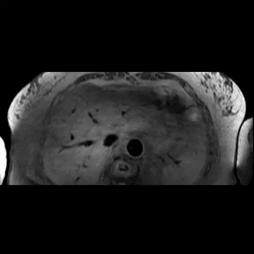
10 Case 1: Female, 77y, histologically confirmed giant-cell arteritis 3D-BB T1 VISTA 3D-BB T1 VISTA CE PPU-gating + Navigator; Scan Time: 5~6 min

11 Case 1: Female, 77y, histologically confirmed giant-cell arteritis 3D-BB T1 VISTA 3D-BB T1 VISTA CE


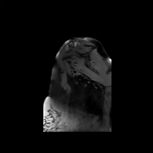
12 Case 2: Female, 27y with Takayasu Arteritis 3D-BB T1 VISTA 3D-BB T1 VISTA CE 18F-FDG PET/CT


13 Case 2: Follow-up after 4 weeks of intensive immunosuppressive therapy (Tocilizumab & high dose steroids) Before high-dose therapy 3D-BB T1 T1 CE 3D-BB After high-dose therapy CE 3D-BB T1


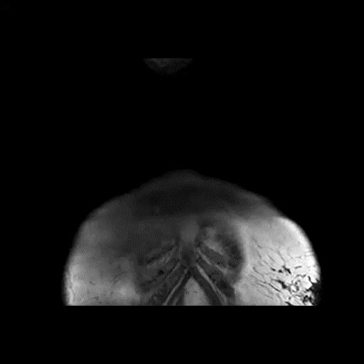
14 Case 3: 36y, female with Takayasu-Aortitis 3D-BB T1 VISTA CE 3D-BB T1 VISTA
15 RSNA 2014 Conclusion Navigated, PPU-triggered T1w-3D VISTA black-blood MRI of the thoracic vessels is feasible in 5 to 6 minutes scan time Image quality is good to excellent in >95% of exams T1w-3D VISTA allows to visualize concentric wall thickening and contrast enhancement in patients with vasculitis Future studies are necessary to evaluate T1w-3D VISTA for monitoring of anti-inflammatory therapies and for a direct comparison with PET/CT and ultrasound Black-Blood MRI could be particularly useful in young patients (e.g. Takayasu arteritis) in which ionizing radiation should be used with caution
16 Plaque Imaging Group Institut für Klinische Radiologie Prof. Dr. med. Dr. h.c. M. Reiser PD Dr. med. T. Saam PD Dr. med. C. Cyran Dr. med. F. Schwarz PD Dr. med. A. Helck Dr. med. H. Hetterich Dr. med. F. Strobl Dr. rer. nat. O. Dietrich Dr. rer. nat. M. Ingrisch Cand. med. S. Fill Cand. med. C. Habbel Cand. med. S. Hörterer Cand. med. T. Obenhuber Cand. med. S. Maurus Cand. med. N. Webber Prof. Dr. med. Dr. h.c. D. Clevert Dr. med. E. Coppenrath Dr. med. K. M. Treitl Dr. med. N. Kammer Interdisziplinäres Zentrum für Schlaganfall- und Demenzforschung Prof. Dr. M. Dichgans (Co-PI CAPIAS) Dr. med. A. Beyer-Karpinska Neurologische Klinik und Poliklinik Prof. Dr. M. Dieterich Neuroradiologie Prof. Dr. H. Brückmann Dr. med. N. Lummel Klinik und Poliklinik für Nuklearmedizin Prof. Dr. P. Bartenstein PD Dr. med. A. Rominger Vascular Imaging Lab, Seattle, USA Prof. Dr. rer. nat. C. Yuan Prof. Dr. med. T. Hatsukami Angiologie Prof. Dr. med. U. Hoffmann Dr. med. M. Czihal Technische Universität München (Physik E17) Prof. Dr. rer. nat. F. Pfeiffer Dr. rer. nat. J. Herzen Marian Willner Marco Stockmar Klinikum Rechts der Isar (Neurologie) PD Dr. med. H. Poppert Dr. med. D. Sepp Klinikum Rechts der Isar (Neuroradiologie) Dr. med. T. Boeck-Behrens Universitätsklinikum Freiburg (Neurlogie) PD Dr. med. A. Harloff Support - MAP Dr. rer. nat. S. Auweter Dr. rer. nat. T. Hendel Visiting Fellows: Dr. med. M. de Trelles
The risk of MR-detected carotid plaque hemorrhage on recurrent or first-time stroke: a meta-analysis of individual patient data
 The risk of MR-detected carotid plaque hemorrhage on recurrent or first-time stroke: a meta-analysis of individual patient data Schindler A 1, Bonati LH 2, Schinner R 1, Altaf N 3, Hosseini AA 3, Esposito-Bauer
MR Advance Techniques. Vascular Imaging. Class II
 MR Advance Techniques Vascular Imaging Class II 1 Vascular Imaging There are several methods that can be used to evaluate the cardiovascular systems with the use of MRI. MRI will aloud to evaluate morphology
In vivo diffusion tensor imaging (DTI) of articular cartilage as a biomarker for osteoarthritis
 of articular cartilage as a biomarker for osteoarthritis") In vivo diffusion tensor imaging (DTI) of articular cartilage as a biomarker for osteoarthritis Jose G. Raya 1, Annie Horng 2, Olaf Dietrich 2, Svetlana Krasnokutsky 3, Luis S. Beltran 1, Maximilian F.
In vivo diffusion tensor imaging (DTI) of articular cartilage as a biomarker for osteoarthritis Jose G. Raya 1, Annie Horng 2, Olaf Dietrich 2, Svetlana Krasnokutsky 3, Luis S. Beltran 1, Maximilian F.
Aortic Vessel Wall Imaging Using 3D Phase Sensitive Inversion Recovery in Children and Young Adults
 Aortic Vessel Wall Imaging Using 3D Phase Sensitive Inversion Recovery in Children and Young Adults Animesh Tandon, MD, MS 1,2, Tarique Hussain, MD, PhD 1,2, Andrew Tran, MD, MS 3, René M Botnar, PhD 4,
Assessment of wall shear stress in patients without aortic disease and with aortic dissection using velocity encoding 4D MRI
 Assessment of wall shear stress in patients without aortic disease and with aortic dissection using velocity encoding 4D MRI Poster No.: C-0841 Congress: ECR 2015 Type: Scientific Exhibit Authors: J. P.
F-Fludeoxyglucose Positron Emission Tomography (FDG-PET) in Patients with Large Vessel Vasculitis (LVV)
 in Patients with Large Vessel Vasculitis (LVV)") Assessment of Treatment Response by 18 F-Fludeoxyglucose Positron Emission Tomography (FDG-PET) in Patients with Large Vessel Vasculitis (LVV) Shubhasree Banerjee 1, Sara Alehashemi 1, Mark A. Ahlman 2,
Assessment of Treatment Response by 18 F-Fludeoxyglucose Positron Emission Tomography (FDG-PET) in Patients with Large Vessel Vasculitis (LVV) Shubhasree Banerjee 1, Sara Alehashemi 1, Mark A. Ahlman 2,
Oak foundation for donating the 3T Siemens Verio scanner. Board of directors BBH and Frh Hospitals for supporting the
 Knee pain and inflammation in the infrapatellar fat pad estimated by conventional and dynamic contrast-enhanced magnetic resonance imaging in obese patients with osteoarthritis: a crosssectional study
Thrombectomy with the preset stent-retriever. Insights from the ARTESp* trial
 Thrombectomy with the preset stent-retriever Insights from the ARTESp* trial Wiebke Kurre, MD Klinikum Stuttgart - Germany * Acute Recanalization of Thrombo-Embolic Ischemic Stroke with preset (ARTESp)
Feasibility of a New C-arm angiography (DYNA-CT) based three-dimensional algorithm in combination with myocardial perfusion assessment
 based three-dimensional algorithm in combination with myocardial perfusion assessment") Feasibility of a New C-arm angiography (DYNA-CT) based three-dimensional algorithm in combination with myocardial perfusion assessment H. Rittger*, A.M.Sinha, J. Rieber±, G. Lauritsch+, J. Brachmann *Universitätsklinik
Feasibility of a New C-arm angiography (DYNA-CT) based three-dimensional algorithm in combination with myocardial perfusion assessment H. Rittger*, A.M.Sinha, J. Rieber±, G. Lauritsch+, J. Brachmann *Universitätsklinik
Non Contrast MRA. Mayil Krishnam. Director, Cardiovascular and Thoracic Imaging University of California, Irvine
 Non Contrast MRA Mayil Krishnam Director, Cardiovascular and Thoracic Imaging University of California, Irvine No disclosures Non contrast MRA-Why? Limitations of CTA Radiation exposure Iodinated contrast
Magnetic Resonance Angiography
 Magnetic Resonance Angiography 1 Magnetic Resonance Angiography exploits flow enhancement of GR sequences saturation of venous flow allows arterial visualization saturation of arterial flow allows venous
MR Tumor Staging for Treatment Decision in Case of Wilms Tumor
 MR Tumor Staging for Treatment Decision in Case of Wilms Tumor G. Schneider, M.D., Ph.D.; P. Fries, M.D. Dept. of Diagnostic and Interventional Radiology, Saarland University Hospital, Homburg/Saar, Germany
High-Resolution MR Angiography: Results in Diseased Arteries
 IAGS Proceedings NEW IMAGING FOR NEW AND OLD DISEASES High-Resolution MR Angiography: Results in Diseased Arteries Peter Gonschior, M D, Ingo Pragst, M D, Gregor Valassis, M D, Claudia Vo g e l - Wiens,
Essentials of Clinical MR, 2 nd edition. 99. MRA Principles and Carotid MRA
 99. MRA Principles and Carotid MRA As described in Chapter 12, time of flight (TOF) magnetic resonance angiography (MRA) is commonly utilized in the evaluation of the circle of Willis. TOF MRA allows depiction
Applications of PET/CT in Rheumatology. Role of PET/CT. Annibale Versari, MD Nuclear Medicine PET Center S.Maria Nuova Hospital Reggio Emilia Italy
 CME Session Associazione Italiana di Medicina Nucleare ed Imaging Molecolare Applications of PET/CT in Rheumatology Role of PET/CT Annibale Versari, MD Nuclear Medicine PET Center S.Maria Nuova Hospital
CAN WE REPLACE TEMPORAL ARTERY BIOPSY WITH CRANIAL ULTRASOUND FOR THE DIAGNOSIS OF GIANT CELL ARTERITIS?
 CAN WE REPLACE TEMPORAL ARTERY BIOPSY WITH CRANIAL ULTRASOUND FOR THE DIAGNOSIS OF GIANT CELL ARTERITIS? Adam P. Croft (ST3 Rheumatology) Susan Mollan, Paresh Jobunputra Speaker has no disclosures TAB
6/23/2009. Inversion Recovery (IR) Techniques and Applications. Variations of IR Technique. STIR, FLAIR, TI and TI Null. Applications of IR
 Techniques and Applications. Variations of IR Technique. STIR, FLAIR, TI and TI Null. Applications of IR") The Anatomy of Basic R Pulse Sequences Inversion Recovery () Techniques and Applications Chen Lin, PhD Indiana University School of edicine & Clarian Health Partners agnetization Preparation Section Chemical
The Anatomy of Basic R Pulse Sequences Inversion Recovery () Techniques and Applications Chen Lin, PhD Indiana University School of edicine & Clarian Health Partners agnetization Preparation Section Chemical
Raja Muthupillai, PhD. Department of Diagnostic and Interventional Radiology St. Luke s Episcopal Hospital. Research Support: Philips Healthcare
 3D Cardiac Imaging Raja Muthupillai, PhD Department of Diagnostic and Interventional Radiology St. Luke s Episcopal Hospital Houston, TX Disclosures Research Support: Philips Healthcare This presentation
Case Presentation VASCULITIS. Case Presentation. Case Presentation. Vasculitis
 Case Presentation VASCULITIS The patient is a 24 year old woman who presented to the emergency room with left-sided weakness. She was confused and complained of a severe headache. She was noted to have
VASCULITIS. Case Presentation. Case Presentation
 VASCULITIS Case Presentation The patient is a 24 year old woman who presented to the emergency room with left-sided weakness. She was confused and complained of a severe headache. She was noted to have
GIANT CELL ARTERITIS. Page 1 of 6 Reproduction of this material requires written permission of the Vasculitis Foundation. Copyright 2018.
 What is giant cell arteritis (GCA)? Giant cell arteritis (GCA) is a form of vasculitis a family of rare disorders characterized by inflammation of the blood vessels, which can restrict blood flow and damage
UNFOLDING NATURE S ORIGAMI: MEDICAL TREATMENT OF TAKAYASU ARTERITIS AND GIANT CELL ARTERITIS
 UNFOLDING NATURE S ORIGAMI: MEDICAL TREATMENT OF TAKAYASU ARTERITIS AND GIANT CELL ARTERITIS CanVasc meeting Montreal Nov 22 2012 Patrick Liang Service de rhumatologie Centre Hospitalier Universitaire
Takayasu s arteritis. Justin Mason. Professor of Vascular Rheumatology Imperial College London Hammersmith Hospital
 Takayasu s arteritis Justin Mason Professor of Vascular Rheumatology Imperial College London Hammersmith Hospital I have nothing to disclose. Takayasu s clinical features Early phase non-specific features
Contrast-enhanced Breast MRI RSSA 2013
 Contrast-enhanced Breast MRI RSSA 2013 Prof. dr. Maurice van den Bosch University Medical Center Utrecht, the Netherlands Index 1) Breast cancer 2) Why MRI of the breast 3) Technique 4) Interpretation
Repeatability of 2D FISP MR Fingerprinting in the Brain at 1.5T and 3.0T
 Repeatability of 2D FISP MR Fingerprinting in the Brain at 1.5T and 3.0T Guido Buonincontri 1,2, Laura Biagi 1,3, Alessandra Retico 2, Michela Tosetti 1,3, Paolo Cecchi 4, Mirco Cosottini 1,4,5, Pedro
Functional Chest MRI in Children Hyun Woo Goo
 Functional Chest MRI in Children Hyun Woo Goo Department of Radiology and Research Institute of Radiology Asan Medical Center, University of Ulsan College of Medicine, Seoul, Korea No ionizing radiation
Preventing blindness: Ultrasound in Giant cell arteritis
 Preventing blindness: Ultrasound in Giant cell arteritis Elizabeth Jernberg, MD Associate Clinical Professor of Medicine Division of Rheumatology University of Washington Virginia Mason Medical Center
D. J. Margolis 1, S. Natarajan 2, D. Kumar 3, M. Macairan 4, R. Narayanan 3, and L. Marks 4
 Biopsy Tracking and MRI Fusion to Enhance Imaging of Cancer Within the Prostate D. J. Margolis 1, S. Natarajan 2, D. Kumar 3, M. Macairan 4, R. Narayanan 3, and L. Marks 4 1 Dept. of Radiology, UCLA, Los
Non-invasive continuous blood pressure monitoring based on radial artery tonometry (T-Line TL-200pro device) in the intensive care unit
 in the intensive care unit") Non-invasive continuous blood pressure monitoring based on radial artery tonometry (T-Line TL-200pro device) in the intensive care unit Bernd Saugel; Agnes S Meidert; Alexander Hapfelmeier; Florian Eyer;
Non-invasive continuous blood pressure monitoring based on radial artery tonometry (T-Line TL-200pro device) in the intensive care unit Bernd Saugel; Agnes S Meidert; Alexander Hapfelmeier; Florian Eyer;
1Pulse sequences for non CE MRA
 MRI: Principles and Applications, Friday, 8.30 9.20 am Pulse sequences for non CE MRA S. I. Gonçalves, PhD Radiology Department University Hospital Coimbra Autumn Semester, 2011 1 Magnetic resonance angiography
Clinical Applications
 C H A P T E R 16 Clinical Applications In selecting pulse sequences and measurement parameters for a specific application, MRI allows the user tremendous flexibility to produce variations in contrast between
Case Reports: Tumor Detection by Diffusion-Weighted MRI and ADC-Mapping with Correlation to PET/CT Results
 Case Reports: Tumor Detection by Diffusion-Weighted MRI and ADC-Mapping with Correlation to PET/CT Results Matthias Philipp Lichy, M.D.; Philip Aschoff, M.D.; Christina Pfannenberg, M.D.; Schlemmer Heinz-Peter,
MRI Abdomen Protocol Pancreas/MRCP with Contrast
 MRI Abdomen Protocol Pancreas/MRCP with Contrast Reviewed By: Brett Mollard, MD; Anna Ellermeier, MD Last Reviewed: July 2018 Contact: (866) 761-4200 Standard uses: 1. Characterization of cystic and solid
FieldStrength. Tips for abdomen/pelvis oncology imaging. SPECIAL ISSUE MR in oncology. Publication for the Philips MRI Community
 FieldStrength Publication for the Philips MRI Community Issue 41 September 2010 Tips for abdomen/pelvis oncology imaging SPECIAL ISSUE MR in oncology This article is part of Field Strength issue 41, September
NON-ATHEROSCLEROTIC PATHOLOGY OF THE CAROTID ARTERIES
 NON-ATHEROSCLEROTIC PATHOLOGY OF THE CAROTID ARTERIES Leslie M. Scoutt, MD, FACR Professor of Diagnostic Radiology & Surgery Vice Chair, Dept of Radiology & Biomedical Imaging Chief, Ultrasound Section
Liver Fat Quantification
 Liver Fat Quantification Jie Deng, PhD, DABMP Department of Medical Imaging May 18 th, 2017 Disclosure Research agreement with Siemens Medical Solutions 2 Background Non-alcoholic fatty liver diseases
Using Radial k-space Sampling and Steady-State Free Precession Imaging
 MRI of Coronary Vessel Walls Cardiac Imaging Original Research A C D E M N E U T R Y L I A M C A I G O F I N G Marcus Katoh 1 Elmar Spuentrup 1 Arno Buecker 1 Tobias Schaeffter 2 Matthias Stuber 3 Rolf
Takayasu s Arteritis: A Case Report With Global Arterial Involvement
 1 Case Report Takayasu s Arteritis: A Case Report With Global Arterial Involvement Waqas Ahmed, Zeeshan Ahmad* From Shifa International Hospital H-8/4, Islamabad, Pakistan Correspondence: Dr Waqas Ahmed,
MRI enters Emergency Department for fast, confident decisions
 MRI enters Emergency Department for fast, confident decisions FieldStrength MRI magazine User experiences - November 2016 www.philips.com/fieldstrength When MRI is the preferred choice, ED patients can
F-2-fluoro-2-deoxyglucose uptake in or adjacent to blood vessel walls
 15 REVIEW 18 F-2-fluoro-2-deoxyglucose uptake in or adjacent to blood vessel walls Yoichi Otomi 1, Hideki Otsuka 2, Kaori Terazawa 1, Hayato Nose 1,3, Michiko Kubo 1, Kazuhide Yoneda 1, Kaoru Kitsukawa
Tips and Tricks of State of the art MRA
 Tips and Tricks of State of the art MRA Mayil Krishnam, MD,MBA, MRCP,FRCR(UK) Professor of Radiology Director, Cardiovascular and Thoracic Imaging University of California, Irvine Objectives Technical
UK Biobank. Imaging modality Cardiovascular Magnetic Resonance (CMR) Version th Oct 2015
 Version th Oct 2015") Imaging modality Cardiovascular Magnetic Resonance (CMR) Version 1.0 http://www.ukbiobank.ac.uk/ 30 th Oct 2015 This document details the procedure for the CMR scan performed at an Imaging assessment centre
Imaging modality Cardiovascular Magnetic Resonance (CMR) Version 1.0 http://www.ukbiobank.ac.uk/ 30 th Oct 2015 This document details the procedure for the CMR scan performed at an Imaging assessment centre
Vasculitides in Surgical Neuropathology Practice
 Vasculitides in Surgical Neuropathology Practice USCAP requires that all faculty in a position to influence or control the content of CME disclose any relevant financial relationship WITH COMMERCIAL INTERESTS
Vasculitis local: systemic
 Vasculitis Inflammation of the vessel wall. Signs and symptoms: 1- local: according to the involved tissue 2- systemic:(fever, myalgia, arthralgias, and malaise) Pathogenesis 1- immune-mediated inflammation
Automatic Ascending Aorta Detection in CTA Datasets
 Automatic Ascending Aorta Detection in CTA Datasets Stefan C. Saur 1, Caroline Kühnel 2, Tobias Boskamp 2, Gábor Székely 1, Philippe Cattin 1,3 1 Computer Vision Laboratory, ETH Zurich, 8092 Zurich, Switzerland
How I do it: Non Contrast-Enhanced MR Angiography (syngo NATIVE)
") Clinical How-I-do-it Cardiovascular How I do it: Non Contrast-Enhanced MR Angiography (syngo NATIVE) Manuela Rick, Nina Kaarmann, Peter Weale, Peter Schmitt Siemens Healthcare, Erlangen, Germany Introduction
Clinical How-I-do-it Cardiovascular How I do it: Non Contrast-Enhanced MR Angiography (syngo NATIVE) Manuela Rick, Nina Kaarmann, Peter Weale, Peter Schmitt Siemens Healthcare, Erlangen, Germany Introduction
Vasculitis local: systemic
 Vasculitis Inflammation of the vessel wall. Signs and symptoms: 1- local: according to the involved tissue 2- systemic:(fever, myalgia, arthralgias, and malaise) Pathogenesis 1- immune-mediated 2- infectious
Original Report. Delayed Contrast-Enhanced MRI of the Aortic Wall in Takayasu s Arteritis: Initial Experience
 MR Imaging Desai et al. MRI of Takayasu s rteritis Milind Y. Desai 1 John H. Stone 2 Thomas K. F. Foo 3 David. Hellmann João. C. Lima David. luemke 4 Desai MY, Stone JH, Foo T, Hellmann D, Lima JC, luemki
Improved Noninvasive Assessment of Coronary Artery Bypass Grafts With 64-Slice Computed Tomographic Angiography in an Unselected Patient Population
 Journal of the American College of Cardiology Vol. 49, No. 9, 2007 2007 by the American College of Cardiology Foundation ISSN 0735-1097/07/$32.00 Published by Elsevier Inc. doi:10.1016/j.jacc.2006.10.066
An evaluation of a superfast MRI sequence in the diagnosis of suspected acute appendicitis
 Brief Report An evaluation of a superfast MRI sequence in the diagnosis of suspected acute appendicitis Bin Zhu 1, Bing Zhang 1, Ming Li 1, Shifu Xi 2, Decai Yu 2, Yitao Ding 2 1 Department of Radiology,
Bone PET/MRI : Diagnostic yield in bone metastases and malignant primitive bone tumors
 Bone PET/MRI : Diagnostic yield in bone metastases and malignant primitive bone tumors Lars Stegger, Benjamin Noto Department of Nuclear Medicine University Hospital Münster, Germany Content From PET to
Supplementary Online Content
 Supplementary Online Content Gregg NM, Kim AE, Gurol ME, et al. Incidental cerebral microbleeds and cerebral blood flow in elderly individuals. JAMA Neurol. Published online July 13, 2015. doi:10.1001/jamaneurol.2015.1359.
The Most Thorough Health Checks
 Klinikum rechts der Isar Technische Universität München Zentrum für Prävention und Sportmedizin The Most Thorough Health Checks Comprehensive Prevention from a Single Source Our highly qualified and motivated
Fulfilling the Promise
 Fulfilling the Promise of Cardiac MR Non-contrast, free-breathing technique generates comprehensive evaluation of the coronary arteries By Maggie Fung, MR Cardiovascular Clinical Development Manager; Wei
Photon Attenuation Correction in Misregistered Cardiac PET/CT
 Photon Attenuation Correction in Misregistered Cardiac PET/CT A. Martinez-Möller 1,2, N. Navab 2, M. Schwaiger 1, S. G. Nekolla 1 1 Nuklearmedizinische Klinik der TU München 2 Computer Assisted Medical
MRI protocol for post-repaired TOF
 2012 NASCI MRI protocol for post-repaired TOF Taylor Chung, M.D. Associate Director, Body and Cardiovascular Imaging Department of Diagnostic Imaging Children s Hospital & Research Center Oakland Oakland,
Advantages of the Spin Echo-type Radial Scan of the Cranial Region
 Advantages of the Spin Echo-type Radial Scan of the Cranial Region Poster No.: C-0969 Congress: ECR 2013 Type: Scientific Exhibit Authors: T. Gomi 1, M. Hasegawa 2, N. Murata 2, A. Tabata 2, M. Tsunoo
Why Talk About Technique? MRI of the Knee:
 Why Talk About Technique? MRI of the Knee: Part 1 - Imaging Techniques Mark Anderson, M.D. University of Virginia Health Sciences Center Charlottesville, Virginia Always had an interest teach our fellows
Supplementary information Detailed Materials and Methods
 Supplementary information Detailed Materials and Methods Subjects The experiment included twelve subjects: ten sighted subjects and two blind. Five of the ten sighted subjects were expert users of a visual-to-auditory
Subclavian artery Stenting
 Subclavian artery Stenting Etiology Atherosclerosis Takayasu s arteritis Fibromuscular dysplasia Giant Cell Arteritis Radiation-induced Vascular Injury Thoracic Outlet Syndrome Neurofibromatosis Incidence
Neuroradiology MR Protocols
 Neuroradiology MR Protocols Brain protocols N 1: Brain MRI without contrast N 2: Pre- and post-contrast brain MRI N 3 is deleted N 4: Brain MRI without or pre-/post-contrast (seizure protocol) N 5: Pre-
Takayasu disease Is it still a room for intervention? NO YES. BUT
 Takayasu disease Is it still a room for intervention? NO YES. BUT Z. Tazi Mezalek Internal Medicine Department Mohammed V University Ibn Sina Hospital - Rabat - Maroc Disclosure Speaker name: TAZI MEZALEK
Year In Review: VasculitisPers. Disclosures. Learning Objectives. none 4/16/2018. Describe new medications for the treatment of vasculitis
 Year In Review: VasculitisPers Cailin Sibley, M.D., M.H.S. Director, Vasculitis Clinic April 27 th, 2018 NTEREST DISCLOSURE Disclosures none Learning Objectives Describe new medications for the treatment
MR Advance Techniques. Cardiac Imaging. Class IV
 MR Advance Techniques Cardiac Imaging Class IV Heart The heart is a muscular organ responsible for pumping blood through the blood vessels by repeated, rhythmic contractions. Layers of the heart Endocardium
CARDIAC MRI. Cardiovascular Disease. Cardiovascular Disease. Cardiovascular Disease. Overview
 CARDIAC MRI Dr Yang Faridah A. Aziz Department of Biomedical Imaging University of Malaya Medical Centre Cardiovascular Disease Diseases of the circulatory system, also called cardiovascular disease (CVD),
FieldStrength. Achieva 3.0T enables cutting-edge applications, best-in-class MSK images
 FieldStrength Publication for the Philips MRI Community Issue 33 December 2007 Achieva 3.0T enables cutting-edge applications, best-in-class MSK images Palo Alto Medical Clinic Sports Medicine Center employs
Matthias Stuber, PhD Associate Professor Division of MRI Research Johns Hopkins University Baltimore, MD
 Coronary Magnetic Resonance Imaging Matthias Stuber, PhD Associate Professor Division of MRI Research Johns Hopkins University Baltimore, MD The Need for MRI Background X-ray coronary angiograpy (gold
Magnetic Resonance Imaging. Basics of MRI in practice. Generation of MR signal. Generation of MR signal. Spin echo imaging. Generation of MR signal
 Magnetic Resonance Imaging Protons aligned with B0 magnetic filed Longitudinal magnetization - T1 relaxation Transverse magnetization - T2 relaxation Signal measured in the transverse plane Basics of MRI
Ultrasound. Computed tomography. Case studies. Utility of IQon Spectral CT in. cardiac imaging
 Ultrasound Computed tomography Case studies Utility of IQon Spectral CT in cardiac imaging Cardiac imaging is a challenging procedure where it is necessary to image a motion-free heart. This requires a
Speed, Comfort and Quality with NeuroDrive
 Speed, Comfort and Quality with NeuroDrive Echelon Oval provides a broad range of capabilities supporting fast, accurate diagnosis of brain conditions and injuries. From anatomical depiction to vascular
Previous talks. Clinical applications for spiral flow imaging. Clinical applications. Clinical applications. Coronary flow: Motivation
 for spiral flow imaging Joao L. A. Carvalho Previous talks Non-Cartesian reconstruction (2005) Spiral FVE (Spring 2006) Aortic flow Carotid flow Accelerated spiral FVE (Fall 2006) 2007? Department of Electrical
Go With The Flow: Role of 4D Flow Imaging
 4D Flow Go With The Flow: Role of 4D Flow Imaging Niti R. Aggarwal, MD Associate Director of Cardiac MRI Assistant Professor of Medicine & Radiology University of Wisconsin Madison Disclosures GE Healthcare
Fast and easy diagnostic imaging from head to toe
 Publication for the Philips MRI Community ISSUE 50 2014 / 1 Fast and easy diagnostic imaging from head to toe Ingenia 1.5T with dstream provides speed and convenience, IntelliSpace Portal provides flexibility
Atlas of the Vasculitic Syndromes
 CHAPTER e40 Atlas of the Vasculitic Syndromes Carol A. Langford Anthony S. Fauci Diagnosis of the vasculitic syndromes is usually based upon characteristic histologic or arteriographic findings in a patient
Vessel Wall Imaging of Intracranial Arterial Disease Commercial Interests
 Vessel Wall Imaging of Intracranial Arterial Disease Commercial Interests Disclosures No relevant commercial interests Off Label / Investigational Use No off label / investigational use Daniel Mandell,
Supplementary Online Content
 Supplementary Online Content Hooshmand B, Magialasche F, Kalpouzos G, et al. Association of vitamin B, folate, and sulfur amino acids with brain magnetic resonance imaging measures in older adults: a longitudinal
TAKAYASU S ARTERITIS. Second-stage symptoms include:
 What is Takayasu s arteritis (TAK)? Takayasu s arteritis (TAK) is a form of vasculitis a family of rare disorders characterized by inflammation of the blood vessels, which can restrict blood flow and damage
Cortical hypoperfusion in Parkinson's disease assessed with arterial spin labeling MRI
 Cortical hypoperfusion in Parkinson's disease assessed with arterial spin labeling MRI Poster No.: C-0609 Congress: ECR 2013 Type: Scientific Exhibit Authors: S. Aoki, K. Kamagata, Y. Motoi, K. Kamiya,
ROLE OF CONTRAST ENHANCED MR ANGIOGRAPHY IN AORTIC COARCTATION
 ROLE OF CONTRAST ENHANCED MR ANGIOGRAPHY IN AORTIC COARCTATION By Adel El Badrawy, Ahmed Abdel Razek, Nermin Soliman, Hala El Marsafawy *, Sameh Amer** From Radiodiagnosis, Pediatric Cardiology* & Cardiothoracic
Orthopedic Hardware Imaging Part II: MRI v. Metal
 Orthopedic Hardware Imaging Trent Roth, MD And Lauren Ladd, MD Indiana University School of Medicine IU Health Physicians-Radiology Recap: Imaging Techniques Radiography Standard for initial and surveillance
MultiTransmit 4D brings robustness to 3.0T cardiac imaging
 Publication for the Philips MRI Community Issue 44 SUMMER 2011 MultiTransmit 4D brings robustness to 3.0T cardiac imaging Bonn clinicians see a reduction in artifacts and inhomogeneities as a significant
Vasculitis Prof. Dr. med. Katharina Glatz Pathologie
 Vasculitis 08-21-2018 Prof. Dr. med. Katharina Glatz Pathologie Agenda Anatomy and histology Vasculitis: Chapel Hill Classification Examples Giant cell arteritis Single organ vasculitis Artery or Vein?
Cover Page. The handle holds various files of this Leiden University dissertation.
 Cover Page The handle http://hdl.handle.net/1887/35124 holds various files of this Leiden University dissertation. Author: Wokke, Beatrijs Henriette Aleid Title: Muscle MRI in Duchenne and Becker muscular
KNEE ALIGNMENT SYSTEM (KAS) MRI Protocol
 MRI Protocol") KNEE ALIGNMENT SYSTEM (KAS) MRI Protocol Sample referral sticker Referral Sticker Insert here Corin 17 Bridge Street Pymble NSW Australia 2073 P: +61 (0)2 9497 7400 F: +61 (0)2 9497 7498 E: KAS.customerservice@coringroup.com
KNEE ALIGNMENT SYSTEM (KAS) MRI Protocol Sample referral sticker Referral Sticker Insert here Corin 17 Bridge Street Pymble NSW Australia 2073 P: +61 (0)2 9497 7400 F: +61 (0)2 9497 7498 E: KAS.customerservice@coringroup.com
High spatial resolution reveals excellent detail in pediatric neuro imaging
 Publication for the Philips MRI Community Issue 46 2012/2 High spatial resolution reveals excellent detail in pediatric neuro imaging Achieva 3.0T with 32-channel SENSE Head coil has become the system
MR coronary artery imaging with 3D motion adapted gating (MAG) in comparison to a standard prospective navigator technique
 in comparison to a standard prospective navigator technique") Journal of Cardiovascular Magnetic Resonance (2005) 7, 793 797 Copyright D 2005 Taylor & Francis Inc. ISSN: 1097-6647 print / 1532-429X online DOI: 10.1080/10976640500287547 ANGIOGRAPHY MR coronary artery
Journal of Cardiovascular Magnetic Resonance (2005) 7, 793 797 Copyright D 2005 Taylor & Francis Inc. ISSN: 1097-6647 print / 1532-429X online DOI: 10.1080/10976640500287547 ANGIOGRAPHY MR coronary artery
An excellent fit to expand the imaging center, Prodiva 1.5T
 An excellent fit to expand the imaging center, Prodiva 1.5T FieldStrength MRI magazine User experiences - December 2017 www.philips.com/fieldstrength High quality MR imaging combined with a simplified,
Quantitative Imaging of Transmural Vasa Vasorum Distribution in Aortas of ApoE -/- /LDL -/- Double Knockout Mice using Nano-CT
 Quantitative Imaging of Transmural Vasa Vasorum Distribution in Aortas of ApoE -/- /LDL -/- Double Knockout Mice using Nano-CT M. Kampschulte 1, M.D.; A. Brinkmann 1, M.D.; P. Stieger 4, M.D.; D.G. Sedding
Non-Contrast MRA. How and When 1996! Why Non-Contrast MRA? Angiography: What are our goals? Inflow Techniques Differences in excitation hx
 A major teaching hospital of Harvard Medical School Angiography: What are our goals? Non-Contrast MRA: How and When Neil M. Rofsky, M.D. Professor of Radiology, Harvard Medical School Director of MRI &
Case Report: Knee MR Imaging of Haemarthrosis in a Case of Haemophilia A
 Clinical > Pediatric Imaging Case Report: Knee MR Imaging of Haemarthrosis in a Case of Haemophilia A M. A. Weber, J. K. Kloth University Hospital Heidelberg, Department of Diagnostic and Interventional
Liver imaging takes a step forward with Ingenia
 Publication for the Philips MRI Community ISSUE 49 2013 / 2 Liver imaging takes a step forward with Ingenia Lyon South Hospital strives to move from several studies first CT, then MR or PET to using just
Comparison of Image quality in temporal bone MRI at 3T using 2D selective RF excitation versus a routine SPACE sequence
 Comparison of Image quality in temporal bone MRI at 3T using 2D selective RF excitation versus a routine SPACE sequence Poster No.: C-1065 Congress: ECR 2015 Type: Authors: Keywords: DOI: Scientific Exhibit
AORTIC ANEURYSM. howmed.net
 AORTIC ANEURYSM howmed.net ANATOMY It is important to understand the anatomy of the aorta Need to know the extent of the aneurysm Need to know the vessels involved This helps with Medical or Surgical management
Title:Giant cell arteritis exclusively detected by 18F-fluorodeoxyglucose positron emission tomography: a case report
 Author's response to reviews Title:Giant cell arteritis exclusively detected by 18F-fluorodeoxyglucose positron emission tomography: a case report Authors: Markus Brückner (markus.brueckner@ukmuenster.de)
The Effects of Music intervention on Functional connectivity. Supplemental Information
 Yang et al. 0 The Effects of Music intervention on Functional connectivity strength of Brain in Schizophrenia Supplemental Information Mi Yang,#, Hui He #, Mingjun Duan,, Xi Chen, Xin Chang, Yongxiu Lai,
The Use and Pitfalls of Intracranial Vessel Wall Imaging: How We Do It 1
 This copy is for personal use only. To order printed copies, contact reprints@rsna.org Reviews and Commentary n How I Do It Arjen Lindenholz, MD Anja G. van der Kolk, MD, PhD Jaco J. M. Zwanenburg, PhD
High Field MR of the Spine
 Department of Radiology University of California San Diego 3T for MR Applications Advantages High Field MR of the Spine Increased signal-to-noise Better fat suppression Increased enhancement with gadolinium
High-Resolution MRI in Giant Cell Arteritis: Imaging of the Wall of the Superficial Temporal Artery
 Vascular Imaging Bley et al. MRI of Vessel Wall in Giant Cell Arteritis Thorsten A. Bley 1 Oliver Wieben 2 Markus Uhl 1 Jens Thiel 3 Dieter Schmidt 4 Mathias Langer 1 Bley TA, Wieben O, Uhl M, Thiel J,
MR imaging of the knee in marathon runners before and after competition
 Skeletal Radiol (2001) 30:72 76 International Skeletal Society 2001 ARTICLE W. Krampla R. Mayrhofer J. Malcher K.H. Kristen M. Urban W. Hruby MR imaging of the knee in marathon runners before and after
Chest X-ray Interpretation
 Chest X-ray Interpretation Introduction Routinely obtained Pulmonary specialist consultation Inherent physical exam limitations Chest x-ray limitations Physical exam and chest x-ray provide compliment