History and Science of CO 2 Digital Subtraction Angiography
|
|
|
- Marlene Cameron
- 6 years ago
- Views:
Transcription
1 History and Science of CO 2 Digital Subtraction Angiography Kyung Cho, M.D. University of Michigan
2 The First Use of CO 2 as a Nonvascular Contrast Agent 1920s Carelli and Sordelli of Buenos Aires performed retroperitoneal pneumography with CO 2 for visualization of the kidneys and adrenal masses Presacral insufflation with CO 2 has replaced air or oxygen insufflation, thus eliminating the hazard of air emboli Carelli HH and Sordelli E. Un Nuevo procedimento para explorar el ninon Rev Assoc Med Argent 34: , 1921

3 CO 2 Angiocardiogram 1950s to 1970s Antecubital vein injection of 100 cc of CO 2 with pt in LLD CO 2 Radiolucent gas trapped in right atrium Opaque band above gas represents pericardial space Normal band thickness is 3 mm

4 CO 2 Capnocavography CO 2 inferior venacavography with the intravenous injection of 80 cc of CO 2 CO 2 Radiology 92:606,1969


5 Pioneer of CO 2 Angiography Dick Hawkins, M.D CO 2 Aortogram

6 Celiac Arteriogram with Air Inadvertent room air injection (70 cc) in celiac artery with good visualization of major arteries AJR 139:19,1982

7 Renal CO 2 Arteriogram AJR139:19,1982 CO 2 arteriogram obtained in 1971 with 20 cc of CO 2.

8 Renal CO 2 DSA CO 2 aortogram in severely hypertensive man. Aortic injection of 35 cc/sec for a total of 75 cc. Digital subtraction imaging AJR 139:19, 1982


9 The Evolution of CO 2 Delivery Techniques Hand held Syringe Standard Angio Injector CO 2 Cylinder (animals only) different dedicated models


10 A B (A) Early model hand-held CO 2 injector. (B) Angiojet computerized CO 2 injector (Angiodynamics)

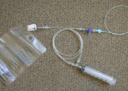
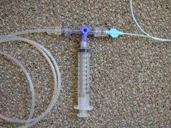
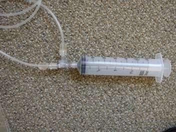
11 The plastic bag CO 2 delivery system AJR 176:229, 2001

12 AngioFlush Bag System AngioDynamics (discontinued)
13 MERITMEDICAL Custom Waste Bag and Contrast Delivery Set

14 CO 2 mmander and AngiAssist Delivery system
15 Science of CO 2 Angiography

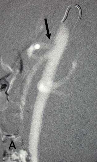
16 Unique CO 2 Properties Negative contrast High solubility Low viscosity Buoyancy Compressibility
17 Properties of CO 2, O 2, N 2, Iodine and Gd CO2 O2 N2 Molecular wt solubility Atomic Number CO 2 C = 6, O =8 Iodine 53 Gd 64


18 Comparative Radiopacity of Saline, Iodine, Gd and CO 2 Saline Omnipaque 240 Gd CO 2 in arm vein

19 CO 2 Angioscopy CO 2 angioscopy shows origin of right renal artery Total displacement of blood without arterial flow from branches

20 CO 2 Cholangiogram CO 2 injection in bile duct fills intra-and extrahepatic ducts Peripheral displacement of contrast medium

21 CVIR;29:637,2006 Air Volume Percent Changes over Time in 20 cc CO 2 -filled Syringes Air Volume Percent of Syringe cc Upright 20 cc Inverted 20 cc Horizontal Time (min.)

22 The Solubility of CO 2 5 cc of CO 2 injected into IVC in L lateral decubitus Cross-table lateral DSA of CO 2 trapped in right atrium CO 2 Complete gas absorption in 45 sec

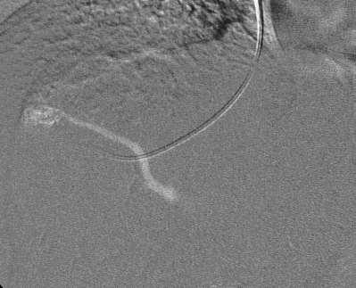
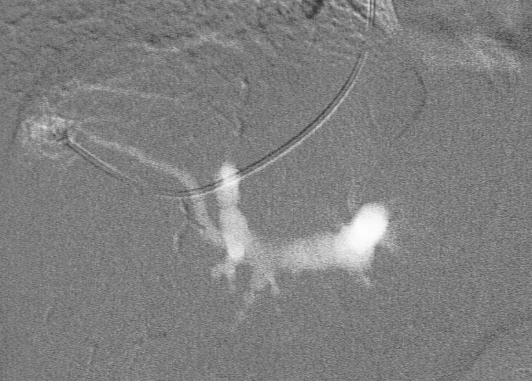
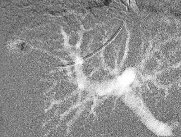
23 Aortoiliac Occlusive Disease: CO 2 DSA 3F dilator inserted into L external iliac artery (arrow) Injection of 30 cc of CO 2 in L external iliac artery CO 2 filling iliac, collaterals, inflow and outflow vessels
 Celiac DSA with contrast")
 CO 2 injection fills both celiac and SMA")
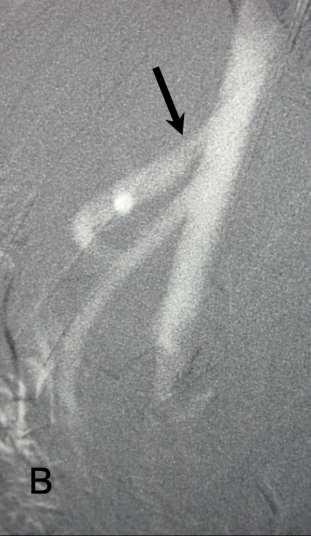
24 Celiac Stenosis: Median Arcuate Ligament Compression A B (A) Celiac DSA with contrast medium showing flow defect at origin of GDA. (B) CO 2 injection fills both celiac and SMA branches


25 CT with intra-aortic injection of CO 2 and contrast medium: CO 2 displaces blood whereas contrast is mixed with blood CO 2 Contrast


26 Buoyant CO 2 is trapped in AAA CO 2 A CO 2 aortogram X-table Lat

27 Lateral CO 2 Aortogram:Median Arcuate Ligament Compression EX INS X-table lateral

28 CO 2 Bubble Flow in a 9.5 mm Tube of a Pulsatile Flow Model 30 0 inclination CO2 Blood


29 CO 2 Dispersal from Different Catheter Design Endhole Halo Pigtail


30 CO 2 Filling of 15.9 mm Tube in a Pulsatile Flow Model: Supine vs elevation 65% CO 2 CO 2 84 %


31 Superior venacavogram with intravenous CO 2 injection Supine LLD: X-table Lat CO 2 in PA CO 2 in RA

32 Buoyant CO 2 injected into right hepatic vein filling anterior, middle and left hepatic veins

33 Buoyant, less viscous CO 2 injected into wedged right hepatic vein fills the portal, right, middle and left hepatic veins

34 Diagram of the hepatic microcirculation
35 CO2 compression during Wedged Hepatic Venogram

36 Buoyant, compressible CO 2 injected into celiac artery flows backwards, filling the aorta, superior mesenteric and renal arteries


37 CO 2 Renal DSA CO 2 Renal DSA Renal artery aneurysm CO 2 injection via microcatheter

38 Safety and Tolerance Study of CO 2 as a Venous Contrast Agent in Swine The hemodynamic and ventilatory responses to intracaval injections of ascending doses of CO 2

39 Polygraph tracing of blood pressure, pulmonary artery pressure and CVP following intracaval injection of CO 2 at 3.2 cc/kg in swine
40 Average percent changes in systemic blood pressure following intracaval injections of ascending doses of CO 2 in swine Supine 80 BP % Change in SBP CO 2 ( cc/kg) Time (min.)
41 Average percent changes of ETCO 2 following intracaval injections of ascending doses of CO 2 in swine Supine ET CO 2 CO2 (cc/kg) 0.0 % change in ET CO Time (min.)
42 Continuous Monitoring during CO 2 Angiography Pulse oximetry (oxygenation) ECG/HR (Circulation) BP (Circulation) Respirations (oxygenation) Capnography (Ventilation & Perfusion)

-a late sign of respiratory compromise in")
43 End-tidal CO 2 monitoring: Increasing Patient Safety during Procedural Sedation and CO 2 angiography EtCO 2 Detect early signs of hypoventilation Information on RR, depth, & apnea Desaturation (SpO 2 )-a late sign of respiratory compromise in hypoventilation.
44 Conclusions CO 2 has been used as a contrast agent in the nonvascular system since 1920s, in the venous system since 1950s and in the arterial system since 1970s. Intravenous CO 2 in doses of cc/kg caused no cardiopulmonary effects in swine. CO 2 is the only safe, proven contrast agent in patients with contrast allergy and renal failure.
45 A thorough knowledge of CO 2 properties, and facile catheterization and imaging techniques are essential in obtaining a successful CO 2 angiogram for the vascular diagnosis and intervention. Blood pressure monitoring and capnography provide the earliest sign of vapor lock in the pulmonary artery from the inadvertent injection of large volume of CO 2 or air.
46 CO 2 is useful as a contrast agent in various vascular diagnosis and interventions, including angioplasty, stenting, thrombolysis, embolization, filter placement, TIPS, and EVAR. Both the plastic bag system and CO 2 mmander with AngiAssist allow for a safe, simple CO 2 delivery for CO 2 angiography.

47 CO 2 Angiography

48 University of Michigan
49 History and Science of CO 2 Digital Subtraction Angiography Kyung Cho, M.D. University of Michigan
Carbon Dioxide Angiography: Scientific Principles and Practice
 Vascular Specialist International Vol. 31, No. 3, September 2015 pissn 2288-7970 eissn 2288-7989 Carbon Dioxide Angiography: Scientific Principles and Practice Original Review Article Kyung Jae Cho Department
Safety of carbon dioxide digital subtraction angiography.
 See discussions, stats, and author profiles for this publication at: https://www.researchgate.net/publication/221789662 Safety of carbon dioxide digital subtraction angiography. ARTICLE in ARCHIVES OF
Arterial Map of the Thorax, Abdomen and Pelvis 2017 Edition
 Arterial Map of the Thorax, Abdomen and Pelvis Angiography 75605 (-26) Aortography, thoracic 75625 (-26) Aortography, abdominal by serialography 75630 (-26) Aortography, abdominal + bilat iliofemoral 75705
BILLING BULLETIN. Re: Interventional Cardiology. Bulletin #: 1. Date Issued: November 10, Background
 BILLING BULLETIN Re: Interventional Cardiology Bulletin #: 1 Date Issued: November 10, 2016 Background This Billing Bulletin provides billing guidance when submitting claims to Manitoba Health, Seniors
CLINICAL PRESENTATION AND RADIOLOGY QUIZ QUESTION
 Donald L. Renfrew, MD Radiology Associates of the Fox Valley, 333 N. Commercial Street, Suite 100, Neenah, WI 54956 6/23/2012 Radiology Quiz of the Week # 78 Page 1 CLINICAL PRESENTATION AND RADIOLOGY
Basics of Interventional Radiology Coding 2018
 Basics of Interventional Radiology Coding 2018 Prepared and Published By: MedLearn Publishing A Division of MedLearn Media, Inc. 445 Minnesota Street, Suite 514 St. Paul, MN 55101 1-800-252-1578 medlearnmedia.com
Basics of Interventional Radiology Coding 2017
 Basics of Interventional Radiology Coding 2017 Prepared and Published By: MedLearn Publishing A Division of Panacea Healthcare Solutions, Inc. 287 East Sixth Street, Suite 400 St. Paul, MN 55101 1-800-252-1578
Intro: Slide 1. Slide 2. Slide 3. Basic understanding of interventional radiology. Gain knowledge of key terms and phrases
 Slide 1 Intro: PRESENTED BY: Selena M. Moore, AAS, CCS, CPC HIMS Physician Liaison Coder This is a modified/updated presentation that was originally written by: Rosemary Waligorski, RHIT, CCS, RCC and
CT angiography techniques. Boot camp
 CT angiography techniques Boot camp Overview Basic concepts Contrast administration arterial opacification Time scan acquisition during the arterial phase Protocol examples Helical non-gated CTA Pulmonary
RadRx Your Prescription for Accurate Coding & Reimbursement Copyright All Rights Reserved.
 Interventional Radiology Coding Case Studies Prepared by Stacie L. Buck, RHIA, CCS-P, RCC, CIRCC, AAPC Fellow President & Senior Consultant Week of November 19, 2018 Abdominal Aortogram, Bilateral Runoff
Case #1. Case #1- Possible codes. Unraveling the -59 modifier. Principles of Interventional. CASE 1: Simple angioplasty
 Unraveling the -59 modifier Principles of Interventional Coding Donald Schon, MD, FACP Debra Lawson, CPC, PCS Distinct or independent from other services performed on the same day Normally not reported
RadRx Your Prescription for Accurate Coding & Reimbursement Copyright All Rights Reserved.
 Interventional Radiology Coding Case Studies Prepared by Stacie L. Buck, RHIA, CCS-P, RCC, CIRCC, AAPC Fellow President & Senior Consultant Week of October 29, 2018 Mesenteric Arteriogram & Thrombectomy/Thrombolysis
Lab Monitor Images Dissection of the Abdominal Vasculature + Lower Digestive System
 Lab Monitor Images Dissection of the Abdominal Vasculature + Lower Digestive System Stomach & Duodenum Frontal (AP) View Nasogastric tube 2 1 3 4 Stomach Pylorus Duodenum 1 Duodenum 2 Duodenum 3 Duodenum
Visceral Vascular Ultrasound. Joel Thompson, MD, MPH Borg & Ide Imaging
 Visceral Vascular Ultrasound Joel Thompson, MD, MPH Borg & Ide Imaging Objectives: Review major abdominal vascular structures Identify normal peak systolic velocity (PSV) for major abdominal arteries.
Sample page. POWER UP YOUR CODING with Optum360, your trusted coding partner for 32 years. Visit optum360coding.com.
 2018 Complete Guide for Interventional Radiology An in-depth guide to interventional radiology coding, billing, and reimbursement for facilities and physicians POWER UP YOUR CODING with Optum360, your
An Overview of Post-EVAR Endoleaks: Imaging Findings and Management. Ravi Shergill BSc Sean A. Kennedy MD Mark O. Baerlocher MD FRCPC
 An Overview of Post-EVAR Endoleaks: Imaging Findings and Management Ravi Shergill BSc Sean A. Kennedy MD Mark O. Baerlocher MD FRCPC Disclosure Slide Mark O. Baerlocher: Current: Consultant for Boston
Interventional Radiology in Trauma. Vikash Prasad, MD, FRCPC Vascular and Interventional Radiology The Moncton Hospital
 Interventional Radiology in Trauma Vikash Prasad, MD, FRCPC Vascular and Interventional Radiology The Moncton Hospital Disclosures None relevant to this presentation Shareholder Johnson and Johnson Goal
RadRx Your Prescription for Accurate Coding & Reimbursement Copyright All Rights Reserved.
 Interventional Radiology Coding Case Studies Prepared by Stacie L. Buck, RHIA, CCS-P, RCC, CIRCC, AAPC Fellow President & Senior Consultant INDICATION: Abdominal aortic aneurysm. INTERVENTIONAL RADIOLOGIST:
2017 Cardiology Survival Guide
 2017 Cardiology Survival Guide Chapter 2: Angioplasty/Atherectomy/Stent The term angioplasty literally means "blood vessel repair." During an angioplasty procedure, the physician inserts a catheter, with
BC Vascular Day. Contents. November 3, Abdominal Aortic Aneurysm 2 3. Peripheral Arterial Disease 4 6. Deep Venous Thrombosis 7 8
 BC Vascular Day Contents Abdominal Aortic Aneurysm 2 3 November 3, 2018 Peripheral Arterial Disease 4 6 Deep Venous Thrombosis 7 8 Abdominal Aortic Aneurysm Conservative Management Risk factor modification
Abdominal Aortic Aneurysm (AAA)
") Abdominal Aortic Aneurysm (AAA) Vascular Workshop: Objectives Anatomy Keith VanHaltren Indications Technique Cases Abdominal Aorta: Normal Size Abdominal aortic aneurysm: Definition Normal diameter of
Abdominal Aortic Aneurysm (AAA) Vascular Workshop: Objectives Anatomy Keith VanHaltren Indications Technique Cases Abdominal Aorta: Normal Size Abdominal aortic aneurysm: Definition Normal diameter of
C3, 4, 5, 6, & 7 Worksheet. C3 Describe the inter-relationships of the structures of the heart
 Name: Date: C3, 4, 5, 6, & 7 Worksheet C3 Describe the inter-relationships of the structures of the heart 1. Label and give the functions of the following: a. left and right atrium: b. left and right ventricle:
Technique. Technique. Technique. Monitoring 1. Local anesthetic? Aseptic technique Hyper-extend (if radial)
") Critical Care Monitoring Hemodynamic Monitoring Arterial Blood Pressure Cannulate artery Uses 2 Technique Sites Locate artery, prep 3 1 Technique Local anesthetic? Aseptic technique Hyper-extend (if radial)
Critical Care Monitoring Hemodynamic Monitoring Arterial Blood Pressure Cannulate artery Uses 2 Technique Sites Locate artery, prep 3 1 Technique Local anesthetic? Aseptic technique Hyper-extend (if radial)
Patient Management Code Blue in the CT Suite
 Patient Management Code Blue in the CT Suite David Stultz, MD November 28, 2001 Case Presentation A 53-year-old woman experienced acute respiratory distress during an IV contrast enhanced CT scan of the
Visceral aneurysm. Diagnosis and Interventions M.NEDEVSKA
 Visceral aneurysm Diagnosis and Interventions M.NEDEVSKA History 1953 De Bakeyand Cooley Visceral aneurysm VAAs rare, reported incidence of 0.01 to 0.2% on routine autopsies. Clinically important Potentially
2013 Coding Changes. Diagnostic Radiology. Nuclear Medicine
 2013 Coding Changes The principal coding changes affecting Radiologists in 2013 occur in the Interventional Radiology Section of the AMA/CPT Manual. As in the past, we continue to see the Relative Update
CT Imaging of Blunt and Penetrating Vascular Trauma DENNIS FOLEY MEDICAL COLLEGE WISCONSIN
 CT Imaging of Blunt and Penetrating Vascular Trauma DENNIS FOLEY MEDICAL COLLEGE WISCONSIN THORACO ABDOMINAL TRAUMA 0 10 20 30 40 50 60 5 cc/sec 30 secs 1.25 mm/ 55 mm Z1.375 2.5 mm/ 55 mm Z 1.375 Grade
MESENTERIC ISCHEMIA. Phillip J Bendick, PhD
 MESENTERIC ISCHEMIA Phillip J Bendick, PhD Arterial Celiac - Hepatic - Splenic Superior Mesenteric Artery Inferior Mesenteric Artery Venous Mesenteric system Porto - hepatic system Inferior Vena Cava Acute
Nasogastric tube. Stomach. Pylorus. Duodenum 1. Duodenum 2. Duodenum 3. Duodenum 4
 Esophagus Barium Swallow Stomach and Duodenum 4 year old Upper GI Nasogastric tube Stomach and Duodenum 4 year old Upper GI Nasogastric tube Stomach Pylorus Duodenum 1 Duodenum 2 Duodenum 3 Duodenum 4
RadRx Your Prescription for Accurate Coding & Reimbursement Copyright All Rights Reserved.
 Interventional Radiology Coding Case Studies Prepared by Stacie L. Buck, RHIA, CCS-P, RCC, CIRCC, AAPC Fellow President & Senior Consultant Week of June 4, 2018 Thrombolysis, Thrombectomy & Angioplasty
TEACHING CASE # 5. Reocclusion Of Transverse And Sigmoid Venous Sinuses Mechanical and Chemical Thrombectomy
 TEACHING CASE # 5 Reocclusion Of Transverse And Sigmoid Venous Sinuses Mechanical and Chemical Thrombectomy CASE PRESENTATION 22M with right transverse and sigmoid venous sinuses occlusion s/p transvenous
Vascular Technology Examination Content Outline
 Vascular Technology Examination Content Outline (Outline Summary) # Domain Subdomain Percentage 1 Normal Anatomy, Perfusion, and Function Evaluate normal anatomy, perfusion, function 2 Pathology, Perfusion,
Surgical Privileges Form: Vascular Surgery
 Surgical Form: Vascular Surgery Clinical Request Applicant s Name:. License No. (If Any):... Date:... Scope of Practice:. Facility:.. Place of Work:. CATEGORY I: GENERAL PRIVILEGES 1. Admitting privileges
Primary to non-coronary IVUS
 codes 2018 2018 codes Primary to non-coronary IVUS Page 2 All coding, coverage, billing and payment information provided herein by Philips is gathered from third-party sources and is subject to change.
Final MPFS 2014 Summary SIR
 Final MPFS 2014 Summary SIR The CY 2014 PFS CF is $27.2006 (p531) Impact Tables (p1285) Refinement Panel Recommendations (p183) Table 23 presents information on the work RVUs for the codes considered by
Obliterative hepatocavopathy ultrasound and cavography findings
 doi:10.2478/v10019-008-0020-6 case report Obliterative hepatocavopathy ultrasound and cavography findings Ramazan Kutlu Department of Radiology, Inonu University School of Medicine, Malatya, Turkey ackgound.
Naviga&ng the Road Map of Vascular Families
 Naviga&ng the Road Map of Vascular Families AAPC Regional Conference Chicago, IL October 26, 2012 Presented by: David Dunn, MD, FACS CIRCC, CCVTC, CPC- H, CCC, CCS, RCC Na&onal Coding Standards Sources
Saphenous Vein Autograft Replacement
 Saphenous Vein Autograft Replacement of Severe Segmental Coronary Artery Occlusion Operative Technique Rene G. Favaloro, M.D. D irect operation on the coronary artery has been performed in 180 patients
The discovery of nephrogenic systemic fibrosis
 CO 2 Angiography in Lower Extremity Arterial Disease Techniques for improving safety and efficacy in the use of carbon dioxide as a contrast agent for evaluation and treatment of lower extremity arterial
Abdominal Doppler Mastering the next level of vascular anatomy in the belly. Cindy A. Owen, RDMS, RVT
 Abdominal Doppler Mastering the next level of vascular anatomy in the belly Cindy A. Owen, RDMS, RVT Introduction Abdominal Doppler is a tough exam Success is dependent on: Patient body habitus Patient
Artery 1 Head and Thoracic Arteries. Arrange the parts in the order blood flows through them.
 Artery 1 Head and Thoracic Arteries 1. Given the following parts of the aorta: 1. abdominal aorta 2. aortic arch 3. ascending aorta 4. thoracic aorta Arrange the parts in the order blood flows through
Endoleak Visualized With Carbon Dioxide Angiography During Endovascular Aneurysm Repair Using the Endurant Stent-Graft
 172 J ENDOVASC THER 2014;21:172 176 CASE REPORT Endoleak Visualized With Carbon Dioxide Angiography During Endovascular Aneurysm Repair Using the Endurant Stent-Graft Robert P. Garvin, MD; Evan J. Ryer,
Case Report 1. CTA head. (c) Tele3D Advantage, LLC
 Tele3D Advantage, LLC") Case Report 1 CTA head 1 History 82 YEAR OLD woman with signs and symptoms of increased intra cranial pressure in setting of SAH. CT Brain was performed followed by CT Angiography of head. 2 CT brain Extensive
Case Report 1 CTA head 1 History 82 YEAR OLD woman with signs and symptoms of increased intra cranial pressure in setting of SAH. CT Brain was performed followed by CT Angiography of head. 2 CT brain Extensive
Place for Interventional Radiology in Acute Stroke
 Place for Interventional Radiology in Acute Stroke Dr Lakmalie Paranahewa MBBS, MD(Radiology), FRCR Consultant Interventional Radiologist Asiri Group of Hospitals Objectives Imaging in Stroke Neurovascular
ASPIRUS WAUSAU HOSPITAL, INC. Passion for excellence. Compassion for people. SUBJECT: END TIDAL CARBON DIOXIDE MONITORING (CAPNOGRAPHY)
") Passion for excellence. Compassion for people. P&P REF : NEW 7-2011 ONBASE POLICY ID: 13363 REPLACES: POLICY STATUS : FINAL DOCUMENT TYPE: Policy EFFECTIVE DATE: 4/15/2014 PROPOSED BY: Respiratory Therapy
Passion for excellence. Compassion for people. P&P REF : NEW 7-2011 ONBASE POLICY ID: 13363 REPLACES: POLICY STATUS : FINAL DOCUMENT TYPE: Policy EFFECTIVE DATE: 4/15/2014 PROPOSED BY: Respiratory Therapy
How to Determine Tolerance for Branch Vessel Coverage
 How to Determine Tolerance for Branch Vessel Coverage Venita Chandra, MD Clinical Assistant Professor of Surgery Division of Stanford Medical School, Stanford, CA PNEC May 25 th, 2017 DISCLOSURES Venita
Anaesthetic considerations for laparoscopic surgery in canines
 Vet Times The website for the veterinary profession https://www.vettimes.co.uk Anaesthetic considerations for laparoscopic surgery in canines Author : Chris Miller Categories : Canine, Companion animal,
SCHOOL OF RADIOGRAPHY
 Course Syllabus Instructor: Dan Bernard 305-9005 Office hours: By appointment. Prerequisites: Acceptance to V.I. School and in good standing with A.R.R.T. Course Description This course will present to
Case 37 Clinical Presentation
 Case 37 73 Clinical Presentation The patient is a 62-year-old woman with gastrointestinal (GI) bleeding. 74 RadCases Interventional Radiology Imaging Findings () Image from a selective digital subtraction
CY2015 Hospital Outpatient: Endovascular Procedure APCs and Complexity Adjustments
 CY2015 Hospital Outpatient: Endovascular Procedure APCs Complexity Adjustments Comprehensive Ambulatory Payment Classifications (c-apcs) CMS finalized the implementation of 25 Comprehensive APC to further
SEVERAL INSTITUTIONS HAVE DEscribed. Safety of Carbon Dioxide Digital Subtraction Angiography ONLINE FIRST POSTER SESSION
 ONLINE FIRST POSTER SESSION Safety of Carbon Dioxide Digital Subtraction Angiography John M. Moos, MD; Sung W. Ham, MD; Sukgu M. Han, MD; Wesley K. Lew, MD; Hong T. Hua, MD; Douglas B. Hood, MD; Vincent
CY2017 Hospital Outpatient: Vascular Procedure APCs and Complexity Adjustments
 CY2017 Hospital Outpatient: Vascular Procedure APCs and Complexity Adjustments Comprehensive Ambulatory Payment Classifications (c-apcs) In CY2015 and in an effort to help pay providers for quality, not
Approaches to type II Endoleaks: Transcaval, transarterial, translumbar. Saher Sabri,MD University of Virginia
 Approaches to type II Endoleaks: Transcaval, transarterial, translumbar Saher Sabri,MD University of Virginia Saher Sabri, M.D. Speakers Bureau: W.L.Gore & Associates, Abbott Type 2 Endoleaks after EVAR
Renal vascular evaluation with 64 Multislice Computerized Tomography Daniela Stoisa, Fabrizzio E. Galiano, Andrés Quaranta, Roberto L.
 Renal vascular evaluation with 64 Multislice Computerized Tomography Daniela Stoisa, Fabrizzio E. Galiano, Andrés Quaranta, Roberto L. Villavicencio Footnote Diagnóstico Médico Oroño. Bv. Oroño 1515. 2000.
CPT 2018 Radiology Code Changes
 CPT 2018 Radiology Code Changes CPT 2018 Radiology Code Changes The following is a listing of new Current Procedural Terminology (CPT ) codes and their descriptors as described in the CPT 2018 codebook.
Abdominal Ultrasonography
 Abdominal Ultrasonography David A. Masneri, DO, FACEP, FAAEM Assistant Professor of Emergency Medicine Assistant Director, Emergency Medicine Residency Medical Director, Operational Medicine Division Center
3 Circulatory Pathways
 40 Chapter 3 Circulatory Pathways Systemic Arteries -Arteries carry blood away from the heart to the various organs of the body. -The aorta is the longest artery in the body; it branches to give rise to
Peripheral and Cardiology Coder 2018
 Peripheral and Cardiology Coder 2018 Cardiovascular Services and Procedures Prepared and Published By: MedLearn Publishing A Division of MedLearn Media, Inc. 445 Minnesota Street, Suite 514 St. Paul, MN
Vascular Sonography Examination
 Vascular Sonography Examination The purpose of The American Registry of Radiologic Technologists (ARRT ) Vascular Sonography Examination is to assess the knowledge and cognitive skills underlying the intelligent
Delineation Of Privileges Vascular Surgery Privileges
 CATEGORY 1 - VASCULAR SURGERY PRIVILEGES Criteria: New Applicants must meet one of the following: a) Board Certification or qualified for certification by the American Board of Vascular Surgery; b) Completion
Image Formation (10) 2 Evaluation and Selection of Representative Images (10)
 2 Evaluation and Selection of Representative Images (10)") STRUCTURED SELF ASSESSMENT CONTENT SPECIFICATIONS SSA LAUNCH DATE: JANUARY 1, 2018 Vascular Sonography The purpose of continuing qualifications requirements (CQR) is to assist registered technologists
STRUCTURED SELF ASSESSMENT CONTENT SPECIFICATIONS SSA LAUNCH DATE: JANUARY 1, 2018 Vascular Sonography The purpose of continuing qualifications requirements (CQR) is to assist registered technologists
Complete Guide for Interventional Radiology
 2015 Complete Guide for Interventional Radiology Contents Introduction... 1 CPT Codes and Descriptions...1 Procedure Codes...2 Chapter 1: The Basics... 5 APC Basics Why Is This Important?...5 CCI Edits
IFT1 Interfacility Transfer of STEMI Patients. IFT2 Interfacility Transfer of Intubated Patients. IFT3 Interfacility Transfer of Stroke Patients
 IFT1 Interfacility Transfer of STEMI Patients IFT2 Interfacility Transfer of Intubated Patients IFT3 Interfacility Transfer of Stroke Patients Interfacility Transfer Guidelines IFT 1 TRANSFER INTERFACILITY
Adult Echocardiography Examination Content Outline
 Adult Echocardiography Examination Content Outline (Outline Summary) # Domain Subdomain Percentage 1 2 3 4 5 Anatomy and Physiology Pathology Clinical Care and Safety Measurement Techniques, Maneuvers,
Harlem Hospital Center Department of Radiology. Residency Training Program. VASCULAR AND INTERVENTIONAL RADIOLOGY: Goals and Objectives:
 Harlem Hospital Center Department of Radiology Residency Training Program VASCULAR AND INTERVENTIONAL RADIOLOGY: Goals and Objectives: ROTATION 1 (Radiology Year 1) MEDICAL KNOWLEDGE To learn normal and
VESSELS: GROSS ANATOMY
 ACTIVITY 10: VESSELS AND CIRCULATION OBJECTIVES: 1) How to get ready: Read Chapter 23, McKinley et al., Human Anatomy, 4e. All text references are for this textbook. 2) Observe and sketch histology slide
Lab Activity 25. Blood Vessels & Circulation. Portland Community College BI 232
 Lab Activity 25 Blood Vessels & Circulation Portland Community College BI 232 Artery and Vein Histology Walls have 3 layers: Tunica intima Tunica media Tunica externa 2 Tunica Intima Is the innermost layer
Expanding Horizons: AngioVac Suction Thrombectomy at UTHealth
 Expanding Horizons: AngioVac Suction Thrombectomy at UTHealth Naveed Saqib, MD Assistant Professor Department of Cardiothoracic and Vascular Surgery McGovern Medical School The University of Texas Science
Hemodynamic Monitoring
 Perform Procedure And Interpret Results Hemodynamic Monitoring Tracheal Tube Cuff Pressure Dean R. Hess PhD RRT FAARC Hemodynamic Monitoring Cardiac Rate and Rhythm Arterial Blood Pressure Central Venous
Transducer Selection. Renal Artery Duplex Exam. Renal Scan. Renal Scan Echogenicity. How to Perform an Optimal Renal Artery Doppler Examination
 How to Perform an Optimal Renal Artery Doppler Examination Director of Ultrasound Education & Quality Assurance Baylor College of Medicine Division of Maternal-Fetal Medicine Maternal Fetal Center Imaging
Vascular Surgery Rotation Objectives for Junior Residents (PGY-1 and 2)
") Vascular Surgery Rotation Objectives for Junior Residents (PGY-1 and 2) Definition Vascular surgery is the specialty concerned with the diagnosis and management of congenital and acquired diseases of the
Vascular Surgery Rotation Objectives for Junior Residents (PGY-1 and 2) Definition Vascular surgery is the specialty concerned with the diagnosis and management of congenital and acquired diseases of the
Coral Trials: A personal experience that challenges its results in patients with uncontrolled blood pressure.
 Coral Trials: A personal experience that challenges its results in patients with uncontrolled blood pressure.. Dr. Javier Ruiz Aburto, FACS, FICS Assistant Professor Ponce School of Medicine Puerto Rico
Technologist Error Patient Dynamics Anomalies Leaks & Computer Errors
 Daisha Marsh RT (R)(CT) Technologist Error Patient Dynamics Anomalies Leaks & Computer Errors Poor ROI placement Example: PE Placed in the wrong anatomy -We placed the ROI in the incorrect anatomy -We
STRUCTURED EDUCATION REQUIREMENTS IMPLEMENTATION DATE: JULY 1, 2016
 STRUCTURED EDUCATION REQUIREMENTS Vascular Sonography The purpose of structured education is to provide the opportunity for individuals to develop mastery of discipline-specific knowledge that, when coupled
Endovascular therapy for Ischemic versus Nonischemic complicated acute type B aortic dissection (catbad).
.") Endovascular therapy for Ischemic versus Nonischemic complicated acute type B aortic dissection (catbad). AS. Eleshra, MD 1, T. Kölbel, MD, PhD 1, F. Rohlffs, MD 1, N. Tsilimparis, MD, PhD 1,2 Ahmed Eleshra
Endovascular therapy for Ischemic versus Nonischemic complicated acute type B aortic dissection (catbad). AS. Eleshra, MD 1, T. Kölbel, MD, PhD 1, F. Rohlffs, MD 1, N. Tsilimparis, MD, PhD 1,2 Ahmed Eleshra
Circulatory System Review
 Circulatory System Review 1. Know the diagrams of the heart, internal and external. a) What is the pericardium? What is myocardium? What is the septum? b) Explain the 4 valves of the heart. What is their
Risk factors for DVT. Venous thrombosis & pulmonary embolism. Anticoagulation (cont d) Diagnosis 1/5/2018. Ahmed Mahmoud, MD
 Diagnosis 1/5/2018. Ahmed Mahmoud, MD") Risk factors for DVT Venous thrombosis & pulmonary embolism Ahmed Mahmoud, MD Surgery ; post op especially for long cases, pelvic operations (THR), Trauma ; long bone fractures, pelvic fractures (posterior
Risk factors for DVT Venous thrombosis & pulmonary embolism Ahmed Mahmoud, MD Surgery ; post op especially for long cases, pelvic operations (THR), Trauma ; long bone fractures, pelvic fractures (posterior
Venous thrombosis & pulmonary embolism. Ahmed Mahmoud, MD
 Venous thrombosis & pulmonary embolism Ahmed Mahmoud, MD Risk factors for DVT Surgery ; post op especially for long cases, pelvic operations (THR), Trauma ; long bone fractures, pelvic fractures (posterior
Educational Exhibit Authors:
 Endoleaks in Abdominal Aortic Aneurysm Endoprosthesis: What radiologists need to know about Diagnostic, Characterization and Basic Management Strategies Poster No.: C-0150 Congress: ECR 2013 Type: Educational
Chimney technique combined with aortoiliac stenting for the treatment. disease. of juxtarenal aortoiliac occlusive
 Chimney technique combined with aortoiliac stenting for the treatment of juxtarenal aortoiliac occlusive disease Suwanruangsri Veera,MD Kaviros Pruesttipong,MD Department of Surgery, Maharat Nakhon Ratchasima
Large veins of the thorax Brachiocephalic veins
 Large veins of the thorax Brachiocephalic veins Right brachiocephalic vein: formed at the root of the neck by the union of the right subclavian & the right internal jugular veins. Left brachiocephalic
Handzettel 1. CT Contrast Media. Agenda. Contrast Media Definition. Agenda. Why we need contrast media? Agenda
 Agenda CT Contrast Media Weena Swatdiswanee Factorinvolvein contrast enchancement Senior Application Specialist, CT Regional Headquarter Asia Australia weena.swat@siemens.com Page 1 Page 2 Agenda Contrast
What is. InSpectra StO 2?
 What is InSpectra StO 2? www.htibiomeasurement.com What is InSpectra StO 2? Hemoglobin O 2 saturation is measured in three areas: 1) Arterial (SaO 2, SpO 2 ) Assesses how well oxygen is loading onto hemoglobin
CIRCULATION Blood and Blood Vessels
 CIRCULATION Blood and Blood Vessels Blood Vessels The 5 Main Types 1. Arteries 2. Arterioles 3. Capillaries 4. Venules 5. Veins http://www.youtube.com/watch?v=pgi80ue-amo Arteries: 1) Arteries Function
Contents. Page 1. Homework 11 Chapter Blood Vessels Due: Week 6 Lec 11
 Page 1 Homework 11 Chapter 18-19 Blood Vessels Due: Week 6 Lec 11 Contents When printing, make sure that you specify the page range that you want to print out! Learning objectives for Lecture 11:...pg
BACHELOR OF SCIENCE IN CARDIO VASCULAR TECHNOLOGY
 BACHELOR OF SCIENCE IN CARDIO VASCULAR TECHNOLOGY BCVT 101 Eco Cardiography BCVT 102 ECG-Stream-Holter BCVT 103 Cat lab BCVT 104 Anatomy 1 st YEAR BCVT 105 Physiology BCVT 106 Pathology & Pathophysiology
TRACE A DROP OF BLOOD FROM RIGHT EAR TO LEFT OCULOMOTOR NERVE
 TRACE A DROP OF BLOOD FROM RIGHT EAR TO LEFT OCULOMOTOR NERVE KEY: TRACE A DROP OF BLOOD FROM RIGHT EAR TO LEFT OCULOMOTOR NERVE RIGHT EAR RIGHT ATRIUM LEFT SUBCLAVIAN ARTERY RIGHT EXTERNAL JUGULAR VEIN
Assessing Cardiac Anatomy With Digital Subtraction Angiography
 485 JACC Vol. 5, No. I Assessing Cardiac Anatomy With Digital Subtraction Angiography DOUGLAS S., MD, FACC Cleveland, Ohio The use of intravenous digital subtraction angiography in the assessment of patients
An unusual source of right upper quadrant pain
 Originally Posted: Month, 00, 20xx An unusual source of right upper quadrant pain Resident(s): Ashish R. Vyas MD (PGY-V), Dominic T. Semaan M.D., J.D. (PGY-V) Attending(s): Dr. Denis Lincoln Program/Dept(s):
Interesting Cases - A Case Report: Renal Cell Carcinoma With Tumor Mass In IVC And Heart. O Wenker, L Chaloupka, R Joswiak, D Thakar, C Wood, G Walsh
 ISPUB.COM The Internet Journal of Thoracic and Cardiovascular Surgery Volume 3 Number 2 Interesting Cases - A Case Report: Renal Cell Carcinoma With Tumor Mass In IVC And Heart O Wenker, L Chaloupka, R
CT abdomen and pelvis
 CT abdomen and pelvis General indications: Assessment of vague abdominal symptoms (pain, colics,distenstion,...) Varifecation of a lesion discovered by other diagnostic modalities as US, barium,ivp, Staging
Challenges. 1. Sizing. 2. Proximal landing zone 3. Distal landing zone 4. Access vessels 5. Spinal cord ischemia 6. Endoleak
 Disclosure I have the following potential conflicts of interest to report: Consulting: Medtronic, Gore Employment in industry Stockholder of a healthcare company Owner of a healthcare company Other(s)
Diagnosis of Renal Artery Stenosis (RAS)
") May 2001 Diagnosis of Renal Artery Stenosis (RAS) Kurt Fink, Harvard Medical School, Year III Epidemiology Hypertension -Affects 60 million Americans Essential HTN >95% of cases Secondary HTN 1-5% of cases
May 2001 Diagnosis of Renal Artery Stenosis (RAS) Kurt Fink, Harvard Medical School, Year III Epidemiology Hypertension -Affects 60 million Americans Essential HTN >95% of cases Secondary HTN 1-5% of cases
VISCERAL ANEURYSM MANAGEMENT WHICH ENDOVASCULAR OPTION? PATRICE MWIPATAYI
 VISCERAL ANEURYSM MANAGEMENT WHICH ENDOVASCULAR OPTION? PATRICE MWIPATAYI FCS (SA), MMed, FRACS Professor of Vascular surgery Royal Perth Hospital, University of Western Australia, Perth, WA Conflict of
1. Long images of aorta (prox, mid, and dist) with AP measurements. 2. Trans images of aorta (prox, mid, and dist) with R/L measurements.
 with AP measurements. 2. Trans images of aorta (prox, mid, and dist) with R/L measurements.") Aorta 1. Long images of aorta (prox, mid, and dist) with AP measurements. 2. Trans images of aorta (prox, mid, and dist) with R/L measurements. 3. Long images of R/L common iliac arteries with AP measurements.
Aorta 1. Long images of aorta (prox, mid, and dist) with AP measurements. 2. Trans images of aorta (prox, mid, and dist) with R/L measurements. 3. Long images of R/L common iliac arteries with AP measurements.
The posterior abdominal wall. Prof. Oluwadiya KS
 The posterior abdominal wall Prof. Oluwadiya KS www.oluwadiya.sitesled.com Posterior Abdominal Wall Lumbar vertebrae and discs. Muscles opsoas, quadratus lumborum, iliacus, transverse, abdominal wall
Reimbursement Guide Zenith Fenestrated AAA Endovascular Graft
 MEDICAL Reimbursement Guide Zenith Fenestrated AAA Endovascular Graft Disclaimer: The information provided herein reflects Cook s understanding of the procedure(s) and/or device(s) from sources that may
ROLE OF CONTRAST ENHANCED MR ANGIOGRAPHY IN AORTIC COARCTATION
 ROLE OF CONTRAST ENHANCED MR ANGIOGRAPHY IN AORTIC COARCTATION By Adel El Badrawy, Ahmed Abdel Razek, Nermin Soliman, Hala El Marsafawy *, Sameh Amer** From Radiodiagnosis, Pediatric Cardiology* & Cardiothoracic
Key words: celiac occlusive disease, pancreaticoduodenectomy, abdominal aorta-celiac bypass
 Key words: celiac occlusive disease, pancreaticoduodenectomy, abdominal aorta-celiac bypass 51(2023) Table 1 Laboratory data on admission Fig. 2 Percutaneous transhepatic cholangiogram shows tapering obstruction
HOSPITAL PROCEDURE Collaborative Practice Committee
 Title: Capnography (ETC0 2 ) Monitoring Code: CPC-2012AUG-1.C.35 HOSPITAL PROCEDURE Collaborative Practice Committee Title of Responsible Party: Director of Medical-Surgical Services Origination Date: