|
|
|
- Arline Oliver
- 6 years ago
- Views:
Transcription
 The influence of gender on tissue at risk in acute stroke: A diffusion-weighted magnetic resonance imaging study in a rat model of focal cerebral ischaemia.")
1 n Baskerville, T. A., Macrae, I. M., Holmes, W. M., and McCabe, C. (2015) The influence of gender on tissue at risk in acute stroke: A diffusion-weighted magnetic resonance imaging study in a rat model of focal cerebral ischaemia. Journal of Cerebral Blood Flow and Metabolism, 36(2), pp There may be differences between this version and the published version. You are advised to consult the publisher s version if you wish to cite from it. Deposited on: 23 February 2016 Enlighten Research publications by members of the University of Glasgow
2 The influence of gender on tissue at risk in acute stroke: an MRI DWI study in a rat model of focal cerebral ischaemia. Tracey A Baskerville (PhD), I Mhairi Macrae (PhD), William M Holmes (PhD) & Christopher McCabe (PhD). Glasgow Experimental MRI Centre (GEMRIC), Institute of Neuroscience & Psychology, College of Medical, Veterinary & Life Sciences (MVLS), University of Glasgow. Correspondence: Professor I Mhairi Macrae Institute of Neuroscience & Psychology, College of Medical, Veterinary & Life Sciences, Wellcome Surgical Institute, University of Glasgow, Glasgow, G61 1QH, UK Fax: Telephone: Mhairi.Macrae@glasgow.ac.uk Cover Title: Evolution of the penumbra in male and female rats. Sources of funding: This work was supported by an award (Ref: NS-GU-122) from the Translational Medicine Research Collaboration a consortium of the Universities of Aberdeen, Dundee, Edinburgh and Glasgow, Grampian, Tayside, Lothian and Greater Glasgow & Clyde NHS Health Boards, Scottish Enterprise and Wyeth Pharmaceuticals.
3 Abstract This is the first study to assess the influence of sex on the evolution of ischaemic injury and penumbra. Permanent middle cerebral artery occlusion (MCAO) was induced in male (n=9) and female (n=10) Sprague-Dawley rats. Diffusion-weighted imaging was acquired over 4h and infarct determined from T 2 images at 24h post-mcao. Penumbra was determined retrospectively from serial ADC lesions and T 2 -defined infarct. ADC lesion volume was significantly smaller in females from 0.5-4h post-mcao as was infarct volume. Penumbral volume and its loss over time, was not significantly different despite the sex difference in acute and final lesion volumes. Keywords: Acute stroke, Diffusion weighted MRI, Gender, MCAO
4 Introduction The ischaemic penumbra is a well established concept in stroke, defined as a region of hypoperfused, metabolically active tissue surrounding the ischaemic core 1. If blood flow is not restored, this tissue becomes incorporated into the ischaemic core and progresses to infarction 2, 3. Therefore, it is crucial that the spatiotemporal evolution of ischaemic damage and penumbra is characterised in animal models of focal cerebral ischaemia to determine if there are differences in the optimal time for therapeutic intervention with regard to sex and in the presence of known stroke risk factors. MR diffusion-weighted imaging (DWI) identifies tissue where diffusion is restricted as a result of cytotoxic oedema leading to a reduction in the apparent diffusion coefficient (ADC). Assessment of penumbral tissue can be determined retrospectively from the growth of the DWI or ADC lesion over time and the final infarct, a method used previously for both preclinical and clinical data sets 4. In pre-clinical research, across the biological disciplines, there is still a considerable sex bias with studies on male animals outnumbering those in females and in particular this bias is most prominent in neuroscience research 5. Reasons often cited for the prevalence of males over females are due to the concerns over contributions of the oestrus cycle, however a recent meta-analysis in female mice demonstrated that they were no more variable when tested throughout their oestrus cycle than males 6, 6. Sex is a key factor influencing various aspects of human stroke including stroke outcome and response to treatment 7. Likewise, in animal models of stroke, differences in molecular mechanisms of ischaemic cell death, stroke outcome and response to therapy have been observed between males and females 8. To date, the acute spatiotemporal progression of ischaemic damage and fate of penumbra has not been characterised in female rats. The study aims were: 1. to establish MRI diffusion thresholds of abnormality in male and female rats following permanent middle cerebral artery occlusion (MCAO); 2. Apply these thresholds to assess differences in the spatiotemporal evolution of ischaemic damage; 3. using retrospective analysis of ADC lesion and infarct volume to calculate penumbra, and compare penumbra volume and lifespan in males & females over the first 4h post-stroke. Materials and Methods
5 All experiments were performed in age matched (12-16weeks) Sprague-Dawley rats (males: g, females: g, Harlan, UK) under licence from the UK Home Office, were subject to the Animals (Scientific Procedures) Act, The report was carried out in accordance with the ARRIVE guidelines ( Rats were randomly assigned to surgery using a random list generator ( Model of middle cerebral artery occlusion Rats were intubated, ventilated and permanent MCAO carried out under isoflurane anaesthesia (5% induction, 2-2.5% maintenance in 70% N 2 O 30% O 2 ). Body temperature maintained at 37 C and both femoral arteries were cannulated for mean arterial blood pressure (MABP) measurement and blood gas analysis. MABP and heart rate were continuously recorded under general anaesthesia, including scanning (AcqKnowledge, Biopac Systems, CA, USA). Permanent MCAO was induced with an intraluminal filament (diameter: mm, tip length: 5-6mm, Doccol, CA, USA) as previously described 9. MRI scanning MRI was performed on a Bruker Biospec 7-T/30-cm system with a gradient insert (121 mm ID, 400 mt/m) and a 72-mm birdcage resonator. A 4 channel phased array rat head surface coil was used for brain imaging. Arterial blood gas analysis was determined hourly (0-4h) during scanning. Diffusion weighted imaging (DWI) was performed at 0.5h and hourly for 4hpost-MCAO to generate quantitative ADC maps and allow assessment of ischaemic injury. At 24h post-mcao, animals were re-anaesthetised and scanned with a RARE T 2 weighted sequence for assessment of infarct volume. For full details of imaging protocol see supplementary file. Image analysis Quantitative ADC maps, (x 10-3 mm 2 /s) generated from raw DWI images using Paravision 5 software (Bruker, Germany) were subsequently processed using Image J software ( Infarct volume was calculated by manually delineating the hyperintense region on T 2 slices which corresponded anatomically to the ADC slices acquired acutely. Area of infarct was
6 summed over all slices and multiplied by slice thickness to calculate total infarct volume which was corrected for brain swelling (Gerriets et al, 2004). ADC thresholds were calculated as previously described 10. Penumbral tissue was assessed retrospectively from ADC-derived lesion growth from 30 minutes and the oedema-corrected infarct volume at 24h. ADC lesion growth at each time point was also expressed as a percentage of final infarct volume to determine the potential impact of intervention at each time point. All analyses were undertaken, blind to the sex of rats. Statistical analysis Data are presented as mean ± SD. Infarct volume, ADC threshold values were compared between males and females using a Student s unpaired t-test. Changes in ADC lesion volume and penumbra over time and between sexes were assessed by a two-way ANOVA (betweensubjects factor: sex; within-subjects factor: time) with Bonferroni s post-test. P<0.05 was considered statistically significant and statistical tests performed using GraphPad Prism v6. Results Mortality and physiological parameters Two of twelve female rats were excluded from analysis due to incomplete MCA occlusion. Three of twelve male rats died overnight before T 2 - weighted images could be acquired. Physiological variables remained within normal levels during stroke surgery and throughout MRI scanning and were comparable between the sexes. Temporal evolution of ADC lesion and final infarct Oedema-corrected infarct volume at 24hr post-mcao was significantly larger in males (males 217±49 mm 3, females 151±56mm 3 ; P<0.05). Brain swelling was not significantly different between groups (males: 18.2±3.0%, females: 17.3±1.9% increase of ipsilateral hemisphere, P=0.3). Brain volume at 4h post-mcao calculated across eight coronal slices, was not statistically different between sexes (males: 901±38mm 3, females: 892±35mm 3, P=0.6). Similarly, there were no differences in brain volume at 24h post MCAO between sexes (males: 976±45mm 3, females: 951±33mm 3 ). There was no evidence of brain swelling as hemisphere volume did
7 not change significantly over the 0-4h time course. Mean contralateral ADC values were similar between the sexes (males: 0.78±0.02, females: 0.77±0.02 x 10⁻³ mm 2 /s, P=0.7) and did not change significantly throughout the 4h scanning protocol. The absolute ADC threshold of abnormality for male rats was 0.58±0.05 x10⁻³ mm 2 /s, a 25 ± 2% reduction from mean contralateral ADC values at 4h post-mcao. In female rats the equivalent ADC threshold was 0.53± 0.03 x10⁻³ mm 2 /s, a 29±2% reduction from mean contralateral ADC values. Both the absolute and relative ADC thresholds were similar between the sexes (P>0.05). From the two-way ANOVA the main effect of sex was significant (P=0.0037, F (1, 19) = 10.97) as was the main effect of time (P<0.0001, F (4, 76) = 112.1). The interaction between the two factors was not significant (P=0.3757). Using the ADC thresholds for each sex, the ADC derived lesions increased significantly over time in males (from 98±64mm 3 at 30min to 223±50mm 3 at 4h; P<0.001) and females (45±28mm 3 at 30min to 147±50mm 3 ; P<0.001). ADC lesions were significantly smaller in female rats from as early as 60min post-mcao and this effect was maintained throughout the 4h time course (Figure 1A). Temporal change of the ischaemic penumbra Penumbral tissue was assessed retrospectively from the growth of the ADC lesion into the final infarct at 24h post-stroke. Figure 2 illustrates the growth of the ADC lesion at each time point in the median animal. The absolute volume of penumbra determined by comparing the ADC lesion at each time point with 24h infarct decreased significantly over time in both male (from 121 ± 41mm 3 at 0.5hr to 0 ± 12mm 3 at 4hr, P<0.0001) and female rats (from 105 ± 44mm 3 at 0.5hr to 4 ± 14mm 3 at 4hr, P<0.0001) with no significant differences between sexes (F (1, 17) = , P=0.7216, Figure 1B). Figure 1C shows the amount of penumbral tissue at each time point expressed as a percentage of the final infarct in males and females reflecting the potential impact of intervention at these time points. For instance, at 30min following MCAO, 59±23% of the final infarct in males was potentially salvageable while in females this equated to 70±13% (Figure 1C).
8 Discussion The STAIR recommendations highlight the importance of investigating sex differences in experimental stroke studies 11. It is well established that sex differences exist in relation to infarct size following experimental stroke with females exhibiting smaller infarcts and improved behavioural outcome compared to males 8. However, to our knowledge, sex differences in the amount of salvageable penumbra and its lifespan have not been studied. This is the first study to (1) define sex-specific diffusion viability thresholds and (2) investigate if sex influences the acute evolution of ischaemic damage, penumbra volume and its loss during the critical first hours following stroke. The major findings are: (1) Calculated diffusion thresholds of tissue abnormality were comparable between the sexes. (2) The acute ADC lesion volume and final infarct were smaller in female rats from as early as 60 minutes post-stroke. (3) Penumbra volume, and its loss over time, was not significantly different between sexes. (1) Our ADC thresholds of 0.58 and 0.53x10⁻³ mm 2 /sec (25% & 29% reduction) for male and female rats, respectively, were similar to previously published thresholds for male Sprague Dawley rats and co-morbid strains such as the spontaneously hypertensive stroke prone (SHRSP) rat (21% reduction) and its normotensive control the WKY (23% reduction) 10, 15. This suggests that rat strain and sex do not have a significant influence on the ADC threshold following MCAO. Using these ADC thresholds, although the absolute ADC lesion volume was smaller in females, the growth in ADC lesion volume (and consequent loss of penumbra over time) was similar in males and females. Previous studies have established sex-specific differences in infarct volume following MCAO with female sex hormones, oestrogen and progesterone having neuroprotective influences 8, 16. Our data show that ischaemic damage was smaller in female rats from as early as 30 min after MCAO and this was maintained throughout the acute scanning protocol and similarly reflected in the final infarct at 24h. Retrospective assessment of penumbra volume revealed no significant sex difference in the absolute volume of penumbra nor in loss of penumbra to the ischaemic core suggesting that the therapeutic time window for intervention is similar. However, absolute volumes do not inform on potential impact of an intervention on final outcome, given that final infarct volumes are different. Therefore we calculated penumbra volume as a percentage of the respective final infarct. Within the first hour of stroke onset, an intervention (thrombolysis to
9 induce reperfusion) could potentially have greater benefit in females in terms of the reduction in final infarct. As a comparison our previous data from normotensive WKY and strokeprone spontaneously hypertensive rats (SHRSP) demonstrated that early intervention in SHRSP has much less of an impact on the final infarct volume compared to other strains. One limitation of the present study was that we did not know which stage of the oestrus cycle female rats were within at the time of stroke surgery and this may account for increased variability in this group however a recent meta-analysis challenged this assumption 6. In conclusion, although the amount of brain damage is significantly smaller in females during the acute phase following stroke, there was no difference in the ADC viability threshold, or the amount or lifespan of potentially salvageable penumbral tissue. Acknowledgements The authors thank Mr. Jim Mullin and Mrs. Lindsay Gallagher for technical assistance involving MRI scanning. Authors Contribution Statement: Dr Tracey Baskerville: Contributed to the execution of experiments, MRI scanning, data analysis and preparation of manuscript. Professor I Mhairi Macrae: Contributed to the writing and editing of manuscript, experimental design and interpretation of data Dr William M Holmes: Developed the MRI sequences and contributed to editing of manuscript Dr Christopher McCabe: Contributed to the execution of experiments, MRI scanning, data analysis, preparation of manuscript and experimental design. Disclosure/Conflict of interest None
10 Supplementary information is available at the Journal of Cerebral Blood Flow & Metabolism website
11 References (1) Astrup J, Siesjo BK, Symon L. Thresholds in cerebral ischemia - the ischemic penumbra. Stroke 1981 Nov;12(6): (2) Albers GW. Expanding the window for thrombolytic therapy in acute stroke. The potential role of acute MRI for patient selection. Stroke 1999 Oct;30(10): (3) Robertson CA, McCabe C, Gallagher L, Lopez-Gonzalez MR, Holmes WM, Condon B, et al. Stroke penumbra defined by an MRI-based oxygen challenge technique: 2. Validation based on the consequences of reperfusion. J Cereb Blood Flow Metab 2011 Aug;31(8): (4) Dani KA, Santosh C, Brennan D, McCabe C, Holmes WM, Condon B, et al. T2*- weighted magnetic resonance imaging with hyperoxia in acute ischemic stroke. Ann Neurol 2010 Jul;68(1): (5) Beery AK, Zucker I. Sex bias in neuroscience and biomedical research. Neurosci Biobehav Rev 2011 Jan;35(3): (6) Prendergast BJ, Onishi KG, Zucker I. Female mice liberated for inclusion in neuroscience and biomedical research. Neurosci Biobehav Rev 2014 Mar;40:1-5. (7) Turtzo LC, McCullough LD. Sex differences in stroke. Cerebrovasc Dis 2008;26(5): (8) Hurn PD, Macrae IM. Estrogen as a neuroprotectant in stroke. J Cereb Blood Flow Metab 2000 Apr;20(4): (9) Longa EZ, Weinstein PR, Carlson S, Cummins R. Reversible Middle Cerebral-Artery Occlusion Without Craniectomy in Rats. Stroke 1989 Jan;20(1): (10) Reid E, Graham D, Lopez-Gonzalez MR, Holmes WM, Macrae IM, McCabe C. Penumbra detection using PWI/DWI mismatch MRI in a rat stroke model with and without comorbidity: comparison of methods. J Cereb Blood Flow Metab 2012 Jun 6. (11) Fisher M, Feuerstein G, Howells DW, Hurn PD, Kent TA, Savitz SI, et al. Update of the stroke therapy academic industry roundtable preclinical recommendations. Stroke 2009 Jun;40(6): (12) Meng X, Fisher M, Shen Q, Sotak CH, Duong TQ. Characterizing the diffusion/perfusion mismatch in experimental focal cerebral ischemia. Ann Neurol 2004 Feb;55(2): (13) Shen Q, Meng X, Fisher M, Sotak CH, Duong TQ. Pixel-by-pixel spatiotemporal progression of focal ischemia derived using quantitative perfusion and diffusion imaging. J Cereb Blood Flow Metab 2003 Dec;23(12):
12 (14) Foley LM, Hitchens TK, Barbe B, Zhang F, Ho C, Rao GR, et al. Quantitative temporal profiles of penumbra and infarction during permanent middle cerebral artery occlusion in rats. Transl Stroke Res 2010 Sep 1;1(3): (15) Reid E, Graham D, Lopez-Gonzalez MR, Holmes WM, Macrae IM, McCabe C. Penumbra detection using PWI/DWI mismatch MRI in a rat stroke model with and without comorbidity: comparison of methods. J Cereb Blood Flow Metab 2012 Jun 6. (16) Hurn PD, Brass LM. Estrogen and stroke: a balanced analysis. Stroke 2003 Feb;34(2):
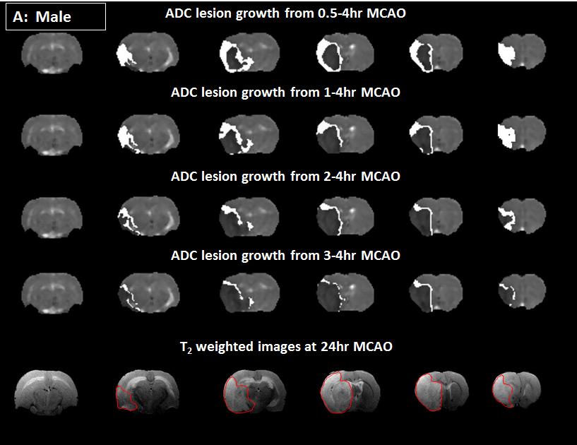
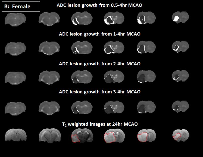
13 Figure legends Figure 1. Acute evolution of (A) ADC-defined ischaemic injury and T 2- derived final infarct volume and (B) penumbra volume as defined by ADC lesion expansion into final infarct in male and female rats. (C) illustrates at each time point the amount of penumbral tissue expressed as a % of the respective final infarct in male & female SD rats and for comparison WKY and SHRSP rats (modified from 15. (*P<0.05, Two way ANOVA, n=10 for both groups). Data are displayed as mean±sd. Figure 2. Images from the median rat from each group (A: Male; B: Female) showing the spatial location of ADC lesion growth (in white) from 0.5-1, 1-2, 2-3 & 3-4 hours following MCAO. The T 2 -weighted infarct (hyperintense region outlined in red) at 24h post- MCAO is shown on the bottom row.
14
15
16
Penumbra detection using PWI/DWI mismatch MRI in a rat stroke model with and without comorbidity: comparison of methods
 & 2012 ISCBFM All rights reserved 0271-678X/12 $32.00 www.jcbfm.com Penumbra detection using PWI/DWI mismatch MRI in a rat stroke model with and without comorbidity: comparison of methods Emma Reid 1,
Copyright 2014 The Authors. Deposited on: 26 August 2014
 Robertson, C. A., McCabe, C., Lopez-Gonzalez, M. R., Deuchar, G. A., Dani, K., Holmes, W. M., Muir, K. W., Santosh, C., and Macrae, I. M. (2014) Detection of ischemic penumbra using combined perfusion
T 2 *-weighted fmri time-to-peak of oxygen challenge in ischemic stroke
 Feature Article T 2 -weighted fmri time-to-peak of oxygen challenge in ischemic stroke Qiang Shen 1,2,3, Shiliang Huang 1 and Timothy Q Duong 1,2,3,4 Journal of Cerebral Blood Flow & Metabolism 216, Vol.
Experimental Assessment of Infarct Lesion Growth in Mice using Time-Resolved T2* MR Image Sequences
 Experimental Assessment of Infarct Lesion Growth in Mice using Time-Resolved T2* MR Image Sequences Nils Daniel Forkert 1, Dennis Säring 1, Andrea Eisenbeis 2, Frank Leypoldt 3, Jens Fiehler 2, Heinz Handels
Remission of diffusion lesions in acute stroke magnetic resonance imaging
 ORIGINAL RESEARCH Remission of diffusion lesions in acute stroke magnetic resonance imaging F. A. Fellner 1, M. R. Vosko 2, C. M. Fellner 1, D. Flöry 1 1. AKH Linz, Institute of Radiology, Austria. 2.
Title: Stability of Large Diffusion/Perfusion Mismatch in Anterior Circulation Strokes for 4 or More Hours
 Author's response to reviews Title: Stability of Large Diffusion/Perfusion Mismatch in Anterior Circulation Strokes for 4 or More Hours Authors: Ramon G. Gonzalez (rggonzalez@partners.org) Reza Hakimelahi
Dong Gyu Na, Vincent N. Thijs, Gregory W. Albers, Michael E. Moseley, and Michael P. Marks. AJNR Am J Neuroradiol 25: , September 2004
 AJNR Am J Neuroradiol 25:1331 1336, September 2004 Diffusion-Weighted MR Imaging in Acute Ischemia: Value of Apparent Diffusion Coefficient and Signal Intensity Thresholds in Predicting Tissue at Risk
MEDICAL POLICY EFFECTIVE DATE: 12/18/08 REVISED DATE: 12/17/09, 03/17/11, 05/19/11, 05/24/12, 05/23/13, 05/22/14
 MEDICAL POLICY SUBJECT: CT (COMPUTED TOMOGRAPHY) PAGE: 1 OF: 5 If the member's subscriber contract excludes coverage for a specific service it is not covered under that contract. In such cases, medical
Applications of Diffusion/Perfusion Magnetic Resonance Imaging in Experimental and Clinical Aspects of Stroke
 Applications of Diffusion/Perfusion Magnetic Resonance Imaging in Experimental and Clinical Aspects of Stroke Timothy Q. Duong, PhD, and Marc Fisher, MD Address Center for Comparative NeuroImaging, University
ACUTE STROKE IMAGING
 ACUTE STROKE IMAGING Mahesh V. Jayaraman M.D. Director, Inter ventional Neuroradiology Associate Professor Depar tments of Diagnostic Imaging and Neurosurger y Alper t Medical School at Brown University
Imaging ischemic strokes: Correlating radiological findings with the pathophysiological evolution of an infarct
 Imaging ischemic strokes: Correlating radiological findings with the pathophysiological evolution of an infarct Jay Chyung,, PhD, HMS III Patient A: history 91 y.o. woman Acute onset R sided weakness and
Update on Early Acute Ischemic Stroke Interventions
 Update on Early Acute Ischemic Stroke Interventions Diana Goodman MD Lead Neurohospitalist Maine Medical Center Assistant Professor of Neurology, Tufts University School of Medicine I have no disclosures
Endovascular Treatment for Acute Ischemic Stroke
 ular Treatment for Acute Ischemic Stroke Vishal B. Jani MD Assistant Professor Interventional Neurology, Division of Department of Neurology. Creighton University/ CHI health Omaha NE Disclosure None 1
Regional and temporal variations in tissue sodium concentration during the. acute stroke phase
 Regional and temporal variations in tissue sodium concentration during the acute stroke phase Authors: Friedrich Wetterling 1,2, Lindsay Gallagher 3, I. Mhairi Macrae 3, Sven Junge 4, Andrew J. Fagan 1,5
Complete Recovery of Perfusion Abnormalities in a Cardiac Arrest Patient Treated with Hypothermia: Results of Cerebral Perfusion MR Imaging
 pissn 2384-1095 eissn 2384-1109 imri 2018;22:56-60 https://doi.org/10.13104/imri.2018.22.1.56 Complete Recovery of Perfusion Abnormalities in a Cardiac Arrest Patient Treated with Hypothermia: Results
The Value of Apparent Diffusion Coefficient Maps in Early Cerebral Ischemia
 AJNR Am J Neuroradiol 22:1260 1267, August 2001 The Value of Apparent Diffusion Coefficient Maps in Early Cerebral Ischemia Patricia M. Desmond, Amanda C. Lovell, Andrew A. Rawlinson, Mark W. Parsons,
Mechanical thrombectomy beyond the 6 hours. Mahmoud Rayes, MD Medical Director, Stroke program Greenville Memorial Hospital
 Mechanical thrombectomy beyond the 6 hours Mahmoud Rayes, MD Medical Director, Stroke program Greenville Memorial Hospital Disclosures None Worldwide statistics 1 IN 6 people will have a stroke at some
Endovascular Therapy: Beyond the Guidelines
 Endovascular Therapy: Beyond the Guidelines Ashutosh P. Jadhav, MD PhD Assistant Professor, Neurology and Neurological Surgery Center for Neuro-endovascular Therapy UPMC Stroke Institute Pittsburgh, PA
Magnetic resonance imaging of blood brain barrier permeability in ischemic stroke using diffusionweighted arterial spin labeling in rats
 Original Article Magnetic resonance imaging of blood brain barrier permeability in ischemic stroke using diffusionweighted arterial spin labeling in rats Journal of Cerebral Blood Flow & Metabolism 0(00)
Where are we heading and where are the big challenges?
 Where are we heading and where are the big challenges? Christopher Levi Neurologist, John Hunter Hospital, Newcastle & Liverpool Hospital, Sydney Executive Director, Sydney Partnership for Health Education
Prediction of Hemorrhage in Acute Ischemic Stroke Using Permeability MR Imaging
 AJNR Am J Neuroradiol 26:2213 2217, October 2005 Technical Note Prediction of Hemorrhage in Acute Ischemic Stroke Using Permeability MR Imaging Andrea Kassner, Timothy Roberts, Keri Taylor, Frank Silver,
AMSER Case of the Month: March 2019
 AMSER Case of the Month: March 2019 62 year-old male with left-sided weakness Ashley Graziano OMS IV, Lake Erie College of Osteopathic Medicine Erik Yannone MD, Charles Q. Li MD, Warren Chang MD, Matthew
Dynamic susceptibility contract-enhanced MRI is increasingly
 Influence of Arterial Input Function on Hypoperfusion Volumes Measured With Perfusion-Weighted Imaging Vincent N. Thijs, MD; Diederik M. Somford, MD; Roland Bammer, PhD; Wim Robberecht, MD, PhD; Michael
Original Research Article
 MAGNETIC RESONANCE IMAGING IN MIDDLE CEREBRAL ARTERY INFARCT AND ITS CORRELATION WITH FUNCTIONAL RECOVERY Neethu Tressa Jose 1, Rajan Padinharoot 2, Vadakooth Raman Rajendran 3, Geetha Panarkandy 4 1Junior
occlusions. Cerebral perfusion is driven fundamentally by regional cerebral
 Appendix Figures Figure A1. Hemodynamic changes that may occur in major anterior circulation occlusions. Cerebral perfusion is driven fundamentally by regional cerebral perfusion pressure (CPP). In response
Stroke Thrombolysis. Dr Peter Anderton (Stroke Consultant DBTH)
") Stroke Thrombolysis Dr Peter Anderton (Stroke Consultant DBTH) Thrombolysis for ischaemic stroke Rationale Restoration of blood flow Salvage of ischaemic penumbra Schematic of the mismatch model for defining
Stroke Thrombolysis Dr Peter Anderton (Stroke Consultant DBTH) Thrombolysis for ischaemic stroke Rationale Restoration of blood flow Salvage of ischaemic penumbra Schematic of the mismatch model for defining
Neuro-vascular Intervention in Stroke. Will Adams Consultant Neuroradiologist Plymouth Hospitals NHS Trust
 Neuro-vascular Intervention in Stroke Will Adams Consultant Neuroradiologist Plymouth Hospitals NHS Trust Stroke before the mid 1990s Swelling Stroke extension Haemorrhagic transformation Intravenous thrombolysis
Sodium-23 Magnetic Resonance Imaging has potential for improving. penumbra detection but not for estimating stroke onset time
 Sodium-23 Magnetic Resonance Imaging has potential for improving penumbra detection but not for estimating stroke onset time Authors: Friedrich Wetterling, PhD, 1 Lindsay Gallagher, HNC, 2 Jim Mullin,
Advanced Neuroimaging for Acute Stroke
 Advanced Neuroimaging for Acute Stroke E. Bradshaw Bunney, MD, FACEP Professor Department Of Emergency Medicine University of Illinois at Chicago Swedish American Belvidere Hospital Disclosures FERNE Board
The Egyptian Journal of Hospital Medicine (July 2018) Vol. 72 (10), Page
 Vol. 72 (10), Page") The Egyptian Journal of Hospital Medicine (July 2018) Vol. 72 (10), Page 5398-5402 The Role of Susceptibility Weighted Imaging (SWI) in Evaluation of Acute Stroke Maha Abdelhamed El Nouby*, Eman Ahmed
The Egyptian Journal of Hospital Medicine (July 2018) Vol. 72 (10), Page 5398-5402 The Role of Susceptibility Weighted Imaging (SWI) in Evaluation of Acute Stroke Maha Abdelhamed El Nouby*, Eman Ahmed
framework for flow Objectives Acute Stroke Treatment Collaterals in Acute Ischemic Stroke framework & basis for flow
 Acute Stroke Treatment Collaterals in Acute Ischemic Stroke Objectives role of collaterals in acute ischemic stroke collateral therapeutic strategies David S Liebeskind, MD Professor of Neurology & Director
Effect of Collateral Blood Flow on Patients Undergoing Endovascular Therapy for Acute Ischemic Stroke
 Effect of Collateral Blood Flow on Patients Undergoing Endovascular Therapy for Acute Ischemic Stroke Michael P. Marks, MD; Maarten G. Lansberg, MD; Michael Mlynash, MD; Jean-Marc Olivot, MD; Matus Straka,
Background. Recommendations for Imaging of Acute Ischemic Stroke: A Scientific Statement From the American Heart Association
 for Imaging of Acute Ischemic Stroke: A Scientific Statement From the American Heart Association An Scientific Statement from the Stroke Council, American Heart Association and American Stroke Association
Assessing Tissue Oxygenation and Predicting Tissue Outcome in Acute Stroke
 The Danish National Research Foundation s Center of Functionally Integrative Neuroscience Aarhus University / Aarhus University Hospital - DENMARK Assessing Tissue Oxygenation and Predicting Tissue Outcome
CT Perfusion is Essential for Stroke Triage. Maarten Lansberg, MD PhD Associate Professor of Neurology Stanford University, Stanford Stroke Center
 CT Perfusion is Essential for Stroke Triage Maarten Lansberg, MD PhD Associate Professor of Neurology Stanford University, Stanford Stroke Center CT Perfusion is Essential for Stroke Triage Disclosures:
Broadening the Stroke Window in Light of the DAWN Trial
 Broadening the Stroke Window in Light of the DAWN Trial South Jersey Neurovascular and Stroke Symposium April 26, 2018 Rohan Chitale, MD Assistant Professor of Neurological Surgery Vanderbilt University
Diagnostic improvement from average image in acute ischemic stroke
 Diagnostic improvement from average image in acute ischemic stroke N. Magne (1), E.Tollard (1), O. Ozkul- Wermester (2), V. Macaigne (1), J.-N. Dacher (1), E. Gerardin (1) (1) Department of Radiology,
STROKE - IMAGING. Dr RAJASEKHAR REDDY 2nd Yr P.G. RADIODIAGNOSIS KIMS,Narkatpalli.
 STROKE - IMAGING Dr RAJASEKHAR REDDY 2nd Yr P.G. RADIODIAGNOSIS KIMS,Narkatpalli. STROKE Describes a clinical event that consists of sudden onset of neurological symptoms Types Infarction - occlusion of
Spatiotemporal dynamics of diffusional kurtosis, mean diffusivity and perfusion changes in experimental stroke
 Available online at www.sciencedirect.com www.elsevier.com/locate/brainres Research Report Spatiotemporal dynamics of diffusional kurtosis, mean diffusivity and perfusion changes in experimental stroke
SUPPLEMENTARY INFORMATION
 SUPPLEMENTARY INFORMATION Supplementary Figure 1. Long-term protection studies. 45 minutes of ischemia was induced in wild type (S1pr2 +/+ ) and S1pr2 -/- by MCAO. A) 5 days later brains were harvested
Diffusion And Perfusion Magnetic Resonance Imaging: Applications To Functional Mri
 Diffusion And Perfusion Magnetic Resonance Imaging: Applications To Functional Mri If looking for the book Diffusion and Perfusion Magnetic Resonance Imaging: Applications to Functional Mri in pdf format,
DIVISION OF CLINCIAL NEUROSCIENCES REGISTER OF INTERESTS
 DIVISION OF CLINCIAL NEUROSCIENCES REGISTER OF INTERESTS JOANNA M WARDLAW INTEREST 10k from Axaron for data supply Funding from Novartis for octreotide studies to department Reading CT scans for ECASS
STATE OF THE ART IMAGING OF ACUTE STROKE
 STATE OF THE ART IMAGING OF ACUTE STROKE Marin Penkov UH St Ivan Rilski Sofia RadiologyTogether 2-3 June 2017 GOALS The concept and significance of penumbra CT and MRI Basic Principles Clinical application
A New Trend in Vascular Imaging: the Arterial Spin Labeling (ASL) Sequence
 Sequence") A New Trend in Vascular Imaging: the Arterial Spin Labeling (ASL) Sequence Poster No.: C-1347 Congress: ECR 2013 Type: Educational Exhibit Authors: J. Hodel, A. GUILLONNET, M. Rodallec, S. GERBER, R. 1
A New Trend in Vascular Imaging: the Arterial Spin Labeling (ASL) Sequence Poster No.: C-1347 Congress: ECR 2013 Type: Educational Exhibit Authors: J. Hodel, A. GUILLONNET, M. Rodallec, S. GERBER, R. 1
Hypoperfusion Intensity Ratio Predicts Infarct Progression and Functional Outcome in the DEFUSE 2 Cohort
 Hypoperfusion Intensity Ratio Predicts Infarct Progression and Functional Outcome in the DEFUSE 2 Cohort Jean Marc Olivot, MD, PhD; Michael Mlynash, MD, MS; Manabu Inoue, MD, PhD; Michael P. Marks, MD;
Diffusion weighted MRI in evaluation of transplanted kidney: Preliminary clinical experience
 African Journal of Nephrology (2009) 13: 26-30 Original Article AJN Diffusion weighted MRI in evaluation of transplanted kidney: Preliminary clinical experience Mohamed Abou El-Ghar; M.D, Huda Refaie;
Stroke Treatment Beyond Traditional Time Windows. Rishi Gupta, MD, MBA
 Stroke Treatment Beyond Traditional Time Windows Rishi Gupta, MD, MBA Director, Stroke and Neurocritical Care Endovascular Neurosurgery Wellstar Health System THE PAST THE PRESENT 2015 American Heart Association/American
Imaging Stroke: Is There a Stroke Equivalent of the ECG? Albert J. Yoo, MD Director of Acute Stroke Intervention Massachusetts General Hospital
 Imaging Stroke: Is There a Stroke Equivalent of the ECG? Albert J. Yoo, MD Director of Acute Stroke Intervention Massachusetts General Hospital Disclosures Penumbra, Inc. research grant (significant) for
Cover Page. The handle holds various files of this Leiden University dissertation.
 Cover Page The handle http://hdl.handle.net/1887/44385 holds various files of this Leiden University dissertation. Author: Ayata, C. Title: Spreading depolarizations : the missing link between mirgraine
Characterization and Evolution of Diffusion MR Imaging Abnormalities in Stroke Patients Undergoing Intra-Arterial Thrombolysis
 AJNR Am J Neuroradiol 25:951 957, June/July 2004 Characterization and Evolution of Diffusion MR Imaging Abnormalities in Stroke Patients Undergoing Intra-Arterial Thrombolysis Pamela W. Schaefer, Alvand
Noninvasive Quantification of Brain Edema and the Space-Occupying Effect in Rat Stroke Models Using Magnetic Resonance Imaging
 Noninvasive Quantification of Brain Edema and the Space-Occupying Effect in Rat Stroke Models Using Magnetic Resonance Imaging T. Gerriets, MD; E. Stolz, MD; M. Walberer, DVM; C. Müller, PhD; A. Kluge,
How can sodium MRI techniques help us understand acute stroke?
 perspective How can sodium MRI techniques help us understand acute stroke? This article addresses the potential usefulness of sodium MRI in the acute phase of stroke, asking whether the additional time
CT Perfusion Parameter Values in Regions of Diffusion Abnormalities
 AJNR Am J Neuroradiol 25:1205 1210, August 2004 CT Perfusion Parameter Values in Regions of Diffusion Abnormalities Marcello Galvez, Gerald E. York, II, and James D. Eastwood BACKGROUND AND PURPOSE: Dynamic
Deposited on: 25 July 2011
 Santosh, C., Brennan, D., McCabe, C., Macrae, I.M., Holmes, W.M., Graham, D.I., Gallagher, L., Condon, B., Hadley, D.M., Muir, K. and Gsell, W. (2008) Potential use of oxygen as a metabolic biosensor in
Comparison of Five Major Recent Endovascular Treatment Trials
 Comparison of Five Major Recent Endovascular Treatment Trials Sample size 500 # sites 70 (100 planned) 316 (500 planned) 196 (833 estimated) 206 (690 planned) 16 10 22 39 4 Treatment contrasts Baseline
5/31/2018. Interventional Therapies that Expand Time Windows for Acute Ischemic Stroke Treatment. Disclosures. Impact of clot burden
 Good Outcome (%) Rankin 0-2 at 90 days 5/31/2018 Interventional Therapies that Expand Time Windows for Acute Ischemic Stroke Treatment Disclosures Cerenovus: I am on Executive Committee for ARISE2 Trial
12/4/2017. Disclosures. Study organization. Stryker Medtronic Penumbra Viz Route 92. Data safety monitoring board Tudor G.
 12/4/2017 Update on Stroke Trials:Extending the Window DWI or CTP Assessment with Clinical Mismatch in the Triage of Wake-Up and Late Presenting Strokes Undergoing Neurointervention with Trevo NP001713
11/1/2018. Disclosure. Imaging in Acute Ischemic Stroke 2018 Neuro Symposium. Is NCCT good enough? Keystone Heart Consultant, Stock Options
 Disclosure Imaging in Acute Ischemic Stroke 2018 Neuro Symposium Keystone Heart Consultant, Stock Options Kevin Abrams, M.D. Chief of Radiology Medical Director of Neuroradiology Baptist Hospital, Miami,
Lecture Outline: 1/5/14
 John P. Karis, MD Lecture Outline: Provide a clinical overview of stroke: Risk Prevention Diagnosis Intervention Illustrate how MRI is used in the diagnosis and management of stroke. Illustrate how competing
RBWH ICU Journal Club February 2018 Adam Simpson
 RBWH ICU Journal Club February 2018 Adam Simpson 3 THROMBOLYSIS Reperfusion therapy has become the mainstay of therapy for ischaemic stroke. Thrombolysis is now well accepted within 4.5 hours. - Improved
Infarct Volume Prediction by Early Magnetic Resonance Imaging in a Murine Stroke Model Depends on Ischemia Duration and Time of Imaging
 Infarct Volume Prediction by Early Magnetic Resonance Imaging in a Murine Stroke Model Depends on Ischemia Duration and Time of Imaging Christoph Leithner, MD; Martina Füchtemeier, PhD; Devi Jorks, MD;
Combined Effect of Hypothermia and Hyperglycemia on Transient Focal Cerebral Ischemia of the Rat
 Combined Effect of Hypothermia and Hyperglycemia on Transient Focal Cerebral Ischemia of the Rat Mei-Zi Jiang, M.D.*, Ja-Seong Koo, M.D.*, Byung-Woo Yoon, M.D.*, Jae-Kyu Roh, M.D.* Department of Neurology,
Assessing Tissue Viability with MR Diffusion and Perfusion Imaging
 AJNR Am J Neuroradiol 24:436 443, March 2003 Assessing Tissue Viability with MR Diffusion and Perfusion Imaging Pamela W. Schaefer, Yelda Ozsunar, Julian He, Leena M. Hamberg, George J. Hunter, A. Gregory
UPSTATE Comprehensive Stroke Center. Neurosurgical Interventions Satish Krishnamurthy MD, MCh
 UPSTATE Comprehensive Stroke Center Neurosurgical Interventions Satish Krishnamurthy MD, MCh Regional cerebral blood flow is important Some essential facts Neurons are obligatory glucose users Under anerobic
Redgrave JN, Coutts SB, Schulz UG et al. Systematic review of associations between the presence of acute ischemic lesions on
 6. Imaging in TIA 6.1 What type of brain imaging should be used in suspected TIA? 6.2 Which patients with suspected TIA should be referred for urgent brain imaging? Evidence Tables IMAG1: After TIA/minor
Title: Brain choline concentration: early quantitative marker of ischemia and infarct
 Karaszewski, MS ID#: NEUROLOGY/2009/3069 Title: Brain choline concentration: early quantitative marker of ischemia and infarct expansion? B Karaszewski,2,3, MD (e-mail: bartosz@karaszewski.org) RGR Thomas,2,
McLean ebasis plus TM
 McLean ebasis plus TM Sample Hospital (0000) Report For Qtr HBIPS Core Measures McLean Hospital 115 Mill Street Belmont, MA 02478 1 2012 Department of Mental Health Services Evaluation Tel: 617-855-3797
Stroke research why does it matter?
 Stroke research why does it matter? Dr Brad Sutherland Radcliffe Department of Medicine Wednesday 23 September 2015 1660 s Oxford Sobering stroke statistics 85% of all strokes ischaemic, 15% haemorrhagic
The follow-up of uterine fibroids treated with HIFU: role of DWI and Dynamic contrast-study MRI
 The follow-up of uterine fibroids treated with HIFU: role of DWI and Dynamic contrast-study MRI Poster No.: C-1137 Congress: ECR 2011 Type: Authors: Keywords: DOI: Scientific Exhibit V. Zampa, V. Vallini,
RESEARCH ARTICLE. Ischemic Core Thresholds Change with Time to Reperfusion: A Case Control Study
 RESEARCH ARTICLE Ischemic Core Thresholds Change with Time to Reperfusion: A Case Control Study Andrew Bivard, PhD, 1 Tim Kleinig, PhD, FRACP, 2 Ferdinand Miteff, FRACP, 1 Kenneth Butcher, FRACP, 3 Longting
Implication of aquaporins in ischemic stroke. New target?
 Implication of aquaporins in ischemic stroke. New target? Balseanu Tudor-Adrian, MD, PhD EXPERIMENTAL RESEARCH CENTER OF NORMAL AND PATHOLOGICAL AGING University of Medicine and Pharmacy of Craiova, Romania
Supplementary Figure 1. Shih, Friedman, Drew, Tsai, Lyden and Kleinfeld
 Supplementary Figure 1. Shih, Friedman, Drew, Tsai, Lyden and Kleinfeld Supplemental Table 1. Physiological parameters Baseline (1 st period) Occlusion (2 nd period) Reperfusion (3 rd period) tmcao:
Evolution of Lesion Volume in Acute Stroke Treated by Intravenous t-pa
 JOURNAL OF MAGNETIC RESONANCE IMAGING 22:23 28 (2005) Original Research Evolution of Lesion Volume in Acute Stroke Treated by Intravenous t-pa Jean-Baptiste Pialat, MD, 1 * Marlene Wiart, PhD, 1 Norbert
MR and CT Perfusion in Acute Ischemic Stroke The uses, methods, and risks associated with these alternative imaging modalities.
 MR and CT Perfusion in Acute Ischemic Stroke The uses, methods, and risks associated with these alternative imaging modalities. BY CATALINA C. IONITA, MD; EVE A. GUTERMAN; AND LEE R. GUTERMAN, PHD, MD
8.0 Take Home Naloxone
 8.0 Take Home Naloxone 8.1 Population characteristics For the financial year (FY) 2016/17, 721 take home naloxone (THN) kits were issued in the City of Edinburgh. Of the 323 individuals who received naloxone
Activity Report April 2012 to March 2013
 North, South East and West of Scotland Cancer Networks Brain/Central Nervous System Tumours National Managed Clinical Network Activity Report April 2012 to March 2013 Professor Roy Rampling Emeritus Professor
The association between neurological deficit in acute ischemic stroke and mean transit time
 Neuroradiology (2006) 48: 69 77 DOI 10.1007/s00234-005-0012-9 DIAGNOSTIC NEURORADIOLOGY Peter D. Schellinger Lawrence L. Latour Chen-Sen Wu Julio A. Chalela Steven Warach The association between neurological
Imaging in Stroke. D Nagaraja, N Karthik
 Imaging in Stroke D Nagaraja, N Karthik Cerebro-vascular disease (stroke) is the second leading cause of death. Prior to CT era, diagnosis was essentially clinical supported by angio and lumbar puncture.
Cerebral Revascularization For Stroke
 Cerebral Revascularization For Stroke If you are looking for the ebook Cerebral Revascularization for Stroke in pdf form, then you've come to loyal site. We present full edition of this ebook in txt, doc,
Intravenous thrombolysis with recombinant tissue plasminogen
 ORIGINAL RESEARCH BRAIN Susceptibility-Diffusion Mismatch Predicts Thrombolytic Outcomes: A Retrospective Cohort Study M. Lou, Z. Chen, J. Wan, H. Hu, X. Cai, Z. Shi, and J. Sun ABSTRACT BACKGROUND AND
Functional aspects of anatomical imaging techniques
 Functional aspects of anatomical imaging techniques Nilendu Purandare Associate Professor & Consultant Radiologist Tata Memorial Centre Functional/metabolic/molecular imaging (radioisotope scanning) PET
Perfusion-weighted imaging (PWI) and diffusionweighted
 and diffusionweighted") Apparent Diffusion Coefficient Thresholds Do Not Predict the Response to Acute Stroke Thrombolysis Poh-Sien Loh, FRACP; Ken S. Butcher, MD, PhD, FRCP(C), FRACP; Mark W. Parsons, PhD, FRACP; Lachlan MacGregor,
Apparent Diffusion Coefficient Thresholds Do Not Predict the Response to Acute Stroke Thrombolysis Poh-Sien Loh, FRACP; Ken S. Butcher, MD, PhD, FRCP(C), FRACP; Mark W. Parsons, PhD, FRACP; Lachlan MacGregor,
Article ID: WMC ISSN
 Article ID: WMC004795 ISSN 2046-1690 Correlation of Alberta Stroke Program Early Computed Tomography Score on CT and Volume on Diffusion Weighted MRI with National Institutes of Health Stroke Scale Peer
Neuroprotective Effect of Delayed Moderate Hypothermia After Focal Cerebral Ischemia
 Neuroprotective Effect of Delayed Moderate Hypothermia After Focal Cerebral Ischemia An MRI Study R. Kollmar, MD; W.R. Schäbitz, MD; S. Heiland, PhD; D. Georgiadis, MD; P.D. Schellinger, MD; J. Bardutzky,
ESCAPE Endovascular treatment for Small Core and Anterior circulation Proximal occlusion with Emphasis on minimizing CT to recanalization times
 ESCAPE Endovascular treatment for Small Core and Anterior circulation Proximal occlusion with Emphasis on minimizing CT to recanalization times Michael D Hill, Mayank Goyal on behalf of the ESCAPE Trial
CT INTERPRETATION COURSE
 CT INTERPRETATION COURSE Refresher Course ASTRACAT October 2012 Stroke is a Clinical Diagnosis A clinical syndrome characterised by rapidly developing clinical symptoms and/or signs of focal loss of cerebral
Guideline scope Stroke and transient ischaemic attack in over 16s: diagnosis and initial management (update)
") NATIONAL INSTITUTE FOR HEALTH AND CARE EXCELLENCE Guideline scope Stroke and transient ischaemic attack in over s: diagnosis and initial management (update) 0 0 This will update the NICE on stroke and
NATIONAL INSTITUTE FOR HEALTH AND CARE EXCELLENCE Guideline scope Stroke and transient ischaemic attack in over s: diagnosis and initial management (update) 0 0 This will update the NICE on stroke and
Clinical Sciences. Arterial Spin Labeling Versus Bolus-Tracking Perfusion in Hyperacute Stroke
 Clinical Sciences Arterial Spin Labeling Versus Bolus-Tracking Perfusion in Hyperacute Stroke Andrew Bivard, PhD; Venkatesh Krishnamurthy, MRCP, FRACP; Peter Stanwell, PhD; Christopher Levi, MBBS, FRACP;
Endovascular Treatment Updates in Stroke Care
 Endovascular Treatment Updates in Stroke Care Autumn Graham, MD April 6-10, 2017 Phoenix, AZ Endovascular Treatment Updates in Stroke Care Autumn Graham, MD Associate Professor of Clinical Emergency Medicine
Imaging Modalities in Acute Stroke: Time is Brain
 April 2001 Imaging Modalities in Acute Stroke: Time is Brain Jeremiah Scharf, Harvard Medical School, MS IV Beth Israel-Deaconess Medical Center Department of Radiology Stroke - Definition and Statistics
What is the best imaging protocol for LVO screening when outside of 0-6h window?
 Klinik und Poliklinik für Neuroradiologische Diagnostik und Intervention Zentrum für Radiologie und Endoskopie WLNC, Los Angeles/CA, USA, May 15-17, 2017 What is the best imaging protocol for LVO screening
Optimal definition for PWI/DWI mismatch in acute ischemic stroke patients
 & 08 ISCBFM All rights reserved 0271-678X/08 $30.00 www.jcbfm.com Brief Communication Optimal definition for PWI/DWI mismatch in acute ischemic stroke patients Wataru Kakuda 1, Maarten G Lansberg 2, Vincent
Populations Interventions Comparators Outcomes Individuals: With acute stroke who are being evaluated for thrombolysis.
 Protocol Computed Tomography Perfusion Imaging of the Brain (60149) Medical Benefit Effective Date: 01/01/16 Next Review Date: 03/19 Preauthorization Yes Review Dates: 11/14, 11/15, 11/16, 03/17, 03/18
The Language of Stroke
 The Language of Stroke Examination / Imaging / Diagnosis / Treatment Dr Suzanne Busch A lot of letters! CBF CVA ICH CVD CBV DWI US MRI/MRA CAA CTA CTP ICA MCA SAH WMD TIA MCA Agnosia A lot of big words!
PRESERVE: How intensively should we treat blood pressure in established cerebral small vessel disease? Guide to assessing MRI scans
 PRESERVE: How intensively should we treat blood pressure in established cerebral small vessel disease? Guide to assessing MRI scans Inclusion Criteria Clinical syndrome Patients must have clinical evidence
The translational roadblock, characterized by beneficial
 Special Report Methodological Quality of Experimental Stroke Studies Published in the Stroke Journal Time Trends and Effect of the Basic Science Checklist Jens Minnerup, MD*; Verena Zentsch, MD*; Antje
TRANSFORMING STROKE CARE IN THE CAPITAL: THE LONDON STROKE STRATEGY
 TRANSFORMING STROKE CARE IN THE CAPITAL: THE LONDON STROKE STRATEGY LUCY GROTHIER Director South London Cardiac and Stroke Network lucy.grothier@slcsn.nhs.uk 27 th May 2011 Gaps in London stroke care GAPS
Characterising hepatitis C virus transmission dynamics in a highrisk incarcerated population
 Characterising hepatitis C virus transmission dynamics in a highrisk incarcerated population Neil Bretaña, Lies Boelen, Rowena Bull, Suzy Teutsch, Peter White, Andrew Lloyd, Fabio Luciani on behalf of
1 TI - Stroke mortality in blacks. Disturbing trends. AU - Gillum RF SO - Stroke Aug;30(8): Review. IDS - PMID: UI:
: Review. IDS - PMID: UI:") 1 TI - Stroke mortality in blacks. Disturbing trends. AU - Gillum RF SO - Stroke. 1999 Aug;30(8):1711-5. Review. IDS - PMID: 10436126 UI: 99365468 2 TI - Mapping the ischaemic penumbra with PET: implications
1 TI - Stroke mortality in blacks. Disturbing trends. AU - Gillum RF SO - Stroke. 1999 Aug;30(8):1711-5. Review. IDS - PMID: 10436126 UI: 99365468 2 TI - Mapping the ischaemic penumbra with PET: implications