Objectives. Vascular Access
|
|
|
- Norma Glenn
- 6 years ago
- Views:
Transcription
1
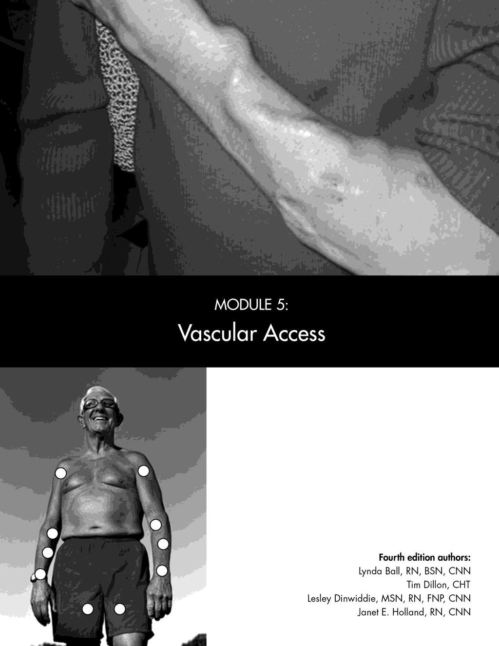
2 Vascular Access Objectives After completing this module, the learner will be able to: 1. Describe the three main types of vascular access. 2. Identify the predialysis assessments for all types of vascular access. 3. Describe the methods of needle insertion for AVFs and grafts. 4. Describe the predialysis assessment, accessing procedure, exit site care, and monitoring of catheters. Module 5 cover photo credit: Photograph of fistula by Stephen Z. Fadem, MD, FACP Used with permission. 122
3 Introduction Vascular access makes chronic hemodialysis possible because it allows the care team to access the patient s blood. An access can be internal (inside the body) or external (outside the body). It must: n Allow repeat access to the blood n Handle blood flow rates that will ensure effective treatments n Be made of materials that are not prone to causing reactions or infections Cephalic vein Basilic vein Antecubital vein Basilic vein Distal cephalic vein Ulnar artery Radial artery Axillary artery Brachial artery Figure 1: Venous/arterial anatomy of the arm The three main types of access are fistulae, grafts, and catheters. To create a fistula, a surgeon sews an artery and a vein together, most often in an arm. Arteries carry oxygen-rich blood from the heart and lungs to the rest of the body. The vessels selected for a fistula are large and have good blood flow, but are deep below the skin and hard to reach with needles. Veins bring blood back to the heart and lungs; they are easy to reach, but too small and too slow flowing for dialysis (see Figure 1). Linking an artery and a vein is the best of both worlds. In 4 6 weeks, high-pressure blood flow from the artery thickens the vein wall and makes it dilate (enlarge) so large needles can be used. Because a fistula is below the skin and is the patient s own tissue, it is less prone to infection and clotting than other types of access. A fistula can last for years even decades and research shows it is the best type of access now available. New surgical techniques and ways to assess and preserve blood vessels have made fistulae an option for more patients. To insert a graft, a surgeon links an artery and vein with a piece of artificial blood vessel. Like a fistula, a graft allows access to the large volume of blood needed for dialysis. Grafts are more prone to stenosis (narrowing of blood vessels), which can cause thrombosis (blood clots). Grafts are also more prone to infection than fistulas, and have a shorter useful lifespan (less than 5 years on average). 1 Grafts are an option for patients who do not have blood vessels suited to create a fistula. A catheter is a plastic, hollow tube placed in a deep central vein in the chest or leg (see Figure 2). Internal jugular vein External jugular vein Subclavian vein Superior vena cava Inferior vena cava Cephalic vein Femoral vein Figure 2: Anatomy of veins Basilic vein 123 3
4 Vascular Access They allow short-term or long-term access to patients blood. Deep central veins have a blood flow rate that allows adequate treatment. Catheters are made of plastic that is foreign to the body, and they pass through the skin, creating a portal for bacteria. They are prone to stenosis, blood clots, and infection. Due to these problems, catheters must often be replaced in the same or new vessels. Catheters are used for patients who: n Can t have a fistula or graft n Are waiting for a fistula or graft to be placed or to mature n Have acute kidney failure and may soon recover kidney function n Are waiting for a peritoneal dialysis catheter n Are waiting for a live donor kidney transplant Despite more than 65 years of effort, access is still the one greatest challenge to the success of dialysis. Between 25 50% of all hemodialysis patient hospital stays are due to access problems. The costs add more than $1 billion to Medicare inpatient bills each year. 2 Patients whose accesses don t work well don t get enough dialysis. They may become uremic and feel ill and tired. They don t feel well enough to work, exercise, or do hobbies that could improve their quality of life. When patients feel ill, it affects their family and friends and the care team. Access problems frustrate the care team and the patient. Trouble cannulating (putting needles into) fistulae and grafts is a source of stress for both. Poor cannulation can lead to problems that may cause access failure. Access failure means loss of the dialysis lifeline. The access must be repaired or replaced if the patient has a site left. Access problems cause hospital stays, surgery, illness, loss of limb, even death. Access problems also use staff time, disrupt schedules, and reduce center income while patients are in the hospital. All forms of access involve some compromise and have problems. Research is being done to seek the best vascular access for dialysis patients. The NKF Kidney Disease Outcomes Quality Initiative (KDOQI) and the Fistula First program (see page 127) are two, ongoing efforts to improve access outcomes. The guidelines include assessment and preservation of blood vessels for fistulae, and promote early fistula placement when possible. 1 In this module, we ll tell you about fistulae, grafts, catheters, and other devices. Each section includes definitions, assessment, and monitoring. We also cover the KDOQI guidelines, patient teaching, and complications of each type of access. You have the vital task of helping patients to keep their lifelines. Proper access care and use can improve quality of life for your patients and job satisfaction for the whole care team. Fistulae HOW FISTULAE ARE CREATED A native arteriovenous fistula (AVF) is made by surgically linking an artery to a vein. This link is the anastomosis, and the site is marked by a scar. It takes 1 3 months for an AVF to be strong enough to use large-gauge needles, so it is best to create one long before dialysis is needed. 124
5 Figure 3: Examples of AVF locations Distal cephalic vein Radial artery Basilic vein Ulnar artery Great saphenous vein Femoral artery As soon as the surgery is done, rapid and strong arterial blood flow sent to the vein starts to enlarge the fistula vein and make it tougher. This is called arterialization, and we say the fistula is maturing. About a week after the surgery, patients should start to do exercises that can help their fistulae to mature, like squeezing a rubber ball or lifting light weights. 3,4 The most common type of native AVF links the radial artery and the cephalic vein in the distal forearm (between the wrist and the elbow). This is called a radiocephalic fistula (see Figure 3). The brachiocephalic (brachial artery and cephalic vein) fistula is also used, and is the most common AVF of the upper arm. If these vessel pairs can t be used for some reason, others can be used instead. These may include: n Basilic vein n Transposed basilic vein (the deep vein is brought closer to the surface of the skin and the vein is moved to the anterior [front] surface of the upper arm for easier needle insertion) n Transposed one of the brachial veins (a pair of veins closely accompanying brachial artery and draining to the axillary vein) n Perforated vein in the antecubital fossa anastomosed to the brachial artery (perforating veins connect superficial and deep veins) n Ulnar artery n Proximal radial artery While the AVF is the best type of access, not every patient can have one. Surgeons must be sure there will be good blood flow for the hand once the fistula is created. The chosen veins must be healthy, straight, large enough to allow for large-gauge needles, and long enough to permit a number of needle sites. Patients must also be able to handle a 10% or more increase in cardiac output (the amount of blood passing through the heart) to have a fistula. A new access strains the heart, because arterial blood quickly short-circuits through the fistula instead of passing slowly through tiny capillary blood vessels. The heart must work harder due to the rapid blood flow. There are many reasons why a patient may not be able to have an AVF. These include: n Damage to veins due to intravenous drugs n Previous surgeries on the arteries and/or veins 125 5
6 Vascular Access Figure 4: Types of anastomosis n Atherosclerosis: plaque or waxy cholesterol that blocks the vessels n Poor quality arteries due to peripheral vascular disease (PVD) or advanced diabetes n Only one working artery to bring blood to the hand n Damage to blood vessels due to intravenous drugs FISTULA PROCEDURE Before surgery, patients should have vessel mapping done to find the best vessels for a fistula. To create a fistula, blood vessel sites are marked on the skin. An incision is made in the skin over the chosen vessels. Then the vessels are sewn together. There are four ways that arteries and veins can be joined to create an AVF (see Figure 4). Each has pros and cons: n The side-to-side (artery-side to vein-side) anastomosis is easiest for a surgeon to do. Side-to-side Side-to-end End-to-side End-to-end It is also the most likely to cause venous hypertension. This is a problem in which the hand fills with fluid due to high pressure in the veins. Sometimes the surgeon will do a side-to-side anastomosis, then tie off one or more of the vessels leading to the hand. n The side-to-end (artery-side to vein-end) anastomosis is preferred by many surgeons, even though it is hardest to do. This method gives high blood flows with few complications. n The end-to-side (artery-end to vein-side) anastomosis has slightly lower blood flow rates than the side-to-side. n The end-to-end (artery-end to vein-end) anastomosis permits less blood flow through the access. After the incision is closed, a thrill, or purring vibration, should be present over the new fistula. You should be able to hear a whooshing bruit with a stethoscope along the course of the vein. The bruit should be continuous and low-pitched. Both the thrill and the bruit help prove that the new fistula is patent (open). PROS AND CONS OF FISTULAE Pros The AVF is the gold standard for hemodialysis access. In general, it lasts longest and has the fewest problems, including infection. Using the patient s own vessels is always best when it can be done. Cons The main disadvantage of an AVF is how long it can take to mature: 4 6 weeks or more. Some fistulae also fail to mature at all, a problem 126
7 called early or primary failure. 5,6 A fistula may not mature if: n The anastomosis is too small, so less blood flows into the fistula n A stenosis develops at the inflow between the anastomosis and the fistula n Side veins off of the AVF (accessory veins) reduce pressure in the fistula, so it does not arterialize n The vessel chosen by the surgeon was too small (<2 mm) Vessel mapping can help a surgeon choose good vessels so this problem is less likely. ASSESSING MATURITY OF A FISTULA As a new technician, you will not be cannulating brand new fistulae. 1 But, you will still need to assess a new fistula at each treatment to be sure it is developing. To do this, you will need to: n Look for signs of infection redness, drainage or abscess formation. n Look for signs of wound healing of the surgical incision. n Feel for a thrill it should be continuous and feel like purring or vibration, but not a strong pulsation. n Feel the diameter of the vessel it should start growing larger immediately after surgery and the growth should be evident within 2 weeks. Note any flat spots. n Listen for a bruit the pitch should be low, and one sound should connect to the next sound. n After the first week, apply a tourniquet and feel for firmnessof the fistula vein; this will tell you that the vessel walls are getting thicker/stronger. Fistula First: Increasing Use of Fistulae in the U.S. The Centers for Medicare and Medicaid Services (CMS) started Fistula First in It is the first breakthrough initiative of CMS, and its goals are to raise the use of AVFs to 40% of hemodialysis patients and decrease the use of catheters. Fistula First includes nephrologists, vascular surgeons, interventional nephrologists, nurses, primary care doctors, patients, and others. These partners are working to change practice and assure that AVFs are the first choice for all patients who can have one. The ESRD Networks and CMS run the program. 7 Fistula First has 11 Change Concepts that centers can use to increase the use of fistulae: 8 1. Routine continuous quality improvement (CQI) review of vascular access 2. Timely referral to a nephrologist 3. Early referral to a surgeon for AVF only evaluation and timely placement 4. Surgeon choice based on best outcomes, willingness, and ability to provide access services 5. Full range of appropriate surgical approaches to AVF evaluation and placement 6. Secondary AVF placement in patients with AV grafts 7. AVF placement in patients with catheters where indicated 8. AVF cannulation training for patient care staff 9. Monitoring and maintenance to ensure adequate access function 10. Education for caregivers and patients 11. Outcomes feedback to guide practice Your role in Fistula First includes being on your center s vascular access CQI team, learning to correctly cannulate fistulae, and taking continuing education on vascular access
8 Vascular Access Figure 5: Types of needles 128 After 2 3 weeks if there is no change in the size of the fistula vein, the nephrologist and surgeon should be told. Dialysis patients should have a post-op visit 4 6 weeks after fistula surgery. According to access expert Gerald Beathard, MD, if access development is not seen by the second week after surgery, the access will not mature unless an evaluation is done and treatment occurs. 9 When the vessel wall is firm and the diameter has grown, the doctor will order the fistula to be cannulated. To cannulate a new AVF, smaller needles (17-gauge) and low blood flows ( ml/min) should be used for the first week of treatment. 10 This will help reduce cuts in the vessel from the needle, help reduce pressure, and help prevent infiltration (pushing the needle through both fistula walls so blood leaks out into the tissues). After the first week, needle gauge size (see Figure 5) can be increased, and blood pump speeds can be turned up. INITIATING DIALYSIS WITH A FISTULA Wash Your Hands Washing your hands is always the first step before you touch any dialysis access. Clean hands and clean gloves help prevent bacteria on Bevel Shaft Butterfly needle Gauge size Hub 17, 16, 15, or the skin s surface from being pushed into the patient s bloodstream by the needle. Change your gloves if they get contaminated by such things as touching your face or hair, the chair, or any other surface. The Occupational Safety and Health Administration (OSHA) requires handwashing and gloves to protect both you and patients from infections. 11 The Centers for Disease Control and Prevention (CDC) recommend the use of hemodialysis infection control precautions (gloves, gown, eye protection, and face mask) any time there may be a risk of blood splatters 12 (see Module 6: Hemodialysis Procedures and Complications for more information on handwashing and hemodialysis infection control precautions). Examine the Fistula At each treatment, you will need to assess a patient s fistula to be sure it has no problems and will work well and give the patient the best dialysis possible. You will need to learn how to inspect (look), auscultate (listen), and palpate (feel) the access, as we describe below. Look for: n Signs and symptoms of infection: Redness, drainage, pus, abscesses, open skin, fever n Steal syndrome (not enough blood flow to the hand): Pale, bluish nail beds or skin n Stenosis (narrowing): Swollen access arm, pale skin, small blue or purple veins on the chest wall where the arm meets the body n Cannulation sites: Scabs from needles, the anastomosis, curves, flat spots, aneurysms (ballooning of the blood vessels) and their width, height, and appearance
9 Listen for: n Bruit: The sound and pitch of the whooshing noise (a higher or louder pitch may mean stenosis) n Deep access location: Place the stethoscope flat over the access and listen for the bruit. Then, move stethoscope from side to side and listen for the bruit to stop. This will help you find the exact location of the access. Feel for: n Skin temperature: Note warmth (possible infection) or cold (decreased blood supply) n Thrill: Should be present and continuous, but not a strong pulse n Stenosis: Feel for flat spots, check flat spots for a thrill n Vein diameter: Start at the anastomosis with your thumb and forefinger on either side of the fistula. Is the diameter the same along the whole fistula? If there are aneurysms, how wide are they? Are there any flat spots? The diameter of the vessel needs to exceed the gauge of the needle. How deep is the access? This will determine your angle of needle entry. n Identify sites for cannulation: Stay 1.5 inches away from the anastomosis, keep the needles at least 1.5 inches apart, avoid curves, flat spots, and aneurysms. When rotating sites, avoid prior cannulation sites (scabs). n Steal syndrome: Cold hand temperature in the patient s access hand (compare with the other hand); have the patient squeeze your hand note any changes in motor skills. Assess Blood Flow The next step before inserting needles into an AVF is to check the blood flow. Each fistula should have a strong flow of blood from the artery through the access and into the vein. The fistula should have a strong thrill at the anastomosis, as arterial blood is pumped by the heart through the fistula. You will also need to check the bruit: place your stethoscope on top of the access and listen to the whooshing sound. It should be strong and continuous, with each sound linked to the one before. A change in the bruit to a high-pitched or louder whooshing may mean that stenosis is present. Teach patients to listen and report any changes to the charge nurse and/or nephrologist right away. A change in the thrill or in the volume of the bruit can also mean that blood flow through the access is slowing down. This is a sign that the fistula may be clotting. Tell the nurse in charge so he or she can assess it before the needles are inserted. You will need to learn how each patient s access normally sounds. Alerting the nurse to a change in the thrill or bruit may allow a failing access to be repaired in time. Prepare Access Skin Clean the patient s access site before needle insertion to keep bacteria from getting into the bloodstream through the skin. Staphylococcus aureus, or staph infection, is common in dialysis patients because: 13 n Patients are at a high risk for infection n Many have diabetes n There are large numbers of catheters n Patients have frequent hospital stays and surgeries, exposing them to infectious agents n Dialysis is a community-based setting 129 9
10 Vascular Access A study by Kaplowitz et al found that staph in the nose and the skin is common in hemodialysis patients. 14 This is why it is vital to teach patients to wash their access sites using antibacterial soap and water or an alcoholbased gel before they come to their chairs. This will help reduce the number of bacteria on the skin before you insert the needles, and lower the patient s risk of infection. Clean and prep the patient s skin with a solution of 70% alcohol, 10% povidone iodine, or chlorhexidine gluconate with 70% alcohol, following your center s procedures: n Alcohol kills bacteria only while it is wet use a 60 second circular rub on each site 15 n Povidone iodine (Betadine ) kills bacteria only after it dries wait 3 5 minutes 15 n Chlorhexidine gluconate (ChloraPrep ) with 70% alcohol kills bacteria only after it dries wait 30 seconds 16 n Sodium hypochlorite (ExSept Plus) the manufacturer recommends you wait 2 minutes before cannulating 17 Start on top of the sites you will put needles into and move outward in a circular, rubbing motion. 2 This will carry bacteria away from the point of insertion. Apply a Tourniquet Always use a tourniquet when you insert needles into a fistula, even when the size of the vessels does not seem to need it. The tourniquet helps you to see the fistula better, holds it in place (prevents rolling), and gives you a better feel for cannulation. The tighter skin also promotes a cleaner entry. Apply the tourniquet as far from the fistula as you can (just below the armpit) to help distribute pressure evenly along the vein and decrease the risk of infiltration. Tourniquets should never be so tight that they cause pain, tingling, or cut off blood flow to the fingers. They should only be used for cannulation, not during dialysis. Insert Needles Feel for the depth of the access before you insert your needles into a fistula. The angle of entry for a fistula will vary based on the depth of the access. The deeper the access, the steeper the angle of entry should be for you to have most of the needle inside the access. This will prevent infiltration if the patient moves the access limb during dialysis. Your center should have a written cannulation training program, with a checklist to be sure that you have learned all of the steps: proper site cleaning, needle insertion, and taping. Practice these skills in a lab setting, on a practice arm, before you try to cannulate a patient. It takes a lot of practice to be a good cannulator. New patient care staff should never cannulate brand new AVFs they are delicate and require an experienced, expert cannulator. 1 Only the most skilled staff in your center should cannulate new fistulae. 18 The key point to remember about cannulation is that it is a gentle technique. You choose your angle of insertion based on the depth of the vein, push the needle through the skin and tissue until you feel a release of pressure, check that you get a flashback (blood in the tubing), drop the angle down, and advance the needle. This sequence should be a fluid motion with no jabbing, digging, or probing with needles. 130
11 Do not flip the needle (turn it 180 after it is in the vessel). 19,20 Flipping the needle can: n Stretch the needle hole, which can lead to oozing during treatment after the patient is heparinized n Tear the lining of the vessel n Cause infiltration By doing a complete assessment, you will know just where to go and how deep the access is there should be no guessing. Determine where you will place both needles ahead of time. Try to leave space beyond the venous needle to place another needle in case the first attempt is not successful or infiltrates. The venous needle is usually closest to the heart. 21 Remember how much is at stake for your patient, and how vital it is for you to learn good technique to help preserve their lifelines. Depending on your center s procedures and how easy or hard it is to insert needles into a certain patient s access, you may do wet cannulation or dry cannulation. Wet cannulation is done with a saline-filled syringe, and can be useful for hard insertions or patients who clot very quickly. Dry cannulation uses the dialysis needles without saline. Follow your center s procedure for inserting needles after properly cleansing the needle puncture sites. 22 Note: If you cannot insert a needle successfully, find another staff person to do it. Most patients can tell you who has most success cannulating their accesses. Antegrade and retrograde needle direction You may wonder which way to point the needles. The venous needle should always be placed antegrade (in the direction of blood flow). This helps prevent turbulence when the blood returns from the extracorporeal circuit. 21,23 It is important to be sure that this needle is downstream from the arterial needle to prevent recirculation of the newly dialyzed blood back through the dialyzer circuit. 23 The arterial needle is so called because it is the one placed closest to the anastomosis and receives blood from the artery. This needle can be placed antegrade or retrograde (into the flow of blood). 23 No matter which way your center suggests for the needles, always keep the needle tips at least inches apart and stay at least inches away from the anastomosis. These rules will prevent recirculation and decreased adequacy. 23 Rope ladder technique (rotating sites) Each time a needle pierces a vein, it makes a hole. When you take the needle out, a clot forms to close up the hole until the vessel heals. When the patient comes in for the next treatment, you will look for the scabs from the last holes and choose different sites so the first ones can heal. This is called rotating sites, or the rope ladder technique. Picture a ladder with rungs the first day you put needles on two different rungs. Then, at each treatment, you chose two new rungs until you reach the top of the ladder, then start over again at the bottom. Sites are rotated to help prevent aneurysms (weak spots in the vessel wall that balloon out). It may seem easier and faster to place needles in the same general area at each treatment, but this weakens the wall of the blood vessel. If you spread out the sites, you will reduce the risk of


 The buttonhole technique has been used in Europe and Japan for more than 25 years, 24 and is becoming more popular in the United States.")
12 Vascular Access Figure 6: Buttonhole technique Drawings used with permission from Medisystems Corporation Using a sharp AV fistula needle, grasp the needle wings, and remove the tip protector. Align the needle cannula, with the bevel facing up, over the cannulation site and pull the skin taut. Cannulate the site. It is important to cannulate the developing constant-site in the exact same place, using the same insertion angle and depth of penetration each time. This requires that a single cannulator perform all cannulations until the sites are well established. A flashback of blood indicates the needle is in the access. Lower the angle of insertion. Continue to advance the needle into the AV fistula until it is appropriately positioned within the vessel. Securely tape the AV fistula needle and proceed with dialysis treatment per center protocol. aneurysms along the fistula. Patients may ask you to cannulate aneurysms because it hurts less. Explain that aneurysms can rupture (break open) because the skin gets very thin; they could lose a lot of blood and need surgery to repair their access. Buttonhole technique (constant-site) The buttonhole technique has been used in Europe and Japan for more than 25 years, 24 and is becoming more popular in the United States. It was first used on an access that had limited surface area for needles. Dr. Zbylut Twardowski, who invented the technique, found that it has fewer infections, missed needle insertions, hematomas (bruises), and infiltrations. 25 Both needles (arterial and venous) should be inserted in an antegrade direction to aid hemostasis after needle removal. To use this technique, remove the scabs from the last cannulation sites. Moisten the scabs first so they don t break into little pieces. To remove the old scab: n Apply saline or alcohol-based gels to a 2 x 2 gauze pad and lay one over each site, then use antiseptic tweezers n Provide alcohol squares and tape for the patient to take home, and ask him or her to tape an alcohol square over each scab before coming to dialysis. After the scab is gone, prepare the sites according to your center s policy. Once the sites are ready, you will reinsert sharp needles at the same angle into the same two holes (see Figure 6). Over the course of 3 4 weeks, a well-healed scar track/tunnel forms like a pierced earring hole. During this time, the same person should place the needles to be sure the same angle is used each time. This person can be the patient, since he or she is always there at each treatment. Once the scar tracks/tunnels are formed, blunt needles (see Figure 7) should be used to avoid cutting the track/tunnel, which can cause oozing during dialysis
13 Securing the Needles After Insertion After the needles are inserted, they need to be secured. The butterfly tape technique is a method of securing the needles (see Figure 6). Carefully place a piece of 1-inch wide adhesive tape, 6 inches or greater in length, under the fistula needle and then fold it so it crosses over the needle site. Then place a bandage or 2 x 2 gauze pad over the needle and secure it with another 6-inch piece of tape. You must secure the needles to keep them from moving or pulling out of the access. Monitor the needles for movement during the dialysis treatment. 23 Coping with Patients Needle Fear In the general public, at least 1 person in 10 has a physical fear of needles an injection, blood, or injury type of specific phobia. 26,27 People with this fear have an involuntary vasovagal response to needles, the sight of blood, surgery, etc.: 27 n First, the pulse speeds up and blood pressure rises n Then, the pulse slows, blood pressure drops, stress hormones are released, and heart rhythms may change n Patients may become pale, sweaty, nauseated, dizzy, and may pass out Some of these fearful patients may choose peritoneal dialysis (PD) to avoid needles, but PD patients may one day need to switch to hemodialysis. Patients who have needle phobia may be able to learn to short-circuit the vasovagal response. Some tips that may help: Buttonhole needle (blunt) Regular needle (sharp) n Lay the chair flat to keep blood in the brain so the patient doesn t pass out. n With the patient s doctor s permission, have the patient tense the muscles of his or her non-access limbs for 10 or 20 seconds, relax, then re-tense them until the needles are in. This can temporarily raise the blood pressure and prevent the vasovagal response. 27 n Reduce needle pain, using the techniques listed in the next section. Pain is part of the cause for the fear. n Teach patients how to insert their own needles. This distracts them from the pain and replaces it with control. Reduce pain from needle insertion Dialysis needles are quite large to allow enough blood flow. So, needle insertion can be painful. The goal is to insert the fistula needles easily and as painlessly as possible, while causing the least amount of trauma to the access. The 3-point technique can help reduce the pain of needle insertion and aid in successful cannulation. 28 First, apply a tourniquet to stabilize the fistula vein. To minimize vein Figure 7: Buttonhole versus regular needle Drawing adapted with permission from Lynda Ball

14 Vascular Access Figure 8: Three-point technique for needle insertion Drawing adapted with permission from Lynda Ball Figure 9: Intradermal injection of local anesthetic movement, place the thumb and forefinger of your non-needle hand on either side of the fistula vein, just above where the needle will go. Then, with your pinky or ring finger of the needle hand, pull the skin taut (tight) and press down on the skin (see Figure 8). The tighter the skin, the more easily the needle can puncture it, which will reduce pain. Pressing on the skin will temporarily block the pain-to-brain sensation for up to 20 seconds, giving staff enough time to insert a needle. 29 Patients who insert their own needles say that they feel much less pain than if someone else does the job. Patients who learn to put in their own needles are taking a vital role in their own care that can help them to feel better. They also help to preserve their access sites. Because they can feel the access from both the outside and the inside, it may be easier for patients to avoid infiltration. Another way to help patients with needle phobia if they have AVFs is to use the buttonhole technique, which causes less pain. 25 Other ways that patients can reduce pain include breathing techniques, guided imagery, and listening to music. Distraction can work quite well. Have the patient in the next chair or a staff person talk with your patient while you insert the needles. Patients may be given the choice of a local anesthetic (a product to numb the skin). Options include intradermal (injected into the skin) lidocaine, ethyl chloride spray, and topical creams or gels that are applied to the skin s surface. 23 The KDOQI Clinical Practice Recommendations for Vascular Access say that patients who are capable and whose access is suitably positioned should be encouraged to self-cannulate and the preferred method is the buttonhole technique. 1 Lidocaine injection An injection of 1% intradermal lidocaine can be used to numb the tissue (see Figure 9). The needle sites must be prepped first. Use a separate 1-cc or tuberculin syringe and needle for each site. Inject the lidocaine just below the skin into the tissue above the graft or fistula. (Never inject lidocaine into the patient s fistula vein; this would allow it to enter the bloodstream.) 23 The lidocaine will form a bubble or wheal just under the skin. Lidocaine burns, so only a small amount should be used. 23 The lidocaine may leak back out from the injection site and/or bleeding may occur at the injection site. Use a sterile gauze pad to wipe away any leakage or bleeding. Note: Because lidocaine is injected with needles, it may not be helpful for patients who have needle fear. 134
15 Lidocaine is a vasoconstrictor that can make the fistula vein smaller in diameter and pull it a little deeper under the skin. This may make cannulation more difficult. Patients whose fistula vein is very close to the skin s surface may have less pain when needles are inserted without lidocaine. The patient can compare the amount of pain if you use lidocaine with one needle and not the other. Per your center s policy, allow the patient to choose which method he or she prefers. Ethyl chloride spray Ethyl chloridespray can be used to numb the skin. The spray creates a cold feeling. It does not numb the tissue under the skin, so a patient with a deep access will still feel the needle enter the tissue and will feel pain. Ethyl chloride spray is not sterile. The site must be cleaned by the patient, sprayed, and then prepped by staff prior to needle insertion. Topical anesthetics Patients can use topical anesthetics (gels or creams that numb skin and tissue). These products must be applied to the skin, then wrapped in plastic wrap at home by the patient at least an hour before the treatment. Topical anesthetics work by contact time, not by the amount applied. In order for the top 3-mm of tissue to be numb, apply the cream 60 minutes before treatment. If you want the top 5-mm of tissue to be numb (for deeper accesses), have the patient apply the cream 120 minutes before treatment. 30 Some examples are: n Prescription EMLA cream (2.5% lidocaine/2.5% prilocaine) n Over-the-counter Less-n-pain (4% lidocaine) n Over-the-counter L.M.X. (4% lidocaine) n Over-the-counter Topicaine (4% or 5% lidocaine) At the center, the patient takes off the plastic wrap and washes off the cream. Remind patients to wash their hands after putting on the cream and to keep their hands away from their eyes to prevent damage to their mucous membranes. Like injected lidocaine, the creams cause vasoconstriction of the fistula. FISTULA CARE POSTDIALYSIS At the end of a treatment, you will untape and remove the needles per your center s policy. Make sure you have completely removed the needle before applying pressure to the skin you could cut the patients access if you press too early. Follow your center s policy to apply the right amount of pressure to the puncture sites. The goal is to stop bleeding, but not damage the access or stop blood flow through Tips to Increase Fistula Life To help an access last longer: Use the buttonhole technique or rotate needle sites at each treatment. Do not keep cannulating a fistula in the same general area. This could cause an aneurysm. Teach the patient not to permit IVs, routine blood draws, or blood pressure checks on the access arm. A Save the Vein card is helpful for patients to carry and give to medical staff if blood draws are needed. Keep accurate and detailed records of each treatment. If you see any problems with the patient s fistula, tell the nurse or the doctor
16 Vascular Access it, which could raise the risk of a blood clot. Teach your patients how to hold their own sites after a treatment. FISTULA COMPLICATIONS For patients, access problems can lead to access failure, inadequate dialysis, hospital stays, and even early death. If an access is lost, a new one must be created. This means surgery and recovery, which disrupt the patient s routine and reduce quality of life. With only about 10 sites in the body that can be used for an access, each new surgery limits the patient s future options. Each year, some patients die because they have run out of access sites. Access problems also affect the care team. They disrupt schedules and make treatment more difficult. Treatment of access problems uses valuable staff time. You need to know about common access problems and how they are treated so you can help preserve your patients dialysis lifelines and quality of life. By learning how to prevent access complications, you can help your patients keep their accesses longer. Infection Never cannulate an AVF that looks infected. The fistula needle can transfer an infection on the skin into the patient s bloodstream. This can cause sepsis blood poisoning one of the leading causes of death in people on dialysis. Tell the nurse right away if you see signs of infection so he or she can call the nephrologist. The nephrologist will give orders about cannulation, follow-up, and antibiotics. DIALYSIS-RELATED COMPLICATIONS OF FISTULAE Certain problems with fistulae occur only during a treatment. Line Separation Exsanguination (severe loss of blood) can occur if a needle comes out, the lines come apart, or the fistula ruptures (bursts). To keep needles from being pulled out, follow your center s procedures for taping (see Securing the Needles After Insertion on page 133). Fasten the bloodlines securely and set the arterial and venous pressure monitor limits so you will know right away if a problem occurs. The air/foam detector and venous and arterial pressure monitors can help prevent exsanguination if they are armed and working. But, sometimes a dislodged needle can cause a slow loss of blood over the course of a treatment. In this case, the drop in venous pressure may not be enough to set off the alarm and you may not see the blood if the patient s arm is hidden by a blanket. If a patient is losing blood through the tubing, turn off the blood pump and clamp the bloodlines. Apply pressure to the needle site if the needles were pulled out. Oxygen, saline, or blood volume expanders may be needed if a lot of blood was lost. If needed, start emergency procedures (e.g., call 911) per center policy. Air Embolism Air that enters the patient s bloodstream can stop the flow of blood, much like a blood clot. 136

17 If enough air enters the bloodstream, the heart pumps foam instead of fluid blood. This reduces cardiac efficiency and can cause cardiac arrest. Bloody foam in the lungs makes it hard to breathe. An air embolus in the brain can mimic a stroke. Depending on where the air goes, the patient may be highly anxious; have trouble breathing; become cyanotic (turn blue); have vision problems or low blood pressure; or become confused, paralyzed, or unconscious. Newer dialysis machines do not permit alarm overrides. If your center has older machines, always check to be sure that the air/foam detector is armed and working before each treatment and when you return the patient s blood after dialysis. If the air/foam detector alarm sounds, look at the venous line to see if any air is present before you override the alarm. Tape or Luer-Lok all line connections securely to keep them from coming apart. Look at the blood tubing injection ports for micro bubbles after you draw samples or give IV solutions. You can also teach patients to watch their bloodlines to be sure air does not enter the blood tubing. No air should be present in the tubing between the air/foam detector (or below the venous drip chamber) and the patient. Air in the arterial line before the dialyzer should be caught in the arterial drip chamber before it can enter the dialyzer. The air/foam detector should stop the blood pump if there is air in the venous drip chamber. If you suspect that a large amount of air entered the venous system, have the patient lie on his or her left side and tell a nurse. Lying on the left side decreases the chance that air will travel to the brain and the pulmonary artery. Tips to Prevent Blood Loss During Dialysis To prevent blood loss during dialysis: Never let patients cover their needles or bloodlines with sheets or blankets. You must always be able to see the access. Secure all bloodlines and access connections before you start a treatment, and tape the needles so they can t be pulled loose. Don t let blood tubing touch the floor; it could be stepped on and pulled apart. Check to be sure that the air/foam detector and arterial and venous pressure monitors are working before you start a treatment, and that the monitors are armed once dialysis begins. Infiltration/Hematoma An infiltration occurs when the needle tip goes into the vein and out the other side, or nicks the side of the vessel, letting blood escape into the patient s tissues (see Figure 10). Infiltration is the most common complication of needle insertion. It occurs less often once staff have more practice placing needles. Infiltration harms the access, which can lead to access failure. For patients, infiltration causes pain, bruising, the need for an extra cannulation, and loss of trust in the staff. Blood that leaks into the tissue around the Figure 10: Needle infiltration Skin Soft tissue Vein
18 Vascular Access blood vessel (hematoma) makes the area swollen, hard, and sometimes red. An infiltrated venous needle will raise the venous pressure, which will set off the venous pressure alarm to stop the blood pump. An infiltration of the arterial needle will cause the arterial pressure to become more negative. To prevent infiltration, closely follow the needle placement technique used at your center, and: n Use a gentle technique. n Do not rush. n Develop a feel for the least bit of resistance in the vessel. n Level the needle to the surface of the skin. Advance it slowly up to the hub as soon as you feel a pressure change and can see a flashback of blood in the needle tubing. n Don t flip the needles. n Flush the needles with saline after insertion to check for good placement (no pain, swelling, or resistance to the saline flush). n Use the wet needle cannulation technique. Remove a needle that infiltrates before heparin is used. 22 Have the patient apply pressure to the site, just as he or she would at the end of a treatment. If infiltration occurs after the patient receives heparin, the nurse may tell you to leave the needle in place. You will then need to insert another one beyond the site of the infiltration (usually above). 19 If a hematoma forms, give the patient an ice pack and something to act as a barrier (i.e., washcloth) between the ice and the skin to hold in place during treatment, to help reduce the swelling. The patient should keep ice on for 20 minutes, off for 20 minutes, on for 20 minutes, etc. 22 Finally, do not subtract the time you spend taking care of a needle infiltration from the patient s total dialysis run time. Lost minutes lead to inadequate treatment. Add the lost time to the end of the treatment to be sure that the patient receives the prescribed dialysis dose. Bleeding During Dialysis Bleeding around the needles during a treatment may be a minor problem (oozing at needle sites) or a life-threatening emergency (needle falling out with the blood pump running). Frequent loss of small amounts of blood during dialysis adds to the risk of anemia, a shortage of red blood cells. Do not flip the dialysis needles. This practice causes the hole to stretch until it is larger than the needle size, causing oozing during dialysis. (If oozing does occur, place a sterile dressing over the site.) Use a back eye needle for the arterial draw to eliminate the need to flip the needle. Profuse bleeding may suggest a tear in the blood vessel or bleeding from a remote site within the access. Uncontrolled bleeding is a life-threatening situation. Call the nurse or doctor right away. Recirculation Recirculation occurs when dialyzed blood returning through the venous needle mixes with blood entering the arterial needle. The mixing means that already-dialyzed blood goes back through the dialyzer to be cleaned again, while the rest of the patient s blood is not cleaned enough. So, recirculation makes 138
19 treatment less efficient. 21 Over time, poor dialysis can lead to symptoms of uremia. Recirculation can occur when: n The blood flow within the AVF is lower than that in the dialyzer (< ml/min) n The needles are placed too close together n Lines are reversed n A stenosis is present In severe cases, recirculation will re-clean the same blood so much that it turns dark from lack of oxygen ( black blood syndrome ). More often, recirculation may not cause any shortterm symptoms. Recirculation studies may be done if the delivered dialysis dose drops as noted by a decrease in URR or Kt/V the blood flow rate drops due to higher venous pressures, or the team thinks stenosis is present. These steps can help ensure proper needle placement to prevent recirculation: 22 n Palpate the patient s access to find the direction of blood flow. n Assure that the tips of the arterial and venous needles are at least 1.5 inches apart. LONG-TERM COMPLICATIONS OF FISTULAE Steal Syndrome Steal syndrome is a set of symptoms caused by hypoxia (lack of oxygen to the tissues). When an access is created, sometimes it steals too much blood away from the hand and sends it through the access. Patients feel pain that can range from minor to severe. 31 In most patients, steal syndrome pain lessens over time because extra blood vessels grow and supply blood to the area (collateral circulation). 31 Vascular surgeons say that people with diabetic neuropathy or peripheral vascular disease must be watched very closely. Their symptoms may get worse, and they will often need some form of intervention. Some of the symptoms to watch for or ask your patients about include: n Pain in the access limb n Tingling in the access limb n Cold feeling in the access limb n A change in motor skills in the hand n Nail beds that are blue in color n Necrotic (dead, blackened) spots on the skin n Decreased feeling in the access limb Tell the nurse or nephrologist if you suspect steal syndrome so they can call the vascular surgeon. Try to keep the patient s hand warm during dialysis, perhaps with a mitten, blanket, or tube sock. Changing the position of the patient s arm may help increase blood flow in the hand. Henriksson and Bergqvist stated that 5% of AVFs cause steal syndrome. 31 They have also found that steal syndrome can be treated by reducing blood flow through the fistula, making the vessels larger, or tying off some blood vessels surgically. You need to know that steal syndrome can be treated, so tell the nurse as soon as you notice any signs of it. Early follow-up with the surgeon is critical
20 Vascular Access Figure 11: Stenosis in a fistula Cephalic vein Aneurysm Putting needles in the access in the same general area time after time can cause an aneurysm. This pattern of cannulation weakens the vessel wall and causes a bulge or gumdrop or ballooning to form on the access. With time, flow in an otherwise normal AVF continues to increase, and the vein may keep enlarging. Aneurysms are more likely to occur upstream (retrograde) from a venous stenosis, especially at sites of repeat needle insertion. The sites are easy to see. Follow their progress and note any associated skin changes. 32 Rotate needle puncture sites or use the buttonhole technique to prevent aneurysms. Do not insert needles into areas of aneurysm. Aneurysms leave less surface area for cannulation. Surgery is needed when the skin over the fistula has signs of impending access rupture such as marked thinning, ulceration, or bleeding. 32 Stenosis Stenosis is a narrowing of the blood vessel that slows the flow of blood through the access (see Figure 11). The KDOQI Clinical Practice Guidelines for Vascular Access say that reduced blood flow through a fistula predicts clotting and access failure. 1 Radial artery Stenosis There are three major sites where stenosis is likely to form: 1. Inflow the most common type of inflow stenosis is called juxta-anastomotic stenosis (JAS). It is found in the vein right next to the anastomosis. A JAS will keep a fistula from maturing because it does not allow enough blood flow to enter the fistula. A JAS may be due to stretching, twisting, or other trauma during the fistula surgery. 32 You may be able to feel a JAS as a flat spot just past the anastomosis. 2. Outflow stenosis can occur anywhere along the outflow vein. It may occur in an area where the patient has had an IV placed in the past. The area past the stenosis may appear smaller, making needle insertion more difficult for fear of infiltration. 3. Central vein central venous stenosis occurs in the large draining vein of the arm, often in the shoulder area. If a stenosis is suspected, the venous system should be checked from the fistula to the heart, so a central stenosis can be found. These stenoses are most likely caused by catheter placement in the past. All patient care staff need to watch for these symptoms: n A high-pitched or louder-pitched bruit n Harsh or water-hammer pulse n Discontinuous bruit (each sound separate whoosh whoosh whoosh) n Decreased thrill n Trouble inserting or threading the dialysis needles 140
21 n Swelling of the patient s access limb n Increased venous pressure during treatment, forcing you to turn the blood pump down n Recirculation n Extracorporeal system clotting off during treatment n Increased bleeding after needles are removed postdialysis n Black blood syndrome n Decrease in Kt/V and URR n Inability to obtain prescribed blood flow rate Stenosis is caused by injury of the blood vessel lining that scars the blood vessel and creates flow turbulence. This leads to overgrowth of smooth muscle cells or aneurysm formation. Stenoses may recur after treatment. To find venous or arterial stenoses, dye is injected into the vessel. With the dye, any narrowing will show up on an x-ray (fistulogram, venogram). Stenosis may also be found by color Doppler ultrasound. 1 Ultrasound is a non-invasive way to look at blood vessels and blood flow. 33 These methods allow the doctor to pinpoint the narrowing. Some cases of stenosis can be treated with angioplasty, which is an outpatient procedure. The doctor threads a catheter with an inflatable balloon tip into the vessel. Once the balloon is in place, he or she inflates it to expand the vessel lumen. In other cases, access revision surgery may be needed to try to repair the access. Thrombosis Thrombosis (formation of a thrombus, or blood clot) occurs in all types of vascular access, but less than 1/6 as often in AVFs than in grafts. 9 Blood has a number of components to stop a wound from bleeding by forming a clot. These include clotting proteins called plasma coagulants and platelets tiny blood cells that stick together to seal off damaged blood vessels. Platelets clump when they are activated by contact with damaged blood vessel walls or by turbulence inside a blood vessel. Activated platelets and damaged tissues signal blood clotting proteins to form a strong net of fibers (fibrin). This net traps more platelets and red blood cells, so the clot gets more solid as it Patient Actions to Prevent Thrombosis To avoid thrombosis, patients should: Prevent medical staff from using the access or access limb for routine blood draws, IVs, or blood pressure checks Feel for the thrill and listen to the bruit daily to ensure patency Think about learning how to self-cannulate to reduce the risk of mispuncture Patients should not: Sleep on the access arm Put too much pressure on the puncture site after removing the dialysis needles Wear tight-fitting clothing or jewelry that squeeze the access arm Carry heavy objects across the arms that compress the access, such as a purse Use the access for IV drugs Wear a watch on the hand that has a radiocephalic or lower arm fistula
22 Vascular Access grows bigger (see Figure 12). A clot may start to form any time there is low blood flow due to low blood pressure, dehydration, or too much pressure on the access. These conditions let blood pool at a damaged surface, like a needle site. In a fistula, blood flow through an area of stenosis may cause enough turbulence to activate platelets, so they stick to the vessel wall. Repeated cannulations also damage vessel walls, causing rough surfaces that activate platelets. Early thrombosis is most often caused by surgical problems with the anastomosis or twisting of the blood vessel. It can also be caused by stenosis, reduced blood flow due to hypotension at dialysis, a cardiac arrest, or compression of the vessel. The vessel can be compressed after surgery if blood leaks into the tissues causing swelling and hematoma. Hematomas may also form at the site of an infiltration, or if an access is used too soon after surgery. Applying too much pressure for too long to the puncture site after treatment may also lead to thrombosis. Pressure should not be placed on a puncture site for more than 20 minutes. If bleeding lasts longer than 20 minutes, the nurse should look at the heparin dose and check for an access problem, like stenosis. Late thrombosis may develop in a fistula that was working. It can be caused by increased turbulence from stenosis. Untreated thrombosis can destroy an access. According to interventional radiologist Dr. Perry Arnold, a thrombosed fistula can be rescued for up to 14 days after it clots. 34 Thrombosis is most often caused by stenosis and low blood flow. The following signs may be present in a patient with pending access thrombosis: n Reduced thrill and bruit n Poor blood flow through the access n Inability to obtain prescribed blood flow rate n Sudden swelling of the hand on the access arm in a patient with a history of stenosis and blood flow problems n Abnormally high venous pressure readings during dialysis n A high recirculation rate; always check if the needle placement may have had an effect before calling the nephrologist n Increased transmembrane pressure (TMP) Suspect thrombosis if you feel no pulse or thrill and hear no bruit along the outflow vein. Figure 12: Blood clotting at a needle puncture site Surface clot (scab) Clot 142 Needle insertion Platelet activation Fibrin trapping platelets and blood cells
23 Thrombosis in a previously working access often follows stenosis. Early detection and repair of stenoses may help save the access. You can offer the team key information about signs of stenosis and thrombosis. Report signs of poor blood flow through the access, including reduced bruit or thrill and swelling of the hand. When you follow proper technique to insert needles and apply moderate, but not heavy, pressure to the puncture site posttreatment, you help reduce the risk of thrombosis. Vascular access monitoring can identify patients at risk for developing thrombosis. 35 The KDOQI Clinical Practice Guidelines for Vascular Access recommend that centers have an access monitoring program. 1 AVFs can be monitored through measurement of dynamic and static venous pressure, access flow measurement, and duplex ultrasound. An access monitoring program helps to improve patients vascular access outcomes by identifying problems early. 35 Thrombosis can be treated by surgical, mechanical, or chemical (use of a drug to dissolve the clot) thrombectomy. In more than 90% of cases, a stenosis is the cause of the clot. 36 This may be corrected by surgery to revise the access, or by angioplasty after the clot is removed. High Output Cardiac Failure An AVF can be one cause of high output cardiac failure. This is a complex condition in which: 1. The fistula brings more blood to the heart 2. The heart works harder, reducing resistance in the arteries 3. Arterial blood pressure falls 4. The drop in arterial blood pressure triggers the renin-angiotensin system 37 Patients with high output cardiac failure may have rapid pulses as their hearts try to make up for the extra blood flow (20% or greater) caused by the access. They may be short of breath if the blood does not contain enough oxygen. Swelling may occur in the hands and feet because return circulation to the heart is poor. Over time, if the problem is not treated, it can cause chest pain, fluid in the lungs, abnormal heart rhythms, and death. High output cardiac failure can also be caused by anemia or by heart disease the patient had before the vascular access. So, prevention is best aimed at treating anemia and creating an access that will not tax the patient s heart. If high output cardiac failure occurs, limiting fluid gain between treatments will reduce the strain on the heart. Longer and/or more frequent treatments may help with fluid gains, reducing the strain. Medication may also be given to help the heart beat more strongly. Instruct patients to report any changes in their overall health and energy level. Surgery may be needed to reduce blood flow through the access. In this case, surgery is done either to band the arterial anastomosis or to tie off the fistula. Grafts An arteriovenous graft (AVG) is an artificial blood vessel used to connect an artery and a vein. Grafts can be long enough to connect vessels in very different parts of the body, if
24 Vascular Access Figure 13: Examples of graft locations Straight graft in upper arm needed. This can be important for patients who have few usable veins left for dialysis access. GRAFT MATERIALS Many types of materials can be used for AVGs. These materials can be divided into biologic and synthetic categories. Biologic Human and animal materials have been used for grafts, but are rarely used today. Some human biologic AVGs were made from a vein from the patient s leg. Veins from the umbilical cords of newborn infants were also used. Umbilical veins had high rates of infection and Looped graft in forearm Straight graft in forearm aneurysms. Bovine (cow) and ovine (sheep) carotid arteries used for grafts were treated to remove proteins that would cause the human body to reject them. These grafts also had high rates of infection and aneurysm. Synthetic Synthetic materials are now used for nearly all grafts. The most widely used synthetic graft material today is expanded polytetrafluoroethylene, or eptfe. Collagen is another material that is used to make grafts. Grafts may be straight, curved, or looped. Some designs provide a larger surface area for needle insertion. GRAFT PROCEDURE Construction of a graft is a surgical procedure that bridges an artery and a vein. During the surgery, incisions are made over the vessel entry sites. The vein is checked to ensure that there is enough blood flow. A tunnel is then made under the skin, and the graft is attached to one vessel, passed through the tunnel, and attached to the other vessel (anastomosis) (see Figure 13). Straight grafts are usually placed in the forearm (radial artery to basilic vein). Loop grafts are placed in either the forearm (brachial artery to basilic vein), the upper arm, or the thigh. 38 The most common graft in the upper arm is curved from the brachial artery to either the basilic or axillary vein. A new graft should be placed at least 3 6 weeks before use, unless a graft material is used that can be cannulated right away Looped graft in thigh GRAFT PROS AND CONS Pros Grafts take less time to mature before the first cannulation. Graft size and blood flows don t
25 depend on the maturation. Grafts may also have larger cannulation areas than fistulae, and be easier for staff to cannulate. 22 Grafts are often used in patients who are not good candidates for native fistulae due to advanced age or other health problems, like diabetes, that damage blood vessels. Grafts make dialysis possible for patients whose veins are not suited for a fistula. They are readily available and come in a variety of sizes and shapes. Cons The biggest problems with all grafts are infection and thrombosis. Grafts develop stenosis at the venous anastomosis most commonly, 1 and clot at a much higher rate than native fistulae. No graft material now exists that is as good as a native blood vessel. STARTING DIALYSIS WITH A GRAFT Wash Your Hands Washing your hands is always the first step before you touch any dialysis access. Clean hands and clean gloves help keep bacteria on the skin s surface from being pushed into the patient s bloodstream by the needle (see Module 6: Hemodialysis Procedures and Complications for more information on handwashing and hemodialysis infection control precautions). Graft Assessment As with fistulae, grafts must be assessed before each dialysis treatment. Below is a general outline for assessment of an AVG: 22 Things to look for when assessing a graft include: n Swelling and redness n Pain and tenderness n Drainage from puncture sites, or from the skin around the graft n Bruises n Healing at previous cannulation sites n Localized warmth and fever Something to listen for when assessing a graft is: n Bruit should be low-pitched and continuous Things to feel for when assessing a graft include: n Pulse feel for a soft, compressible pulse n Thrill continuous (feel without compression only) n Skin temperature should be normal, not hot n Hardness or pain Confirm the Direction of Blood Flow To find the direction of the blood flow, feel the entire length of the graft. Compress the middle of the graft with two middle fingers and feel for the pulse and/or thrill on both sides of the area being compressed. You will feel the strongest pulse on the arterial side. The pulse or thrill will be faint or not palpable at the venous end. 22 Assess Blood Flow Every graft should have a strong flow of blood from the artery through the access and into the vein. The pulse of a graft feels like a pounding or buzzing with each heartbeat, as arterial blood is pumped by the heart to the rest of the body. This pulse, or thrill, should be strong, and you should be able to palpate it over the entire length of the access. The thrill should decrease over the venous portion of the graft
26 Vascular Access Listen to the sound of the blood flow through the graft. The blood flow creates a bruit that you can hear by placing your stethoscope over the access and listening to the swooshing sound. The sound should be strong and steady. Like the thrill, the bruit should decrease over the venous portion of the graft. It is important to learn how each patient s access normally sounds. Alerting the charge nurse to a change in the thrill or bruit may allow early intervention to save a failing graft. PREPARING TO USE A GRAFT The steps to prepare access skin, reduce pain from injection, and insert needles used for grafts (see Figure 14) are the same as for fistulae (see Initiating Dialysis with a Fistula on page 128). Refer to the specific graft material manufacturer s recommendations for graft care and use. Different graft materials may have unique instructions for use. Never use a tourniquet on a graft! INSERTING NEEDLES Selecting a Cannulation Site Prior to cannulation, you need to choose a site for needle insertion. Select a site that is at least a half inch away from any previous needle sites. Do not cannulate near anastomoses. The site should be at least one inch from an anastomosis, obstructions, or restrictions. Keep arterial and venous needles at least 2 inches apart. 39 Site Rotation of a Straight Graft To decide the needle site rotation in a straight graft, first divide the graft into equal halves at its middle. Use the middle as a reference. Cannulate the arterial half of the graft by moving toward the arterial anastomosis. Cannulate the venous half by moving towards the venous anastomosis. Each time you use a graft, space the needle sites equally along the length of the graft. Each needle insertion should be.25.5 inches from the last site. Place needles along all three sides of the graft, not just along the top. 39 Site Rotation of Loop Graft To decide the needle site rotation in a loop graft, first divide the graft into equal halves at the middle of the loop. Use the middle as a reference. Cannulate the arterial half by moving towards the arterial anastomosis. Cannulate the venous half by moving towards the venous anastomosis. Rotate sites by equally spacing the sites along the graft. The space should be between.25.5 inches away from last sites. Place needles along all three sides of the graft, not just along the top. 39 Needle Direction You will need to know how to find the direction of blood flow in the graft. The venous needle is inserted in antegrade (in the direction of blood flow) placement. This is needed to prevent excessive venous return pressure and damage to the blood cells. The arterial needle can be placed antegrade or retrograde (against the direction of blood flow). Retrograde placement of the arterial needle is preferred. 39 GRAFT CARE POSTDIALYSIS Postdialysis graft care is the same as postdialysis fistula care (see page 135.) GRAFT COMPLICATIONS Complications associated with AVGs include infection, stenosis, thrombosis, and 146
27 pseudoaneurysm. The rate of infection and thrombosis is higher in AVGs than in native AVFs. Figure 14: Needle insertion in a graft Infection Infection is more common in grafts than in fistulae. A break in the use of aseptic technique is the main cause of infection. Your center has policies and procedures on how to prepare the cannulation site using aseptic technique. You must follow these steps to lower the risk of infection. Signs and symptoms of an infected graft include redness, swelling, pain, fever, and chills. Tell the nurse about a possible infection right away so he or she can call the nephrologist. The nephrologist will give orders about cannulation, follow-up, and antibiotics. Stenosis Stenosis at the venous end of a graft anastomosis is a common problem. Stenosis can also develop along the length of a graft. Graft-vein stenosis develops when smooth muscle cells at the venous anastomosis grow more than they should. The cells form extra layers that fill up the graft lumen, reducing blood flow. This process is called neointimal hyperplasia. Turbulence at either anastomosis and/or within the graft may play a role in this problem. KDOQI Clinical Practice Guidelines for Vascular Access recommend that grafts be checked for stenosis at least monthly. 1 Grafts can be tested by measuring venous pressure or access flow. Learn your center s policies and procedures on the test(s) used to monitor for stenosis (see Stenosis on page 140 for more information on the causes, signs and symptoms, and treatment of stenosis.) Needle punctures the skin Needle travels through subcutaneous tissue Needle enters the vessel Needle is leveled out and threaded into vessel lumen Thrombosis Thrombosis is the most common reason for AVGs to fail. A decreased or absent thrill/ pulse is a sign of thrombosis. To check for thrombosis, look at the graft and palpate for a thrill/pulse throughout the length of the graft. Teach patients how to palpate for the thrill/pulse in their access and report any Skin Soft tissue Vessel lumen


28 Vascular Access changes to dialysis staff right away (see Thrombosis on page 141 for more information on the causes, prevention, and treatment of thrombosis). Steal Syndrome Steal syndrome can occur with grafts as well as fistulae. Some symptoms to watch for include pain, tingling, coldness, a change in motor skills in the hand, blue nail beds, and/ or decreased sensation in the access hand. The access surgeon should be told, so alert the nurse or nephrologist if you suspect steal syndrome. Try to keep the patient s hand warm during dialysis, perhaps with a mitten or tube sock. Changing the position of the patient s arm may help increase blood circulation in the hand. DIALYSIS-RELATED COMPLICATIONS OF GRAFTS The same dialysis-related complications that happen with fistulas also occur with grafts (see Dialysis-related Complications of Fistulae on page 136 for complete information about line separation, air embolism, bleeding, infiltration/hematoma, and recirculation). COMPLICATIONS RELATED TO POOR NEEDLE SITE ROTATION Pseudoaneurysm (a bubble-like blister in the graft caused by weakness in the graft wall) and graft collapse can occur if needle sites are not rotated well. For a pseudoaneurysm to develop, the graft needs a defect and increased pressure from a venous stenosis. Each time a graft is cannulated, the needle cuts a hole in the graft. If the cannulation sites are not rotated, the holes will come together to form larger holes (see Figure 15). 32 Informally termed one-site-itis, this overuse of the graft can have grave consequences. The graft may start to come apart, which leads to graft collapse. 39 At the same time, the frequent placement of needles in the same area damages the tissue above the graft, slowing healing and weakening all the protective skin layers. In time, the pressure of the blood flow following the path of least resistance will be stronger than the tissue covering it, and the pseudoaneurysm may rupture. If this occurs, the patient can die in a matter of minutes if he or she is alone. No graft Figure 15: One-site-itis photos Photos reprinted with permission from Bard Peripheral Vascular 148

29 should be allowed to reach this point. Careful predialysis assessment and consultation with other team members can prevent pseudoaneurysms from becoming life-threatening problems. Central Venous Catheters Some patients start hemodialysis without a permanent access, and these patients will most often have a catheter. For example, patients may have an urgent need for treatment and no access due to: n Acute renal failure n Peritonitis from peritoneal dialysis n Uremia from chronic kidney disease (CKD) with no matured fistula n A wait for a scheduled transplant n An agreement with a doctor to have a trial of dialysis n Refusal of fistula surgery n Access failure or infection All of these events occur often, so it is good that we can access the blood quickly using a percutaneous (through the skin) catheter or a subcutaneous (under the skin) port/catheter device. These devices are made for short-term use days to weeks but some patients may need them for months or even years. Unfortunately, patients who use catheters are more likely to develop blood infections that can be fatal. 40 Central venous catheters can damage the central blood vessels as well as the heart. So, patients with catheters should be encouraged to be assessed for a fistula as soon as possible. Some patients resist fistulae due to a fear of needles and/or of unsightly scarring and vein enlargement, but the longer they wait, the higher the risk of sepsis. 41 TYPES OF CATHETER AND PORT/CATHETER DEVICES A central venous catheter is a relatively large tube placed into a high-flowing central vein that leads into the heart. Because hemodialysis removes and returns blood at the same time, the tube has two side-by-side chambers called lumens. The end of the catheter that enters the patient s bloodstream is the tip; it has holes for blood entry and exit (see Figure 16). The other end, the tail, is outside the body with the two lumens apart. Each lumen has an adapter-connector on the end. The connectors attach to the bloodlines through specially-designed needles that are placed at the start of a treatment. The exit site, where the catheter comes out through the skin, is covered by a sterile dressing, especially when the catheter is not in use. Short-term Catheter Sometimes a patient needs only a few treatments with a catheter. For example, Collarbone Subclavian vein Exit site Internal jugular Dual lumens Catheter tip Figure 16: Internal jugular catheter

30 Vascular Access Figure 17: Vasca LifeSite Hemodialysis Access System Drawing used with permission from Vasca, Inc. patients with acute renal failure, infection, or a clotted fistula or graft may use a short-term catheter. This type of catheter is short in length because it exits the skin directly over the venotomy (the site where the catheter enters the vein). If the patient still needs a catheter upon discharge from the hospital, this catheter should be exchanged for a tunneled catheter. Tunneled, Cuffed Catheter A tunneled, cuffed catheter (TCC) is longer than a short-term catheter because the tail is tunneled under the skin from the venotomy to the exit site, a few centimeters away. Most tunneled catheters have a cuff. The cuff of the catheter is a fibrous material about 5 mm wide, wrapped around the catheter. The cuff is placed about 1 cm up the tunnel from the exit site. Healing tissue inside the tunnel bonds with the cuff; this keeps the catheter from pulling out and bacteria from migrating up the tunnel and into the bloodstream. For this reason, TCCs are safer and are the preferred type. Another style of TCC is the single lumen catheter. Two single lumen catheters must be placed to do conventional hemodialysis; one to take blood to the dialyzer and the other to return it. These catheters are called twin catheters. Port/Catheter Device A port/catheter device is usually a combination of a port and a single lumen catheter. The catheter has a tip like a percutaneous single catheter, but the tail connects to a metal port placed under the skin (see Figure 17). Two port/catheter devices are needed to do hemodialysis. The shorter catheter is the pull, or arterial, line; the longer one is the return, or venous, line. The bloodstream is accessed by placing a needle into the metal port and opening the valve. The dialysis needles are treated like extensions of the catheters. The heparin is removed and the needles are flushed with saline prior to connection with the lines. At the end of treatment, the procedure is reversed. Removing the needles at the end of each treatment closes a valve that seals each port device internally. At press time, no port/catheter devices were on the market, though some previously placed port/catheter devices were still in use. CATHETER AND PORT/ CATHETER DEVICE SITES Catheters for hemodialysis are always placed in veins that are able to support the high blood flows needed for dialysis. The very large veins in the neck and chest that empty into the right 150
31 atrium of the heart are the most suitable. The largest of these, the superior vena cava, collects blood from the head and neck veins (internal and external jugular) and from the arms via the subclavian veins. The right internal jugular (RIJ) vein is the very best because: n In most people, it is the largest vessel n In most people, it is the shortest and straightest distance to the right atrium of the heart n The RIJ has the lowest rate of stenosis and/or clots, that would prevent return blood flow from a future fistula or graft in the arm. NOTE: A catheter should not be placed on the same side as a working fistula or graft. The left internal jugular (LIJ) is the second choice, but it is longer and has two large curves to navigate. This reduces blood flow. The subclavian veins should not be used for a catheter unless a surgeon has found that no fistula or graft can ever be placed in the ipsilateral (same side) arm or there is a lifethreatening emergency. 1 The femoral veins in the groin can be used for catheter access (see Figure 18) when: n Temporary access is needed for urgent dialysis and the RIJ cannot be used n Long-term catheter access is needed and none of the upper central veins can be used There are other, more exotic catheter placements such as translumbar or transhepatic for patients who no longer have any of the usual placement sites. See a vascular access text for more details. Telling an IJ Catheter Apart from a Subclavian The subclavian vein is, as its name suggests, below the clavicle (collarbone). Because IJ TCC exit sites are often in the same place as the exit site of a short-term or TCC subclavian catheter, there can be confusion over which vein the catheter enters: If a TCC is seen and/or felt over the clavicle, it is IJ. If the catheter disappears under the clavicle, it is subclavian. percutaneous or subcutaneous. The tips of all catheters inserted into the chest should be placed well into the right atrium of the heart and the tips of all femoral veins should be well up into the inferior vena cava (IVC). This tip placement assures the best flow into the catheter, for the best dialysis. PLACEMENT OF CATHETERS AND PORT/ CATHETER DEVICES Most catheters are placed in vascular interventional outpatient centers by a radiologist or a nephrologist. Exit site Figure 18: Femoral catheter placement The following principles of venous entry are true for all hemodialysis catheters, whether Venous Arterial
32 Vascular Access Vascular, transplant, and general surgeons also place them, often at the same time as they create a fistula or place a graft. Most port/catheter devices are placed in hospitals. The main advantage of using a vascular interventional suite or operating room is that X-rays can be done during the procedure, to reduce placement problems. Catheters must be placed with strict aseptic technique. Procedures done at the patient s bedside or in a doctor s office are less than ideal, and must be followed by a chest X-ray to confirm catheter placement before starting hemodialysis. All newly placed catheters are tested by saline flushing to assure function and then locked with heparin to prevent clotting. Local anesthetics and conscious sedation IV medications to relieve pain and sedate the patient are used during catheter placements. Uncomplicated catheter placements take less than 30 minutes. Port/catheter device placement takes longer and is more complex. IJ Placement The IJ vein is 2 3 cm above the clavicle. It is found by an ultrasound device, then punctured with a large needle. Dark, venous blood is pulled back to prove that the vein was reached. A guide wire is inserted through the needle, the needle is removed, and the catheter is advanced into the vessel over the wire. Once the catheter is in the vein, the guidewire is removed. The tunnel under the skin for a TCC placement may be formed first, depending on the type of catheter used. The tunnel is usually brought over the clavicle, with the exit site 2 3 cm below. Stitches are placed around the catheter at the exit sites of both temporary and tunnel-cuffed catheters. The stitches can be removed from a TCC after days, but should not be removed from a shortterm catheter until the catheter itself is removed. Placement of any chest catheter can cause: n Bleeding n Hematoma (blood that has collected under the skin) n Air embolus n Pneumothorax n Hemothorax Subclavian Placement As stressed before, the subclavian vein the large vein in the shoulder that drains the majority of blood from the arm on the same side should be the catheter site of last resort. It is much more likely than an IJ to become stenosed. If it must be used, general placement technique and procedure is the same as for an IJ catheter. The doctor must take care to prevent kinking of the catheter due to the sharp angle needed to enter the vein under the clavicle. The risk of pneumothorax and hemothorax may be higher due to the need to puncture directly over the lungs. Femoral Placement The femoral vein is next to the femoral artery in the groin. The method for finding the vessel and placing femoral catheters is similar to IJ placement. The most frequent complication of femoral placement is femoral artery puncture and hematoma. Port/Catheter Device Placement The port is inserted through an incision into a pocket that is formed under the skin. After the catheter s placement and tunneling, it is 152
33 connected securely to the vein. 42 Prior to closing with sutures, all pocket bleeding must be stopped. The pocket may be flushed with an antibiotic solution to prevent infection. PROS AND CONS OF CATHETERS AND PORT/ CATHETER DEVICES Pros n Patients can have vascular access for urgent treatment. n Catheters and port/catheter devices can be hidden under most clothing. n The patient s hands and arms can move freely during dialysis. n Patients can shower or swim with port/catheter devices, once the incision is healed and the stitches are removed. n Patients will say that no needles is the main advantage of catheters. This must be weighed against the proven higher risk of death and severe illness from long-term catheter use. Cons n Catheters and port/catheter devices are more likely to cause life-threatening bacteremias (blood infections). n Catheters are foreign bodies in the bloodstream and can cause inflammation and clotting in the blood vessels, leading to stenosis and occlusion (blockages). n Catheters generally have lower blood flows over time, for less adequate dialysis and more frequent procedures. Port/catheter devices usually have more reliably high flows over time. 42 CARE OF CATHETERS AND PORT/CATHETER DEVICES Catheter and port/catheter cannulation and care are usually done by registered nurses. Refer to your center s protocol for care instructions. Preventing Infection The following steps will help prevent infection in catheters and port/catheter devices: n Always make sure you have washed your hands and changed your gloves prior to touching the patient and his or her equipment. n Always make sure you and the patient each wear a mask covering both the mouth and nose whenever the catheter is opened or the exit site exposed. Airborne bacteria that can infect catheters and therefore the patient s blood can be found in mouths and noses of both patients and staff. n Assess the catheter exit site or port site before starting treatment. Look for any signs of redness, or drainage or pus. n Always use aseptic technique when opening, handling, cleaning, or cannulating the port, or connecting and dressing the catheter (as per center protocol). n Teach patients how to protect the catheter and its dressing. Most dialysis centers change the dressing at each treatment, and patients shouldn t need to do this between treatments. If the dressing gets wet, it is best to remove the wet dressing, dry carefully around the catheter exit site, and cover the site with a large elastic bandage. To prevent the dressing from getting wet, most centers suggest no
34 Vascular Access showers or swimming for patients with catheters. However, there are devices that are made to keep catheters dry in the shower. Check with your center s cathetercare protocol. n Patients must also be taught not to use sharp objects such as pins or scissors around their catheters. Pin holes put the patient at risk for infection and also require catheter changing before the next treatment. Accidentally cutting the catheter can cause severe blood loss or air entry. The patient should be shown how to pinch off the catheter securely and seek medical help immediately. n Report any problems with the catheter to the nursing staff as soon as possible. Preparing to Use a Catheter Predialysis assessment n Ask the patient about any problems with the catheter during previous treatments or at home. n Remove the dressing and note any changes in catheter position, such as the cuff becoming visible at the exit site. n Observe for any redness and drainage (fluid or material coming out around the catheter) such as pus. n Assess the ease of heparin removal and saline flushing before connecting the bloodlines. Presence of clots and/or sluggish saline flushing suggests that the catheter is blocked or compressed, or that catheter position has changed. This must be corrected before connecting the bloodlines. The nursing staff will further assess the problem and may instill a thrombolytic enzyme to dissolve the clot. 43 Considerations for accessing catheters and cleansing exit sites n Prepare procedure site using dialysis precautions. n Conduct procedures using aseptic technique (correct handwashing, masks for patient and staff, no-touch technique, and disposable clean gloves). n Chlorhexidine 2% with 70% alcohol is the preferred solution for cleansing of central venous catheter sites (check catheter manufacturer s warnings about the effect of disinfectants on catheter material). n For patients who are sensitive to chlorhexidine 2% with 70% alcohol, chlorhexidine aqueous may be used instead (check catheter manufacturer s warnings about the effect of disinfectants on catheter material). n For patients who are sensitive to chlorhexidine aqueous, povidone solution may be used (use according to manufacturer s directions). n Skin cleansing should include the following steps: 1. Apply solution/swab in a circular motion working from catheter exit site outwards. 2. Cover an area 10 cm in diameter. 3. Perform this step twice. Do not rinse off or blot excess solution from skin. 154
35 4. Allow solution to dry completely before applying dressing. n To cleanse the connection between any CVC catheter hub and cap use two swabs: 1. Hold up the catheter at the connection site with one swab. 2. Use second swab to clean from catheter connection up catheter for 10 cm. 3. Cleanse hub connection site and cap vigorously with the first swab. Discard swab. 4. Do not drop a connection site once it is cleaned. n To cleanse the section of the catheter that lies next to the skin, gently swab the top and underside of the catheter starting at the exit site and working outwards. n Always be sure the clamps are closed before opening the catheter to air when pulling off the catheter caps, removing a syringe, or undoing the Luer-Lok connectors in the lines. Monitoring Catheters During the Treatments If pressure alarms signal that the flow cannot be maintained at the prescribed rate, you must: 1. Look at the patient and the lines to make sure there is no major bleeding or air entry. Call immediately for help if there is. 2. Next, assess for mechanical obstruction. Are the lines kinked anywhere along the circuit? Check again that the catheter is in place. 3. Reposition the patient lower the head of the chair, have the patient turn his head, cough, etc., to help move the tip of the catheter for better flow. 4. Flush with saline to help further assess the condition of the catheter. 5. Switch or reverse the lines so that blood is pulled through the venous port and returned through the arterial port. This should be done to complete a treatment only with the permission of the charge nurse. Line reversing creates recirculation of the blood, which leads to less adequate dialysis and does not correct the problem. It is also another chance for infection, and all the precautions taken to start dialysis must be repeated. 6. Ask the nurse to assess the problem. He or she may give a thrombolytic enzyme to clear the catheter of a clot. If these measures do not restore the prescribed blood flow, the patient should be referred to the interventional doctors or surgeons to correct the problem. This may require a new catheter placement. Monitor blood flow through the dialyzer, noting the amount of arterial and venous pressure exerted on the catheter to achieve the prescribed blood flow. Always keep the prepump arterial monitor connected and open. It tells you the condition of the arterial catheter lumen, as does the venous pressure monitor for the returning lumen. Patient safety is our main concern during dialysis. A catheter accident can quickly become fatal. For this reason, staff must pay a great deal of attention to securing the connections and lines and making sure the catheter or needle site are visible during the treatment. Patients can bleed to death under
 Issues related to vascular access are often a challenge to manage.")

36 Vascular Access a blanket, or an air embolus can occur if air is sucked in through a dislodged catheter or connection. Good catheter care is good patient care. Improving Vascular Access Outcomes CONTINUOUS QUALITY IMPROVEMENT (CQI) Issues related to vascular access are often a challenge to manage. Continuous quality improvement (CQI) can be a powerful tool to help reduce the rate of vascular access problems and provide the best outcomes (see Figure 19). Collecting data and forming a CQI team are the first steps toward making a plan. For example, it might be helpful to begin a log or computer database to track vascular access problems. The log could include: n Access type n Date of placement n Surgeon n Type of complication n Action taken n Data from monitoring tests Figure 19: CQI process 156
Evaluation of AVF and AVG
 Evaluation of AVF and AVG 2013 Nephrology Nursing Symposium Albuquerque Vascular Access Leading cause of hospitalization in the ESRD population Annual cost approaching $1.5 billion (USRDS, 2004) Current
UW MEDICINE PATIENT EDUCATION. Hemodialysis. A treatment option for kidney disease. Treatment Options for Kidney Disease
 UW MEDICINE PATIENT EDUCATION Hemodialysis A treatment option for kidney disease Class Goals 1. Understand the purpose and care of blood access. 2. Understand the purpose and basic principles of hemodialysis.
Table of Contents. Dialysis Port Care Chemotherapy Port Care G-Tube Care Colostomy Bags Wound Dressings
 Table of Contents Dialysis Port Care Chemotherapy Port Care G-Tube Care Colostomy Bags Wound Dressings Dialysis Port Care Know What Type of Vascular Access You Have. Fistula: An artery in your forearm
It is important to learn all you can about your access so you can take better care of yourself.
 PEP CONNECT PEP Talk Outline Topic Vascular Access P A T I E N T E D U C A T I O N P R O G R A M An adequate permanent vascular access is the key to living long and well on dialysis. There are three kinds
BLEEDING EMERGENCY. Always apply pressure directly over the bleeding site with your thumb or use an inverted bottle top to localise the pressure
 Bleeding between dialysis sessions is very rare and there are almost always warning signs before this happens. Warning signs include:- Infection of the fistula Bulging area, usually with shiny skin and
CARE OF YOUR FISTULA
 CARE OF YOUR FISTULA 1 . What is a fistula? An arteriovenous fistula, or AVF, is a vessel that is formed by joining a vein to an artery in your arm during an operation to form an accessible blood vessel
BUTTONHOLE CANNULATION
 BUTTONHOLE CANNULATION What is a Buttonhole? Technique in which an AV fistula is cannulated in the exact same spot, at the same angle and at the same depth of penetration every time. Benefits Reduction
The Buttonhole Technique for AV Fistula Cannulation
 The Buttonhole Technique for AV Fistula Cannulation Lynda K. Ball, RN, MSN, CNN October 16 & 17, 2008 Trends in Nephrology Nursing Kaiser Permanente Acute Dialysis Symposium 1 CMS Disclaimer This publication
Treatment Options Not Not access
 Treatment Options Treatment options for Chronic Kidney Disease (CKD) CKD is a permanent condition. Treatment will: Help the patient feel better Not cure the disease Not make the kidneys start working again
Module 10 Troubleshooting Guide
 Module 10 Troubleshooting Guide Your safety and wellbeing are our priority. Issues can occur during your treatment and it is important that you recognize the symptoms. This guide will teach you how to
St George Hospital Renal Department Internal Only
 RENAL VASCULAR ACCESS CANNULATION POLICY AND PROCEDURE SUMMARY: A functioning arteriovenous fistula (AVF) or arteriovenous graft (AVG) is paramount in the maintenance of regular and optimal haemodialysis
Cannulation Techniques Webinar
 Cannulation Techniques Webinar March 28, 2012 ESRD Network of Texas Lynda K. Ball, MSN, RN, CNN Objectives Discuss assessment skills inspection, palpation, and auscultation of an AV fistula to determine
Fistula for Hemodialysis
 St. Joseph s Healthcare Hamilton Kidney and Urinary Program Fistula for Hemodialysis What is a fistula? A fistula is the joining of an artery and a vein by a surgeon. The fistula is called an access. It
MANITOBA RENAL PROGRAM
 SUBJECT Venipuncture of Arteriovenous Fistula/Graft MANITOBA RENAL PROGRAM SECTION CODE 30.20.01 30.20 Vascular Access AUTHORIZATION Professional Advisory Committee, Manitoba Renal Program Nursing Practice
Cannulating AV Fistula using Buttonhole Technique
 Nephrology Directorate Subject: Objective: Prepared by: Cannulating AV Fistula using Buttonhole Technique To enable the safe insertion and removal of needles into arteriovenous fistulae using the buttonhole
Nursing Care of the Dialysis Patient. Adrian Hordon, MSN, RN
 Nursing Care of the Dialysis Patient Adrian Hordon, MSN, RN Understand principles of hemodialysis Recognize different access ports Identify side effects and complications Discuss nursing care for pre and
MANITOBA RENAL PROGRAM
 SUBJECT Venipuncture of Arteriovenous Fistula/Graft MANITOBA RENAL PROGRAM SECTION CODE 30.30.01 30.30 Vascular Access AUTHORIZATION Professional Advisory Committee, Manitoba Renal Program Nursing Practice
AMMAR SERAWAN, MD. Ain Wzain Hospital. April 21, 2012 Vascular Access Study Workshop
 AMMAR SERAWAN, MD Ain Wzain Hospital April 21, 2012 Vascular Access Study Workshop 1 Inspection for signs of: - infection (redness, discharge, edema) -aneurysms (may be cannulated using the lateral side
Arteriovenous (AV) fistulae for haemodialysis
 fistulae for haemodialysis") Great Ormond Street Hospital for Children NHS Foundation Trust: Information for Families Arteriovenous (AV) fistulae for haemodialysis This information sheet explains about the arterio-venous fistula method
Great Ormond Street Hospital for Children NHS Foundation Trust: Information for Families Arteriovenous (AV) fistulae for haemodialysis This information sheet explains about the arterio-venous fistula method
PREPARATION OF FISTULA NEEDLES TO COMMENCE HAEMODIALYSIS VIA AVF OR GRAFT AND REMOVAL OF NEEDLES AT END OF DIALYSIS SESSION
 Nephrology Directorate Lower Lane Fazakerly L9 7AL PREPARATION OF FISTULA NEEDLES TO COMMENCE HAEMODIALYSIS VIA AVF OR GRAFT AND REMOVAL OF NEEDLES AT END OF DIALYSIS SESSION A) PREPARATION Wash hands
How did you learn to cannulate
 Cannulation Camp: Basic Needle Cannulation Training for Dialysis Staff Deborah J. Brouwer, RN, CNN At the time of this article s publication, the author was with Dialysis Clinic, Inc., in Pittsburgh, Pennsylvania.
Fistula/Graft Protection. Leslie Dork Renal Medicine Associates
 + Fistula/Graft Protection Leslie Dork Renal Medicine Associates + Disclaimer Renal Medicine Associates employee I have no conflicts of interest. + Access failure Infections Infiltrations Stenoses/Thrombosis
Graft for Hemodialysis
 St. Joseph s Healthcare Hamilton Kidney and Urinary Program Graft for Hemodialysis What is a graft? A graft is a man-made tube that joins an artery and a vein in your arm. The graft is called an access.
Introduction to the Native Arteriovenous Fistula: A primer for medical students and radiology residents
 Introduction to the Native Arteriovenous Fistula: A primer for medical students and radiology residents Jesus Contreras, D.O. PGY-4 John Yasmer, D.O. Department of Radiology No Disclosures Objectives Introduce
Haemodialysis access with an arteriovenous fistula
 Haemodialysis access with an arteriovenous fistula NHS Logo here Health & care information you can trust The Information Standard Certified Member Working together for better patient information If you
Procedures/Risks:central venous catheter
 Procedures/Risks:central venous catheter Central Venous Catheter Placement Procedure: Placement of the central venous catheter will take place in the Interventional Radiology Department (IRD) at The Ohio
ACCESS CARE: YOUR LIFELINE
 1 ACCESS CARE: YOUR LIFELINE This brochure, Access Care: Your Lifeline, was developed by the Patient Advisory Council with the help of Sally Rice, A.C.S.W., a social worker at the University Louisville
The arteriovenous fistula for haemodialysis
 The arteriovenous fistula for haemodialysis Department of Renal Medicine Patient Information Leaflet Introduction The information contained in this booklet is for: Patients waiting to go on the haemodialysis
ANGIOPLASTY AND STENTING
 ANGIOPLASTY AND STENTING What is angioplasty and stenting? During an angioplasty, your vascular surgeon inflates a small balloon inside a narrowed blood vessel. This balloon helps to widen your blood vessel
Sichol sooksee,rn. Hemodialysis Unit Rajavej Chiang Mai Hospital
 Sichol sooksee,rn. Hemodialysis Unit Rajavej Chiang Mai Hospital Button hole or Ladder?? Vascular Access Cannulation It s a Life Line of Hemodialysis patient Arterio-venous fistula(avf) is the K/DOQI
Central Venous Catheter Care and Maintenance (includes catheter troubleshooting guide)
") Central Venous Catheter Care and Maintenance (includes catheter troubleshooting guide) A Guide for Patients in the Home Phone Number: Nurse/Contact: Central Venous Catheters This manual is a guide for
Central Venous Catheter Care and Maintenance (includes catheter troubleshooting guide) A Guide for Patients in the Home Phone Number: Nurse/Contact: Central Venous Catheters This manual is a guide for
Catheter-directed Thrombolysis
 Scan for mobile link. Catheter-directed Thrombolysis Catheter-directed thrombolysis treats vascular blockages and improves blood flow by dissolving abnormal blood clots. A blood clot, or thrombus, can
Exercises After Breast Surgery
 PATIENT & CAREGIVER EDUCATION Exercises After Breast Surgery This information describes how to do arm and shoulder exercises, a breathing exercise, and scar massage after your breast surgery. Starting
Who is this leaflet for? What is a fistula? How is the fistula formed?
 Your fistula Who is this leaflet for? Your doctor has told you that your kidneys are not working properly. One of the many functions of the kidneys is to get rid of waste products from your body. If your
About your graft for dialysis. Information for patients Sheffield Kidney Institute (Renal Unit)
") About your graft for dialysis Information for patients Sheffield Kidney Institute (Renal Unit) page 2 of 12 You will have discussed with your doctor that your kidney condition means that you need to have
About your graft for dialysis Information for patients Sheffield Kidney Institute (Renal Unit) page 2 of 12 You will have discussed with your doctor that your kidney condition means that you need to have
VASCULAR ACCESS. Arterio-venous fistula. Central venous catheter. Arterio-venous graft
 VASCULAR ACCESS 1 Vascular access is the route into your bloodstream that allows you to be connected to the dialysis machine which removes and returns your blood after it has been cleaned. There are three
Home Health Foundation, Inc. To create more permanent IV access for patients undergoing long term IV therapy.
 PROCEDURE ORIGINAL DATE: 06/99 Revised Date: 09/02 Home Health Foundation, Inc. SUBJECT: PURPOSE: MIDLINE CATHETER INSERTION To create more permanent IV access for patients undergoing long term IV therapy.
About your fistula for dialysis. Information for patients Sheffield Kidney Institute (Renal Unit)
") About your fistula for dialysis Information for patients Sheffield Kidney Institute (Renal Unit) You will have discussed with your doctor that your kidney condition means that you need to have regular
About your fistula for dialysis Information for patients Sheffield Kidney Institute (Renal Unit) You will have discussed with your doctor that your kidney condition means that you need to have regular
Successful IV Starts Revised February 2014
 Successful IV Starts Revised February 2014 Why Intravenous Therapy? Used for access to the body s circulation Indications: Administer fluids, blood, medications, and nutrition Obtain laboratory specimens
WHAT DO I NEED TO KNOW ABOUT MY VASCULAR ACCESS? FISTULA/GRAFT
 WHAT DO I NEED TO KNOW ABOUT MY VASCULAR ACCESS? FISTULA/GRAFT WHY DO I NEED VASCULAR ACCESS? For dialysis to work properly we need to create a way to take blood from your body, pass it through the dialyzer,
The temporary haemodialysis catheter
 The temporary haemodialysis catheter Department of Renal Medicine Patient Information Leaflet Introduction The information in this booklet is for: People have renal failure and who require urgent haemodialysis.
Children's (Pediatric) PICC Line Placement
 PICC Line Placement") Scan for mobile link. Children's (Pediatric) PICC Line Placement A peripherally inserted central catheter (PICC line) is most often used to deliver medication over a long period. The doctor or nurse inserts
Scan for mobile link. Children's (Pediatric) PICC Line Placement A peripherally inserted central catheter (PICC line) is most often used to deliver medication over a long period. The doctor or nurse inserts
EDUCATION. Peripheral Artery Disease
 EDUCATION Peripheral Artery Disease Peripheral Artery Disease You may have circulation problems that have to do with your blood vessels. You may feel aches, pains, cramps, numbness or muscle fatigue when
Cardiac Catheterization
 Cardiac Catheterization What is cardiac catheterization? Cardiac catheterization is a procedure used to diagnose and treat cardiovascular conditions. During cardiac catheterization, a long thin tube called
Central venous access devices for children with lysosomal storage disorders
 Great Ormond Street Hospital for Children NHS Foundation Trust: Information for Families Central venous access devices for children with lysosomal storage disorders This information explains about central
Intravascular Ultrasound
 Scan for mobile link. Intravascular Ultrasound Intravascular ultrasound (IVUS) uses a transducer or probe to generate sound waves and produce pictures of the coronary arteries. IVUS can show the entire
Patient Information Publications Warren Grant Magnuson Clinical Center National Institutes of Health
 Warren Grant Magnuson Clinical Center National Institutes of Health What is a subcutaneous injection? A subcutaneous injection is given in the fatty layer of tissue just under the skin. A subcutaneous
Cardiac Catheterization Lab Procedures
 Patient Education Cardiac Catheterization Lab Procedures This booklet describes cardiac catheterization. It also details how to prepare for the procedure and the care needed after it is done. Before You
2. Need for serial arterial blood gas determinations. 2. Anticipation of the initiation of thrombolytic therapy
 I. Subject: Arterial Cannulation II. Policy: Arterial cannulation will be performed upon a physician's order by Cardiopulmonary and Respiratory Therapy personnel certified in the arterial catheterization
Sid Bhende MD Sentara Vascular Specialists April 28 th Dialysis Access Review: Understanding the Access Options our Patients Face
 Sid Bhende MD Sentara Vascular Specialists April 28 th 2018 Dialysis Access Review: Understanding the Access Options our Patients Face Disclosures Dialysis Background Why is it important? Outline National
AV Fistula for Dialysis
 AV Fistula for Dialysis Introduction Before starting regular hemodialysis sessions, you must first prepare a vascular access. A vascular access is the site on your body where blood is removed and returned
Revised 12/10. Copyright 2003 St. Jude Children's Research Hospital Page 1 of 5
 Some treatments, such as starting an IV or putting a needle in a port, are painful. Whenever possible, your child should have the option of reducing this pain as much as possible. One way to not feel the
Aortic and Iliac Artery Occlusive Disease (Updated 10.08)
") S PATIENT INFORMATION oregon surgical specialists Aortic and Iliac Artery Occlusive Disease (Updated 10.08) Abdominal aortic and iliac artery occlusive disease occurs when the arteries in your abdomen
S PATIENT INFORMATION oregon surgical specialists Aortic and Iliac Artery Occlusive Disease (Updated 10.08) Abdominal aortic and iliac artery occlusive disease occurs when the arteries in your abdomen
Deep Vein Thrombosis
 Deep Vein Thrombosis Introduction Deep vein thrombosis (DVT) is a blood clot in a vein. This condition can affect men and women of any age and race. DVT is a potentially serious condition. If not treated,
Critical Thinking. Beat the Clock!
 Critical Thinking Beat the Clock! Critical Thinking Making good choices by using information from several sources o Training o Policies and procedures o Communicating/collaborating with other dialysis
1. Brouwer DJ. Cannulation camp: basic needle cannulation training for dialysis staff. Dialysis & Transplantation 1995;24(11):
:") The information provided is intended to be general guidance based on current practices in the field. 1 The steps provided here may not be complete, and are not intended to be a replacement for the education,
The information provided is intended to be general guidance based on current practices in the field. 1 The steps provided here may not be complete, and are not intended to be a replacement for the education,
Going home after an AV Fistula or AV Graft
 Going home after an AV Fistula or AV Graft TGH Information for patients and families During your hospital stay, your surgeon created an: AV Fistula AV Graft Read this booklet to learn: how to care for
Why Can't I Cannulate This Fistula? Fistula Immaturity: The Simple But Critical Steps for a Functioning (Mature) AVF
 AVF") Why Can t I Cannulate This Access? Steven J. Bander M.D. Adjunct Professor of Nephrology St. Louis University Director, Vascular Access Center, St. Luke s Hospital Saint Louis, MO Why Can't I Cannulate
Why Can t I Cannulate This Access? Steven J. Bander M.D. Adjunct Professor of Nephrology St. Louis University Director, Vascular Access Center, St. Luke s Hospital Saint Louis, MO Why Can't I Cannulate
All about your anaesthetic
 Patient information leaflet All about your anaesthetic Regional anaesthesia 4 and associated risks For patients having a surgical procedure at a Care UK independent diagnostic and treatment entre This
If viewing a printed copy of this policy, please note it could be expired. Got to to view current policies.
 If viewing a printed copy of this policy, please note it could be expired. Got to www.fairview.org/fhipolicies to view current policies. Department Policy Code: D: PC-5575 Entity: Fairview Pharmacy Services
The Power of Purple* Polyurethane PICC. Patient Guide. Access Systems
 The Power of Purple* Polyurethane PICC Patient Guide Access Systems Preamble Your doctor is giving you a PowerPICC* catheter so that you can easily get the intravenous (IV) medicines you need. This catheter
Angiogram and angioplasty
 Angiogram and angioplasty The femoral arteries run from the groin to the thigh, delivering blood to your legs. When there is a narrowing or blockage in these arteries, the blood supply to the legs is reduced,
Gangrene. Introduction Gangrene is the death of tissues in your body. It happens when a part of your body loses its blood supply.
 Gangrene Introduction Gangrene is the death of tissues in your body. It happens when a part of your body loses its blood supply. Gangrene can happen on the surface of the body, such as on the skin. It
Ileal Conduit Diversion Surgery
 Here are some words and pictures to help you understand this surgery: Bladder: the bladder stores urine that is made by the kidneys Bowels: the bowels are the parts of the body that digest food and fluids.
Cardiac Catheterization Lab Procedures
 Patient Education Cardiac Catheterization Lab Procedures This handout describes how cardiac catheterization works. It also explains how to prepare for your procedure and the self-care needed after you
PEER MENTORSHIP TRAINING PROGRAM ESRD National Coordinating Center (NCC)
") National Patient/Family Engagement Learning and Action Network (N-PFE LAN) Published: July 2015 PEER MENTORSHIP TRAINING PROGRAM ESRD National Coordinating Center (NCC) Peer Mentorship Training Program
National Patient/Family Engagement Learning and Action Network (N-PFE LAN) Published: July 2015 PEER MENTORSHIP TRAINING PROGRAM ESRD National Coordinating Center (NCC) Peer Mentorship Training Program
HEMODIALYSIS. Nursing 246 Adapted from Lewis Chapter 47 Donna Geiger, RN, MSN, FNP
 HEMODIALYSIS Nursing 246 Adapted from Lewis Chapter 47 Donna Geiger, RN, MSN, FNP Dialysis Begun when patient s uremia can no longer be adequately managed conservatively What is uremia?? What lab test
Introduction What Causes Peripheral Vascular Disease? How Do Doctors Treat Peripheral Vascular Disease?... 9
 Patient Information Table of Contents Introduction... 3 What is Peripheral Vascular Disease?... 5 What Are Some of the Symptoms of Peripheral Vascular Disease?... 7 What Causes Peripheral Vascular Disease?...
Patient Information. Preventing and treating blood clots
 Patient Information Preventing and treating blood clots 1_Clexane_Patient_Booklet_AW05.indd 1 8/04/2016 11:26 a The information provided in this document is for patients prescribed CLEXANE, and should
KNEE ARTHROSCOPY SURGERY
 KNEE ARTHROSCOPY SURGERY SUMMARY OF PROCEDURE Arthroscopy involves looking at the inside of the knee joint with a small telescope and camera (arthroscope). The image is projected onto a television monitor
PROVINCIAL STANDARDS & GUIDELINES
 PROVINCIAL STANDARDS & GUIDELINES CARE OF NEEDLING SITES POST-HEMODIALYSIS FOR FISTULAS & GRAFTS (HEMOSTASIS) Updated July 2015 Approved by the BCPRA Hemodialysis Committee Table of Contents 1.0 Practice
Peripherally Inserted Central Catheter (PICC) Booklet
 Booklet") Aintree University Hospital FT PICC Booklet: a real world example This local booklet is an example used in the NICE medical technology guidance adoption support resource for SecurAcath for securing percutaneous
Aintree University Hospital FT PICC Booklet: a real world example This local booklet is an example used in the NICE medical technology guidance adoption support resource for SecurAcath for securing percutaneous
CAST CARE. Helping Broken Bones Heal
 CAST CARE Helping Broken Bones Heal When You Need a Cast Are you injured and in need of a cast? Don t worry, you ll get through it. Wearing a cast will help your injured body part heal. Healing takes time,
LAPAROSCOPIC HERNIA REPAIR
 LAPAROSCOPIC HERNIA REPAIR Treating Your Hernia with Laparoscopy When You Have a Hernia Anyone can have a hernia. This is a weakness or tear in the wall of the abdomen. It often results from years of wear
Interstitial Breast Brachytherapy
 Patient Education Questions? Your questions are important. Call your doctor or health care provider if you have questions or concerns. UWMC clinic staff are also available to help at any time. During normal
Understanding your HICKMAN CATHETER
 Understanding your HICKMAN CATHETER Patient Information What is the purpose of this information sheet? This information sheet has been written by patients, members of the public and health professionals.
Coronary angioplasty and stents
 Tests and Procedures Coronary angioplasty and stents By Mayo Clinic Staff Coronary angioplasty (AN-jee-o-plas-tee), also called percutaneous coronary intervention, is a procedure used to open clogged heart
Drainage Frequency: PATIENT GUIDE. Dressing Frequency: Every Drainage Weekly Drainage. Physician Contact Information. Dr. Phone:
 Drainage Frequency: PATIENT GUIDE Dressing Frequency: Every Drainage Weekly Drainage Physician Contact Information Dr. Phone: CHEST DRAINAGE Pleural Space Insertion Site Cuff Exit Site Catheter Valve Connector
Case #1. Case #1- Possible codes. Unraveling the -59 modifier. Principles of Interventional. CASE 1: Simple angioplasty
 Unraveling the -59 modifier Principles of Interventional Coding Donald Schon, MD, FACP Debra Lawson, CPC, PCS Distinct or independent from other services performed on the same day Normally not reported
Chapter 14: Arterial Puncture Procedures
 Objectives Chapter 14: Arterial Puncture Procedures 1. Define the key terms and abbreviations listed at the beginning of this chapter. 2. State the primary reason for performing arterial punctures and
A child in the emergency room has an order for a blood draw. You would approach the child in which of the following scenarios?
 Phlebotomy Career Training Test Name: Being a phlebotomist requires a great amount of concentration. Given the following situations select the best response. 1. A child in the emergency room has an order
Home Care for Your Nephrostomy Catheter - The James
 PATIENT EDUCATION patienteducation.osumc.edu Home Care for Your Nephrostomy Catheter - The James This handout tells you how to care for your nephrostomy catheter. If you have any questions about this information,
If viewing a printed copy of this policy, please note it could be expired. Got to to view current policies.
 If viewing a printed copy of this policy, please note it could be expired. Got to www.fairview.org/fhipolicies to view current policies. Department Policy Code: D: PC-5555 Entity: Fairview Pharmacy Services
PNEUMONIA. Your Treatment and Recovery
 PNEUMONIA Your Treatment and Recovery Understanding Pneumonia Symptoms of Pneumonia Do you feel feverish and tired, with a cough that won t go away? If so, you may have pneumonia. This is a lung infection
How to Set Up and Infuse Your TPN
 Page 1 of 10 How to Set Up and Infuse Your TPN Important: Do not change any of the supplies listed here. Keep supplies away from children. Only your home care nurse or persons trained by Fairview Home
Session 3 or 6: Being Active: A Way of Life.
 Session 3 or 6: Being Active: A Way of Life. You can find the time to be active. Set aside one block of time every day to be active. When can you set aside 20 to 30 minutes to do an activity you like?
ATI Skills Modules Checklist for Central Venous Access Devices
 For faculty use only Educator s name Score Date ATI Skills Modules Checklist for Central Venous Access Devices Student s name Date Verify order Patient record Assess for procedure need Identify, gather,
Total Thyroidectomy. Post-Surgery Instructions for: WHAT YOU SHOULD KNOW:
 Post-Surgery Instructions for: Orlando Health Surgical Group 14 West Gore Street Orlando, FL 32806 321-843-5001 Total Thyroidectomy Michael Kahky, MD, F.A.C.S Marc Demers, MD, F.A.C.S Jeffrey R. Smith,
Instructions for Use. IJ Catheter Ultrasound Model Version 2400 Series
 Instructions for Use IJ Catheter Ultrasound Model Version 2400 Series 3601 Sagamore Parkway North, Suite B Lafayette, IN 47904 Phone 765.742.4813 www.hemocleanse.com Instructions Revision 3.0 2014 Table
1. Medications: a. A pain medicine (narcotic): b. An anti-inflammatory medicine: c. An antibiotic:
: b. An anti-inflammatory medicine: c. An antibiotic:") Post Operative Instructions: 1. Medications: Three medications have been prescribed for you to make you more comfortable, decrease the swelling in the knee following surgery, and to reduce the risk of
Post Operative Instructions: 1. Medications: Three medications have been prescribed for you to make you more comfortable, decrease the swelling in the knee following surgery, and to reduce the risk of
Safer injecting Reducing your risk. Includes free DVD for injectors!
 Safer injecting Reducing your risk Includes free DVD for injectors! When a needle feels blunt, this is what it looks like. Imagine what it could do to your veins. This needle has been through the skin
Ligation with Stripping
 Ligation with Stripping Understanding Problem Leg Veins Do your legs feel tired and achy at the end of the day? Have you stopped wearing shorts because you don t like the way your legs look? Vein problems
Insertion of a totally implantable vascular access device (TIVAD)
") Insertion of a totally implantable vascular access device (TIVAD) What is a TIVAD? A TIVAD is a long hollow tube that is inserted into one of the large veins in your body. One end of the tube sits in a
Insertion of a totally implantable vascular access device (TIVAD) What is a TIVAD? A TIVAD is a long hollow tube that is inserted into one of the large veins in your body. One end of the tube sits in a
Tips for Successful Venipuncture
 Page 1 of 5 Patient Positioning Have patient lie or sit down. Never draw blood on a patient who is standing. Make the patient as comfortable as you possibly can. Always watch and ask patient if he/she
Education for Self Administration of Intravenous Therapy HOME IV THERAPY PICC. Portacath
 HOME IV THERAPY PICC Portacath Who To contact Cardio-Respiratory Integrated Specialist Services (CRISS) Office hours 0800 1630 hours Ph: 364 0167 Weekends and after hours, phone Christchurch Hospital operator
Information for patients. Vena Cava Filters. Sheffield Vascular Institute. Northern General Hospital
 Information for patients Vena Cava Filters Sheffield Vascular Institute Northern General Hospital You have been given this leaflet because you need a procedure known as Vena Cava Filter insertion. This
A VIDEO SERIES. living WELL. with kidney failure HOW KIDNEY FAILURE AFFECTS YOUR BODY
 A VIDEO SERIES living WELL with kidney failure HOW KIDNEY FAILURE AFFECTS YOUR BODY Contents 2 Introduction 3 What will I learn? 4 How does kidney failure affect my body? 5 How can I protect my heart
Traumatic Instability Understanding your shoulder injury and its repair
 Patient Education Traumatic Instability Understanding your shoulder injury and its repair Your upper arm bone popped out of the socket of your shoulder blade. When this happened, it tore your ligaments
CLINICAL GUIDELINES SUPPORTING THE USE OF BUTTONHOLE NEEDLING at SaTH NHS Trust
 CLINICAL GUIDELINES SUPPORTING THE USE OF BUTTONHOLE NEEDLING at SaTH NHS Trust Lead Clinician: Dr Steve Davies Renal Consultant Division: 1 (Unscheduled Care) Implementation date: 16 th September 2014