Total Anomalous Pulmonary Venous Connections: Anatomy and Diagnostic Imaging
|
|
|
- Tiffany Bryant
- 6 years ago
- Views:
Transcription
1 Total Anomalous Pulmonary Venous Connections: Anatomy and Diagnostic Imaging Timothy Slesnick, MD March 12, 2015 Congenital Cardiac Anesthesia Society Annual Meeting
2 Disclosures I will discuss the use of a medical device/drug (Gadolinium) that is classified by the Food and Drug Administration as investigational for the intended use. I do not have relationships with financial interests.
3 Goals TAPVR anatomy and subtypes Spectrum of presentations Key echocardiographic features Alternative diagnostic imaging options (and when to use them)
4 There s a new blue baby coming to the NICU they say the heart was normal on the outside echo but we ll need you to take a look
5 Acutely cyanotic infant in shock 2 hour old neonate Term delivery APGARs Deeply cyanotic in the DR Decreased perfusion
6 Acutely cyanotic infant with shock Normal prenatal care Outside hospital placed lines and performed a quick echo which was interpreted as normal
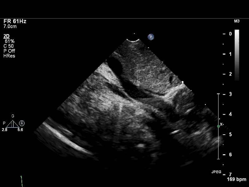
7 What you want to see
8 What you do see X
 Poor perfusion Gallop rhythm, no")
9 Our Neonate HR 195, on ventilator, BP 45/25, SaO2 40 s (pre and post-ductal) Poor perfusion Gallop rhythm, no murmurs
10 Differential diagnosis? Meconium aspiration / CDH / HMD Tetralogy of Fallot Transposition of the Great Arteries Tricuspid Atresia Truncus Arteriosus Terrible HLHS Total Anomalous Pulmonary Venous Connection

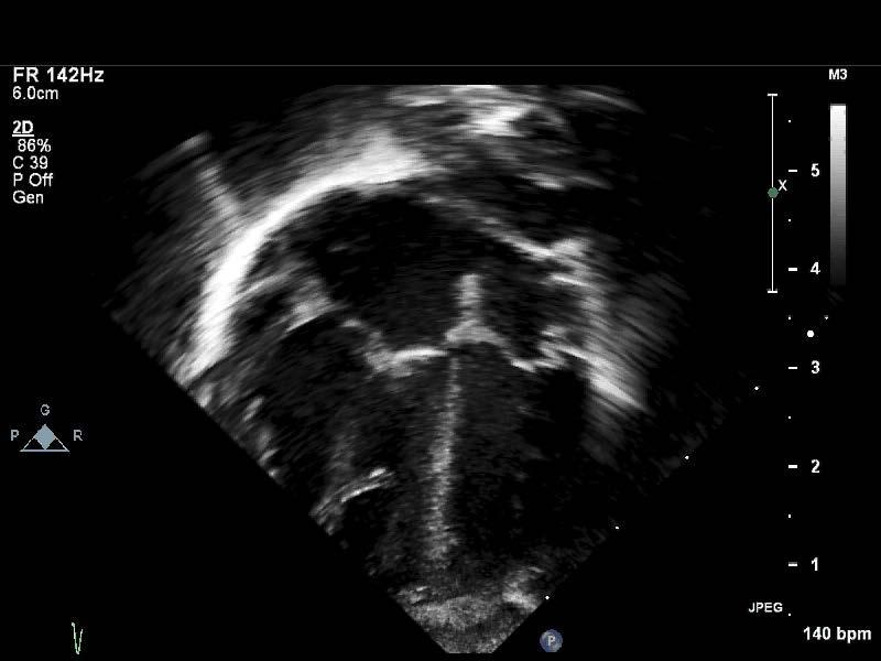
11 What you want to see

12 What you do see
13 Key Features of Our Echo Small appearing left heart

14 Key Features of Our Echo Pure R -> L shunt at PFO

15 Key Features of Our Echo P Veins to confluence


16 Key Features of Our Echo
17 TAPVR and Its Subtypes
18 Definition All pulmonary venous blood flow returns anomalously to the systemic veins or directly to the right atrium Prevalence estimated at 1 in 10,000 CDC, heartdefects/tapvr.html
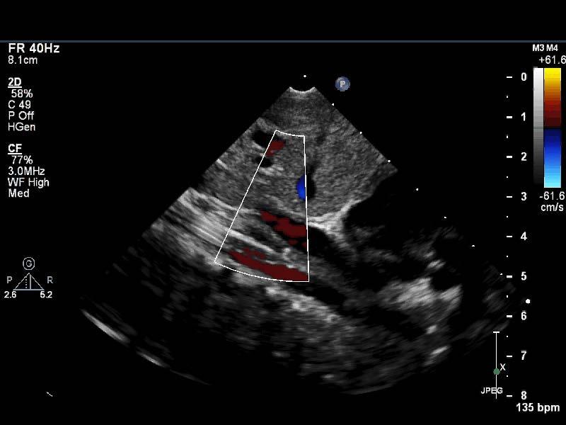
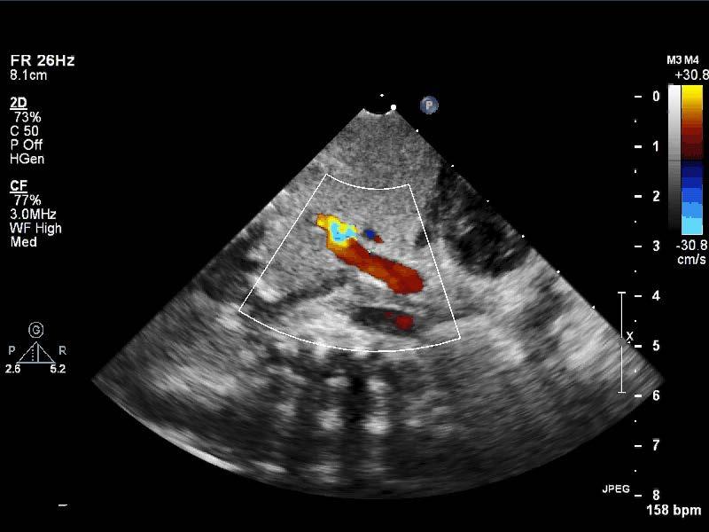
19 Embryology Early gestation lung buds drain to the common cardinal systems (right forms SVC and azygous, left forms LSVC and CS) and the umbilicovitelline system (forms the IVC, ductus venosus, and portal system) Day the common pulmonary vein (CPV) begins to develop, and by day 30 connects with pulmonary venous plexus
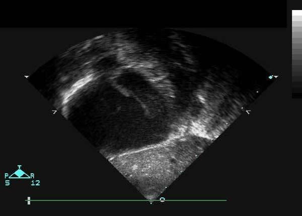
20 Embryology Failure of the CPV to connect to pulmonary venous plexus leads to persistence of one of the primitive connections Failure of the septum primum to normally form can lead to pulmonary veins connecting directly to the RA Failure to incorporate the common pulmonary vein leads to cor triatriatum

21 Anatomic Subtypes Type 1: Supracardiac (43-50%) Type 2: Cardiac (18-20%) Type 3: Infracardiac (20-27%) Type 4: Mixed (10-12%) Non-obstructed vs Obstructed (for all types)
22 Type 1: Supracardiac Vertical Vein most often drains to LIV CDC, heartdefects/tapvr.html
 CDC, http://www.cdc.gov/ncbddd/ heartdefects/tapvr.")
23 Type 1: Supracardiac Vertical Vein most often drains to LIV Can course between LPA and left mainstem bronchus - > VICE May present obstructed (around 50%) CDC, heartdefects/tapvr.html

24 Typically to the Coronary Sinus Least likely to be obstructed Can present later (few months) Type 2: Cardiac University of Chicago, total-anomalous-pulmonary-venous-connection-tapvc
")

25 Descending vein to portal vein, IVC, hepatic vein, or ductus venosus (umbiicovitelline venous system) Classic teaching is they are ALWAYS obstructed -> present at birth University of Chicago, Type 3: Infracardiac (Infradiaphragmatic)
26 The Importance of the PFO/ ASD ALL preload to the LV (aka systemic cardiac output) is supplied by right to left shunting across the atrial communication
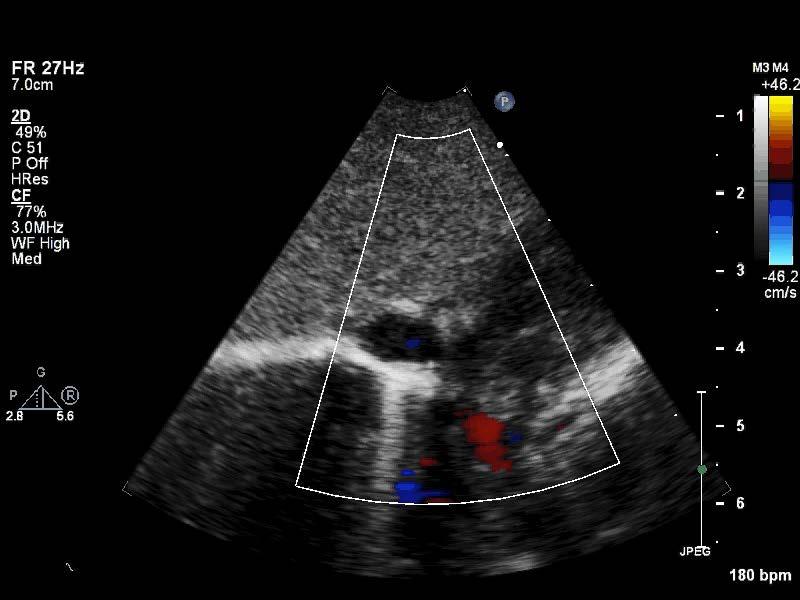
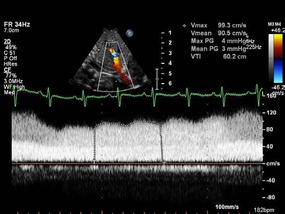
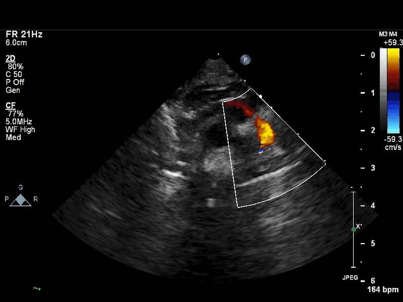
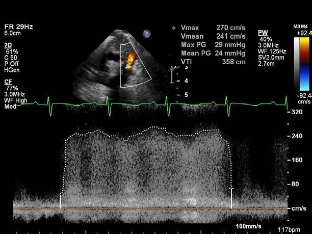
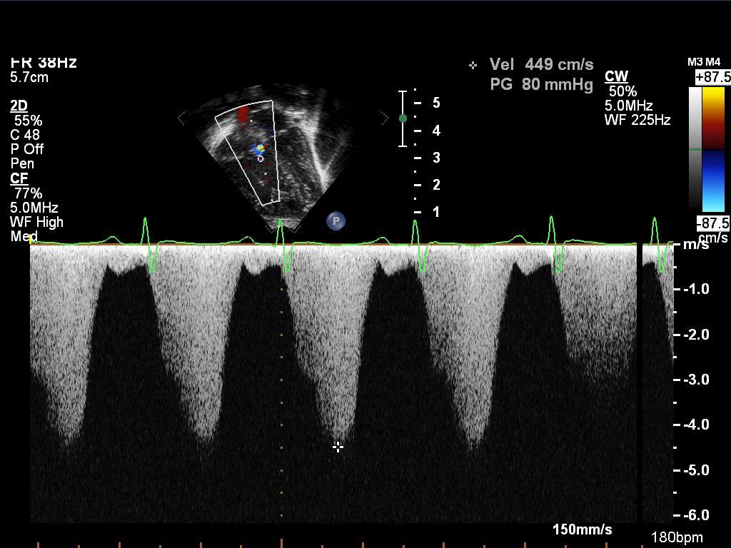
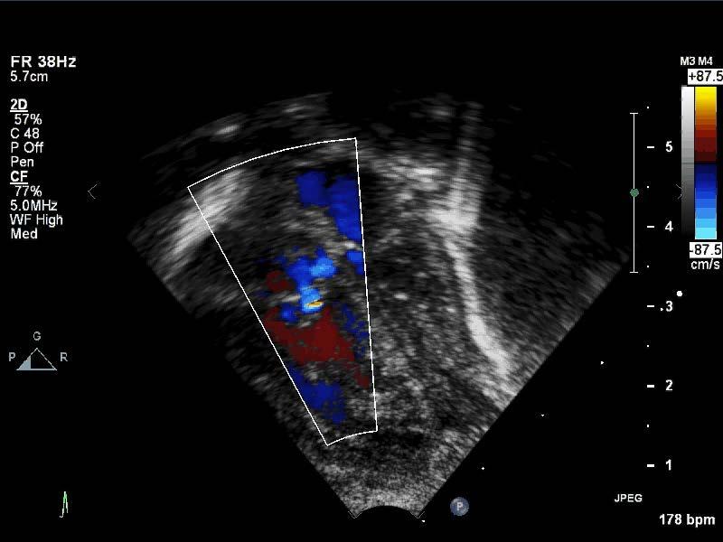
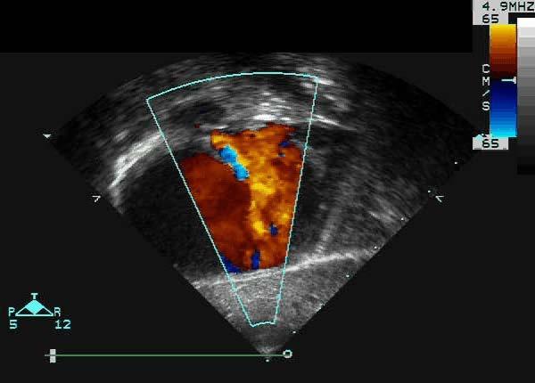
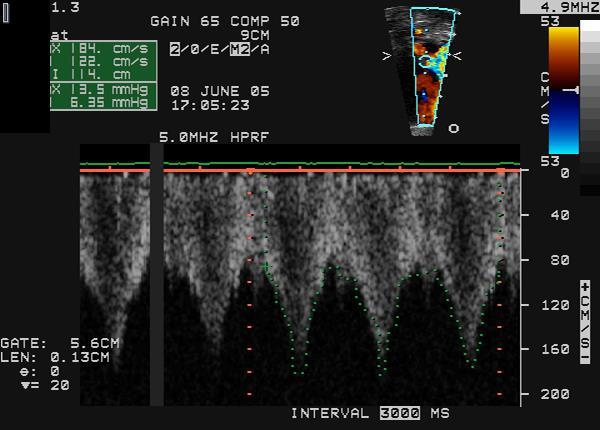
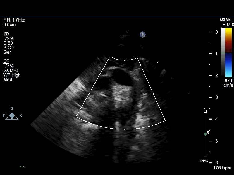
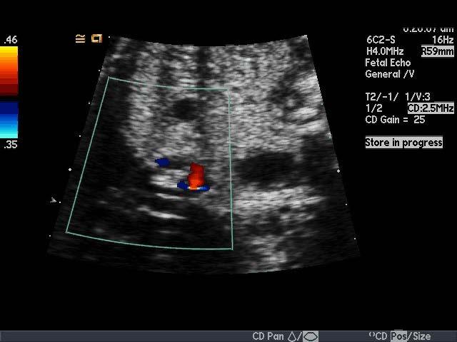
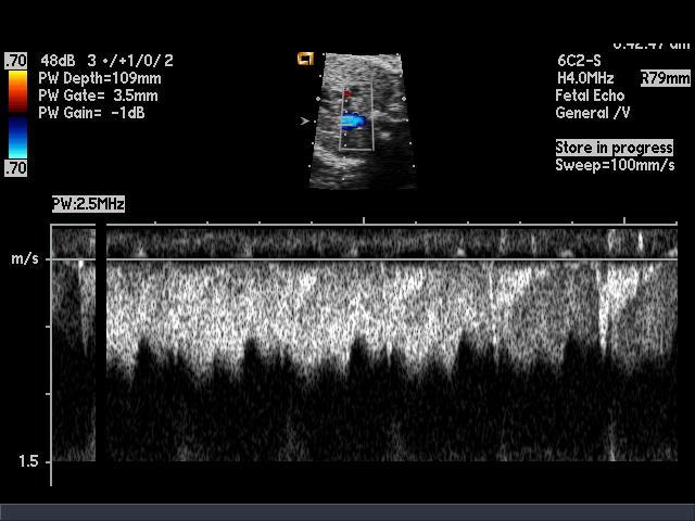
27 Echocardiographic Features

28 Confluence


29 Visualize All Pulmonary Veins RUPV LUPV RLPV LLPV

30 Type 1 to the LIV


31 Type 2 to the CS
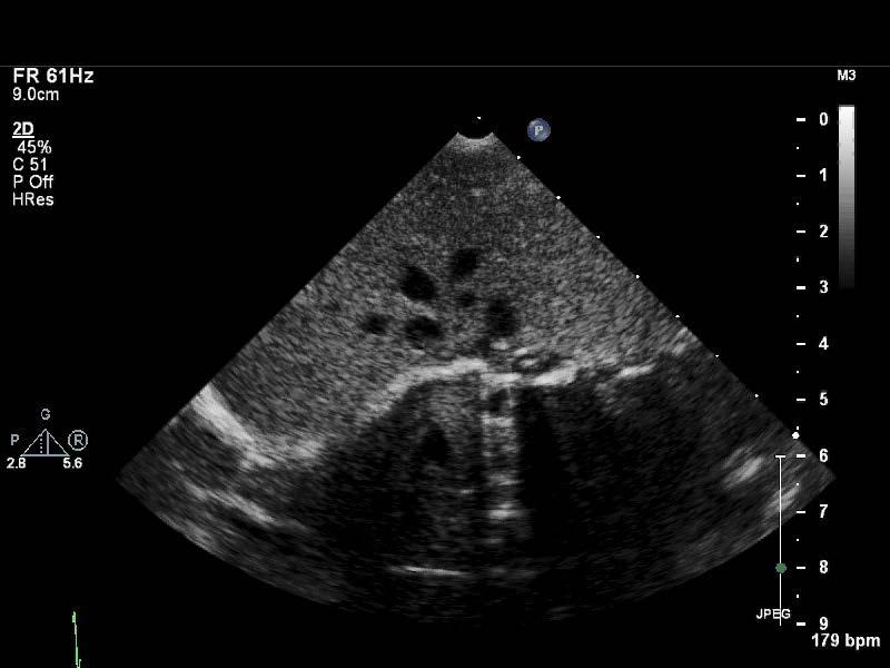

32 Type 3 - Infradiaphragmatic
33 Type 3 Infradiaphragmatic Unobstructed?
34 Type 3 Infradiaphragmatic Obstructed
35 Type 2 Again?


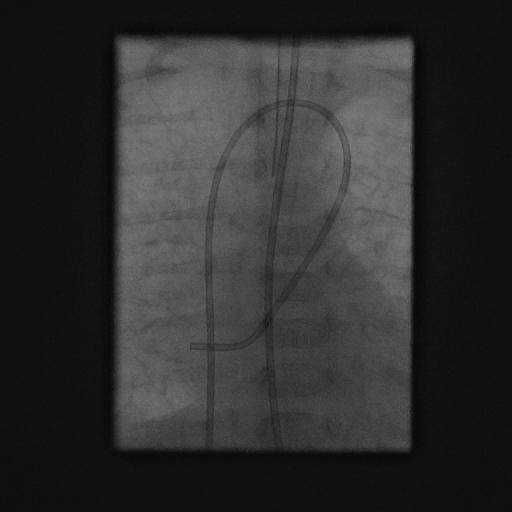
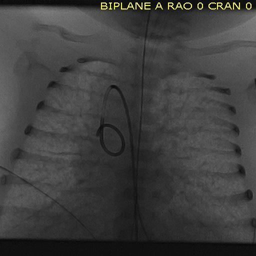
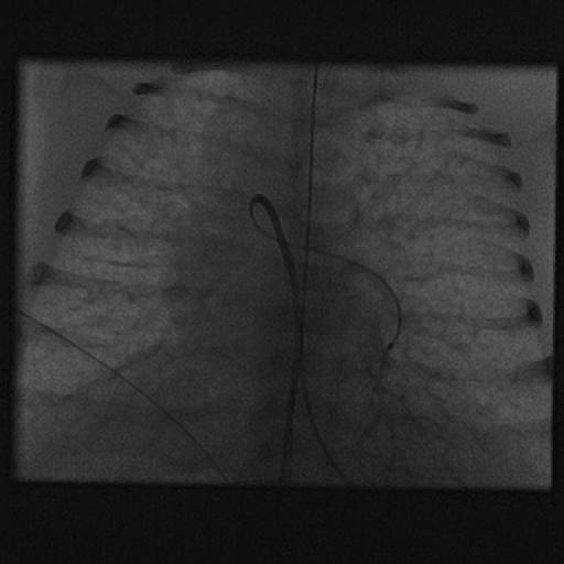
36 Type 4 Mixed

37 All types: Doppler to Assess for Obstruction
38 Small Left Heart

39 Pulmonary Hypertension
40 Atrial Communication
41 Atrial Communication
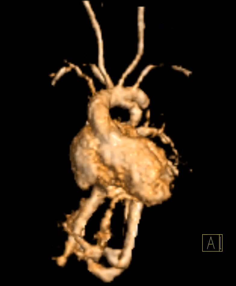
42 PDA
43 Alternative Imaging Options


44 Very Challenging to Diagnose Prenatally Estimated that pulmonary veins carry only ~5% of pulmonary blood flow in utero

45 In Utero Diagnosis
46 Cardiac Catheterization


47 Cardiac Catheterization X
48 Cardiac Catheterization Stenting VV
49 Cardiac Catheterization CPV Atresia
50 Cardiac Catheterization CPV Atresia

51 TEE Not utilized much as can obstruct the confluence / drainage


52 CT Angiogram Images courtesy of Dr. Tony Hlavacek, MUSC

53 Cardiac MRI

54 Follow up Cardiac MRI
55 Back to Our Patient
56 Emergent Surgery More to come in the next talk


57 Clinical Course ICU for 16 days, total LOS 20 days Discharged on room air, full oral feeds, on twice daily diuretics as only medication
58 Clinical Course Now 3 years old Some mild developmental delays Clinically asymptomatic Mild flow acceleration at pulmonary venous anastomosis, no PH, normal function
59 Conclusions TTE remains the mainstay in diagnosis of TAPVR Venous flow away from the heart is BAD! aka Red venous flow signals on either suprasternal notch or abdominal imaging planes are BAD! Visualization of the individual veins, confluence, and course of drainage are critical to accurate diagnosis
60 Conclusions Always assess the PFO, pure right to left shunting is BAD! Supracardiac TAPVR is the most common, and roughly 50% present obstructed Infracardiac are nearly all obstructed at birth For mixed TAPVR or if the diagnosis is unclear, CTA and Cardiac MRI can be helpful Cath is primarily reserved for rare cases where pre-operative intervention is needed
61 Thank You
Surgical Management Of TAPVR. Daniel A. Velez, M.D. Congenital Cardiac Surgeon Phoenix Children s Hospital
 Surgical Management Of TAPVR Daniel A. Velez, M.D. Congenital Cardiac Surgeon Phoenix Children s Hospital No Disclosures Goals Review the embryology and anatomy Review Surgical Strategies for repair Discuss
Screening for Critical Congenital Heart Disease
 Screening for Critical Congenital Heart Disease Caroline K. Lee, MD Pediatric Cardiology Disclosures I have no relevant financial relationships or conflicts of interest 1 Most Common Birth Defect Most
5.8 Congenital Heart Disease
 5.8 Congenital Heart Disease Congenital heart diseases (CHD) refer to structural or functional heart diseases, which are present at birth. Some of these lesions may be discovered later. prevalence of Chd
The Physiology of the Fetal Cardiovascular System
 The Physiology of the Fetal Cardiovascular System Jeff Vergales, MD, MS Department of Pediatrics Division of Pediatric Cardiology jvergales@virginia.edu Disclosures I serve as the medical director for
Congenital Heart Disease: Physiology and Common Defects
 Congenital Heart Disease: Physiology and Common Defects Jamie S. Sutherell, M.D, M.Ed. Associate Professor, Pediatrics Division of Cardiology Director, Medical Student Education in Pediatrics Director,
Congenital Heart Disease
 Congenital Heart Disease Mohammed Alghamdi, MD, FRCPC, FAAP, FACC Associate Professor and Consultant Pediatric Cardiology, Cardiac Science King Fahad Cardiac Centre King Saud University INTRODUCTION CHD
Cardiovascular Pathophysiology: Right to Left Shunts aka Cyanotic Lesions
 Cardiovascular Pathophysiology: Right to Left Shunts aka Cyanotic Lesions Ismee A. Williams, MD, MS iib6@columbia.edu Pediatric Cardiology Learning Objectives To discuss the hemodynamic significance of
Cardiovascular Pathophysiology: Right to Left Shunts aka Cyanotic Lesions Ismee A. Williams, MD, MS Pediatric Cardiology
 Cardiovascular Pathophysiology: Right to Left Shunts aka Cyanotic Lesions Ismee A. Williams, MD, MS iib6@columbia.edu Pediatric Cardiology Learning Objectives To discuss the hemodynamic significance of
Cardiac Catheterization Cases Primary Cardiac Diagnoses Facility 12 month period from to PRIMARY DIAGNOSES (one per patient)
") PRIMARY DIAGNOSES (one per patient) Septal Defects ASD (Atrial Septal Defect) PFO (Patent Foramen Ovale) ASD, Secundum ASD, Sinus venosus ASD, Coronary sinus ASD, Common atrium (single atrium) VSD (Ventricular
PRIMARY DIAGNOSES (one per patient) Septal Defects ASD (Atrial Septal Defect) PFO (Patent Foramen Ovale) ASD, Secundum ASD, Sinus venosus ASD, Coronary sinus ASD, Common atrium (single atrium) VSD (Ventricular
ECHOCARDIOGRAPHIC APPROACH TO CONGENITAL HEART DISEASE: THE UNOPERATED ADULT
 ECHOCARDIOGRAPHIC APPROACH TO CONGENITAL HEART DISEASE: THE UNOPERATED ADULT Karen Stout, MD, FACC Divisions of Cardiology University of Washington Medical Center Seattle Children s Hospital NO DISCLOSURES
When is Risky to Apply Oxygen for Congenital Heart Disease 부천세종병원 소아청소년과최은영
 When is Risky to Apply Oxygen for Congenital Heart Disease 부천세종병원 소아청소년과최은영 The Korean Society of Cardiology COI Disclosure Eun-Young Choi The author have no financial conflicts of interest to disclose
Cardiac CT in Infants with Congenital heart disease Sunrise Session. LaDonna Malone, MD May 17, 2018
 Cardiac CT in Infants with Congenital heart disease Sunrise Session LaDonna Malone, MD May 17, 2018 None Disclosures Objectives Describe cardiac CT techniques used in infants with congenital heart disease.
Atrial Septal Defects
 Supplementary ACHD Echo Acquisition Protocol for Atrial Septal Defects The following protocol for echo in adult patients with atrial septal defects (ASDs) is a guide for performing a comprehensive assessment
Adult Congenital Heart Disease: What All Echocardiographers Should Know Sharon L. Roble, MD, FACC Echo Hawaii 2016
 1 Adult Congenital Heart Disease: What All Echocardiographers Should Know Sharon L. Roble, MD, FACC Echo Hawaii 2016 DISCLOSURES I have no disclosures relevant to today s talk 2 Why should all echocardiographers
Congenital Heart Disease An Approach for Simple and Complex Anomalies
 Congenital Heart Disease An Approach for Simple and Complex Anomalies Michael D. Pettersen, MD Director, Echocardiography Rocky Mountain Hospital for Children Denver, CO None Disclosures 1 ASCeXAM Contains
Critical Heart Disease in the Newborn. What you need to know
 Critical Heart Disease in the Newborn What you need to know DISCLOSURES Nothing to report OBJECTIVES DESCRIBE NEONATAL CARDIOVASCULAR PHYSIOLOGY RECOGNIZE NEONATAL CARDIAC EMERGENCIES FORMULATE TREATMENT
Hypoplastic Left Heart Syndrome: Echocardiographic Assessment
 Hypoplastic Left Heart Syndrome: Echocardiographic Assessment Craig E Fleishman, MD, FACC, FASE Director, Non-invasive Cardiac Imaging The Hear Center at Arnold Palmer Hospital for Children, Orlando SCAI
Infradiaphragmatic Total Anomalous Pulmonary Venous Return
 Infradiaphragmatic Total Anomalous Pulmonary Venous Return Report of a New and Correctable Variant J. Kent Trinkle, M.D., Gordon K. Danielson, M.D., Jacqueline A. Noonan, M.D., and Charles Stephens, M.D.
PULMONARY VENOLOBAR SYNDROME. Dr.C.Anandhi DNB Resident, Southern Railway Headquarters Hospital.
 PULMONARY VENOLOBAR SYNDROME Dr.C.Anandhi DNB Resident, Southern Railway Headquarters Hospital. Presenting complaint: 10 yrs old girl with recurrent episodes of lower respiratory tract infection from infancy.
Glenn Shunts Revisited
 Glenn Shunts Revisited What is a Super Glenn Patricia O Brien, MSN, CPNP-AC Nurse Practitioner, Pediatric Cardiology No Disclosures Single Ventricle Anatomy Glenn Shunt Cavopulmonary Anastomosis Anastomosis
3/14/2011 MANAGEMENT OF NEWBORNS CARDIAC INTENSIVE CARE CONFERENCE FOR HEALTH PROFESSIONALS IRVINE, CA. MARCH 7, 2011 WITH HEART DEFECTS
 CONFERENCE FOR HEALTH PROFESSIONALS IRVINE, CA. MARCH 7, 2011 MANAGEMENT OF NEWBORNS WITH HEART DEFECTS A NTHONY C. CHANG, MD, MBA, MPH M E D I C AL D I RE C T OR, HEART I N S T I T U T E C H I LDRE N
Heart and Soul Evaluation of the Fetal Heart
 Heart and Soul Evaluation of the Fetal Heart Ivana M. Vettraino, M.D., M.B.A. Clinical Associate Professor, Michigan State University College of Human Medicine Objectives Review the embryology of the formation
Common Defects With Expected Adult Survival:
 Common Defects With Expected Adult Survival: Bicuspid aortic valve :Acyanotic Mitral valve prolapse Coarctation of aorta Pulmonary valve stenosis Atrial septal defect Patent ductus arteriosus (V.S.D.)
Congenital Heart Defects
 Normal Heart Congenital Heart Defects 1. Patent Ductus Arteriosus The ductus arteriosus connects the main pulmonary artery to the aorta. In utero, it allows the blood leaving the right ventricle to bypass
Absent Pulmonary Valve Syndrome
 Absent Pulmonary Valve Syndrome Fact sheet on Absent Pulmonary Valve Syndrome In this condition, which has some similarities to Fallot's Tetralogy, there is a VSD with narrowing at the pulmonary valve.
CONGENITAL HEART DISEASE (CHD)
") CONGENITAL HEART DISEASE (CHD) DEFINITION It is the result of a structural or functional abnormality of the cardiovascular system at birth GENERAL FEATURES OF CHD Structural defects due to specific disturbance
CONGENITAL HEART DISEASE (CHD) DEFINITION It is the result of a structural or functional abnormality of the cardiovascular system at birth GENERAL FEATURES OF CHD Structural defects due to specific disturbance
Pediatric Board Review Congenital Heart Disease. Steven H. Todman, M.D. Pediatric Cardiologist Louisiana State University
 Pediatric Board Review Congenital Heart Disease Steven H. Todman, M.D. Pediatric Cardiologist Louisiana State University Our Mission To discuss various types of congenital heart disease that are commonly
Anomalous pulmonary venous connection: the journey from theory to practice!
 Anomalous pulmonary venous connection: the journey from theory to practice! Poster No.: C-1245 Congress: ECR 2016 Type: Educational Exhibit Authors: C. Schiau, S. Manole; Cluj-Napoca/RO Keywords: Congenital,
The Blue Baby. Network Stabilisation of the Term Infant Study Day 15 th March 2017 Joanna Behrsin
 The Blue Baby Network Stabilisation of the Term Infant Study Day 15 th March 2017 Joanna Behrsin Session Structure Definitions and assessment of cyanosis Causes of blue baby Structured approach to assessing
9/8/2009 < 1 1,2 3,4 5,6 7,8 9,10 11,12 13,14 15,16 17,18 > 18. Tetralogy of Fallot. Complex Congenital Heart Disease.
 Current Indications for Pediatric CTA S Bruce Greenberg Professor of Radiology Arkansas Children s Hospital University of Arkansas for Medical Sciences greenbergsbruce@uams.edu 45 40 35 30 25 20 15 10
Cardiac Emergencies in Infants. Michael Luceri, DO
 Cardiac Emergencies in Infants Michael Luceri, DO October 7, 2017 I have no financial obligations or conflicts of interest to disclose. Objectives Understand the scope of congenital heart disease Recognize
Hybrid Stage I Palliation / Bilateral PAB
 Hybrid Stage I Palliation / Bilateral PAB Jeong-Jun Park Dept. of Thoracic & Cardiovascular Surgery Asan Medical Center, University of Ulsan CASE 1 week old neonate with HLHS GA 38 weeks Birth weight 3.0Kg
NASCI 2012 Segmental Analysis
 NASCI 2012 Segmental Analysis Frandics Chan, M.D., Ph.D. Stanford University Medical Center Lucile Packard Department Children s of Radiology Hospital Menagerie of Congenital Cardiac Lesions 1. Absent
Isolated major aortopulmonary collateral as the sole pulmonary blood supply to an entire lung segment
 Washington University School of Medicine Digital Commons@Becker Open Access Publications 2017 Isolated major aortopulmonary collateral as the sole pulmonary blood supply to an entire lung segment Hannah
Heart and Lungs. LUNG Coronal section demonstrates relationship of pulmonary parenchyma to heart and chest wall.
 Heart and Lungs Normal Sonographic Anatomy THORAX Axial and coronal sections demonstrate integrity of thorax, fetal breathing movements, and overall size and shape. LUNG Coronal section demonstrates relationship
Tracheoesophageal fistula repair in a premature infant with VACTERL, infradiaphragmatic TAPVR, and pulmonary hypertension.
 PBLD Table # 26 Tracheoesophageal fistula repair in a premature infant with VACTERL, infradiaphragmatic TAPVR, and pulmonary hypertension. Moderators: Annette Schure, MD; Lauren Welsh, MD Objectives: 1.
Congenital Heart Disease: Cyanotic Lesions. Amitesh Aggarwal
 Congenital Heart Disease: Cyanotic Lesions Amitesh Aggarwal 12 y/o male admitted because of dyspnea and cyanosis Patient has been cyanotic since few months after birth Has episodes of tachypnea and worsening
ADULT CONGENITAL HEART DISEASE. Stuart Lilley
 ADULT CONGENITAL HEART DISEASE Stuart Lilley More adults than children have congenital heart disease Huge variety of congenital lesions from minor to major Heart failure, re-operation and arrhythmia are
Objectives Part 1. Objectives Part 2. Fetal Circulation Transition to Postnatal Circulation Normal Cardiac Anatomy Ductal Dependence and use of PGE1
 Cardiac Physiology Gia Marzano, AC PNP Pediatric Cardiac Surgery Rush Center for Congenital Heart Disease Rush University Medical Center Objectives Part 1 Fetal Circulation Transition to Postnatal Circulation
Surgical Procedures. Direct suture of small ASDs Patch repair Transcatheter closure with a prosthetic device called occluder
 PEDIATRIC Review Surgical Procedures Atrial Septal Defect repair: Direct suture of small ASDs Patch repair Transcatheter closure with a prosthetic device called occluder Balloon atrial septostomy (Rashkind)
A SURGEONS' GUIDE TO CARDIAC DIAGNOSIS
 A SURGEONS' GUIDE TO CARDIAC DIAGNOSIS PART II THE CLINICAL PICTURE DONALD N. ROSS B. Sc., M. B., CH. B., F. R. C. S. CONSULTANT THORACIC SURGEON GUY'S HOSPITAL, LONDON WITH 53 FIGURES Springer-Verlag
Pediatric Echocardiography Examination Content Outline
 Pediatric Echocardiography Examination Content Outline (Outline Summary) # Domain Subdomain Percentage 1 Anatomy and Physiology Normal Anatomy and Physiology 10% 2 Abnormal Pathology and Pathophysiology
Foetal Cardiology: How to predict perinatal problems. Prof. I.Witters Prof.M.Gewillig UZ Leuven
 Foetal Cardiology: How to predict perinatal problems Prof. I.Witters Prof.M.Gewillig UZ Leuven Cardiopathies Incidence : 8-12 / 1000 births ( 1% ) Most frequent - Ventricle Septum Defect 20% - Atrium Septum
Anatomy & Physiology
 1 Anatomy & Physiology Heart is divided into four chambers, two atrias & two ventricles. Atrioventricular valves (tricuspid & mitral) separate the atria from ventricles. they open & close to control flow
DGPK guideline: PAPVC
 DGPK guideline: PAPVC Partial anomalous pulmonary venous connection (PAPVC) Harald Bertram, Hannover Oliver Dewald, Bonn Angelika Lindinger, Kaiserslautern & Trier DGPK guideline committee No disclosures
Neonatal Cardiac Anomalies
 Objectives Neonatal Cardiac Anomalies Karen Knuth, RNC, MN, NNP-BC, ARNP Seattle Childrens Hospital What is CHD? Normal anatomy and circulation Clinical presentation: signs and symptoms Diagnostics Common
Introduction. Pediatric Cardiology. General Appearance. Tools of Assessment. Auscultation. Vital Signs
 Introduction Pediatric Cardiology An introduction to the pediatric patient with heart disease: M-III Lecture Douglas R. Allen, M.D. Assistant Professor and Director of Community Pediatric Cardiology at
Anomalous Systemic Venous Connection Systemic venous anomaly
 World Database for Pediatric and Congenital Heart Surgery Appendix B: Diagnosis (International Paediatric and Congenital Cardiac Codes (IPCCC) and definitions) Anomalous Systemic Venous Connection Systemic
Congenital heart disease: When to act and what to do?
 Leading Article Congenital heart disease: When to act and what to do? Duminda Samarasinghe 1 Sri Lanka Journal of Child Health, 2010; 39: 39-43 (Key words: Congenital heart disease) Congenital heart disease
Paediatric Cardiology. Acyanotic CHD. Prof F F Takawira
 Paediatric Cardiology Acyanotic CHD Prof F F Takawira Aetiology Chromosomal Down syndrome, T13, T18 Genetic syndromes (gene defects) Velo-Cardio-facial (22 del) Genetic syndromes (undefined aetiology)
ECHOCARDIOGRAPHIC ASSESSMENT OF TOTAL ANOMALOUS PULMONARY VENOUS CONNECTIONS IN PEDIATRIC PATIENTS
 Total nomalous Pulmonary Venous Connection ECHOCRDIOGRPHIC SSESSMENT OF TOTL NOMLOUS PULMONRY VENOUS CONNECTIONS IN PEDITRIC PTIENTS Meng-Luen Lee, Mei-Hwan Wu, 1 Jou-Kou Wang, 1 and Hung-Chi Lue 1 ackground
11/27/2012. Objectives. What is Critical Congenital Heart Disease?
 Screening for Critical Congenital Heart Disease in the Apparently Healthy Newborn A presentation of Texas Pulse Oximetry Project: A Joint Educational Initiative of The University of Texas Health Science
Esophageal varices in congenital heart disease with total anomalous pulmonary venous connection
 The International Journal of Cardiac Imaging 16: 405±409, 2000. Ó 2000 Kluwer Academic Publishers. Printed in the Netherlands. 405 Esophageal varices in congenital heart disease with total anomalous pulmonary
Transposition of the Great Arteries (TGA)
") Transposition of the Great Arteries (TGA) Noelle Layer, HMS III Gillian Lieberman, MD June 2008 Agenda Background of TGA Epidemiology Anatomy Clinical presentation Meet Baby L: Differential diagnosis Menu
Transposition of the Great Arteries (TGA) Noelle Layer, HMS III Gillian Lieberman, MD June 2008 Agenda Background of TGA Epidemiology Anatomy Clinical presentation Meet Baby L: Differential diagnosis Menu
DEVELOPMENT OF THE CIRCULATORY SYSTEM L E C T U R E 5
 DEVELOPMENT OF THE CIRCULATORY SYSTEM L E C T U R E 5 REVIEW OF CARDIAC ANATOMY Heart 4 chambers Base and apex Valves Pericardial sac 3 layers: epi, myo, endo cardium Major blood vessels Aorta and its
MEDICAL MANAGEMENT WITH CAVEATS 1. In one study of 50 CHARGE patients with CHD, 75% required surgery. 2. Children with CHARGE may be resistant to chlo
 CARDIOLOGY IN CHARGE SYNDROME: FOR THE PHYSICIAN Angela E. Lin, M.D. Teratology Program/Active Malformation Surveillance, Brigham and Women's Hospital, Old PBBH-B501, 75 Francis St., Boston, MA 02115 alin@partners.org
Multimodality Imaging of Septal Defects
 Multimodality Imaging of Septal Defects Ohio-ACC 2018 Annual Meeting October 27, 2018 Kan N. Hor, MD Director, Cardiac Magnetic Resonance Imaging Associate Professor of Pediatrics The Heart Center, Nationwide
Slide 1. Slide 2. Slide 3 CONGENITAL HEART DISEASE. Papworth Hospital NHS Trust INTRODUCTION. Jakub Kadlec/Catherine Sudarshan INTRODUCTION
 Slide 1 CONGENITAL HEART DISEASE Jakub Kadlec/Catherine Sudarshan NHS Trust Slide 2 INTRODUCTION Most common congenital illness in the newborn Affects about 4 9 / 1000 full-term live births in the UK 1.5
Objectives. Prenatal Diagnosis of Critical Congenital Heart Disease. The Law. Disclosure. Dylan s Story
 Keeping the Beat: Pediatric Cardiac Screening and Management of Congenital Heart Disease Prenatal Diagnosis of Critical Congenital Heart Disease Robert Koppel, MD Neonatal/Perinatal Medicine, Pediatrics
By Dickens ATURWANAHO & ORIBA DAN LANGOYA MAKchs, MBchB CONGENTAL HEART DISEASE
 By Dickens ATURWANAHO & ORIBA DAN LANGOYA MAKchs, MBchB CONGENTAL HEART DISEASE Introduction CHDs are abnormalities of the heart or great vessels that are present at birth. Common type of heart disease
The Fetal Cardiology Program
 The Fetal Cardiology Program at Texas Children s Fetal Center About the program Since the 1980s, Texas Children s Fetal Cardiology Program has provided comprehensive fetal cardiac care to expecting families
Neonatal Single Ventricle Heart Disease Recognition, Management, Counseling
 Neonatal Single Ventricle Heart Disease Recognition, Management, Counseling Christopher J. Petit MD Assistant Professor, Pediatric Cardiology Director, Single Ventricle Program Baylor College of Medicine,
5/22/2013. Alan Zuckerman 1, Swapna Abhyankar 1, Tiffany Colarusso 2, Richard Olney 2, Kristin Burns 3, Marci Sontag 4
 Alan Zuckerman 1, Swapna Abhyankar 1, Tiffany Colarusso 2, Richard Olney 2, Kristin Burns 3, Marci Sontag 4 1 National Library of Medicine, NIH, Bethesda, MD, USA, 2 Centers for Disease Control and Prevention,
MR Imaging and CT Evaluation of Congenital Pulmonary Vein Abnormalities. Infants 1 PEDIATRIC IMAGING
 Note: This copy is for your personal non-commercial use only. To order presentation-ready copies for distribution to your colleagues or clients, contact us at www.rsna.org/rsnarights. PEDIATRIC IMAGING
DISCLOSURE OBJECTIVES PULMONARY VEIN STENOSIS DIAGNOSTIC TOOLS. Echo with Doppler Catheterization with angiography CT angiography MRI
 1 2 ND INTERNATIONAL CONFERENCE: NEONATAL AND CHILDHOOD PULMONARY VASCULAR DISEASE, MARCH 13-14, 2009, SAN FRANCISCO, USA PATHOPHYSIOLOGY OF PULMONARY VEIN FLOW: IMAGING NORMAL AND ABNORMAL PULMONARY VEIN
Congenital Heart Disease. CCCHD In WI. Critical Congenital Heart Disease. Why Screen? 4/20/2018. Early Detection = Better Outcomes
 Congenital Heart Disease A Positive Screen? What Does it Mean? A Review of Pulse Oximetry Screening for Critical Congenital Heart Disease Elizabeth Goetz MD MPH 8-10/1000 livebirths 3% of all infant mortality
CongHeartDis.doc. Андрій Миколайович Лобода
 CongHeartDis.doc Андрій Миколайович Лобода 2015 Зміст 3 Зміст Зміст 4 A child with tetralogy of Fallot is most likely to exhibit: -Increased pulmonary blood flow -Increased pressure in the right ventricle
2D/3D in Evaluation of Atrial Septum
 2D/3D in Evaluation of Atrial Septum Roberto M Lang, MD OSTIUM SECUNDUM ASD: 2D AND 3D TNSESOPHAGEAL ECHO 1 Biplane views 90 0 3D Acquisi on Acquire 3D volume Lang RM et al. JASE 2012;25:3 46. Right atrial
Data Collected: June 17, Reported: June 30, Survey Dates 05/24/ /07/2010
 Job Task Analysis for ARDMS Pediatric Echocardiography Data Collected: June 17, 2010 Reported: Analysis Summary For: Pediatric Echocardiography Exam Survey Dates 05/24/2010-06/07/2010 Invited Respondents
How to Recognize a Suspected Cardiac Defect in the Neonate
 Neonatal Nursing Education Brief: How to Recognize a Suspected Cardiac Defect in the Neonate https://www.seattlechildrens.org/healthcareprofessionals/education/continuing-medical-nursing-education/neonatalnursing-education-briefs/
CCHD Screening with Pulse Oximetry: A Success Story!
 CCHD Screening with Pulse Oximetry: A Success Story! Nicole Spillane, MD Associate Director of Neonatology, HackensackUMC Hackensack Meridian Health Objective Recognize the contribution of pulse oximetry
ORIGINAL RESEARCH PAPER
 ORIGINAL RESEARCH PAPER ROLE OF CT PULMONARY ANGIOGRAPHY IN CONGENITAL HEART DISEASES IN PAEDIATRIC POPULATION Radiology KEY WORDS: Congenital heart disease, CT pulmonary angiography, pediatric heart disease,
Segmental Analysis. Gautam K. Singh, M.D. Washington University School of Medicine St. Louis
 Segmental Analysis Gautam K. Singh, M.D. Washington University School of Medicine St. Louis Segmental Analysis Segmental Analysis: From Veins to Ventricles Segmental Approach to Evaluation of Congenital
All You Need to Know About Situs and Looping Disorders: Embryology, Anatomy, and Echocardiography
 All You Need to Know About Situs and Looping Disorders: Embryology, Anatomy, and Echocardiography Helena Gardiner Co-Director of Fetal Cardiology, The Fetal Center, University of Texas at Houston Situs
Adults with Congenital Heart Disease
 Adults with Congenital Heart Disease Edward K. Rhee, MD, FACC Director, Pediatric-Adult Congenital Arrhythmia Service SJHMC Disclosures & Disclaimer I have no lucrative financial relationships with industry
Born Blue. Anesthesia and CHD. Kristine Faust, CRNA, MS, MBA, DNAP
 Born Blue Anesthesia and CHD Kristine Faust, CRNA, MS, MBA, DNAP Disclosures Disclosures None to Report Objectives Review all congenital defects in which the patient is blue Describe physiology of the
Atrial Septostomy in HLHS and ECMO: Indications, Technique and Outcome
 Atrial Septostomy in HLHS and ECMO: Indications, Technique and Outcome Dr Damien Kenny, MB, MD Assistant Professor of Pediatrics Director of the Cardiac Catheterization Hybrid Suite Co-Director of the
Survival Rates of Children with Congenital Heart Disease continue to improve.
 DOROTHY RADFORD Survival Rates of Children with Congenital Heart Disease continue to improve. 1940-20% 1960-40% 1980-70% 2010->90% Percentage of children with CHD reaching age of 18 years 1938 First Patent
Cardiology Fellowship Manual. Goals & Objectives -Cardiac Imaging- 1 P a g e
 Cardiology Fellowship Manual Goals & Objectives -Cardiac Imaging- 1 P a g e UNIV. OF NEBRASKA CHILDREN S HOSPITAL & MEDICAL CENTER DIVISION OF CARDIOLOGY FELLOWSHIP PROGRAM CARDIAC IMAGING ROTATION GOALS
Congenital heart disease. By Dr Saima Ali Professor of pediatrics
 Congenital heart disease By Dr Saima Ali Professor of pediatrics What is the most striking clinical finding in this child? Learning objectives By the end of this lecture, final year student should be able
Notes: 1)Membranous part contribute in the formation of small portion in the septal cusp.
Membranous part contribute in the formation of small portion in the septal cusp.") Embryology 9 : Slide 16 : There is a sulcus between primitive ventricular and bulbis cordis that will disappear gradually and lead to the formation of one chamber which is called bulboventricular chamber.
Embryology 9 : Slide 16 : There is a sulcus between primitive ventricular and bulbis cordis that will disappear gradually and lead to the formation of one chamber which is called bulboventricular chamber.
بسم هللا الرحمن الرحيم. The cardio vascular system By Dr.Rawa Younis Mahmood
 بسم هللا الرحمن الرحيم The cardio vascular system By Dr.Rawa Younis Mahmood Introduction Evaluation of the cardio vascular system depend on history and physical examination by : Asking about cyanosis,blueness
Hypoplastic Left Heart Syndrome and Obstructive Total Anomalous Pulmonary Venous Connection: A Rare and Severe Association
 Hypoplastic Left Heart Syndrome and Obstructive Total Anomalous Pulmonary Venous Connection: A Rare and Severe Association Claudia Martins Cosentino, Karen Saori Shiraishi, Ana Karina Spuras Stella, Tamara
SURGICAL TREATMENT AND OUTCOME OF CONGENITAL HEART DISEASE
 SURGICAL TREATMENT AND OUTCOME OF CONGENITAL HEART DISEASE Mr. W. Brawn Birmingham Children s Hospital. Aims of surgery The aim of surgery in congenital heart disease is to correct or palliate the heart
Research article. Primary detection of congenital heart diseases in the Kyrgyz Republic
 Research article Primary detection of congenital heart diseases in the Kyrgyz Republic Irina A. Akhmedova, Gulzada A. Imanalieva, Damir A.Abibillaev, Taalaibek Z. Kudaiberdiev Scientific Research Institute
Objective 2/9/2012. Blood Gas Analysis In The Univentricular Patient: The Need For A Different Perspective. VENOARTERIAL CO2 GRADIENT
 Blood Gas Analysis In The Univentricular Patient: The Need For A Different Perspective. Gary Grist RN CCP Chief Perfusionist The Children s Mercy Hospitals and Clinics Kansas City, Mo. Objective The participant
"Lecture Index. 1) Heart Progenitors. 2) Cardiac Tube Formation. 3) Valvulogenesis and Chamber Formation. 4) Epicardium Development.
 Heart Progenitors. 2) Cardiac Tube Formation. 3) Valvulogenesis and Chamber Formation. 4) Epicardium Development.") "Lecture Index 1) Heart Progenitors. 2) Cardiac Tube Formation. 3) Valvulogenesis and Chamber Formation. 4) Epicardium Development. 5) Septation and Maturation. 6) Changes in Blood Flow during Development.
"Lecture Index 1) Heart Progenitors. 2) Cardiac Tube Formation. 3) Valvulogenesis and Chamber Formation. 4) Epicardium Development. 5) Septation and Maturation. 6) Changes in Blood Flow during Development.
Coarctation of the aorta
 T H E P E D I A T R I C C A R D I A C S U R G E R Y I N Q U E S T R E P O R T Coarctation of the aorta In the normal heart, blood flows to the body through the aorta, which connects to the left ventricle
Preoperative Echocardiographic Assessment of Uni-ventricular Repair
 Preoperative Echocardiographic Assessment of Uni-ventricular Repair Salem Deraz, MD Pediatric Cardiologist, Aswan Heart Centre Magdi Yacoub Heart Foundation Uni-ventricular repair A single or series of
Talking with Parents about a Positive Critical Congenital Heart Disease (CCHD) Screen or Test. Emily Drake, PhD RN May 20, 2014.
 Screen or Test. Emily Drake, PhD RN May 20, 2014.") Talking with Parents about a Positive Critical Congenital Heart Disease (CCHD) Screen or Test Emily Drake, PhD RN May 20, 2014 Disclosure I have no financial or other conflicts of interest 1 Learning Objectives
Talking with Parents about a Positive Critical Congenital Heart Disease (CCHD) Screen or Test Emily Drake, PhD RN May 20, 2014 Disclosure I have no financial or other conflicts of interest 1 Learning Objectives
The Chest X-ray for Cardiologists
 Mayo Clinic & British Cardiovascular Society at the Royal College of Physicians, London : 21-23-October 2013 Cases-Controversies-Updates 2013 The Chest X-ray for Cardiologists Michael Rubens Royal Brompton
Chapter 2 Cardiac Interpretation of Pediatric Chest X-Ray
 Chapter 2 Cardiac Interpretation of Pediatric Chest X-Ray Ra-id Abdulla and Douglas M. Luxenberg Key Facts The cardiac silhouette occupies 50 55% of the chest width on an anterior posterior chest X-ray
Pathophysiology: Left To Right Shunts
 Pathophysiology: Left To Right Shunts Daphne T. Hsu, MD dh17@columbia.edu Learning Objectives Learn the relationships between pressure, blood flow, and resistance Review the transition from fetal to mature
CYANOTIC CONGENITAL HEART DISEASES. PRESENTER: DR. Myra M. Koech Pediatric cardiologist MTRH/MU
 CYANOTIC CONGENITAL HEART DISEASES PRESENTER: DR. Myra M. Koech Pediatric cardiologist MTRH/MU DEFINITION Congenital heart diseases are defined as structural and functional problems of the heart that are
Echocardiography in Adult Congenital Heart Disease
 Echocardiography in Adult Congenital Heart Disease Michael Vogel Kinderherz-Praxis München CHD missed in childhood Subsequent lesions after repaired CHD Follow-up of cyanotic heart disease CHD missed in
Complex Congenital Heart Disease in Adults
 Complex Congenital Heart Disease in Adults Linda B. Haramati, MD Disclosures Complex Congenital Heart Disease in Adults Linda B. Haramati MD, MS Jeffrey M. Levsky MD, PhD Meir Scheinfeld MD, PhD Department
Appendix A.1: Tier 1 Surgical Procedure Terms and Definitions
 Appendix A.1: Tier 1 Surgical Procedure Terms and Definitions Tier 1 surgeries AV Canal Atrioventricular Septal Repair, Complete Repair of complete AV canal (AVSD) using one- or two-patch or other technique,
Admixture lesions in congenital cyanotic heart disease
 HEMODYNAMIC ROUNDS Admixture lesions in congenital cyanotic heart disease Jaganmohan A Tharakan Department of Cardiology, Sree Chitra Tirunal Institute for Medical Sciences and Technology, Trivandrum,
Pathophysiology: Left To Right Shunts
 Pathophysiology: Left To Right Shunts Daphne T. Hsu, MD dh17@columbia.edu Learning Objectives Learn the relationships between pressure, blood flow, and resistance Review the transition from fetal to mature
Totally Anomalous Pulmonary Venous Connection and Complex Congenital Heart Disease
 Article Totally Anomalous Pulmonary Venous Connection and Complex Congenital Heart Disease Prenatal Echocardiographic Diagnosis and Prognosis Chandrakant R. Patel, MBBS, John R. Lane, MD, Michael L. Spector,