Supraventricular Tachycardia (SVT)
|
|
|
- Randolph Fisher
- 6 years ago
- Views:
Transcription
1 Supraventricular Tachycardia (SVT) Daniel Frisch, MD Cardiology Division, Electrophysiology Section Thomas Jefferson University Hospital


2 Short RP Are these the Mid same RP tachycardias? Long RP
3 Outline I. Classification/Clinical II. Circuits III. Pathophysiology IV. ECG Diagnosis
4 SVT Narrow QRS Wide QRS Irregular QRS Regular QRS Aberration Pre-excitation Atrial Fibrillation Atrial Flutter Atrial Tachycardia AV Nodal Reentrant Tachycardia AV Reentrant Tachycardia
5 SVT Narrow QRS Wide QRS Irregular QRS Regular QRS Aberration Pre-excitation Atrial Fibrillation Atrial Flutter Atrial Tachycardia AV Nodal Reentrant Tachycardia AV Reentrant Tachycardia
6 Epidemiology of SVT AF and AFL are the most common arrhythmias affecting >2 million people in the US The most common SVT is AVNRT (60%), followed by AVRT (30%), and AT (10%) AVNRT is more common in women (70%) Mean age of onset 32y AVRT is more common in men Mean age of onset 23y AT is more common with older age and structural heart disease Orejarena, LA. PSVT. JACC 31: , 1998
7 Clinical History in SVT Symptoms Palpitations ( irregular or skipped beats) Exertional fatigue/dyspnea Chest discomfort Near-syncope (rarely syncope) Historical Features Triggers Abruptness of onset and termination Common in AVRT and AVNRT Frequency of episodes Incessant is often AT Ability to stop symptoms Common in AVRT and AVNRT SVT Sinus tachycardia AT AVNRT AVRT Possible Effect of CSM Gradual, temporary slowing of HR AV block Cessation Cessation Adapted from Wellens ECG in Emergency Decision Making 2006
8 Circuits


9 Diagramming SVT P P P P P P P P P P P P P P P 1 beat A 1 beat Next beat AVN His V
10 Sinus Rhythm Atrium AVN HPS Ventricle
11 Atrial Tachycardia Atrium AVN HPS Ventricle
12 Typical AVNRT Atrium AVN HPS Ventricle Retrograde fast pathway Antegrade slow pathway Indicates reentrant mechanism
13 Orthodromic AVRT Atrium AVN HPS Ventricle The curved lines depict extra-hisian depolarization of the atrium due to conduction up an accessory pathway (AP) VA intervals are longer than VA intervals in AVNRT because of sequential activation Retrograde AP conduction Indicates reentrant mechanism
14 RP Intervals in AVNRT, AVRT, and AT AVNRT AVRT AT Atrium AVN HPS Ventricle RP RP RP Retrograde fast pathway Antegrade slow pathway Retrograde AP conduction AT Source Indicates reentry *RP interval is shorter in AVNRT vs. AVRT and AT because of parallel rather than sequential activation of the atria and ventricles. *Cycle length variation in AT is shown intentionally to demonstrate that A- A changes precede H-H or V-V changes.

15 Pathophysiology Courtesy of M. Josephson

16 AVNRT, AVRT, or AT? Courtesy of M. Josephson
17 AVNRT, AVRT, or AT? 1. AVNRT 2. AVRT 3. AT 33% 33% 33% 1 2 3
18 Courtesy of M. Josephson
19 Initiation of Typical AVNRT with an APD Atrium AVN HPS Ventricle An APD blocks in the fast pathway, conducts antegrade over the slow pathway, and then retrograde over the fast pathway Fast pathway Slow pathway Atrial premature depolarization Block Indicates reentrant mechanism VA
20 Initiation Trend review Termination


21 48 M with palpitations Typical or Atypical AVNRT? Baseline Typical AVNRT The P wave and QRS must be simultaneous (because you cannot see the P wave)
22 Simultaneous QRS complexes and P waves QRS complex P wave

23 How Does the Termination of this SVT Help Determine the Mechanism? Termination with a P wave
24 Termination of AVNRT vs. AT AVNRT AT Atrium AVN HPS Ventricle Retrograde fast pathway Antegrade slow pathway AT Source Sinus beat The last beat of AVNRT would be expected to depolarize both A & V The last beat of AT would be expected to depolarize only the V

25 AVNRT, AVRT, or AT? BPM BPM Courtesy of M. Josephson
26 AVNRT, AVRT, or AT? 1) AVNRT 2) AVRT 3) AT 33% 33% 33% 1) 2) 3)

27 AVRT AVNRT Coumel s Law If Right AP and RBBB: Then VA and TCL increase If Left AP and LBBB Then VA and TCL increase Courtesy of M. Josephson
28 AVRT with Left-Sided AP and LBBB Atrium AVN HPS Ventricle When an a bundle branch block develops ipsilateral to the site of an AP (in this case a left bundle [LB] branch block in the presence of a left-sided AP) the VA interval increases with or without an increase in the SVT rate
29 Orthodromic AVRT with Ipsilateral BBB Atrium AVN HPS Ventricle VA VA When an a bundle branch block develops ipsilateral to the site of an AP (in this case a left bundle [LB] branch block in the presence of a left-sided AP) the VA interval increases with or without an increase in the SVT rate

30 42 M with a history of palpitations since his teens AVNRT, AVRT, or AT? Δ Δ Δ Δ Note the Δ wave! An accessory pathway is the likely involved the SVT (AVRT)
31 AVNRT, AVRT, or AT? 1) AVNRT 2) AVRT 3) AT 33% 33% 33% 1) 2) 3)
32 QRS Alternans (a feature of AVRT)

33 AVNRT, AVRT, or AT?
34 AVNRT, AVRT, or AT? 1) AVNRT 2) AVRT 3) AT 33% 33% 33% 1) 2) 3)

35 LS RS LI LA RI RA Right Lateral view showing Posteroinferior RA septal AT

36 ATs Tend to cluster Kistler PM et al. P-Wave Morphology in Focal Atrial Tachycardia. JACC 2006;48:

37 ECG Diagnosis: tbnid=atklgnqc5w3-zm:&tbnh=87&tbnw=133&hl=en&start=3&prev=/images%3fq%3dnormal%2bekg%26svnum%3d10%26hl%3den%26lr%3d
38 Surface ECG Analysis of SVT All cases: 1. Identify the P wave 2. Determine the P wave axis and morphology 3. Determine the P wave relationship to the QRS 4. Determine the RP and PR intervals 5. Determine the presence/absence of QRS alternans Selected cases: 6. Note the influence of VPDs or BBB on the tachycardia 7. Note the initiation and termination of the tachycardia 8. Note the consequence of vagal maneuvers on the SVT
39 ECG Characteristics of SVT ECG Sign AVNRT AVRT AT AV Block Rare Excluded if present Possible QRS Alternans Rare Common Rare P wave location Within QRS Between QRSs. Fixed RP Between QRSs. RP varies P wave polarity Always (-) in II, III, avf Often (-) in II, III, avf Often (+) in II, III, avf P wave width Narrow Variable Variable Aberrancy Rare Common Uncommon Adapted from Wellens ECG in Emergency Decision Making 2006
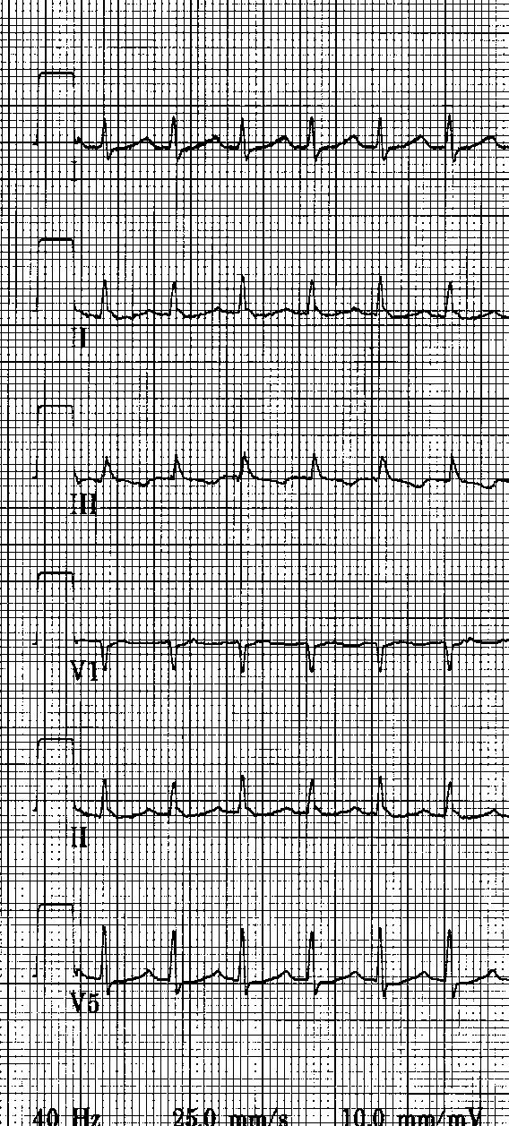
 Speed it up (paper speed 25")
40 ECG Diagnosis: Tips 1.Enhance the view Gain it up (2X or more) Speed it up (paper speed mm/sec) 2.Remember CSM 3.Compare to baseline

41 71M with Fatigue What is the Rhythm?
")
42 Carotid Sinus Massage (Left) Flutter Waves

43 Post Cardioversion Initial
44 Conclusions Pre-Interpretation Interpretation 1. Recall the pathophysiology of the different SVTs 2. Use all available information Epidemiology Response to Rx (adenosine) 3. Enhance the view on ECG Gain it up (2X or more) Speed it up (paper speed mm/sec) 4. Remember CSM 5. Compare to baseline Look for pre-excitation (WPW) 1. Find zones of transition Initiation (APD with long PR?), termination (P wave?) 2. Look for perturbations SVT with BBB, initiation and/or termination with VPD, change in RP interval or change in SVT rate with BBB should raise suspicion for AP 3. Persistence of SVT with AV block excludes AVRT 4. (+) P waves II,III,F suggest AT 5. QRS alternans suggests AVRT
45 Selected References Josephson ME and Wellens HJ. Differential diagnosis of supraventricular tachycardia. Cardiol Clin Aug;8(3): Wellens HJJ, Conover MB. The ECG in Emergency Decision Making. Ed. 2. Philadelphia, Saunders, 2005.

46
47 Supraventricular Tachycardia (SVT) Daniel Frisch, MD Cardiology Division, Electrophysiology Section Thomas Jefferson University Hospital
WPW syndrome and AVRT
 WPW syndrome and AVRT Myung-Yong Lee, MD, PhD Division of Cardiology Department of Internal Medicine School of Medicine Dankook University, Cheonan, Korea Supraventricular tachycardia (SVT) Paroxysmal
Supraventricular Tachycardia (SVT)
") Supraventricular Tachycardia (SVT) Bruce Stambler, MD Piedmont Heart Atlanta, GA Supraventricular Tachycardia Objectives Types and mechanisms AV nodal reentrant tachycardia (AVNRT) AV reciprocating tachycardia
Supraventricular Tachycardia (SVT) Bruce Stambler, MD Piedmont Heart Atlanta, GA Supraventricular Tachycardia Objectives Types and mechanisms AV nodal reentrant tachycardia (AVNRT) AV reciprocating tachycardia
We are IntechOpen, the world s leading publisher of Open Access books Built by scientists, for scientists. International authors and editors
 We are IntechOpen, the world s leading publisher of Open Access books Built by scientists, for scientists 3,500 108,000 1.7 M Open access books available International authors and editors Downloads Our
The Efficient and Smart Methods for Diagnosis of SVT 대구파티마병원순환기내과정병천
 The Efficient and Smart Methods for Diagnosis of SVT 대구파티마병원순환기내과정병천 Differentiation Supraventricular Origin from Ventricular Origin on ECG. QRS-Complex Width. 1. Narrow QRS-Complex Tachycardia (
LONG RP TACHYCARDIA MAPPING AND RF ABLATION
 LONG RP TACHYCARDIA MAPPING AND RF ABLATION Dr. Hayam Eldamanhoury Ain shams univeristy Arrhythmia is a too broad topic SVT is broadly defined as narrow complex ( unless aberrant conduction ) Requires
ECG QUIZ Luc DE ROY Brussels Belgium Disclosure in relation to this topic: none
 ECG QUIZ Luc DE ROY Brussels Belgium Disclosure in relation to this topic: none TEST EXAMPLE What is the colour of this rectangle? 1. Blue? 2. Red? 3. Purple? 4. Green? 5. Yellow? 6. 1 and 3? 7. Any of
Case-Based Practical ECG Interpretation for the Generalist
 Case-Based Practical ECG Interpretation for the Generalist Paul D. Varosy, MD, FACC, FAHA, FHRS Director of Cardiac Electrophysiology VA Eastern Colorado Health Care System Associate Professor of Medicine
Nathan Cade, MD Brandon Fainstad, MD Andrew Prouse, MD
 Nathan Cade, MD Brandon Fainstad, MD Andrew Prouse, MD OBJECTIVES 1. Identify the basic electrophysiology of the four causes of wide complex tachycardia. 2. Develop a simple framework for acute management
Uncommon forms of AV reentry: atrio and fasciculo-ventricular fibers, slow conducting fibers. Jesus Almendral, Madrid, Spain
 Uncommon forms of AV reentry: atrio and fasciculo-ventricular fibers, slow conducting fibers Jesus Almendral, Madrid, Spain Common forms of AV reentry Accessory pathways: Upper insertion: atrium Lower
TACHYARRHYTHMIAs. Pawel Balsam, MD, PhD
 TACHYARRHYTHMIAs Pawel Balsam, MD, PhD SupraVentricular Tachycardia Atrial Extra Systole Sinus Tachycardia Focal A. Tachycardia AVRT AVNRT Atrial Flutter Atrial Fibrillation Ventricular Tachycardia Ventricular
Huseng Vefali MD St. Luke s University Health Network Department of Cardiology
 Huseng Vefali MD St. Luke s University Health Network Department of Cardiology Learning Objectives Establish Consistent Approach to Interpreting ECGs Review Essential Cases for Paramedics and first responders
PAEDIATRIC ECG Dimosthenis Avramidis, MD.
 PAEDIATRIC ECG Dimosthenis Avramidis, MD. Consultant Mitera Children s Hospital Athens Greece S. Associate 1st Cardiology Dpt Evangelismos Hospital Athens Greece 5 y/o with sinus tach Background ECG changes
ECGs on the acute admission ward. - Cardiology Update -
 ECGs on the acute admission ward - Cardiology Update - Dr Simon Fynn Consultant Cardiologist Papworth Hospital, Cambridge RCP London Oct 2017 ECG 1 1. AF with BBB 2. Pre-excited AF 3. SVT with BBB 4.
Pediatrics ECG Monitoring. Pediatric Intensive Care Unit Emergency Division
 Pediatrics ECG Monitoring Pediatric Intensive Care Unit Emergency Division 1 Conditions Leading to Pediatric Cardiology Consultation 12.7% of annual consultation Is arrhythmias problems Geggel. Pediatrics.
1 Cardiology Acute Care Day 22 April 2013 Arrhythmia Tutorial Course Material
 1 Cardiology Acute Care Day 22 April 2013 Arrhythmia Tutorial Course Material Arrhythmia recognition This tutorial builds on the ECG lecture and provides a framework for approaching any ECG to allow the
Validation of right ventricular pacing response during SVT in mechanistic diagnosis. (Transition zone)
") Validation of right ventricular pacing response during SVT in mechanistic diagnosis. (Transition zone) By Marwan Sayed Assistant lecturer Cardiology department Assiut University Under supervision of Prof
Validation of right ventricular pacing response during SVT in mechanistic diagnosis. (Transition zone) By Marwan Sayed Assistant lecturer Cardiology department Assiut University Under supervision of Prof
Emergency Medical Training Services Emergency Medical Technician Paramedic Program Outlines Outline Topic: WPW Revised: 11/2013
 Emergency Medical Training Services Emergency Medical Technician Paramedic Program Outlines Outline Topic: WPW Revised: 11/2013 Wolff-Parkinson-White syndrome (WPW) is a syndrome of pre-excitation of the
You Don t Want to Miss This One! Focus on can t miss EKG tracings
 You Don t Want to Miss This One! Focus on can t miss EKG tracings Renaissance St. Louis Grand Hotel Convention Center October 23, 2014 David K. Tan, M.D., EMT-T, FAAEM EMS Section Chief, Division of Emergency
(living in the fast lane)
") (living in the fast lane) Presented by M.D. Cardiology IT S A group of ECG and Electrophysiological abnormalities in which The atrial impulses are conducted partly or completely, PREMATURELY, to the ventricles
(living in the fast lane) Presented by M.D. Cardiology IT S A group of ECG and Electrophysiological abnormalities in which The atrial impulses are conducted partly or completely, PREMATURELY, to the ventricles
ECG Interactive Session
 SA HEART 2018 PRE-CONGRESS WORKSHOP 4 OCTOBER 2018 ECG Interactive Session Ashley Chin Cardiologist/Electrophysiologist University of Cape Town Groote Schuur Hospital ECG 1 Which ONE of the following is
Paroxysmal Supraventricular Tachycardia PSVT.
 Atrial Tachycardia; is the name for an arrhythmia caused by a disorder of the impulse generation in the atrium or the AV node. An area in the atrium sends out rapid signals, which are faster than those
Supraventricular Tachycardia: From Fetus to Adult. Mohamed Hamdan, MD
 Supraventricular Tachycardia: From Fetus to Adult Mohamed Hamdan, MD Learning Objectives Define type of SVT by age Describe clinical approach Describe prenatal and postnatal management of SVT 2 SVT Across
Dr.Binoy Skaria 13/07/15
 Dr.Binoy Skaria binoyskaria@hotmail.com binoy.skaria@heartofengland.nhs.uk 13/07/15 Acknowledgement Medtronic, Google images & Elsevier for slides Natalie Ryan, Events Manager, HEFT- for organising the
Step by step approach to EKG rhythm interpretation:
 Sinus Rhythms Normal sinus arrhythmia Small, slow variation of the R-R interval i.e. variation of the normal sinus heart rate with respiration, etc. Sinus Tachycardia Defined as sinus rhythm with a rate
a lecture series by SWESEMJR
 Arrhythmias Automaticity- the ability to depolarize spontaneously. Pacemakers: Sinoatrial node: 70 bpm AV-nodal area: 40 bpm His-Purkinje: 20-40 bpm it Mechanisms of arrhythmias 1. Increased automaticity
ECGs and Arrhythmias: Family Medicine Board Review 2009
 Rate Rhythm Intervals Hypertrophy ECGs and Arrhythmias: Family Medicine Board Review 2009 Axis Jess (Fogler) Waldura, MD University of California, San Francisco walduraj@nccc.ucsf.edu Ischemia Overview
How To Think About Rhythms and Conduction
 How To Think About Rhythms and Conduction Frank Yanowitz, MD Professor of Medicine (Cardiology) University of Utah School of Medicine Medical Director, IHC ECG Services Intermountain Healthcare http://ecg.utah.edu
ECG Interpretation Made Easy
 ECG Interpretation Made Easy Dr. A Tageldien Abdellah, MSc MD EBSC Lecturer of Cardiology- Hull University Hull York Medical School 2007-2008 ECG Interpretation Made Easy Synopsis Benefits Objectives Process
I have no conflicts of interest relative to this lecture.
 PATHWAYS AROUND THE PITFALLS OF EKG RHYTHM INTERPRETATION Donald D. Brown, MD April 4, 2019 I have no conflicts of interest relative to this lecture. OBJECTIVES FOR PATHWAYS AROUND PITFALLS OF EKG RHYTHM
Concise Review for Primary-Care Physicians
 Concise Review for Primary-Care Physicians Narrow QRS Complex Tachycardias STEPHEN J. PIEPER, M.D., AND MARSHALL S. STANTON, M.D. Regular narrow QRS complex tachycardias are a common problem encountered
FLB s What Are Those Funny-Looking Beats?
 FLB s What Are Those Funny-Looking Beats? Reading Assignment (pages 27-45 in Outline ) The 5-Step Method ECG #: Mearurements: Rhythm (s): Conduction: Waveform: Interpretation: A= V= PR= QRS= QT= Axis=
Journal of the American College of Cardiology Vol. 36, No. 2, by the American College of Cardiology ISSN /00/$20.
 Journal of the American College of Cardiology Vol. 36, No. 2, 2000 2000 by the American College of Cardiology ISSN 0735-1097/00/$20.00 Published by Elsevier Science Inc. PII S0735-1097(00)00770-1 Diagnostic
Supraventricular Arrhythmias. Reading Assignment. Chapter 5 (p17-30)
") Supraventricular Arrhythmias Reading Assignment Chapter 5 (p17-30) The Supraventricular Rhythms In Our Lives Site of Origin Single Events Slow Rates Intermediate Rates Fast Rates (>100 bpm) Sinus Sinus
Supraventricular Arrhythmias Reading Assignment Chapter 5 (p17-30) The Supraventricular Rhythms In Our Lives Site of Origin Single Events Slow Rates Intermediate Rates Fast Rates (>100 bpm) Sinus Sinus
Differential diagnosis and pacing in maneuvers narrow QRS tachycardia. Richard Schilling
 Differential diagnosis and pacing in maneuvers narrow QRS tachycardia Richard Schilling Differential diagnosis of narrow complex QRS tachycardia Anything that activates the ventricle normally via the His
Repetitive narrow QRS tachycardia in a 61-year-old female patient with recent palpitations
 Journal of Geriatric Cardiology (2018) 15: 193 198 2018 JGC All rights reserved; www.jgc301.com Case Report Open Access Repetitive narrow QRS tachycardia in a 61-year-old female patient with recent palpitations
Tachy. Induction tachycardia lead ECG during Tachy /25/2009. Sinus Rhythm Single His
 12-lead ECG during Tachy 10.30.31 Sinus Rhythm Single His 11.20.02 Induction tachycardia 11.23.23 Tachy 11.25.23 1 I This finding excludes: (a) AVNRT (either typical or atypical) Tachy: Alternating cycle
2) Heart Arrhythmias 2 - Dr. Abdullah Sharif
 Heart Arrhythmias 2 - Dr. Abdullah Sharif") 2) Heart Arrhythmias 2 - Dr. Abdullah Sharif Rhythms from the Sinus Node Sinus Tachycardia: HR > 100 b/m Causes: o Withdrawal of vagal tone & Sympathetic stimulation (exercise, fight or flight) o Fever
2) Heart Arrhythmias 2 - Dr. Abdullah Sharif Rhythms from the Sinus Node Sinus Tachycardia: HR > 100 b/m Causes: o Withdrawal of vagal tone & Sympathetic stimulation (exercise, fight or flight) o Fever
Arrhythmia Management Joshua M. Cooper, MD, FHRS, FACC
 Arrhythmia Management Joshua M. Cooper, MD, FHRS, FACC Professor of Medicine Director of Cardiac Electrophysiology Temple University Health System Plumbing Electrical System Bradyarrhythmias Sinus Node
the ECG, 6 mg of intravenous adenosine was administered as a fast bolus through a large bore intravenous cannula in
 (arrows). Electrocardiography Series Singapore Med.1 2011, 52(3) 146 CME Article Regular narrow complex tachycardia Singh D, Teo S G, Poh K K V Hr JL1 11 VL VF WA, Fig. I ECG shows regular narrow complex
Electrocardiography for Healthcare Professionals
 Electrocardiography for Healthcare Professionals Chapter 7: Junctional Dysrhythmias 2012 The Companies, Inc. All rights reserved. Learning Outcomes 7.1 Describe the various junctional dysrhythmias 7.2
8/20/2012. Learning Outcomes (Cont d)
") 1 2 3 4 Electrocardiography for Healthcare Professionals Chapter 7: Junctional Dysrhythmias Learning Outcomes 7.1 Describe the various junctional dysrhythmias 7.2 Identify premature junctional complexes
1 2 3 4 Electrocardiography for Healthcare Professionals Chapter 7: Junctional Dysrhythmias Learning Outcomes 7.1 Describe the various junctional dysrhythmias 7.2 Identify premature junctional complexes
Case #1. 73 y/o man with h/o HTN and CHF admitted with dizziness and SOB Treated for CHF exacerbation with Lasix Now HR 136
 Tachycardias Case #1 73 y/o man with h/o HTN and CHF admitted with dizziness and SOB Treated for CHF exacerbation with Lasix Now HR 136 Initial Assessment Check Telemetry screen if pt on tele Telemetry
EHRA Accreditation Exam - Sample MCQs Invasive cardiac electrophysiology
 EHRA Accreditation Exam - Sample MCQs Invasive cardiac electrophysiology Dear EHRA Member, Dear Colleague, As you know, the EHRA Accreditation Process is becoming increasingly recognised as an important
The most common. hospitalized patients. hypotension due to. filling time Rate control in ICU patients may be difficult as many drugs cause hypotension
 Arrhythmias in the critically ill ICU patients: Approach for rapid recognition & management Objectives Be able to identify and manage: Atrial fibrillation with a rapid ventricular response Atrial flutter
In certain cases of supraventricular
 Case Report Hellenic J Cardiol 2013; 54: 469-473 A Tachycardia with Varying QRS Morphology and RP Intervals: Differential Diagnosis and Therapy Socrates Korovesis, Eleftherios Giazitzoglou, Demosthenes
Case Report Simultaneous Accessory Pathway and AV Node Mechanical Block
 185 Case Report Simultaneous Accessory Pathway and AV Node Mechanical Block Daniel Garofalo, MD, FRACP, Alfonso Gomez Gallanti, MD, David Filgueiras Rama, MD, Rafael Peinado Peinado, PhD, FESC Unidad de
Return to Basics. ECG Rate and Rhythm. Management of the Hospitalized Patient September 25, 2009
 Management of the Hospitalized Patient September 25, 2009 ECG Refresher and Update 2009 Return to Basics Determine rate and rhythm Determine intervals and axes Define morphology of P-QRS-T-U Compare with
EKG Abnormalities. Adapted from:
 EKG Abnormalities Adapted from: http://www.bem.fi/book/19/19.htm Some key terms: Arrhythmia-an abnormal rhythm or sequence of events in the EKG Flutter-rapid depolarizations (and therefore contractions)
Chapter 16: Arrhythmias and Conduction Disturbances
 Complete the following. Chapter 16: Arrhythmias and Conduction Disturbances 1. Cardiac arrhythmias result from abnormal impulse, abnormal impulse, or both mechanisms together. 2. is the ability of certain
Module 1: Introduction to ECG & Normal ECG
 Module 1: Introduction to ECG & Normal ECG Importance of Correct anatomical positions Measurements & Morphologies ONLY accurate if Precise anatomical positions adhered to Standardised techniques are used
Evaluation and Initial Treatment of Supraventricular Tachycardia
 T h e n e w e ngl a nd j o u r na l o f m e dic i n e clinical practice Evaluation and Initial Treatment of Supraventricular Tachycardia Mark S. Link, M.D. This Journal feature begins with a case vignette
Basic Electrophysiology Protocols
 Indian Journal of Cardiology ISSN-0972-1622 2012 by the Indian Society of Cardiology Vol. 15, (3-4), 27-37 [ 27 Review Article Shomu Bohora Assistant Professor, Deptt. of Cardiology, U.N. Mehta Institute
Ablation Update and Case Studies. Lawrence Nair, MD, FACC Director of Electrophysiology Presbyterian Heart Group
 Ablation Update and Case Studies Lawrence Nair, MD, FACC Director of Electrophysiology Presbyterian Heart Group Disclosures No financial relationships to disclose Objectives At the conclusion of this activity,
ACCESSORY PATHWAYS AND SVT. Neil Grubb Royal Infirmary of Edinburgh
 ACCESSORY PATHWAYS AND SVT Neil Grubb Royal Infirmary of Edinburgh Bypass tracts - properties accessory AV connections usually endocardial may exhibit unidirectional conduction conduction properties similar
Ventricular Preexcitation (Wolff-Parkinson-White Syndrome and Its Variants) 柯文欽醫師 國泰綜合醫院心臟內科主治醫師 臺北醫學大學講師
 柯文欽醫師 國泰綜合醫院心臟內科主治醫師 臺北醫學大學講師") Ventricular Preexcitation (Wolff-Parkinson-White Syndrome and Its Variants) 柯文欽醫師 國泰綜合醫院心臟內科主治醫師 臺北醫學大學講師 The Nobel Prize in Physiology or Medicine 1924 "for his discovery of the mechanism of the electrocardiogram"
Ventricular Preexcitation (Wolff-Parkinson-White Syndrome and Its Variants) 柯文欽醫師 國泰綜合醫院心臟內科主治醫師 臺北醫學大學講師 The Nobel Prize in Physiology or Medicine 1924 "for his discovery of the mechanism of the electrocardiogram"
Return to Basics. Normal Intervals & Axes. ECG Rate and Rhythm
 Return to Basics Management of the Hospitalized Patient October 15, 2010 ECG Refresher and Update 2010 Determine rate and rhythm Determine intervals and axes Define morphology of P-QRS-T-U Compare with
SIMPLY ECGs. Dr William Dooley
 SIMPLY ECGs Dr William Dooley Content Basic ECG interpretation pattern Some common (examined) abnormalities Presenting ECGs in context Setting up an ECG Setting up an ECG 1 V1-4 th Right intercostal space
REtrive. REpeat. RElearn Design by. Test-Enhanced Learning based ECG practice E-book
 Test-Enhanced Learning Test-Enhanced Learning Test-Enhanced Learning Test-Enhanced Learning based ECG practice E-book REtrive REpeat RElearn Design by S I T T I N U N T H A N G J U I P E E R I Y A W A
ECGs and Arrhythmias: Family Medicine Board Review 2012
 Overview ECGs and Arrhythmias: Family Medicine Board Review 2012 Jess Waldura, MD University of California, San Francisco walduraj@nccc.ucsf.edu Bundle branch blocks Quick review of ischemia Arrhythmias
Lecture outline. Electrical properties of the heart. Automaticity. Excitability. Refractoriness. The ABCs of ECGs Back to Basics Part I
 Lecture outline The ABCs of ECGs Back to Basics Part I Meg Sleeper VMD, DACVIM (cardiology) University of Florida Veterinary School Electrical properties of the heart Action potentials Normal intracardiac
Cardiology Flash Cards
 Cardiology Flash Cards EKG in a nut shell www.brain101.info Conduction System www.brain101.info 2 Analyzing EKG Step by step Steps in Analyzing ECG'S 1. Rhythm: - Regular _ Sinus, Junctional or Ventricular.
ECG interpretation basics
 ECG interpretation basics Michał Walczewski, MD Krzysztof Ozierański, MD 21.03.18 Electrical conduction system of the heart Limb leads Precordial leads 21.03.18 Precordial leads Precordial leads 21.03.18
UNDERSTANDING YOUR ECG: A REVIEW
 UNDERSTANDING YOUR ECG: A REVIEW Health professionals use the electrocardiograph (ECG) rhythm strip to systematically analyse the cardiac rhythm. Before the systematic process of ECG analysis is described
Miscellaneous Stuff Keep reading the Outline
 Miscellaneous Stuff Keep reading the Outline Welcome to the 5-Step Method ECG #: Mearurements: Rhythm (s): Conduction: Waveform: Interpretation: A= V= PR= QRS= QT= Axis= 1. Compute the 5 basic measurements:
INTERESTING ECGS. Wide-complex tachycardias 11/13/ yr male with acute ant wall MI, S/P primary PTCA
 INTERESTING ECGS V S Prakash Prof, & Head Dept. of Cardiology M S Ramaiah Hospitals 50 yr male with acute ant wall MI, S/P primary PTCA Pt. underwent successful PAMI An hour later,in the CCU Ventricular
AV Node Dependent SVT:Substrates, Mechanisms, and Recognition
 AV Node Dependent SVT:Substrates, Mechanisms, and Recognition Ching-Tai Tai Taipei Veterans General Hospital National Yang-Ming University AV Node Reentry Anatomy Physiology Anatomic-Physiologic Relation
Core Content In Urgent Care Medicine
 Palpitations/Arrhythmias Ebrahim Barkoudah, MD Clinical Instructor in Internal Medicine Harvard Medical School Assistant in Internal Medicine & Pediatrics Massachusetts General Hospital MGH Chelsea Chelsea,
Return to Basics. ECG Rate and Rhythm. Management of the Hospitalized Patient October 4, 2007
 Management of the Hospitalized Patient October 4, 2007 ECG Refresher for the Hospitalists Return to Basics Determine rate and rhythm Determine intervals and axes Define morphology of P-QRS-T-U Compare
HR: 50 bpm (Sinus) PR: 280 ms QRS: 120 ms QT: 490 ms Axis: -70. Sinus bradycardia with one ventricular escape (*)
 PR: 280 ms QRS: 120 ms QT: 490 ms Axis: -70. Sinus bradycardia with one ventricular escape (*)") 1? HR: 50 bpm (Sinus) PR: 280 ms QRS: 120 ms QT: 490 ms Axis: -70 1 Sinus P waves? 2 sinus cycles The pause (2 sinus cycles) suggests that the sinus fired (?) but did not conduct to the atria (i.e., missing
1? HR: 50 bpm (Sinus) PR: 280 ms QRS: 120 ms QT: 490 ms Axis: -70 1 Sinus P waves? 2 sinus cycles The pause (2 sinus cycles) suggests that the sinus fired (?) but did not conduct to the atria (i.e., missing
Knowing is not enough; we must apply. Willing is not enough; we must do.
 Cardiovascular Boot Camp Basic Training Day 2: Cardiac Arrhythmias and Emergency Treatment Overview Cardiac Arrhythmias: Physiologic Links to Recognition and Management NTI 2014 Denver 2014 Karen Marzlin
Atrioventricular (AV) Nodal Reentry Associated with 2:1 Infra-His Conduction Block during Tachycardia in a Patient with AV Nodal Triple Pathways
 Nodal Reentry Associated with 2:1 Infra-His Conduction Block during Tachycardia in a Patient with AV Nodal Triple Pathways") Atrioventricular (AV) Nodal Reentry Associated with 2:1 Infra-His Conduction Block during Tachycardia in a Patient with AV Nodal Triple Pathways Haruhiko ABE, M.D., Takashi OHKITA, M.D., Masasuke FUJITA,
Atrioventricular (AV) Nodal Reentry Associated with 2:1 Infra-His Conduction Block during Tachycardia in a Patient with AV Nodal Triple Pathways Haruhiko ABE, M.D., Takashi OHKITA, M.D., Masasuke FUJITA,
15 16 September Seminar W10O. ECG for General Practice
 15 16 September 2012 Seminar W10O ECG for General Practice Speaker: Ms Natasha Eaton ECG for General Practice Speaker: Natasha Eaton Cardiac CNC Executive Representative Electrocardiography The graphic
SVT Discriminators. Definition of SVT Discrimination. Identify which patient populations might benefit from these features
 Definition of SVT Discrimination Identify which patient populations might benefit from these features Understand the 4 types of SVT discriminators used by St Jude Medical Be aware of programmable parameters
Arrhythmias on the AMU
 Arrhythmias on the AMU Dr Andrew R. Houghton United Lincolnshire Hospitals NHS Trust @arh_cardio An arrhythmia is present in 10% of admissions to the AMU Case history AMU referral to cardiology 25 yr 2-3
PhD FRCP MESC MEAPCI. Consultant Cardiologist SVT - Supra Ventricular Tachycardia. Coronary Arteries
 SVT - Supra Ventricular Tachycardia Coronary Arteries Overview LMS Supraventricular tachycardia is defined as an abnormally fast heartbeat. It's a describes a group of arrhythmias which all originate from
Advanced ECG Interpretation Pt. II
 Advanced ECG Interpretation Pt. II Dysrhythmias Amal Mattu, MD, FAAEM Professor and Vice Chair Director, Emergency Cardiology Fellowship Department of Emergency Medicine University of Maryland School of
Arrhythmic Complications of MI. Teferi Mitiku, MD Assistant Clinical Professor of Medicine University of California Irvine
 Arrhythmic Complications of MI Teferi Mitiku, MD Assistant Clinical Professor of Medicine University of California Irvine Objectives Brief overview -Pathophysiology of Arrhythmia ECG review of typical
Asymptomatic patient with WPW
 Asymptomatic patient with WPW Dimosthenis Avramidis, MD. Arrythmiologist Mitera Children s Hospital Athens Greece Scientific Associate 1st Cardiology Dpt Evangelismos Hospital Athens Greece Preexcitation
ECGs: Everything a finalist needs to know. Dr Amy Coulden As part of the Simply Finals series
 ECGs: Everything a finalist needs to know Dr Amy Coulden As part of the Simply Finals series Aims and objectives To be able to interpret basic ECG abnormalities To be able to recognise commonly tested
Fast & Slow Tachy & Brady Arrhythmias DAVID STULTZ, MD, FACC KPN HEART & VASCULAR AUGUST 7, 2017
 Fast & Slow Tachy & Brady Arrhythmias DAVID STULTZ, MD, FACC KPN HEART & VASCULAR AUGUST 7, 2017 Normal EKG EKG boxes Heart Rate 1 big box = 200ms 1 small box = 40ms Big Boxes Between QRS complexes Heart
Please check your answers with correct statements in answer pages after the ECG cases.
 ECG Cases ECG Case 1 Springer International Publishing AG, part of Springer Nature 2018 S. Okutucu, A. Oto, Interpreting ECGs in Clinical Practice, In Clinical Practice, https://doi.org/10.1007/978-3-319-90557-0
Emergency treatment to SVT Evidence-based Approach. Tran Thao Giang
 Emergency treatment to SVT Evidence-based Approach Tran Thao Giang Description ECG manifestations: HR is extremely rapid and regular (240bpm ± 40) P wave is: usually invisible When visible: anormal P axis,
TEST BANK FOR ECGS MADE EASY 5TH EDITION BY AEHLERT
 Link download full: http://testbankair.com/download/test-bank-for-ecgs-made-easy-5thedition-by-aehlert/ TEST BANK FOR ECGS MADE EASY 5TH EDITION BY AEHLERT Chapter 5 TRUE/FALSE 1. The AV junction consists
Narrow QRS complex tachycardias rhythms with a
 Dmitriy Kireyev, MD, FACC; Stanley F. Fernandez, MD; Vipul Gupta, MD, MPH; Mikhail V. Arkhipov, MD; Joseph A. Paris, MD, FACC Cambridge Health Alliance, Whidden Memorial Hospital, Everett, Mass (Dr. Kireyev);
PAROXYSMAL SUPRAVENTRICULAR TACHYCARDIA (PSVT)
") PAROXYSMAL SUPRAVENTRICULAR TACHYCARDIA (PSVT) Mary Boudreau Conover It is disappointing how many medical professionals are unable to make the diagnosis of PSVT and to differentiate between the two main
PAROXYSMAL SUPRAVENTRICULAR TACHYCARDIA (PSVT) Mary Boudreau Conover It is disappointing how many medical professionals are unable to make the diagnosis of PSVT and to differentiate between the two main
Conduction Problems / Arrhythmias. Conduction
 Conduction Problems / Arrhythmias Conduction Wolf-Parkinson White Syndrome (WPW) and Lown-Ganong-Levine (LGL): Atrial impulses bypass the AV node through an accessory pathway or bypass tract (bundle of
10 ECGs No Practitioner Can Afford to Miss. Objectives
 10 ECGs No Practitioner Can Afford to Miss Mary L. Dohrmann, MD Professor of Clinical Medicine Division of Cardiovascular Medicine University of Missouri School of Medicine No disclosures Objectives 1.
Electrocardiography for Healthcare Professionals
 Electrocardiography for Healthcare Professionals Kathryn A. Booth Thomas O Brien Chapter 10: Pacemaker Rhythms and Bundle Branch Block Learning Outcomes 10.1 Describe the various pacemaker rhythms. 10.2
December 2018 Tracings
 Tracings Tracing 1 Tracing 4 Tracing 1 Answer Tracing 4 Answer Tracing 2 Tracing 5 Tracing 2 Answer Tracing 5 Answer Tracing 3 Tracing 6 Tracing 3 Answer Tracing 6 Answer Questions? Contact Dr. Nelson
Cardiac rhythm detailed monitoring by an implanted pacemaker: The iecg solution
 Cardiac rhythm detailed monitoring by an implanted pacemaker: The iecg solution Francesco Zanon, MD, FESC, FHRS Arrhythmia and Electrophysiology Unit, Cardiology Dept. Santa Maria della Misericordia General
ECG Interpretation Cat Williams, DVM DACVIM (Cardiology)
") ECG Interpretation Cat Williams, DVM DACVIM (Cardiology) Providing the best quality care and service for the patient, the client, and the referring veterinarian. GOAL: Reduce Anxiety about ECGs Back to
ECG Interpretation Cat Williams, DVM DACVIM (Cardiology) Providing the best quality care and service for the patient, the client, and the referring veterinarian. GOAL: Reduce Anxiety about ECGs Back to
PEDIATRIC SVT MANAGEMENT
 PEDIATRIC SVT MANAGEMENT 1 INTRODUCTION Supraventricular tachycardia (SVT) can be defined as an abnormally rapid heart rhythm originating above the ventricles, often (but not always) with a narrow QRS
Paramedic Rounds. Tachyarrhythmia's. Sean Sutton Dallas Wood
 Paramedic Rounds Tachyarrhythmia's Sean Sutton Dallas Wood Objectives At the end of this session, the paramedic will be able to: State the key components of the cardiac conduction pathway, along with the
Sustained tachycardia with wide QRS
 Sustained tachycardia with wide QRS Courtesy from Prof. Antonio Américo Friedmann. Electrocardiology Service of University of Faculty of São Paulo. Opinions from colleagues Greetings to everyone, In a
Atrial Fibrillation and Common Supraventricular Tachycardias. Sunil Kapur MD
 Atrial Fibrillation and Common Supraventricular Tachycardias Sunil Kapur MD Cardiac Electrophysiology Brigham and Women s Hospital Instructor, Harvard Medical School No disclosures Cardiac Conduction:
ECG pre-reading manual. Created for the North West Regional EMET training program
 ECG pre-reading manual Created for the North West Regional EMET training program Author:- Dr Juan Carlos Ascencio-Lane juan.ascencio-lane@ths.tas.gov.au 1 Disclaimer This handbook has been created for
CASE 10. What would the ST segment of this ECG look like? On which leads would you see this ST segment change? What does the T wave represent?
 CASE 10 A 57-year-old man presents to the emergency center with complaints of chest pain with radiation to the left arm and jaw. He reports feeling anxious, diaphoretic, and short of breath. His past history
Bundle Branch & Fascicular Blocks. Reading Assignment (p53-58 in Outline )
") Bundle Branch & Fascicular Blocks Reading Assignment (p53-58 in Outline ) Objectives 1. QRS analysis of Right and Left BBB 2. Uncomplicated vs complicated BBB 3. Diagnosis of RBBB with LAFB and LPFB 4.
Bundle Branch & Fascicular Blocks Reading Assignment (p53-58 in Outline ) Objectives 1. QRS analysis of Right and Left BBB 2. Uncomplicated vs complicated BBB 3. Diagnosis of RBBB with LAFB and LPFB 4.
Differentiating Slow Fast Atrioventricular Nodal Reentry Tachycardia From Atrioventrcular..
 IOSR Journal of Dental and Medical Sciences (IOSR-JDMS) e-issn: 2279-0853, p-issn: 2279-0861.Volume 16, Issue 1 Ver. X (January. 2017), PP 69-77 www.iosrjournals.org Differentiating Slow Fast Atrioventricular
Bradydysrhythmias and Atrioventricular Conduction Blocks
 Emerg Med Clin N Am 24 (2006) 1 9 Bradydysrhythmias and Atrioventricular Conduction Blocks Jacob W. Ufberg, MD*, Jennifer S. Clark, MD Department of Emergency Medicine, Temple University School of Medicine,