Giovanni Di Salvo MD, PhD, FESC Second University of Naples Monaldi Hospital
|
|
|
- Juliet Smith
- 6 years ago
- Views:
Transcription


1 Giovanni Di Salvo MD, PhD, FESC Second University of Naples Monaldi Hospital

2 VSD is one of the most common congenital cardiac abnormalities in the newborn. It can occur as an isolated finding or in combination with other congenital defects. VSD can occur in any portion of the ventricular septum, but usually along the fusion lines between the different portions.
3 Echocardiographic evaluation of ventricular septal defects includes: Identification of the location of defects on the septum Establishing the number of defects Delineation of associated anatomic features Assessment of the size and hemodynamic significance of the defects
: 5-7%")
4 Perimembranous: 80% Muscular: 5-20% Inlet: 5-8% Supracristal (Double Committed): 5-7%
5 Membranous: Relatively small, bounded superiorly by the aortic valve (RC and NC cusps) and inferiorly by the muscolar septum
6 From the RV the Membranous septum is divided in 2 parts by the septal leaflet of the Tricuspid valve. Pars atrioventricularis, which lies above the tricuspid valve Pars interventricularis, which lies beneath the septal leaflet of the TV
7 1. Inlet Portion 2. Trabecular septum 3. Outlet (or infundibular septum)

8 Inferior to the membranous septum, between the 2 AV valves. It is the 1\3 superior and posterior portion of the muscular septum. Its inferior borders are bounded by the chordal attachments of the AV valve


9 It is the largest portion of the ventricular septum and does not lie in the same plane. It extends from the membranous septum to the cardiac apex.

10 It lies inferior to the great vessels, beneth the RC cusp of the aortic valve and the L cusp of the pulmonary valve.
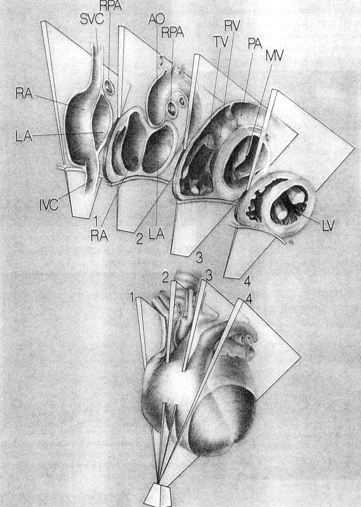
 The trabecular septum (the remaining")
11 The standard PLAX images predominatly: The infundibular septum (superior 1\3 of the septum) The trabecular septum (the remaining portion)
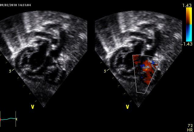
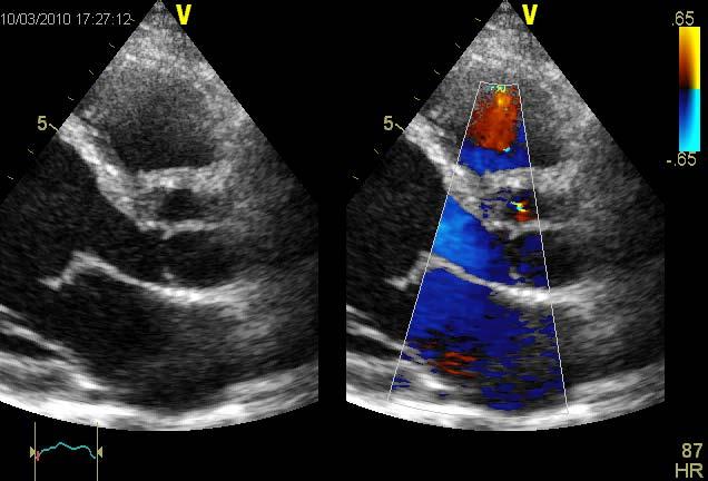
12 From the standard PLAX angulating the trasducer toward the right hip the RV inflow tract can be visualized. As the US plane passes from the aortic valve to the tricuspid valve the membranous septum is imaged.
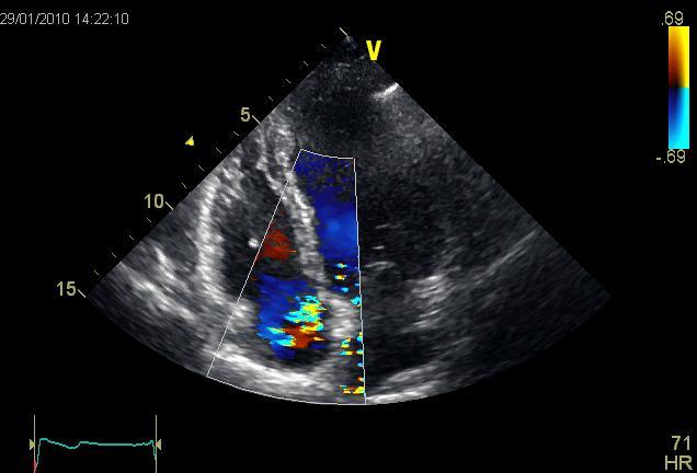
13 Tilting the US plane more anteriorly until the PV the Supracristal portion of the outlet septum can be visualized.

14 At the base of the heart we can see: The membranous septum to the right, beneath the septal leaflet of the TV The subaortic defects in the middle Infundibular defects on the left.

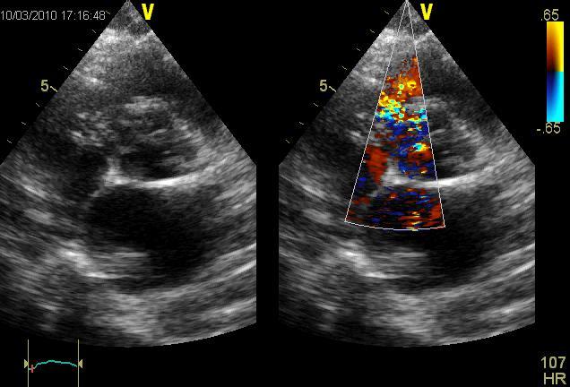
15 Through the 2 AV valves: Inlet ventricular septal defects Mid muscular defects Anterior trabecular defects
16 At the level of Papillary muscles: Trabecular defects At the level of the Apex: Apical Trabecular defects

.")
17 Inlet defects are visualized in the superior 1\3 of the septum Mid muscular defects in the middle half of the septum Apical defects in the apical portion of the septum (distal to moderator band).

 Mid")
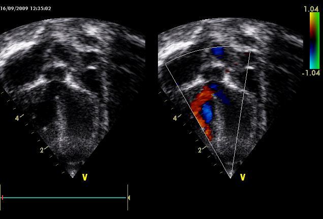
18 Subaortic outlet septum (infracristal portion) Mid muscular septum Apical septum


19 Standard echo cut Subcostal views Subcostal coronal view (4 chamber)

20 Outlet septum Trabecular septum

21 Perimembranous ventricular septal defect Subcostal left oblique view

22 Perimembranous ventricular septal defect Short Axis view

23 Perimembranous ventricular septal defect Long Axis view

24 Double committed ventricular septal defect Subcostal coronal view
25 Double committed ventricular septal defect Subcostal view

26 Inlet ventricular septal defect Apical 4 Chambers view (patients affected by cavc)
27 Inlet ventricular septal defect Short axis view (patients affected by cavc)

28 Apical 4 Chamber view Multiple muscular VSD

29 Trabecular ventricular septal defect Short Axis view

30


31 Antero-superior deviation of infundibular septum creates a sub pulmonary obstruction. PA LA Ao RA RV VSD Infundibular septum Right oblique subcostal view


32 Posterior deviation of infundibular septum can lead to a sub-aortic stenosis. This form can be associated with aortic coarctation. Posterior infundibular septal deviation VSD Ao RV LV IVS Zoomed view of an apical 5 chamber cut
33
34
35
36 Gerbode defect is a rare form of VSD that allows for communication between the left ventricle and the right atrium.
37

38

39 Normal diastolic pulmonary arterial pressure using PR jet is very useful to distinguish the true pulmonary arterial hypertension from high velocity jet in the right atrium caused by Gerbode type defect.

40

41


42 VSD diameter Trans-ventricular pressure gradient estimation Right ventricular pressure estimation LV shape L/T ratio= 1.45


: Moderate (Qp/Qs > 2.")
43 Calculate: LVOT diameter RVOT diameter VTI in LVOT using PW velocities VTI in RVOT using PW velocities (Qp/Qs < 1.4): Mild (Qp/Qs ): Moderate (Qp/Qs > 2.3): Severe

44 Position of device or patch Residual or accessory shunt Aortic Regurgitation Left atrial and ventricular volumes normalization
HISTORY. Question: What category of heart disease is suggested by this history? CHIEF COMPLAINT: Heart murmur present since early infancy.
 HISTORY 18-year-old man. CHIEF COMPLAINT: Heart murmur present since early infancy. PRESENT ILLNESS: Although normal at birth, a heart murmur was heard at the six week check-up and has persisted since
Perimembranous VSD: When Do We Ask For A Surgical Closure? LI Xin. Department of Cardiothoracic Surgery Queen Mary Hospital Hong Kong
 Perimembranous VSD: When Do We Ask For A Surgical Closure? LI Xin Department of Cardiothoracic Surgery Queen Mary Hospital Hong Kong Classification (by Kirklin) I. Subarterial (10%) Outlet, conal, supracristal,
Cardiac ultrasound protocols
 Cardiac ultrasound protocols IDEXX Telemedicine Consultants Two-dimensional and M-mode imaging planes Right parasternal long axis four chamber Obtained from the right side Displays the relative proportions
Most common fetal cardiac anomalies
 Most common fetal cardiac anomalies Common congenital heart defects CHD % of cardiac defects Chromosomal Infants Fetuses anomaly (%) 22q11 deletion (%) VSD 30 5~10 20~40 10 PS 9 5 (PA w/ VSD) HLHS 7~9
British Society of Echocardiography
 British Society of Echocardiography Affiliated to the British Cardiac Society A Minimum Dataset for a Standard Adult Transthoracic Echocardiogram From the British Society of Echocardiography Education
Atrial Septal Defects
 Supplementary ACHD Echo Acquisition Protocol for Atrial Septal Defects The following protocol for echo in adult patients with atrial septal defects (ASDs) is a guide for performing a comprehensive assessment
ADULT CONGENITAL HEART DISEASE. Stuart Lilley
 ADULT CONGENITAL HEART DISEASE Stuart Lilley More adults than children have congenital heart disease Huge variety of congenital lesions from minor to major Heart failure, re-operation and arrhythmia are
ECHOCARDIOGRAPHIC APPROACH TO CONGENITAL HEART DISEASE: THE UNOPERATED ADULT
 ECHOCARDIOGRAPHIC APPROACH TO CONGENITAL HEART DISEASE: THE UNOPERATED ADULT Karen Stout, MD, FACC Divisions of Cardiology University of Washington Medical Center Seattle Children s Hospital NO DISCLOSURES
FUNCTIONALLY SINGLE VENTRICLE
 MORPHOLOGICAL DETERMINANTS VI TRAN EuroEcho, Budapest, 7 th December 2011 DECLARATION OF CONFLICT OF INTEREST: I have nothing to declare What is the functionally single ventricle? The heart that is incapable
How to assess an adult with a Ventricular Septal Defect. When it should be closed and how?
 TTE AND TEE VDS ASSESSMENT. WHAT S S THE SIZE, WHERE ARE THE MARGINS? How to assess an adult with a Ventricular Septal Defect. When it should be closed and how? Dr Gianfranco Butera, MD, PhD Dr Gianfranco
Echocardiography in Adult Congenital Heart Disease
 Echocardiography in Adult Congenital Heart Disease Michael Vogel Kinderherz-Praxis München CHD missed in childhood Subsequent lesions after repaired CHD Follow-up of cyanotic heart disease CHD missed in
List of Videos. Video 1.1
 Video 1.1 Video 1.2 Video 1.3 Video 1.4 Video 1.5 Video 1.6 Video 1.7 Video 1.8 The parasternal long-axis view of the left ventricle shows the left ventricular inflow and outflow tract. The left atrium
MITRAL STENOSIS. Joanne Cusack
 MITRAL STENOSIS Joanne Cusack BSE Breakdown Recognition of rheumatic mitral stenosis Qualitative description of valve and sub-valve calcification and fibrosis Measurement of orifice area by planimetry
The background of the Cardiac Sonographer Network News masthead is a diagnostic image:
 Number 5 Welcome Number 5 Welcome to the newsletter created just for you: sonographers who perform pediatric echocardiograms in primarily adult echo labs. Each issue features tips on echocardiography of
Appendix II: ECHOCARDIOGRAPHY ANALYSIS
 Appendix II: ECHOCARDIOGRAPHY ANALYSIS Two-Dimensional (2D) imaging was performed using the Vivid 7 Advantage cardiovascular ultrasound system (GE Medical Systems, Milwaukee) with a frame rate of 400 frames
Congenital Heart Disease An Approach for Simple and Complex Anomalies
 Congenital Heart Disease An Approach for Simple and Complex Anomalies Michael D. Pettersen, MD Director, Echocardiography Rocky Mountain Hospital for Children Denver, CO None Disclosures 1 ASCeXAM Contains
cardiac imaging planes planning basic cardiac & aortic views for MR
 cardiac imaging planes planning basic cardiac & aortic views for MR Dianna M. E. Bardo, M. D. Assistant Professor of Radiology & Cardiovascular Medicine Director of Cardiac Imaging cardiac imaging planes
Cases in Adult Congenital Heart Disease
 Cases in Adult Congenital Heart Disease Sabrina Phillips, MD FACC FASE Associate Professor of Medicine The University of Oklahoma Health Sciences Center No Disclosures I Have Palpitations 18 Year old Man
Adel Hasanin Ahmed 1 ASD
 Adel Hasanin Ahmed 1 ASD Atrial septal defect (ASD) is the commonest form of congenital heart disease seen in adults. The commonest form of defect is the secundum ASD, accounting for two thirds of cases,
8/31/2016. Mitraclip in Matthew Johnson, MD
 Mitraclip in 2016 Matthew Johnson, MD 1 Abnormal Valve Function Valve Stenosis Obstruction to valve flow during that phase of the cardiac cycle when the valve is normally open. Hemodynamic hallmark - pressure
Anatomy of Atrioventricular Septal Defect (AVSD)
") Surgical challenges in atrio-ventricular septal defect in grown-up congenital heart disease Anatomy of Atrioventricular Septal Defect (AVSD) S. Yen Ho Professor of Cardiac Morphology Royal Brompton and
Surgical challenges in atrio-ventricular septal defect in grown-up congenital heart disease Anatomy of Atrioventricular Septal Defect (AVSD) S. Yen Ho Professor of Cardiac Morphology Royal Brompton and
Imaging Evaluation of the Ventricular Septum
 Imaging Evaluation of the Ventricular Septum Craig E Fleishman, MD FACC FASE The Heart Center at Arnold Palmer Hospital for Children, Orlando SCAI Fall Fellows Course 2013 Las Vegas Disclosure Information
CARDIAC AND CORONARY ARTERY ANATOMY NO DISCLOSURES. Axial Anatomy of Heart. Axial Anatomy of Heart. Axial Anatomy of Heart
 CARDIAC AND CORONARY ARTERY ANATOMY NO DISCLOSURES NASCI MEETING, ORLANDO FLORIDA 2009 KOSTAKI G. BIS, MD, FACR DEPARTMENT OF RADIOLOGY WILLIAM BEAUMONT HOSPITAL Royal Oak, Michigan OBJECTIVES CARDIAC
Double outlet right ventricle: navigation of surgeon to chose best treatment strategy
 Double outlet right ventricle: navigation of surgeon to chose best treatment strategy Jan Marek Great Ormond Street Hospital & Institute of Cardiovascular Sciences, University College London Double outlet
Heart and Lungs. LUNG Coronal section demonstrates relationship of pulmonary parenchyma to heart and chest wall.
 Heart and Lungs Normal Sonographic Anatomy THORAX Axial and coronal sections demonstrate integrity of thorax, fetal breathing movements, and overall size and shape. LUNG Coronal section demonstrates relationship
Echocardiographic assessment in Adult Patients with Congenital Heart Diseases
 Echocardiographic assessment in Adult Patients with Congenital Heart Diseases Athanasios Koutsakis Cardiologist, Cl. Research Fellow George Giannakoulas Ass. Professor in Cardiology 1st Cardiology Department,
CMR for Congenital Heart Disease
 CMR for Congenital Heart Disease * Second-line tool after TTE * Strengths of CMR : tissue characterisation, comprehensive access and coverage, relatively accurate measurements of biventricular function/
Adult Congenital Heart Disease: What All Echocardiographers Should Know Sharon L. Roble, MD, FACC Echo Hawaii 2016
 1 Adult Congenital Heart Disease: What All Echocardiographers Should Know Sharon L. Roble, MD, FACC Echo Hawaii 2016 DISCLOSURES I have no disclosures relevant to today s talk 2 Why should all echocardiographers
Heart and Soul Evaluation of the Fetal Heart
 Heart and Soul Evaluation of the Fetal Heart Ivana M. Vettraino, M.D., M.B.A. Clinical Associate Professor, Michigan State University College of Human Medicine Objectives Review the embryology of the formation
Adel Hasanin Ahmed 1
 Adel Hasanin Ahmed 1 PERICARDIAL DISEASE The pericardial effusion ends anteriorly to the descending aorta and is best visualised in the PLAX. PSAX is actually very useful sometimes for looking at posterior
Segmental Analysis. Gautam K. Singh, M.D. Washington University School of Medicine St. Louis
 Segmental Analysis Gautam K. Singh, M.D. Washington University School of Medicine St. Louis Segmental Analysis Segmental Analysis: From Veins to Ventricles Segmental Approach to Evaluation of Congenital
What is Ebstein Anomaly?
 Echocardiograpnhic Evaluation of : Definition, Detection and Determinants of Outcome P. W. O Leary, M.D. Division of Pediatric Cardiology Mayo Clinic No Conflicts to Disclose What is? Failure of the TV
Atrioventricular Canal (Septal) Defects. Norman H Silverman MD. D Sc (Med),FACC, FAHA
 Defects. Norman H Silverman MD. D Sc (Med),FACC, FAHA") Atrioventricular Canal (Septal) Defects Norman H Silverman MD. D Sc (Med),FACC, FAHA Embryology of the A-V Canal Looping NHS. Formation of the Atrial Septum Embryology of the A-V Canal NHS. Development
Atrioventricular Canal (Septal) Defects Norman H Silverman MD. D Sc (Med),FACC, FAHA Embryology of the A-V Canal Looping NHS. Formation of the Atrial Septum Embryology of the A-V Canal NHS. Development
ISUOG Basic Training. Obtaining & Interpreting Heart Views Correctly Alfred Abuhamad, USA. Basic training. Editable text here
 ISUOG Basic Training Obtaining & Interpreting Heart Views Correctly Alfred Abuhamad, USA Learning Objectives 6, 7 & 8 At the end of the lecture you will be able to: describe how to assess cardiac situs
Congenital heart disease. By Dr Saima Ali Professor of pediatrics
 Congenital heart disease By Dr Saima Ali Professor of pediatrics What is the most striking clinical finding in this child? Learning objectives By the end of this lecture, final year student should be able
Congenital Heart Defects
 Normal Heart Congenital Heart Defects 1. Patent Ductus Arteriosus The ductus arteriosus connects the main pulmonary artery to the aorta. In utero, it allows the blood leaving the right ventricle to bypass
Transposition of the Great Arteries Preoperative Diagnostic Considerations. John Simpson Evelina Children s Hospital London, UK
 Transposition of the Great Arteries Preoperative Diagnostic Considerations John Simpson Evelina Children s Hospital London, UK Areas to be covered Definitions Scope of occurrence of transposition of the
2) VSD & PDA - Dr. Aso
 VSD & PDA - Dr. Aso") 2) VSD & PDA - Dr. Aso Ventricular Septal Defect (VSD) Most common cardiac malformation 25-30 % Types of VSD: According to position perimembranous, inlet, muscular. According to size small, medium, large.
2) VSD & PDA - Dr. Aso Ventricular Septal Defect (VSD) Most common cardiac malformation 25-30 % Types of VSD: According to position perimembranous, inlet, muscular. According to size small, medium, large.
Normal TTE/TEE Examinations
 Normal TTE/TEE Examinations Geoffrey A. Rose, MD FACC FASE Sanger Heart & Vascular Institute Before you begin imaging... Obtain the patient s Height Weight BP PLAX View PLAX View Is apex @ 9-10 o clock?
VMS Quick Reference Guide
 VMS Quick Reference Guide Connecting to the VMS Workstation Ensure Ultrasound system is up and running prior to starting VMS. Connect the video cable from the ultrasound machine to the VMS Integrated Station.
Right Heart Evaluation ASE Guidelines Review. Chris Mann RDCS, RCS, FASE Faculty, Echocardiography Pitt Community College Greenville, NC
 Right Heart Evaluation ASE Guidelines Review Chris Mann RDCS, RCS, FASE Faculty, Echocardiography Pitt Community College Greenville, NC Objectives Briefly review right atrial and right ventricular anatomy
Echo Doppler Assessment of Right and Left Ventricular Hemodynamics.
 Echo Doppler Assessment of Right and Left Ventricular Hemodynamics. Itzhak Kronzon, MD, FASE, FACC, FESC, FAHA, FACP, FCCP Northwell, Lenox Hill Hospital, New York Professor of Cardiology Hofstra University
ULTRASOUND OF THE FETAL HEART
 ULTRASOUND OF THE FETAL HEART Cameron A. Manbeian, MD Disclosure Statement Today s faculty: Cameron Manbeian, MD does not have any relevant financial relationships with commercial interests or affiliations
Type Size AP/PS RP/RS Qp/Qs. Ia Resistive <0.3 < Ib Resistive <0.3 < IIa Resistive <0.5 >2
 Transcatheter closure of VSD using Duct Occluder device Nguyen Lan Hieu, MD, PhD Hanoi Medical University Vietnam Heart Institute Anatomy of VSD 1. Perimembranous VSD: Aneurysm septal membranous(tv or
Making Sense of Cardiac Views and Imaging Characteristics for 13 Congenital Heart Defects (CHDs)
") Making Sense of Cardiac Views and Imaging Characteristics for 13 Congenital Heart Defects (CHDs) Manny Gaziano, MD, FACOG obimages.net obimages.net@gmail.com Acknowledgements: Krista Wald, RDMS, sonographer,
Making Sense of Cardiac Views and Imaging Characteristics for 13 Congenital Heart Defects (CHDs) Manny Gaziano, MD, FACOG obimages.net obimages.net@gmail.com Acknowledgements: Krista Wald, RDMS, sonographer,
Communication of Mitral Valve with Both Ventricles Associated with Double Outlet Right Ventricle
 Communication of Mitral Valve with Both Ventricles Associated with Double Outlet Right Ventricle By RAJENTDRA TANDON, M.D., JAMES H. MOLLR, MD, AND JESSE E. EDWARDS, M.D. SUMMARY A rare case of an infant
Follow-up after VSD closure- what to look for?
 The 3rd Congress of Congenital heart disease Ventricular Septal Defect from A-Z January 9-11. 2013, Ho Chi Minh City, Vietnam Follow-up after VSD closure- what to look for? Nina Wunderlich University Hospital
Fetal Echocardiography and the Routine Obstetric Sonogram
 JDMS 23:143 149 May/June 2007 143 Fetal Echocardiography and the Routine Obstetric Sonogram SHELLY ZIMBELMAN, RT(R)(CT), RDMS, RDCS ASAD SHEIKH, MD, RDCS Congenital heart disease (CHD) is the most common
2/4/2011. Nathan Kerner, M.D.
 Nathan Kerner, M.D. Definition Elevated pressures - cut off usually >40 mmhg pulmonary artery systolic pressure (PASP) Usually associated with elevated pulmonary vascular resistance (PVR) measured in dynessec/cm
PART II ECHOCARDIOGRAPHY LABORATORY OPERATIONS ADULT TRANSTHORACIC ECHOCARDIOGRAPHY TESTING
 PART II ECHOCARDIOGRAPHY LABORATORY OPERATIONS ADULT TRANSTHORACIC ECHOCARDIOGRAPHY TESTING STANDARD - Primary Instrumentation 1.1 Cardiac Ultrasound Systems SECTION 1 Instrumentation Ultrasound instruments
Imaging Guide Echocardiography
 Imaging Guide Guide to Small Animal Echocardiography using the Vevo Imaging Systems System Compatibility: This guide contains instructions and suggestions for work on the Vevo2100, VevoLAZR, Vevo 3100
5.8 Congenital Heart Disease
 5.8 Congenital Heart Disease Congenital heart diseases (CHD) refer to structural or functional heart diseases, which are present at birth. Some of these lesions may be discovered later. prevalence of Chd
P = 4V 2. IVC Dimensions 10/20/2014. Comprehensive Hemodynamic Evaluation by Doppler Echocardiography. The Simplified Bernoulli Equation
 Comprehensive Hemodynamic Evaluation by Doppler Echocardiography Itzhak Kronzon, MD North Shore LIJ/ Lenox Hill Hospital New York, NY Disclosure: Philips Healthcare St. Jude Medical The Simplified Bernoulli
PROSTHETIC VALVE BOARD REVIEW
 PROSTHETIC VALVE BOARD REVIEW The correct answer D This two chamber view shows a porcine mitral prosthesis with the typical appearance of the struts although the leaflets are not well seen. The valve
Anomalous Systemic Venous Connection Systemic venous anomaly
 World Database for Pediatric and Congenital Heart Surgery Appendix B: Diagnosis (International Paediatric and Congenital Cardiac Codes (IPCCC) and definitions) Anomalous Systemic Venous Connection Systemic
Cardiac Catheterization Cases Primary Cardiac Diagnoses Facility 12 month period from to PRIMARY DIAGNOSES (one per patient)
") PRIMARY DIAGNOSES (one per patient) Septal Defects ASD (Atrial Septal Defect) PFO (Patent Foramen Ovale) ASD, Secundum ASD, Sinus venosus ASD, Coronary sinus ASD, Common atrium (single atrium) VSD (Ventricular
PRIMARY DIAGNOSES (one per patient) Septal Defects ASD (Atrial Septal Defect) PFO (Patent Foramen Ovale) ASD, Secundum ASD, Sinus venosus ASD, Coronary sinus ASD, Common atrium (single atrium) VSD (Ventricular
Hemodynamic Assessment. Assessment of Systolic Function Doppler Hemodynamics
 Hemodynamic Assessment Matt M. Umland, RDCS, FASE Aurora Medical Group Milwaukee, WI Assessment of Systolic Function Doppler Hemodynamics Stroke Volume Cardiac Output Cardiac Index Tei Index/Index of myocardial
가천의대길병원소아심장과최덕영 PA C IVS THE EVALUATION AND PRINCIPLES OF TREATMENT STRATEGY
 가천의대길병원소아심장과최덕영 PA C IVS THE EVALUATION AND PRINCIPLES OF TREATMENT STRATEGY PA c IVS (not only pulmonary valve disease) Edwards JE. Pathologic Alteration of the right heart. In: Konstam MA, Isner M, eds.
Assessment of LV systolic function
 Tutorial 5 - Assessment of LV systolic function Assessment of LV systolic function A knowledge of the LV systolic function is crucial in the undertanding of and management of unstable hemodynamics or a
Can SCMR CMR protocol recommendations
 Can SCMR CMR protocol recommendations V1.3 - April 2009 CanSCMR CMR Protocol and SOP Recommendation 2009 (15 minutes) 2 Planning of LV fct. real time multiple axes Realtime 3 cine long axis 6 long axes
Echocardiography in adult congenital heart disease
 S12 Department of Cardiology, Royal Hospital for Sick Children, Glasgow G3 8SJ, UK A Houston S Lilley T Richens University Department of Medicine and Therapeutics, Western Infirmary, Glasgow G11 6NT, UK
B-Mode measurements protocols:
 Application Note How to Perform the Most Commonly Used Measurements from the Cardiac Measurements Package associated with Calculations of Cardiac Function using the Vevo Lab Objective The Vevo LAB offline
Fig.1 Normal appearance of RV in SAX:
 Tutorial 7 - Assessment of the right heart Assessment of the Right heart The right heart assessment clinically and echocardiographically is not a very important part of mainstream cardiology. In the ICU,
Echocardiography in the Adult with Congenital Heart Disease
 1 1 Echocardiography in the Adult with Congenital Heart Disease Julie A. Kovach Indications for Echocardiography in the Evaluation of the Adult with Congenital Heart Disease........ 279 Indications and
Back to Basics: Common Errors In Quantitation In Everyday Practice
 Back to Basics: Common Errors In Quantitation In Everyday Practice Deborah Agler, ACS, RDCS, FASE October 9, 2017 ASE: Echo Florida Rebecca T. Hahn, MD Director of Interventional Echocardiography Professor
Percutaneous VSD closure
 Percutaneous VSD closure Gianfranco Butera San Donato Milanese - Italy Patients selection Pts having hemodynamically significant VSD Left ventricular enlargement (left ventricular overload),defined as
ICE: Echo Core Lab-CRF
 APPENDIX 1 ICE: Echo Core Lab-CRF Study #: - Pt Initials: 1. Date of study: / / D D M M M Y Y Y Y 2. Type of Study: TTE TEE 3. Quality of Study: Poor Moderate Excellent Ejection Fraction 4. Ejection Fraction
Cardiac MRI in ACHD What We. ACHD Patients
 Cardiac MRI in ACHD What We Have Learned to Apply to ACHD Patients Faris Al Mousily, MBChB, FAAC, FACC Consultant, Pediatric Cardiology, KFSH&RC/Jeddah Adjunct Faculty, Division of Pediatric Cardiology
Comprehensive Hemodynamics By Doppler Echocardiography. The Echocardiographic Swan-Ganz Catheter.
 Comprehensive Hemodynamics By Doppler Echocardiography. The Echocardiographic Swan-Ganz Catheter. Itzhak Kronzon, MD, FASE, FACC, FESC, FAHA, FACP, FCCP North Shore HS, LIJ/Lenox Hill Hospital, New York
Pediatric Echocardiography Examination Content Outline
 Pediatric Echocardiography Examination Content Outline (Outline Summary) # Domain Subdomain Percentage 1 Anatomy and Physiology Normal Anatomy and Physiology 10% 2 Abnormal Pathology and Pathophysiology
AIMI-HF PROCEDURE MANUAL TECHNICAL GUIDE FOR ECHOCARDIOGRAPHY. MHI Core Laboratory E. O Meara - J.C. Tardif J. Vincent, G. Grenier, C.
 AIMI-HF PROCEDURE MANUAL TECHNICAL GUIDE FOR ECHOCARDIOGRAPHY MHI Core Laboratory E. O Meara - J.C. Tardif J. Vincent, G. Grenier, C. Roy February 2016 Montreal Heart Institute HF Research Aude Turgeon,
Mapping and Ablation of Challenging Outflow Tract VTs: Pulmonary Artery, LVOT, Epicardial
 Mapping and Ablation of Challenging Outflow Tract VTs: Pulmonary Artery, LVOT, Epicardial Samuel J. Asirvatham, MD Mayo Clinic Rochester California Heart Rhythm Symposium San Francisco, CA September 8,
How to Assess and Treat Obstructive Lesions
 How to Assess and Treat Obstructive Lesions Erwin Oechslin, MD, FESC, FRCPC, Director, Congenital Cardiac Centre for Adults Peter Munk Cardiac Centre University Health Network/Toronto General Hospital
Blood supply of the Heart & Conduction System. Dr. Nabil Khouri
 Blood supply of the Heart & Conduction System Dr. Nabil Khouri Arterial supply of Heart Right coronary artery Left coronary artery 3 Introduction: Coronary arteries - VASAVASORUM arising from aortic sinuses
Doppler-echocardiographic findings in a patient with persisting right ventricular sinusoids
 Zurich Open Repository and Archive University of Zurich Main Library Strickhofstrasse 39 CH-8057 Zurich www.zora.uzh.ch Year: 1990 Doppler-echocardiographic findings in a patient with persisting right
2D/3D in Evaluation of Atrial Septum
 2D/3D in Evaluation of Atrial Septum Roberto M Lang, MD OSTIUM SECUNDUM ASD: 2D AND 3D TNSESOPHAGEAL ECHO 1 Biplane views 90 0 3D Acquisi on Acquire 3D volume Lang RM et al. JASE 2012;25:3 46. Right atrial
Eponymous cardiovascular abnormalities- Imaging review and historical perspectives
 Eponymous cardiovascular abnormalities- Imaging review and historical perspectives Poster No.: C-2567 Congress: ECR 2015 Type: Educational Exhibit Authors: Y. Ahmed, P. Rajiah; Cleveland, Ohio/US Keywords:
JOINT MEETING 2 Tricuspid club Chairpersons: G. Athanassopoulos, A. Avgeropoulou, M. Khoury, G. Stavridis
 JOINT MEETING 2 Tricuspid club Chairpersons: G. Athanassopoulos, A. Avgeropoulou, M. Khoury, G. Stavridis Similarities and differences in Tricuspid vs. Mitral Valve Anatomy and Imaging. Echo evaluation
NEW GUIDELINES MAJOR ECHOCARDIOGRAPHIC CRITERIA FOR ARVC MINOR ECHOCARDIOGRAPHIC CRITERIA FOR ARVC
 NEW GUIDELINES The Echocardiographic Assessment of the Right Ventricle with particular reference to Arrhythmogenic Right Ventricular Cardiomyopathy A Protocol of the British Society of Echocardiography
An update on technique of fetal echocardiography with emphasis on anomalies detectable in four chambered view.
 An update on technique of fetal echocardiography with emphasis on anomalies detectable in four chambered view. Dr. Ranjitha.G Specialist Radiologist NMC-SH Al ain, UAE Fetal echocardiography is an essential
Certificate in Clinician Performed Ultrasound (CCPU) Syllabus. Rapid Cardiac Echo (RCE)
 Syllabus. Rapid Cardiac Echo (RCE)") Certificate in Clinician Performed Ultrasound (CCPU) Syllabus Rapid Cardiac Echo (RCE) Purpose: Rapid Cardiac Echocardiography (RCE) This unit is designed to cover the theoretical and practical curriculum
Certificate in Clinician Performed Ultrasound (CCPU) Syllabus Rapid Cardiac Echo (RCE) Purpose: Rapid Cardiac Echocardiography (RCE) This unit is designed to cover the theoretical and practical curriculum
3/14/2011 MANAGEMENT OF NEWBORNS CARDIAC INTENSIVE CARE CONFERENCE FOR HEALTH PROFESSIONALS IRVINE, CA. MARCH 7, 2011 WITH HEART DEFECTS
 CONFERENCE FOR HEALTH PROFESSIONALS IRVINE, CA. MARCH 7, 2011 MANAGEMENT OF NEWBORNS WITH HEART DEFECTS A NTHONY C. CHANG, MD, MBA, MPH M E D I C AL D I RE C T OR, HEART I N S T I T U T E C H I LDRE N
IAE performance standards and recommendations for a comprehensive transthoracic echocardiographic study in adults
 IE Guidelines IE performance standards and recommendations for a comprehensive IE performance standards and recommendations for a comprehensive Nitin urkule 1, Manish ansal 2, Rahul Mehrotra 2, shwin Venkateshvaran
HEMODYNAMIC ASSESSMENT
 HEMODYNAMIC ASSESSMENT INTRODUCTION Conventionally hemodynamics were obtained by cardiac catheterization. It is possible to determine the same by echocardiography. Methods M-mode & 2D echo alone can provide
Case 47 Clinical Presentation
 93 Case 47 C Clinical Presentation 45-year-old man presents with chest pain and new onset of a murmur. Echocardiography shows severe aortic insufficiency. 94 RadCases Cardiac Imaging Imaging Findings C
Adult Echocardiography Examination Content Outline
 Adult Echocardiography Examination Content Outline (Outline Summary) # Domain Subdomain Percentage 1 2 3 4 5 Anatomy and Physiology Pathology Clinical Care and Safety Measurement Techniques, Maneuvers,
D ouble chambered right ventricle (DCRV) is a congenital
 is a congenital") 789 CONGENITAL HEART DISEASE The role of echocardiography in diagnosing double chambered right ventricle in adults P Hoffman, A W Wójcik, J Różański, H Siudalska, E Jakubowska, E K Włodarska, M Kowalski...
789 CONGENITAL HEART DISEASE The role of echocardiography in diagnosing double chambered right ventricle in adults P Hoffman, A W Wójcik, J Różański, H Siudalska, E Jakubowska, E K Włodarska, M Kowalski...
Double Outlet Right Ventricle with Anterior and Left-Sided Aorta and Subpulmonary Ventricular Septal Defect
 Case Report Double Outlet Right Ventricle with Anterior and Left-Sided rta and Subpulmonary Ventricular Septal Defect Luciana Braz Peixoto, Samira Morhy Borges Leal, Carlos Eduardo Suaide Silva, Sandra
Congenital heart disease: When to act and what to do?
 Leading Article Congenital heart disease: When to act and what to do? Duminda Samarasinghe 1 Sri Lanka Journal of Child Health, 2010; 39: 39-43 (Key words: Congenital heart disease) Congenital heart disease
Pulmonary Hypertension. Echocardiography: Pearls & Pitfalls
 Pulmonary Hypertension Echocardiography: Pearls & Pitfalls Αθανάσιος Γ. Κουτσάκης Ειδικευόμενος Καρδιολογίας Α Καρδιολογική Κλινική ΑΠΘ Σεμινάρια Ομάδων Εργασίας Ελληνικής Καρδιολογικής Εταιρείας Ιωάννινα,
ECHOCARDIOGRAPHY DATA REPORT FORM
 Patient ID Patient Study ID AVM - - Date of form completion / / 20 Initials of person completing the form mm dd yyyy Study period Preoperative Postoperative Operative 6-month f/u 1-year f/u 2-year f/u
Hypoplastic Left Heart Syndrome: Echocardiographic Assessment
 Hypoplastic Left Heart Syndrome: Echocardiographic Assessment Craig E Fleishman, MD, FACC, FASE Director, Non-invasive Cardiac Imaging The Hear Center at Arnold Palmer Hospital for Children, Orlando SCAI
5 Working With Measurements
 5 Working With Measurements Measurement Overview Measurements accompanying ultrasound images supplement other clinical procedures available to the attending physician. Accuracy of the measurements is determined
PRACTICAL GUIDE TO FETAL ECHOCARDIOGRAPHY IC Huggon and LD Allan
 PRACTICAL GUIDE TO FETAL ECHOCARDIOGRAPHY IC Huggon and LD Allan Fetal Cardiology Unit, Harris Birthright Research Centre for Fetal Medicine, King's College Hospital, London, UK IMPORTANCE OF PRENATAL
Segmental approach to normal and abnormal situs arrangement - Echocardiography -
 Segmental approach to normal and abnormal situs arrangement - Echocardiography - Jan Marek Great Ormond Street Hospital & Institute of Cardiovascular Sciences, University College London No disclosures
UPDATE FETAL ECHO REVIEW
 UPDATE 1 FETAL ECHO REVIEW Study Alert for RDCS Candidates D A V I E S P U B L I S H I N G I N C. Fetal Echo Review Study Alert U P D A T E D A U G U S T 1, 2 0 1 2 Nikki Stahl, RT(R)(M)(CT), RDMS, RVT
MRI (AND CT) FOR REPAIRED TETRALOGY OF FALLOT
 FOR REPAIRED TETRALOGY OF FALLOT") MRI (AND CT) FOR REPAIRED TETRALOGY OF FALLOT Linda B Haramati MD, MS Departments of Radiology and Medicine Bronx, New York OUTLINE Pathogenesis Variants Initial surgical treatments Basic MR protocols
MRI (AND CT) FOR REPAIRED TETRALOGY OF FALLOT Linda B Haramati MD, MS Departments of Radiology and Medicine Bronx, New York OUTLINE Pathogenesis Variants Initial surgical treatments Basic MR protocols
CARDIAC ANATOMY. David McGiffin Director of Cardiothoracic Surgery and Transplantation Alfred Health, Melbourne
 CARDIAC ANATOMY David McGiffin Director of Cardiothoracic Surgery and Transplantation Alfred Health, Melbourne Outline The aorto-ventricular unit The mitral valve Interior of the right ventricle Aorto-ventricular
Pulmonary valve echo motion in pulmonary
 British HeartJournal, I975, 37, ii84-ii90. Pulmonary valve echo motion in pulmonary regurgitation' Arthur E. Weyman, James C. Dillon, Harvey Feigenbaum, and Sonia Chang From the Department of Medicine,
Transposition of the Great Arteries
 SMGr up Transposition of the Great Arteries Hala Mounir Agha 1 *, Mohamed Moustafa 2 and Ibrahim Abou Farag 2 1 Pediatric Cardiology Division, Cairo University Specialized Pediatric Hospital, Egypt 2 Pediatric